Limitations of Recent Studies Dealing with the Antibacterial Properties of Silver Nanoparticles: Fact and Opinion


Abstract
:1. Introduction
2. Ag NPs and Antibacterial Activity
2.1. Nanoparticles Definition
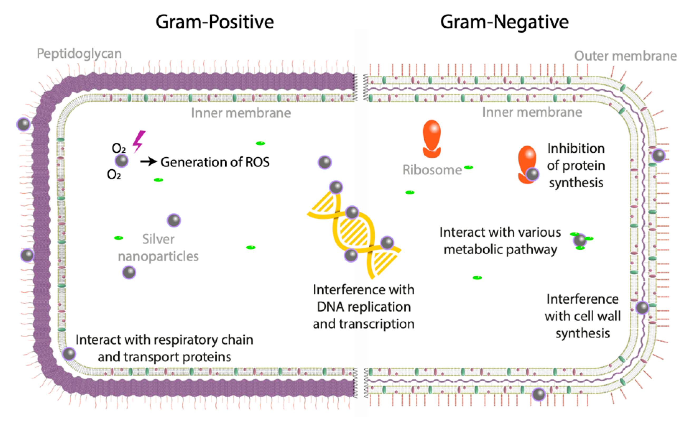
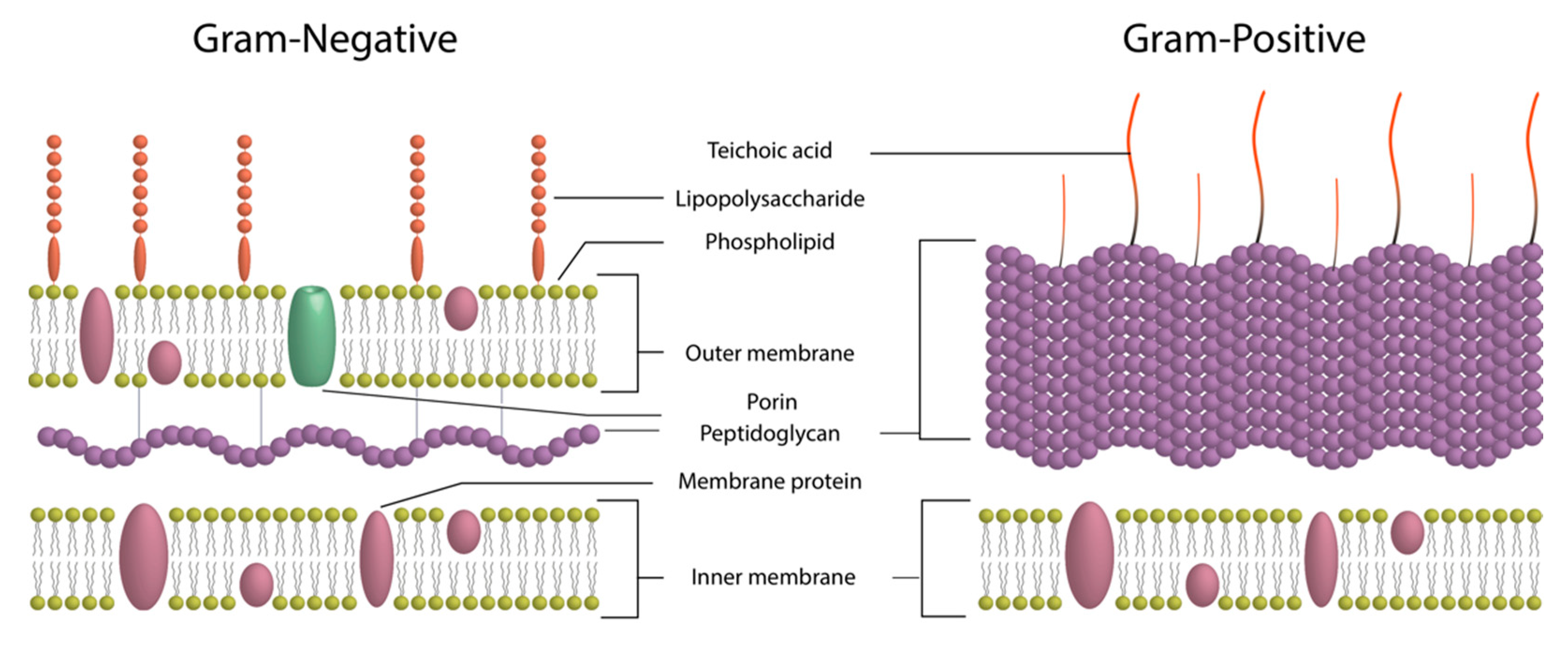
2.2. Antibacterial Mechanism of Silver Colloidal Solutions
2.3. Colloidal Solution Parameters Influencing Antibacterial Activity
2.3.1. NPs-Metallic-Core Size
2.3.2. Shape and Structural Properties
2.3.3. Surface Stability
2.3.4. Surface Accessibility
2.3.5. Other Chemicals and Concentration of Silver
3. How to Evaluate the Antibacterial Properties of Ag NPs?
3.1. Which Bacteria Test?
3.2. Which Technique to Use?
4. Conclusions and Future Perspectives
Supplementary Materials
Funding
Acknowledgments
Conflicts of Interest
References
- CDC Containing Unusual Antibiotic Resistance. Available online: https://www.cdc.gov/vitalsigns/containing-unusual-resistance/index.html (accessed on 1 August 2018).
- Database Scifinder. «Search with “Nanoparticles” and “Antibacterial” Terms». 14454 Publications over 2000–2018. Available online: https://scifinder.cas.org (accessed on 14 November 2019).
- Subbiah, R.; Jeon, S.B.; Park, K.; Ahn, S.J.; Yun, K. Investigation of cellular responses upon interaction with silver nanoparticles. Int. J. Nanomed. 2015, 10, 191–201. [Google Scholar]
- Łysakowska, M.E.; Ciebiada-Adamiec, A.; Klimek, L.; Sienkiewicz, M. The activity of silver nanoparticles (Axonnite) on clinical and environmental strains of Acinetobacter spp. Burns 2015, 41, 364–371. [Google Scholar] [CrossRef] [PubMed]
- Kourmouli, A.; Valenti, M.; van Rijn, E.; Beaumont, H.J.E.; Kalantzi, O.-I.; Schmidt-Ott, A.; Biskos, G. Can disc diffusion susceptibility tests assess the antimicrobial activity of engineered nanoparticles? J. Nanopart. Res. 2018, 20, 62. [Google Scholar] [CrossRef] [PubMed] [Green Version]
- Pompilio, A.; Geminiani, C.; Bosco, D.; Rana, R.; Aceto, A.; Bucciarelli, T.; Scotti, L.; Di Bonaventura, G. Electrochemically Synthesized Silver Nanoparticles Are Active Against Planktonic and Biofilm Cells of Pseudomonas aeruginosa and Other Cystic Fibrosis-Associated Bacterial Pathogens. Front. Microbiol. 2018, 9, 1349. [Google Scholar] [CrossRef] [PubMed] [Green Version]
- Markowska, K.; Grudniak, A.M.; Milczarek, B.; Wolska, K.I. The Effect of Silver Nanoparticles on Listeria monocytogenes PCM2191 Peptidoglycan Metabolism and Cell Permeability. Pol. J. Microbiol. 2018, 67, 315–320. [Google Scholar] [CrossRef] [Green Version]
- Choi, Y.; Kim, H.-A.; Kim, K.-W.; Lee, B.-T. Comparative toxicity of silver nanoparticles and silver ions to Escherichia coli. J. Environ. Sci. 2018, 66, 50–60. [Google Scholar] [CrossRef] [PubMed]
- Manipriya, B.; Tasneem, B.; Prem, K.L.; Kalyani, M. Evaluation of antibacterial activity of silver nanoparticles against methicillin-resistant Staphylococcus aureus and detection of virulence factors-nuclease, phosphatase, and bio film production. Asian J. Pharm. Clin. Res. 2018, 11, 224. [Google Scholar]
- Singh, V.; Tiwari, A. Evaluating the antimicrobal efficiency of chemically silver nanoparticles. Int. J. Curr. Microbiol. Appl. Sci. 2015, 4, 5–10. [Google Scholar]
- Korshed, P.; Li, L.; Ngo, D.-T.; Wang, T. Effect of Storage Conditions on the Long-Term Stability of Bactericidal Effects for Laser Generated Silver Nanoparticles. Nanomaterials 2018, 8, 218. [Google Scholar] [CrossRef] [Green Version]
- Belluco, S.; Losasso, C.; Patuzzi, I.; Rigo, L.; Conficoni, D.; Gallocchio, F.; Cibin, V.; Catellani, P.; Segato, S.; Ricci, A. Silver as Antibacterial toward Listeria monocytogenes. Front. Microbiol. 2016, 7, 307. [Google Scholar] [CrossRef] [Green Version]
- Skіba, M.; Pivovarov, A.; Makarova, A.; Vorobyova, V. Plasma-chemical Synthesis of Silver Nanoparticles in the Presence of Citrate. Chem. J. Mold. 2018, 13, 7–14. [Google Scholar] [CrossRef]
- Wu, Y.; Yang, Y.; Zhang, Z.; Wang, Z.; Zhao, Y.; Sun, L. A facile method to prepare size-tunable silver nanoparticles and its antibacterial mechanism. Adv. Powder Technol. 2018, 29, 407–415. [Google Scholar] [CrossRef]
- Acharya, D.; Singha, K.M.; Pandey, P.; Mohanta, B.; Rajkumari, J.; Singha, L.P. Shape dependent physical mutilation and lethal effects of silver nanoparticles on bacteria. Sci. Rep. 2018, 8, 201. [Google Scholar] [CrossRef] [PubMed] [Green Version]
- Tian, X.; Jiang, X.; Welch, C.; Croley, T.R.; Wong, T.-Y.; Chen, C.; Fan, S.; Chong, Y.; Li, R.; Ge, C.; et al. Bactericidal Effects of Silver Nanoparticles on Lactobacilli and the Underlying Mechanism. ACS Appl. Mater. Interfaces 2018, 10, 8443–8450. [Google Scholar] [CrossRef] [PubMed]
- Rojas-Andrade, M.; Cho, A.T.; Hu, P.; Lee, S.J.; Deming, C.P.; Sweeney, S.W.; Saltikov, C.; Chen, S. Enhanced antimicrobial activity with faceted silver nanostructures. J. Mater. Sci. 2015, 50, 2849–2858. [Google Scholar] [CrossRef]
- Vanitha, G.; Rajavel, K.; Boopathy, G.; Veeravazhuthi, V.; Neelamegam, P. Physiochemical charge stabilization of silver nanoparticles and its antibacterial applications. Chem. Phys. Lett. 2017, 669, 71–79. [Google Scholar] [CrossRef]
- Mostafa, A.A.; Sayed, S.R.M.; Solkamy, E.N.; Khan, M.; Shaik, M.R.; Al-Warthan, A.; Adil, S.F. Evaluation of Biological Activities of Chemically Synthesized Silver Nanoparticles. J. Nanomater. 2015, 2015, 1–7. [Google Scholar] [CrossRef] [Green Version]
- Khatoon, U.T.; Nageswara Rao, G.V.S.; Mohan, K.M.; Ramanaviciene, A.; Ramanavicius, A. Antibacterial and antifungal activity of silver nanospheres synthesized by tri-sodium citrate assisted chemical approach. Vacuum 2017, 146, 259–265. [Google Scholar] [CrossRef]
- Silvan, J.M.; Zorraquin-Peña, I.; Gonzalez de Llano, D.; Moreno-Arribas, M.V.; Martinez-Rodriguez, A.J. Antibacterial Activity of Glutathione-Stabilized Silver Nanoparticles Against Campylobacter Multidrug-Resistant Strains. Front. Microbiol. 2018, 9, 458. [Google Scholar] [CrossRef]
- Vasileva, M.Yu.; Ershov, A.Yu.; Baigildin, V.A.; Lagoda, I.V.; Kuleshova, L.Yu.; Shtro, A.A.; Zarubaev, V.V.; Yakimanskii, A.V. Synthesis of Silver Glyconanoparticles Based on 3-Thiopropionylhydrazones of Mono- and Disaccharides. Russ. J. Gen. Chem. 2018, 88, 109–113. [Google Scholar] [CrossRef]
- Bhargava, A.; Pareek, V.; Roy Choudhury, S.; Panwar, J.; Karmakar, S. Superior Bactericidal Efficacy of Fucose-Functionalized Silver Nanoparticles against Pseudomonas aeruginosa PAO1 and Prevention of Its Colonization on Urinary Catheters. ACS Appl. Mater. Interfaces 2018, 10, 29325–29337. [Google Scholar] [CrossRef] [PubMed]
- Nam, S.; Park, B.; Condon, B.D. Water-based binary polyol process for the controllable synthesis of silver nanoparticles inhibiting human and foodborne pathogenic bacteria. RSC Adv. 2018, 8, 21937–21947. [Google Scholar] [CrossRef] [Green Version]
- Sangsuwan, A.; Kawasaki, H.; Matsumura, Y.; Iwasaki, Y. Antimicrobial Silver Nanoclusters Bearing Biocompatible Phosphorylcholine-Based Zwitterionic Protection. Bioconjug. Chem. 2016, 27, 2527–2533. [Google Scholar] [CrossRef] [PubMed]
- Ajitha, B.; Ashok Kumar Reddy, Y.; Sreedhara Reddy, P. Enhanced antimicrobial activity of silver nanoparticles with controlled particle size by pH variation. Powder Technol. 2015, 269, 110–117. [Google Scholar] [CrossRef]
- Ashkarran, A.A. The effect of visible-light intensity on shape evolution and antibacterial properties of triangular silver nanostructures. Opt. Mater. 2016, 58, 454–460. [Google Scholar] [CrossRef]
- Garuglieri, E.; Cattò, C.; Villa, F.; Zanchi, R.; Cappitelli, F. Effects of sublethal concentrations of silver nanoparticles on Escherichia coli and Bacillus subtilis under aerobic and anaerobic conditions. Biointerphases 2016, 11, 04B308. [Google Scholar] [CrossRef] [Green Version]
- Li, W.-R.; Sun, T.-L.; Zhou, S.-L.; Ma, Y.-K.; Shi, Q.-S.; Xie, X.-B.; Huang, X.-M. A comparative analysis of antibacterial activity, dynamics, and effects of silver ions and silver nanoparticles against four bacterial strains. Int. Biodeterior. Biodegrad. 2017, 123, 304–310. [Google Scholar] [CrossRef]
- Mosselhy, D.A.; El-Aziz, M.A.; Hanna, M.; Ahmed, M.A.; Husien, M.M.; Feng, Q. Comparative synthesis and antimicrobial action of silver nanoparticles and silver nitrate. J. Nanopart. Res. 2015, 17, 1–10. [Google Scholar] [CrossRef]
- Lau, C.P.; Abdul-Wahab, M.F.; Jaafar, J.; Chan, G.F.; Abdul Rashid, N.A. Toxic effect of high concentration of sonochemically synthesized polyvinylpyrrolidone-coated silver nanoparticles on Citrobacter sp. A1 and Enterococcus sp. C1. J. Microbiol. Immunol. Infect. 2017, 50, 427–434. [Google Scholar] [CrossRef]
- Asadi, M.; Khosravi-Darani, K.; Haj-Seyed Javadi, N.; Esmaeili, S.; Azadnia, E. Synthesis of Silver Nanoparticles through Chemical Reduction and its Antibacterial Effect. RRJFPDT 2015, 3, 18–23. [Google Scholar]
- Gurusamy, V.; Krishnamoorthy, R.; Gopal, B.; Veeraravagan, V.; Neelamegam, P. Systematic investigation on hydrazine hydrate assisted reduction of silver nanoparticles and its antibacterial properties. Inorg. Nano-Met. Chem. 2017, 47, 761–767. [Google Scholar] [CrossRef]
- Hong, X.; Wen, J.; Xiong, X.; Hu, Y. Shape effect on the antibacterial activity of silver nanoparticles synthesized via a microwave-assisted method. Environ. Sci. Pollut. Res. 2016, 23, 4489–4497. [Google Scholar] [CrossRef]
- Duffy, L.L.; Osmond-McLeod, M.J.; Judy, J.; King, T. Investigation into the antibacterial activity of silver, zinc oxide and copper oxide nanoparticles against poultry-relevant isolates of Salmonella and Campylobacter. Food Control 2018, 92, 293–300. [Google Scholar] [CrossRef]
- Wang, H.; Jiang, Y.; Zhang, Y.; Zhang, Z.; Yang, X.; Ali, Md.A.; Fox, E.M.; Gobius, K.S.; Man, C. Silver nanoparticles: A novel antibacterial agent for control of Cronobacter sakazakii. J. Dairy Sci. 2018, 101, 10775–10791. [Google Scholar] [CrossRef] [Green Version]
- Yi, J.; Cheng, J. Effects of water chemistry and surface contact on the toxicity of silver nanoparticles to Bacillus subtilis. Ecotoxicology 2017, 26, 639–647. [Google Scholar] [CrossRef]
- Bondarenko, O.M.; Sihtmäe, M.; Kuzmičiova, J.; Ragelienė, L.; Kahru, A.; Daugelavičius, R. Plasma membrane is the target of rapid antibacterial action of silver nanoparticles in Escherichia coli and Pseudomonas aeruginosa. Int. J. Nanomed. 2018, 13, 6779–6790. [Google Scholar] [CrossRef] [Green Version]
- Lv, X.; Wang, H.; Su, A.; Chu, Y. A Novel Approach for Sericin-Conjugated Silver Nanoparticle Synthesis and Their Potential as Microbicide Candidates. J. Microbiol. Biotechnol. 2018, 28, 1367–1375. [Google Scholar] [CrossRef] [Green Version]
- Hoseini-Alfatemi, S.M.; Karimi, A.; Armin, S.; Fakharzadeh, S.; Fallah, F.; Kalanaky, S. Antibacterial and antibiofilm activity of nanochelating based silver nanoparticles against several nosocomial pathogens: Bioactivity of nanochelating based AgNPs. Appl. Organomet. Chem. 2018, 32, e4327. [Google Scholar] [CrossRef]
- Jin, J.-C.; Wu, X.-J.; Xu, J.; Wang, B.-B.; Jiang, F.-L.; Liu, Y. Ultrasmall silver nanoclusters: Highly efficient antibacterial activity and their mechanisms. Biomater. Sci. 2017, 5, 247–257. [Google Scholar] [CrossRef]
- Ajitha, B.; Kumar Reddy, Y.A.; Reddy, P.S.; Jeon, H.-J.; Ahn, C.W. Role of capping agents in controlling silver nanoparticles size, antibacterial activity and potential application as optical hydrogen peroxide sensor. RSC Adv. 2016, 6, 36171–36179. [Google Scholar] [CrossRef]
- Abbaszadegan, A.; Ghahramani, Y.; Gholami, A.; Hemmateenejad, B.; Dorostkar, S.; Nabavizadeh, M.; Sharghi, H. The Effect of Charge at the Surface of Silver Nanoparticles on Antimicrobial Activity against Gram-Positive and Gram-Negative Bacteria: A Preliminary Study. J. Nanomater. 2015, 2015, 1–8. [Google Scholar] [CrossRef] [Green Version]
- Cavassin, E.D.; de Figueiredo, L.F.P.; Otoch, J.P.; Seckler, M.M.; de Oliveira, R.A.; Franco, F.F.; Marangoni, V.S.; Zucolotto, V.; Levin, A.S.S.; Costa, S.F. Comparison of methods to detect the in vitro activity of silver nanoparticles (AgNP) against multidrug resistant bacteria. J. Nanobiotechnol. 2015, 13, 64. [Google Scholar] [CrossRef] [PubMed]
- Padmos, J.D.; Boudreau, R.T.M.; Weaver, D.F.; Zhang, P. Impact of Protecting Ligands on Surface Structure and Antibacterial Activity of Silver Nanoparticles. Langmuir 2015, 31, 3745–3752. [Google Scholar] [CrossRef] [PubMed]
- Long, Y.-M.; Hu, L.-G.; Yan, X.-T.; Zhao, X.-C.; Zhou, Q.-F.; Cai, Y.; Jiang, G.-B. Surface ligand controls silver ion release of nanosilver and its antibacterial activity against Escherichia coli. Int. J. Nanomed. 2017, 12, 3193–3206. [Google Scholar] [CrossRef] [PubMed] [Green Version]
- El-Zahry, M.R.; Mahmoud, A.; Refaat, I.H.; Mohamed, H.A.; Bohlmann, H.; Lendl, B. Antibacterial effect of various shapes of silver nanoparticles monitored by SERS. Talanta 2015, 138, 183–189. [Google Scholar] [CrossRef] [PubMed]
- Niska, K.; Knap, N.; Kędzia, A.; Jaskiewicz, M.; Kamysz, W.; Inkielewicz-Stepniak, I. Capping Agent-Dependent Toxicity and Antimicrobial Activity of Silver Nanoparticles: An In Vitro Study. Concerns about Potential Application in Dental Practice. Int. J. Med. Sci. 2016, 13, 772–782. [Google Scholar] [CrossRef] [PubMed] [Green Version]
- Kujda, M.; Ocwieja, M.; Adamczyk, Z.; Bochenska, O.; Bras, G.; Kozik, A.; Bielanska, E.; Barbasz, J. Charge stabilized silver nanoparticles applied as antibacterial agents. J. Nanosci. Nanotechnol. 2015, 15, 3574–3583. [Google Scholar] [CrossRef]
- Vu, X.H.; Duong, T.T.T.; Pham, T.T.H.; Trinh, D.K.; Nguyen, X.H.; Dang, V.-S. Synthesis and study of silver nanoparticles for antibacterial activity against Escherichia coli and Staphylococcus aureus. Adv. Nat. Sci. Nanosci. Nanotechnol. 2018, 9, 025019. [Google Scholar] [CrossRef]
- Feng, A.; Cao, J.; Wei, J.; Chang, F.; Yang, Y.; Xiao, Z. Facile Synthesis of Silver Nanoparticles with High Antibacterial Activity. Materials 2018, 11, 2498. [Google Scholar] [CrossRef] [Green Version]
- LaMer, V.K.; Dinegar, R.H. Theory, Production and Mechanism of Formation of Monodispersed Hydrosols. J. Am. Chem. Soc. 1950, 72, 4847–4854. [Google Scholar] [CrossRef]
- Zhang, Z.; Shen, W.; Xue, J.; Liu, Y.; Liu, Y.; Yan, P.; Liu, J.; Tang, J. Recent advances in synthetic methods and applications of silver nanostructures. Nanoscale Res. Lett. 2018, 13, 54. [Google Scholar] [CrossRef] [PubMed] [Green Version]
- Mathur, P.; Jha, S.; Ramteke, S.; Jain, N.K. Pharmaceutical aspects of silver nanoparticles. Artif. Cells Nanomed. Biotechnol. 2018, 46, 115–126. [Google Scholar] [CrossRef] [PubMed] [Green Version]
- Lee, S.; Jun, B.-H. Silver Nanoparticles: Synthesis and Application for Nanomedicine. Int. J. Mol. Sci. 2019, 20, 865. [Google Scholar] [CrossRef] [PubMed] [Green Version]
- Halbus, A.F.; Horozov, T.S.; Paunov, V.N. Colloid particle formulations for antimicrobial applications. Adv. Colloid Interface Sci. 2017, 249, 134–148. [Google Scholar] [CrossRef]
- De Matteis, V.; Cascione, M.; Toma, C.; Leporatti, S. Silver Nanoparticles: Synthetic Routes, In Vitro Toxicity and Theranostic Applications for Cancer Disease. Nanomaterials 2018, 8, 319. [Google Scholar] [CrossRef] [Green Version]
- Hühn, J.; Carrillo-Carrion, C.; Soliman, M.G.; Pfeiffer, C.; Valdeperez, D.; Masood, A.; Chakraborty, I.; Zhu, L.; Gallego, M.; Yue, Z.; et al. Selected Standard Protocols for the Synthesis, Phase Transfer, and Characterization of Inorganic Colloidal Nanoparticles. Chem. Mater. 2017, 29, 399–461. [Google Scholar] [CrossRef]
- Cushing, B.L.; Kolesnichenko, V.L.; O’Connor, C.J. Recent Advances in the Liquid-Phase Syntheses of Inorganic Nanoparticles. Chem. Rev. 2004, 104, 3893–3946. [Google Scholar] [CrossRef]
- Yan, X.; He, B.; Liu, L.; Qu, G.; Shi, J.; Hu, L.; Jiang, G. Antibacterial mechanism of silver nanoparticles in Pseudomonas aeruginosa: Proteomics approach. Metallomics 2018, 10, 557–564. [Google Scholar] [CrossRef]
- Ullah Khan, S.; Saleh, T.A.; Wahab, A.; Ullah Khan, M.H.; Khan, D.; Ullah Khan, W.; Rahim, A.; Kamal, S.; Ullah Khan, F.; Fahad, S. Nanosilver: New ageless and versatile biomedical therapeutic scaffold. Int. J. Nanomed. 2018, 13, 733–762. [Google Scholar] [CrossRef] [Green Version]
- Cao, H. (Ed.) Silver Nanoparticles for Antibacterial Devices: Biocompatibility and Toxicity; CRC Press, Taylor & Francis Group: Boca Raton, FL, USA, 2017; ISBN 978-1-4987-2532-3. [Google Scholar]
- Haider, A.; Kang, I.-K. Preparation of Silver Nanoparticles and Their Industrial and Biomedical Applications: A Comprehensive Review. Adv. Mater. Sci. Eng. 2015, 2015, 1–16. [Google Scholar] [CrossRef] [Green Version]
- Khodashenas, B. The Influential Factors on Antibacterial Behaviour of Copper and Silver Nanoparticles. Indian Chem. Eng. 2016, 58, 224–239. [Google Scholar] [CrossRef]
- Franci, G.; Falanga, A.; Galdiero, S.; Palomba, L.; Rai, M.; Morelli, G.; Galdiero, M. Silver Nanoparticles as Potential Antibacterial Agents. Molecules 2015, 20, 8856–8874. [Google Scholar] [CrossRef] [PubMed] [Green Version]
- Fujiwara, K.; Sotiriou, G.A.; Pratsinis, S.E. Enhanced Ag+ Ion Release from Aqueous Nanosilver Suspensions by Absorption of Ambient CO2. Langmuir 2015, 31, 5284–5290. [Google Scholar] [CrossRef] [PubMed]
- Xia, Y.; Xiong, Y.; Lim, B.; Skrabalak, S.E. Shape-Controlled Synthesis of Metal Nanocrystals: Simple Chemistry Meets Complex Physics? Angew. Chem. Int. Ed. 2009, 48, 60–103. [Google Scholar] [CrossRef]
- Hu, G.; Jin, W.; Chen, Q.; Cai, Y.; Zhu, Q.; Zhang, W. Antibacterial activity of silver nanoparticles with different morphologies as well as their possible antibacterial mechanism. Appl. Phys. Mater. Sci. Process. 2016, 122, 1–7. [Google Scholar] [CrossRef]
- Ishikawa, S.; Matsumura, Y.; Katoh-Kubo, K.; Tsuchido, T. Antibacterial activity of surfactants against Escherichia coli cells is influenced by carbon source and anaerobiosis. J. Appl. Microbiol. 2002, 93, 302–309. [Google Scholar] [CrossRef]
- Maneedaeng, A.; Phoemboon, S.; Chanthasena, P.; Chudapongse, N. Synthesis, interfacial properties, and antimicrobial activity of a new cationic gemini surfactant. Korean J. Chem. Eng. 2018, 35, 2313–2320. [Google Scholar] [CrossRef]
- Hepel, M.; Stobiecka, M. Detection of Oxidative Stress Biomarkers Using Functional Gold Nanoparticles. In Fine Particles in Medicine and Pharmacy; Matijević, E., Ed.; Springer US: Boston, MA, USA, 2012; pp. 241–281. ISBN 978-1-4614-0378-4. [Google Scholar]
- Stobiecka, M.; Hepel, M. Rapid functionalization of metal nanoparticles by moderator-tunable ligand-exchange process for biosensor designs. Sens. Actuators B Chem. 2010, 149, 373–380. [Google Scholar] [CrossRef]
- Hepel, M.; Blake, D.; McCabe, M.; Stobiecka, M.; Coopersmith, K. Assembly of Gold Nanoparticles Induced by Metal Ions. In Functional Nanoparticles for Bioanalysis, Nanomedicine, and Bioelectronic Devices, Volume 1; Hepel, M., Zhong, C.-J., Eds.; American Chemical Society: Washington, DC, USA, 2012; Volume 1112, pp. 207–240. ISBN 978-0-8412-2775-0. [Google Scholar]
- Kästner, C.; Thünemann, A.F. Catalytic Reduction of 4-Nitrophenol Using Silver Nanoparticles with Adjustable Activity. Langmuir 2016, 32, 7383–7391. [Google Scholar] [CrossRef]
- Duval, R.E.; Grare, M.; Demoré, B. Fight against Antimicrobial Resistance: We Always Need New Antibacterials but for Right Bacteria. Molecules 2019, 24, 3152. [Google Scholar] [CrossRef] [Green Version]
- Centers for Disease Control and Prevention (CDC). Antibiotic Resistance Threats in the United States. 2013. Available online: https://www.cdc.gov/drugresistance/threat-report-2013/index.html (accessed on 31 July 2018).
- European Centre for Disease Prevention and Control (ECDC). Antimicrobial Resistance Surveillance in Europe 2016. Available online: https://ecdc.europa.eu/en/publications-data/antimicrobial-resistance-surveillance-europe-2016 (accessed on 31 July 2018).
- Anjana, G.; Gowri, M.; Raja, C.S.A.; Prasath, M.; Balakumar, S.; Ganesh, V. Silver Nanoparticles as a Non Alcoholic Hospital Disinfectant to Combat Nosocomial Pathogens. J. Bionanosci. 2015, 9, 102–111. [Google Scholar] [CrossRef]
- Balouiri, M.; Sadiki, M.; Ibnsouda, S.K. Methods for in vitro evaluating antimicrobial activity: A review. J. Pharm. Anal. 2016, 6, 71–79. [Google Scholar] [CrossRef] [Green Version]
- ISO 20776-1. Clinical Laboratory Testing and In Vitro Diagnostic Test Systems—Susceptibility Testing of Infectious Agents and Evaluation of Performance of Antimicrobial Susceptibility Test Devices—Part 1: Reference Method for Testing the In Vitro Activity of Antimicrobial Agents Against Rapidly Growing Aerobic Bacteria Involved in Infectious Diseases. ISO 20776-1: La Plaine Saint Denis, France, 2007. [Google Scholar]
- Clinical Laboratory Standards Institute (CLSI). Methods for Dilution Antimicrobial Susceptibility Tests for Bacteria that Grow Aerobically; Approved Standard-Tenth Ed. M07-A10; CLSI: Wayne, PA, USA, 2015. [Google Scholar]
- Liao, C.; Li, Y.; Tjong, S. Bactericidal and Cytotoxic Properties of Silver Nanoparticles. Int. J. Mol. Sci. 2019, 20, 449. [Google Scholar] [CrossRef] [PubMed] [Green Version]
- Gao, X.; Lowry, G.V. Progress towards standardized and validated characterizations for measuring physicochemical properties of manufactured nanomaterials relevant to nano health and safety risks. NanoImpact 2018, 9, 14–30. [Google Scholar] [CrossRef]
- Cockerill, F.; Clinical and Laboratory Standards Institute. Methods for Dilution Antimicrobial Susceptibility Tests for Bacteria that Grow Aerobically: Approved Standard; Clinical and Laboratory Standards Institute: Wayne, PA, USA, 2015; ISBN 978-1-56238-783-9. [Google Scholar]
- EUCAST (European Committee on Antimicrobial Susceptibility Testing). Routine and Extended Internal Quality Control for MIC Determination and Disk Diffusion as Recommended by EUCAST. Available online: http://www.eucast.org/fileadmin/src/media/PDFs/EUCAST_files/QC/v_8.0_EUCAST_QC_tables_routine_and_extended_QC.pdf (accessed on 31 July 2018).
- ISO 20776-2. Clinical Laboratory Testing and In Vitro Diagnostic Test Systems—Susceptibility Testing of Infectious Agents and Evaluation of Performance of Antimicrobial Susceptibility Test Devices—Part 2: Evaluation of Performance of Antimicrobial Susceptibility Test Devices; ISO 20776-2: La Plaine Saint Denis, France, 2007.


| Stabilizer | NPs Size (nm) | NPs Shape | Stock Suspension Concentration or Mass | Zeta Potential (mV) * | Comment | Bacteria | Bacteria Origin | Protocol | Culture Media | Reference |
|---|---|---|---|---|---|---|---|---|---|---|
| “Naked” | 19.5 ± 7.7 | Nano-sphere | N/A | −18.0 ± 0.6 (in culture media) | NaBH4 + AgNO3 in presence of ultrasonication | E. faecalis S. aureus S. epidermidis B. subtilis E. coli S. typhimurium S. enterica | KCCM 13807 KCTC 1916 KCTC 1971 KCTC 1021 KCTC 1682 KCCM 40253 KACC 10763 | Kirby-Bauer method Microdilution method MIC90 | Mueller Hinton Agar Luria Bertani | [3] |
| 10 | Nano-flake | 50 ppm | N/A | Axonnite Silver suspended in demineralized water | A. baumannii (n = 17) A. baumannii A. nosocomialis (n = 10) | Clinical isolates ATCC 1906 Clinical isolates | Microdilution method | N/A | [4] | |
| 10 20 40 | Nano-sphere | N/A | N/A | Vapor nucleation in N2 gas | E. coli | N/A | Kirby-Bauer method | Mueller Hinton 2 Agar | [5] | |
| 55.6 ± 2.9 (DLS) | Nanosphere | 8.53 mg | −51.5 ± 2.5 | Electrochemically synthesized | P. aeruginosa (n = 3) S. maltophilia (n = 3) B. cepacia (n = 3) S. aureus (n = 3) | Clinical isolates | Kirby-Bauer method Microdilution method TKA | Mueller Hinton Cation-Adjusted | [6] | |
| 2–5 (70–75% TEM) | Nano-sphere | 50 mg/kg | +9.2 | Nano-Tech (Warsaw, Poland) | L. monocytogenes | PCM 2191 | Microdilution method | Tryptone Soy yeast extract broth | [7] | |
| 23.6 (TEM) 57.8 (DLS) | Nano-sphere | N/A | −28.3 (N/A) | Nanoleader (Korea) | E. coli (K-12) | KCTC 1116 | Growth Curves | Luria Bertani | [8] | |
| 10 | Nano-sphere | 1 mg/mL | N/A | Sisco Research lab. | S. aureus S. aureus (n = 30) | ATCC 25923 Clinical isolates | Agar Well diffusion method Microdilution method | Mueller Hinton Agar Luria Bertani | [9] | |
| “Naked” | 40 (TEM) | Nano-sphere | N/A | N/A | AgNO3 + NaBH4 | S. aureus B. cereus P. aeruginosa K. pneumoniae E. coli | N/A | Agar Well diffusion method | Mueller Hinton Agar | [10] |
| Unknown “Naked” | 35 27.2 | Nano-sphere | 20 µg/mL N/A | N/A | Sigma Aldrich (Dorset UK) Laser generated | E. coli (K-12) | JM 109 | Agar Well diffusion method | Mueller Hinton | [11] |
| Citrate | 23 ± 2 (TEM) | Nano-sphere | 2 mM | N/A | Citrate BioPure™ Silver, Nanocomposix (San Diego, CA, USA) | L. monocytogenes (n = 20) L. monocytogenes | Clinical isolates | Colony Forming Units | Mueller Hinton | [12] |
| 6.0–28.2 (XRD) | N/A | −28.2 to −32.0 | Plasma discharge | S. aureus E. coli | ATCC 25923 ATCC 35218 | Kirby-Bauer method | Nutrient Agar | [13] | ||
| 2.3 ± 0.5 12.5 ± 2.2 32.4 ± 6.5 (TEM) | Nano-sphere | N/A1 | N/A1 | NaBH4 + AgNO3 + sodium citrate | S. aureus E. coli | ATCC 35696 ATCC 23282 | Microdilution method Kirby-Bauer method Growth Curves | Broth medium | [14] | |
| 40–50 20 × 20–90 | Nano-sphere Nano-rod | N/A | −28.8 −23.5 | Citrate thermal reduction method | S. aureus B. subtilis P. aeruginosa K. pneumoniae E. coli | ATCC 25923 AST5-2 AL2-14B AWD5 ATCC 25922 | Microdilution method Kirby-Bauer method TKA | Nutrient Agar | [15] | |
| 20.1 ± 4.4 (TEM) 49.3 ± 5.7 (DLS) | Nano-sphere | N/A | −19.2 ± 0.7 | Citrate thermal reduction method | E. coli S. aureus L. bulgaricus L. casei | ATCC 25922 ATCC 25923 CGMCC 1.6970 CGMCC 1.2435 | TKA | Luria Bertani Tryptone Soy MRS | [16] | |
| 10–40 (TEM) | Sharp-tipped triangular, truncated triangular, nanoprisms, decahedra, tetrahedra | N/A | N/A | Photochemical synthesis: 4.76 ± 3.88 nm silver seed nanoparticles (AgNO3 + citrate + NaBH4) + 40 W blue LEDs (Hongke Lighting kem = 455–475 nm) {111} facets | E. coli | ATCC 25922 | Growth Curves | Luria Bertani | [17] | |
| Citrate | 20 ± 9 25 ± 3 11 ± 6 (TEM) | Nano-sphere | N/A | −26.37 −37.95 −28.23 (H2O + salts) | AgNO3 + citrate + NaBH4 | S. aureus | ATCC 25923 | Agar Well diffusion method | Nutrient Agar | [18] |
| 42–58 (TEM) | Nano-sphere | N/A | N/A | AgNO3 + citrate + NaBH4; polydisperse; XRD11: intense reflection at (111) | S. aureus S. pyogenes S. typhi P. aeruginosa | N/A | Kirby-Bauer method | Tryptone Soy | [19] | |
| 15 18 30 30 (DLS) | Nano-sphere | N/A | −38.8 −30.7 −38.5 −42.2 (N/A) | AgNO3 + Citrate + NaBH4 | E. coli B. subtilis | N/A | Kirby-Bauer method | Nutrient Agar | [20] | |
| GSH13 | 10–50 | Nano-sphere | 0.197 mg/mL | N/A | AgBF4 + NaBH4 + glutathione | C. jejuni (n = 22) C. coli (n = 18) C. jejuni | Animal or Human clinical isolates NCTC 11168 | Microdilution method | Brucella Mueller Hinton | [21] |
| D-xylose L-arabinose D-ribose D-glucose D-galactose D-mannose D-lactose D-xylose | 33 30 39 25 28 25 15 18 | Nano-sphere | N/A | N/A | E. coli Klebsiella spp. | N/A | N/A | N/A | [22] | |
| L-fucose | 10.15 ± 3.37 (TEM) | Nano-sphere | N/A | −65.4 −17.7 | AgNO3 + NaBH4 + sodium citrate + mercaptopropionic acid, then L-fucose | P. aeruginosa (PAO1) | N/A | Microdilution method | Luria Bertani without Chloride ions | [23] |
| PEG | 15.8 ± 2.2 (TEM) | Nano-sphere | N/A | −17.2 ± 2.1 | AgNO3 + EG/PEG | S. aureus P. aeruginosa S. enterica E. coli | ATCC 6538 ATCC 15442 ATCC 10708 ATCC 11229 | Microdilution method | Mueller Hinton | [24] |
| PC | 3.3 ± 0.9 4.9 ± 2.9 (TEM) | Nano-sphere | N/A | N/A | Zwitterionic Protection: AgNO3 + NaBH4 + PC-SH | E. coli S. aureus | OW6 Mu50 | Growth curves | Todd Hewitt broth | [25] |
| PVA | 31 (SEM; TEM: 26) 24 (SEM) 19 (SEM) 14 (SEM; TEM: 10) | Nano-sphere | N/A | N/A | PVA + AgNO3 + NaBH4 (pH = 6; 8; 10; 12, respectively) | E. coli Pseudomonas sp. | N/A | Kirby-Bauer method | Nutrient Agar | [26] |
| PVP/citrate | 50–60 70–80 | Semi-triangular and truncated triangular silver nanoparticles + few nano-sphere Triangular silver nanoparticles with sharp corner | N/A | N/A | Citrate + AgNO3 + NaBH4 + PVP + visible-light halogen lamp (50 and 100 W, respectively); bigger nanoparticles (>100 nm) obtained with visible-light halogen lamps with higher intensities | E. coli | N/A | Colony Forming Units | Nutrient Agar | [27] |
| PVP20 | 14.0 ± 0.3 (TEM) | Nano-sphere | 1 mg/mL | −27.3 | Nanocomposix, OECD standard BioPure, PVP20 40kDa | E. coli (K-12) B. subtilis | MG1655 ATCC 6051 | Growth curves | Tryptone Soy | [28] |
| 5 20 | Nano-sphere | 1 mg/mL 2 mg/mL | N/A | Shanghai Institute of Fine Chemical Materials (China) | E. coli P. aeruginosa S. aureus S. epidermidis | ATCC 8739 ATCC 9027 ATCC 6538 ATCC 12228 | Poisoned Food Technique | Mueller Hinton | [29] | |
| 8 29 (TEM) | Nano-sphere | N/A | −22.36 −37.82 | PVP + ethylene glycol + AgNO3; redispersed in water | A. hydrophila P. putida E. coli B. subtilis S. aureus | 4AK4 KT2442 Trans 1-T1 ATCC 28357 N/A | Kirby-Bauer method Microdilution method | N/A Mueller Hinton | [30] | |
| 15.6 (TEM) | Nano-sphere | N/A | N/A | PVP + AgNO3 + NaBH4 in water; polydisperse | Citrobacter sp. Enterococcus sp. | N/A | Colony Forming Units | N/A | [31] | |
| 3–34 (TEM) | Nano-sphere | N/A | N/A | AgNO3 + Ethanol + PVP (55000 molecular mass) in water | S. aureus PTCC No. 1112 E. coli PTCC No. 1330 | ATCC 6537 ATCC 8739 | N/A | Mueller Hinton | [32] | |
| 10–15 (TEM) | Nano-sphere | N/A | N/A | AgNO3 + PVP (k30; Mw. 40000) + hydrazine; influence of AgNO3, PVP and hydrazine concentrations | E. coli S. aureus | ATCC 25922 ATCC 25923 | Kirby-Bauer method | Nutrient Agar | [33] | |
| 60 ± 15 55 ± 10 60 × 2000–4000 | Nano-sphere Nano-cube, right bipyramids Nanowire | N/A | N/A | AgNO3 + PVP (k30) + ethylene glycol + NaCl (0. 1, and 5 mg, respectively) | E. coli | ATCC 25922 | Growth curves Microdilution method | Luria Bertani | [34] | |
| 20.6 ± 3.1 | Nano-sphere | 1 mg/mL | −35 | Nanocomposix (San Diego, CA, USA) | C. jejuni (n = 4) C. jejuni Salmonella spp. (n = 5) | Chicken isolates NCTC 11168 Chicken isolates | Microdilution method | Mueller Hinton Luria Bertani | [35] | |
| PVP Glycerol | 31.2 (TEM) 46.5 (DLS) | Nano-sphere | N/A | +18.7 (N/A) | AgNO3 + PVP or glycerol + sodium citrate (NH4OH, pH = 8) | C. sakazakii | ATCC 29544 ATCC BAA894 ATCC 29004 ATCC 12868 | Microdilution method Oxford cup method | Luria Bertani | [36] |
| Oleylamine | 10 (TEM) | Nano-sphere | N/A | −7.11 | ColdStones Tech. (Suzhou, China) | B. subtilis | ATCC 6633 | Growth curves | Luria Bertani | [37] |
| Casein | 12.5 ± 4 (TEM) 50.0 ± 0.7 (DLS) | Nano-sphere | N/A | −26.6 ± 1.7 {in 3-(N-morpholini) propanesulfonic acid-(hydroxy-methanyl) aminomethane} | Lab. Argenol S. L. (Zaragoza, Spain) | E. coli P. aeruginosa | MC 1061 DS 10-129 | Bioluminescence inhibition assay | Luria Bertani | [38] |
| Sericin | 3.78 ± 1.14 (TEM) | Nano-sphere | N/A | N/A | Silk sericin protein + AgNO3 + NaBH4 | S. aureus E. coli | ATCC 25923 ATCC 25922 | Cell counting (FCM) | Nutrient medium | [39] |
| Thioacetic acid Propionic acid | 20–25 30–35 | Nano-sphere | N/A | N/A | Thioacetic or propionic acid + silver acetate + sodium carbonate (US20120100372A1) | S aureus S. epidermidis A. baumannii P. aeruginosa | ATCC 25923 ATCC 35984 ATCC 19606 ATCC 27853 | Microdilution method | Mueller Hinton | [40] |
| Lipoid acid | 2.0 ± 0.5 (TEM) | Nano-sphere | N/A | N/A | dihydrolipoic acid + NaOH + AgNO3 + NaBH4 | S. aureus E. coli E. coli | N/A DH5α DSM4230 | Growth curves | Luria Bertani | [41] |
| PEG EDTA PVP PVA | 44 39 35 31 (SEM) | Nano-sphere | N/A1 | −17.5 −23.0 −41.0 −47.0 | PEG, EDTA, PVP or PVA + AgNO3 + NaOH + NaBH4 in water | E. coli Pseudomonas spp. | N/A | Kirby-Bauer method | Nutrient Agar | [42] |
| “Naked” Unknown | 7.5 10.1 (TEM) | Nano-sphere | 9.7 × 10⁻⁸ mol/L 4 × 10⁻⁸ mol/L | −38.0 0.0 | AgNO3 + NaBH4 Rice starch + AgNO3 | S. aureus S. mutans S. pyogenes E. coli P. vulgaris | ATCC 29737 ATCC 35668 ATCC 8668 ATCC 15224 ATCC 7829 | Agar Well diffusion method Microdilution method | Brain Heart Infusion Mueller Hinton | [43] |
| PVA Citrate Citrate | 10 nm (SEM) 40 nm (SEM) 60 nm | Nano-sphere | N/A N/A 20 μg/L | −17.0 −48.4 N/A | Chitosan-Ag NPs also prepared and tested (Sigma Aldrich) | A. baumanni (n = 17) P. aeruginosa (n = 13) Enterobacteriaceae (n = 21) S. maltophilia (n = 2) S. aureus (n = 13) S. aureus S. epidermidis Enterococcus sp. (n = 14) | Clinical isolates Clinical isolates Clinical isolates Clinical isolates Clinical isolates ATCC 29213 INCQS 198 Clinical isolates | Agar Well diffusion method Microdilution method Time kill assay | Mueller Hinton Mueller Hinton Cation Adjusted Tryptone Soy Mueller Hinton Cation Adjusted | [44] |
| Cysteine PVP | 7.6 ± 1.5 7.7 ± 1.6 (TEM) | Nano-sphere | N/A | N/A | AgNO3 + NaBH4 + L-cysteine AgNO3 + KOH + PVP (Mw 8000) + NaBH4 | S. aureus E. coli P. aeruginosa | ATCC 29213 ATCC 23716 ATCC 25619 | Microdilution method | Mueller Hinton Cation Adjusted | [45] |
| Citrate MPA MHA MPS | 10.2 ± 2.3 10.2 ± 2.5 10.2 ± 2.2 9.9 ± 2.0 | Nano-sphere | N/A | −47.4 −34.5 −29.1 −32.6 | Citrate + tannic acid + AgNO3 Citrate-Ag NPs + mercaptopropionic acid (ligand exchange) Citrate-Ag NPs + mercaptohexanoic acid (ligand exchange) Citrate-Ag NPs + mercaptopropionic sulfonic acid (ligand exchange) | E. coli | MG 1655 | Growth curves | Luria Bertani | [46] |
| Citrate PVP PEG | 40 (TEM; 10–70) | Nano-sphere Triangular silver nanoparticles with rounded edges Hexagonal silver NPs | N/A | N/A | AgNO3 + citrate PVP (Mw = 25000) + AgNO3 + citrate + H2O2 + NaBH4 PEG (2000) + NaOH + AgNO3 | E. coli | DH5α | Agar Well diffusion method Growth curves | Luria Bertani | [47] |
| Lipoid acid PEG “Naked” | 9.5 ± 1.9 (TEM) 9.8 ± 2.0 11.2 ± 2.1 | Nano-sphere | N/A | −28.6 (Serum free culture medium) −10 (Serum free culture medium) −33.9 (Serum free culture medium) | Nanocomposix, Europe Nanocomposix, Europe Also Tannic acid-NPs US research (Nanomaterilas (Houston, TX, USA) | Actinomyces (n = 1) Bacteroides (n = 4) Bacteroides fragilis Bifidobacterium (n = 1) Bifidobacterium breve Finegoldia (n = 2) Fusobacterium (n = 4) Fusobacterium nucleatum Parabacteroides (n = 1) Parvimonas (n = 2) Peptostreptococcus (n = 1) Peptostreptococcus anaerobius Porphyromonas (n = 3) Porphyromonas levii Prevotella (n = 5) Prevotella loescheii Propionibacterium (n = 2) Tannerella (n = 1) S. aureus S. aureus S. aureus S. epidermidis S. mutans | Clinical isolates Clinical isolates ATCC 25285 Clinical isolates ATCC 15700 Clinical isolates Clinical isolates ATCC 25585 Clinical isolates Clinical isolates Clinical isolates ATCC 25286 Clinical isolates ATCC 29147 Clinical isolates ATCC 15930 Clinical isolates Clinical isolates ATCC 25923 ATCC 6538 ATTC 6538P ATCC 14990 ATCC 29175 | Plate dilution method Microdilution method | Brucella agar supplemented Mueller Hinton | [48] |
| Citrate HH SHSH SHST | 15 ± 4 13 ± 2 13 ± 4 10 ± 6 (TEM) | Nano-sphere | 290 mg/L 290 mg/L 330 mg/L 330 mg/L | −39.8 ± 0.74 (H2O + NaCl) −34.0 ± 1.97 (H2O + NaCl) −34.4 ± 2.03 (H2O + NaCl) −35.9 ± 1.09 (H2O + NaCl) | AgNO3 + Citrate + NaBH4 Hydroxylamine hypochlorite + NaOH + AgNO3 Sodium hypophosphite + sodium hexametaphosphate + AgNO2 Sodium hypophosphite + sodium hexametaphosphate + sodium tripolyphosphate + AgNO2 | E. coli (K-12) E. coli | ATCC 10798 ER2566 | Microdilution method | Mueller Hinton | [49] |
| Starch | 8 ± 4 (TEM) | Nano-sphere | N/A | N/A | AgNO3 + NaBH4 + (C6H10O5)n | S. aureus E. coli | N/A | Kirby-Bauer method | Nutrient agar | [50] |
| AOT | 20 50 | “Nano-sphere” | N/A | N/A | Bis(2-ethylhexyl) sulfosuccinate + AgNO3 + ascorbic acid | E. coli S. aureus | N/A | Microdilution method | Luria Bertani | [51] |
| Nanoparticle Samples | ||
|---|---|---|
| Synthesis | Specification of all chemicals used | [83] |
| Characterizations | Metal core size (TEM) Hydrodynamic size (DLS) Zeta-potential Stock suspension concentration in metal or metal mass (e.g., ICP-AES) | |
| Microbiology | ||
| Antibacterial activity | Specification of the procedure used for antibacterial activity determination:
| [79,80,84,85,86] |
| Without forgetting to specify growing conditions (temperature and time of incubation, shaking or not) | ||
| Bacteria | Specification of the origin of the bacteria: Strains issued from international collections (e.g., ATCC) or Clinical Isolates (with appropriated antibiograms) For example:
| |
| Bacterial medium | Mueller Hinton (MH) | |
| Cation-Adjusted MH (CA-MH) Supplemented in accordance with the bacterial strains studied… | ||
© 2019 by the authors. Licensee MDPI, Basel, Switzerland. This article is an open access article distributed under the terms and conditions of the Creative Commons Attribution (CC BY) license (http://creativecommons.org/licenses/by/4.0/).
Share and Cite
Duval, R.E.; Gouyau, J.; Lamouroux, E. Limitations of Recent Studies Dealing with the Antibacterial Properties of Silver Nanoparticles: Fact and Opinion. Nanomaterials 2019, 9, 1775. https://doi.org/10.3390/nano9121775
Duval RE, Gouyau J, Lamouroux E. Limitations of Recent Studies Dealing with the Antibacterial Properties of Silver Nanoparticles: Fact and Opinion. Nanomaterials. 2019; 9(12):1775. https://doi.org/10.3390/nano9121775
Chicago/Turabian StyleDuval, Raphaël E., Jimmy Gouyau, and Emmanuel Lamouroux. 2019. "Limitations of Recent Studies Dealing with the Antibacterial Properties of Silver Nanoparticles: Fact and Opinion" Nanomaterials 9, no. 12: 1775. https://doi.org/10.3390/nano9121775