Facile Synthesis of Triangular and Hexagonal Anionic Gold Nanoparticles and Evaluation of Their Cytotoxicity


Abstract
:1. Introduction
2. Materials and Methods
2.1. Materials
2.2. E. annuus Leaf Extract Preparation
2.3. Green Synthesis of AuNPs
2.4. Effect of E. annuus Leaf Extract Concentration on Green Synthesis of AuNPs
2.5. Effect of Temperature on Green Synthesis of AuNPs
2.6. Characterisation of Nanoparticles
2.7. Cytotoxicity Assay
2.7.1. Cell Culture
2.7.2. Cell Differentiation
2.7.3. Cell Viability Assay
3. Results and Discussion
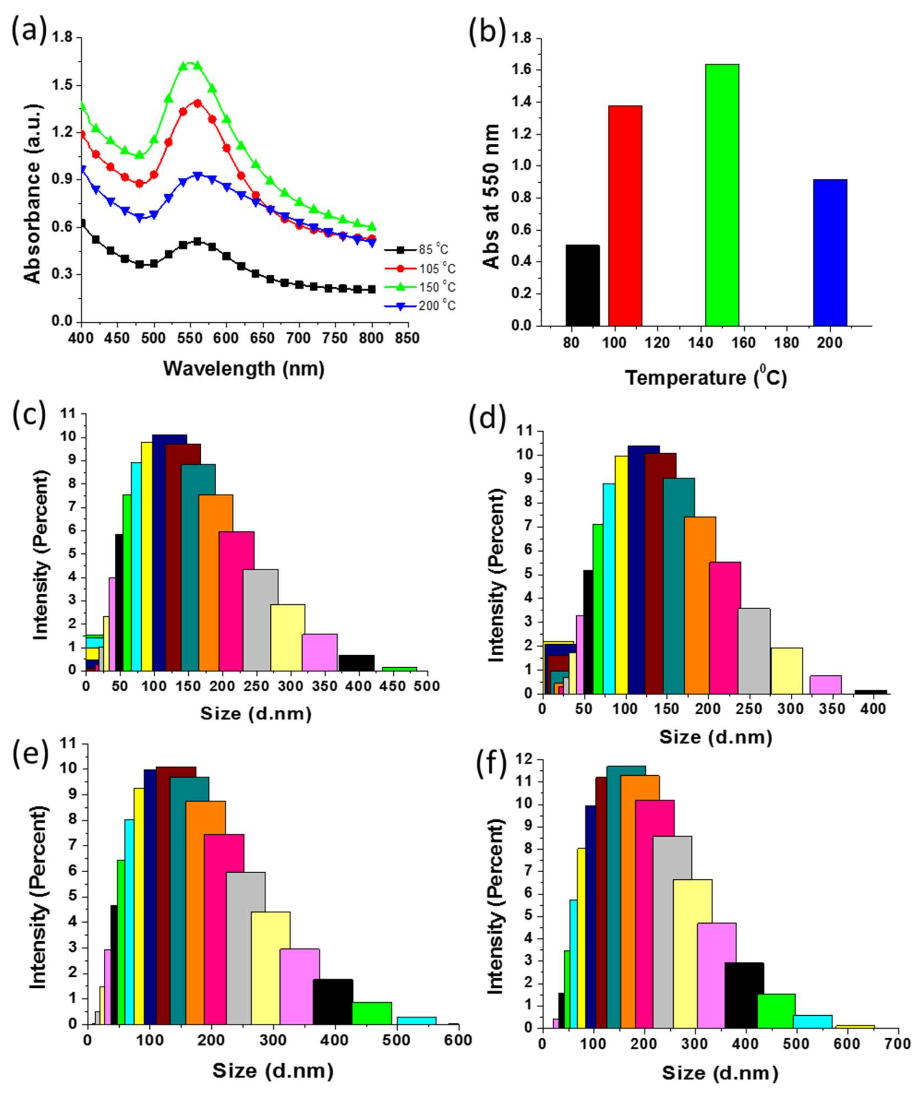
3.1. UV–Vis Spectroscopy Analysis
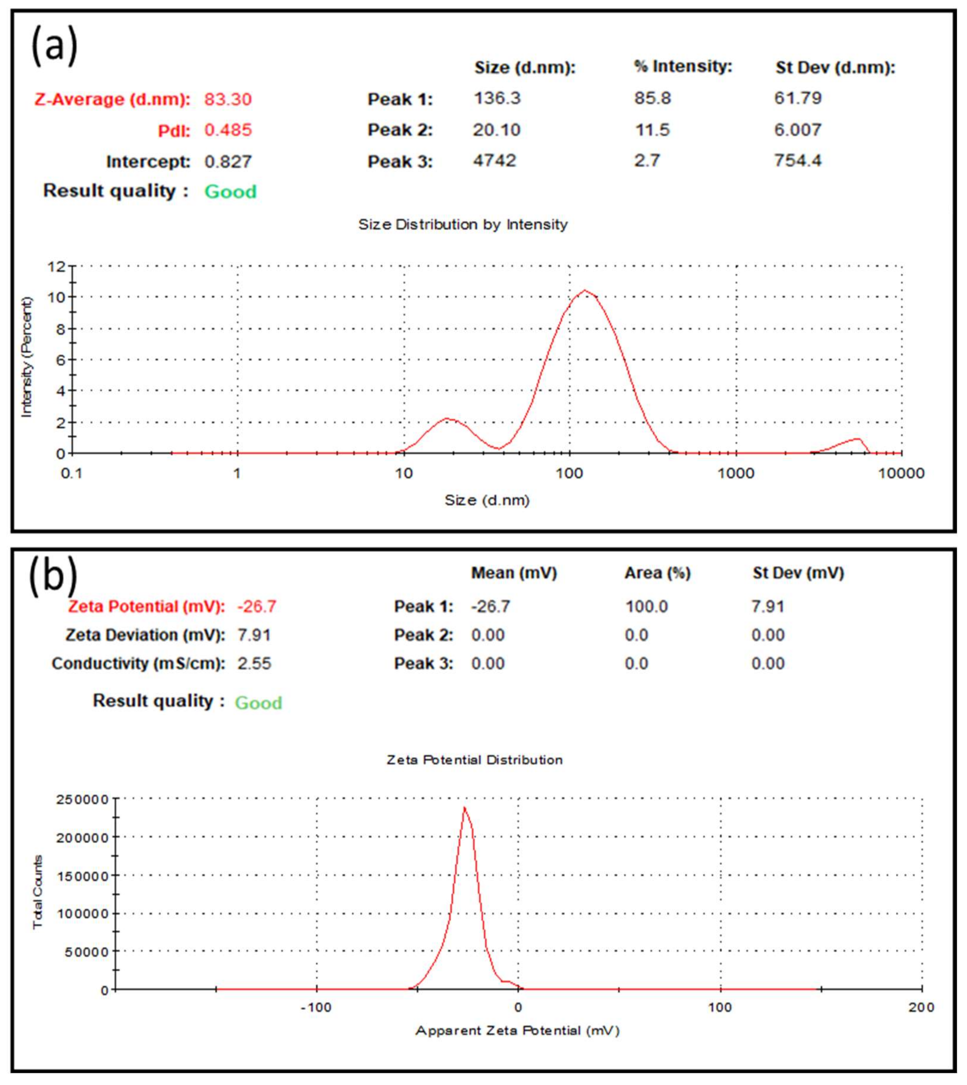
3.2. DLS Analysis
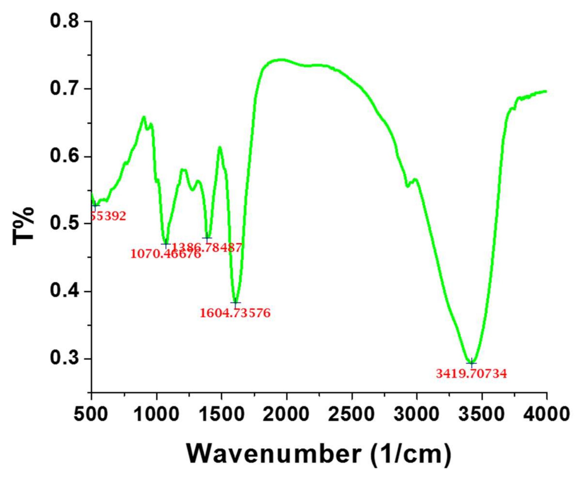
3.3. FTIR Analysis
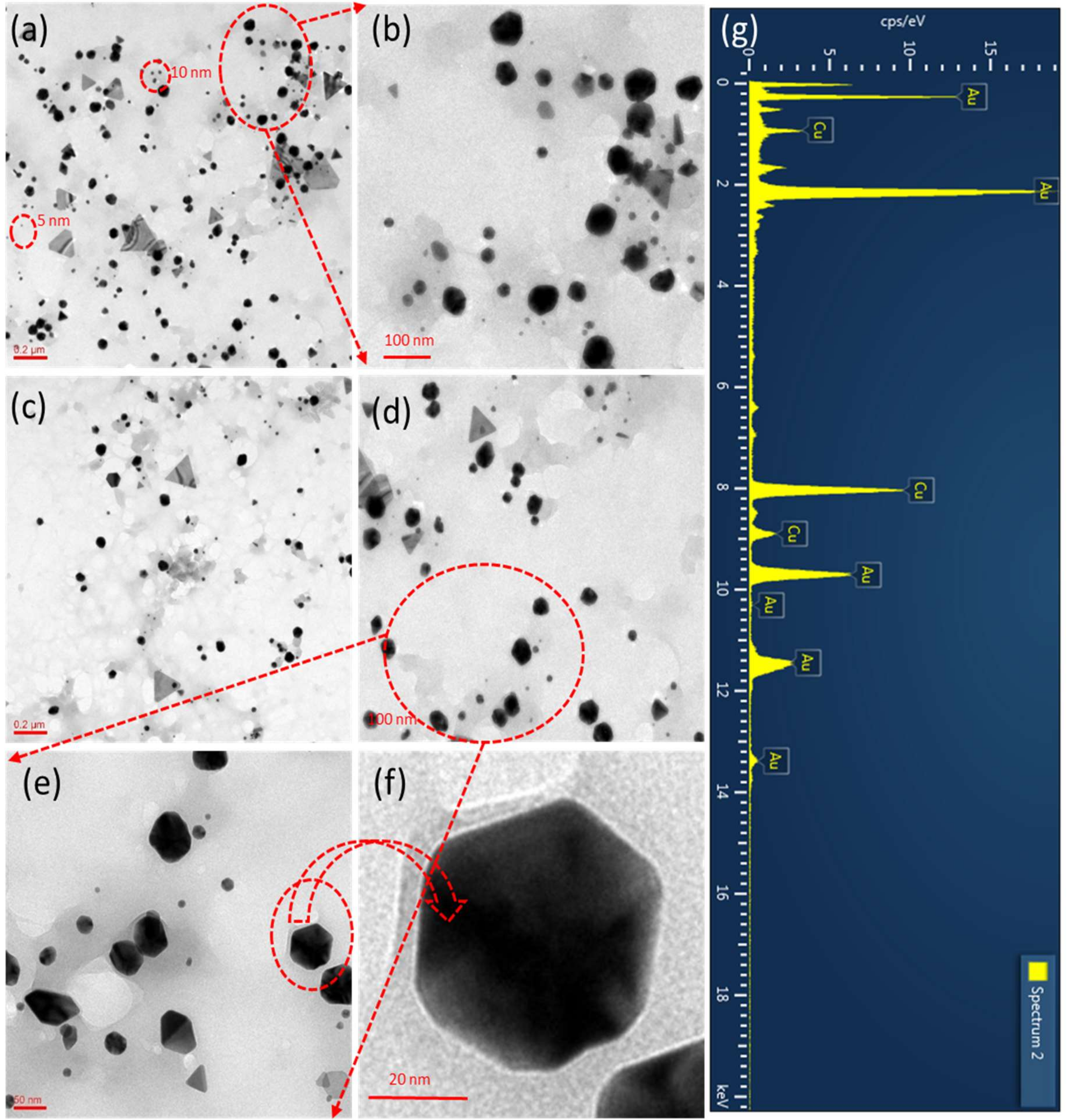
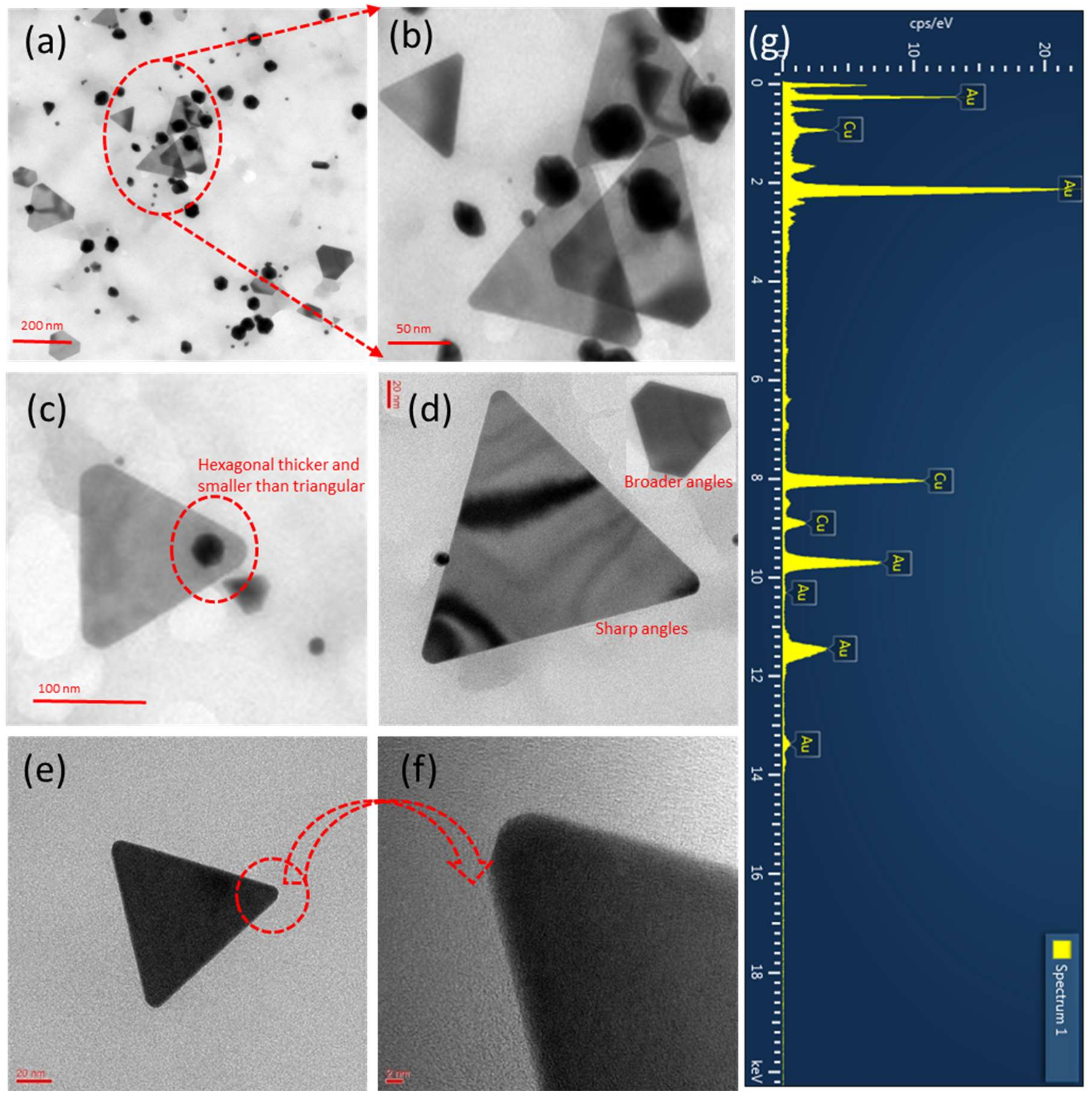
3.4. TEM Analysis
3.5. EDX Analysis
3.6. Possible Green Synthesis Mechanism
3.7. Effect of E. annuus Leaf Extract Concentration on Green Synthesis of AuNPs
3.8. Effect of Reaction Temperature on Green Synthesis of AuNPs
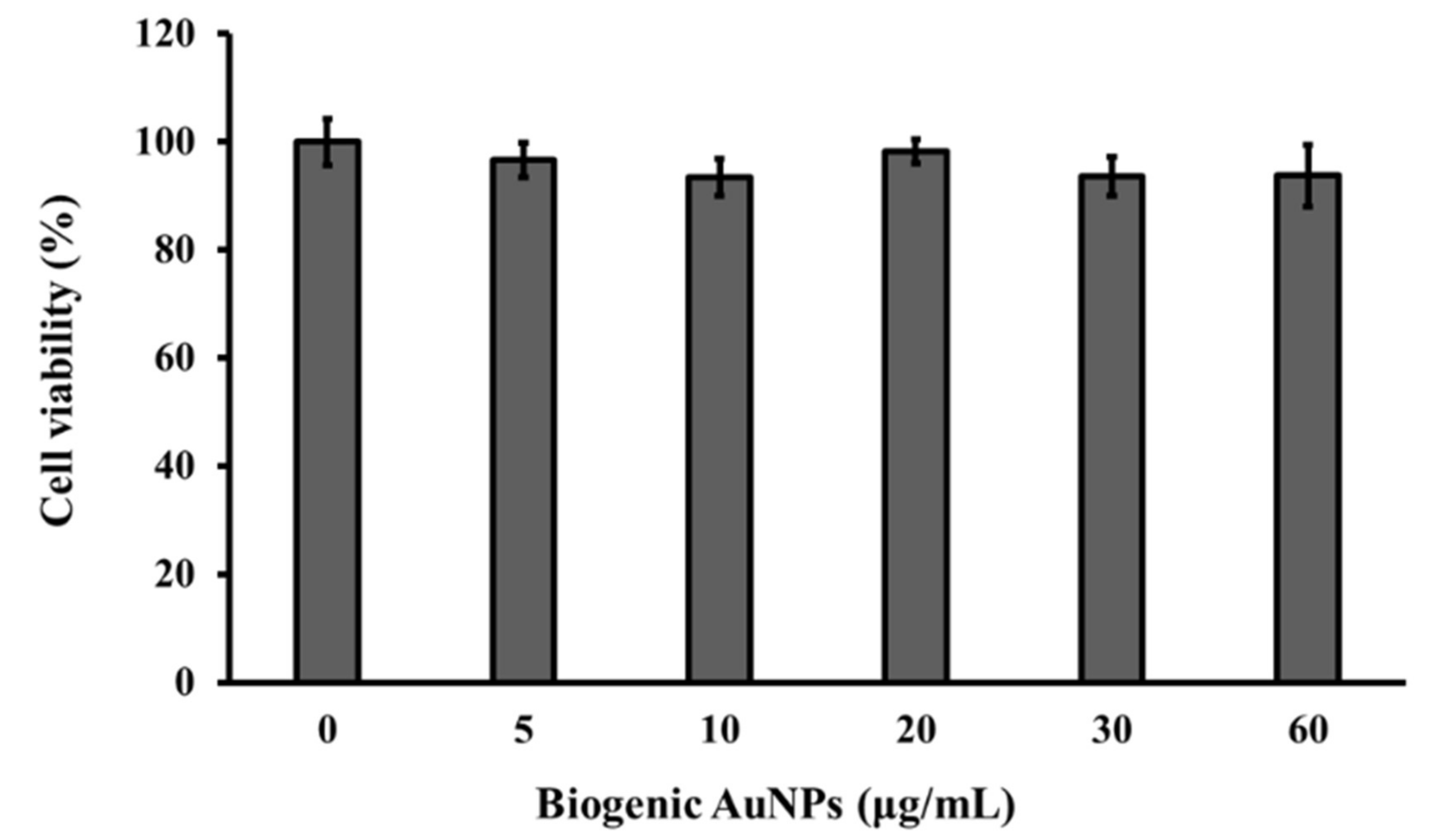
3.9. Cytotoxicity Assays
4. Conclusions
Author Contributions
Funding
Acknowledgments
Conflicts of Interest
References
- Tripathi, R.M.; Shrivastav, B.R.; Shrivastav, A. Antibacterial and catalytic activity of biogenic gold nanoparticles synthesised by Trichoderma harzianum. IET Nanobiotechnol. 2018, 12, 509–513. [Google Scholar] [CrossRef] [PubMed]
- Tripathi, R.; Park, S.H.; Kim, G.; Kim, D.-H.; Ahn, D.; Kim, Y.M.; Kwon, S.J.; Yoon, S.-Y.; Kang, H.J.; Chung, S.J. Metal-induced redshift of optical spectra of gold nanoparticles: An instant, sensitive, and selective visual detection of lead ions. Int. Biodeterior. Biodegrad. 2019, 144, 104740. [Google Scholar] [CrossRef]
- Tripathi, R.; Gupta, R.K.; Singh, P.; Bhadwal, A.S.; Shrivastav, A.; Kumar, N.; Shrivastav, B. Ultra-sensitive detection of mercury (II) ions in water sample using gold nanoparticles synthesized by Trichoderma harzianum and their mechanistic approach. Sens. Actuators B Chem. 2014, 204, 637–646. [Google Scholar] [CrossRef]
- Tripathi, R.; Chung, S.J. Biogenic nanomaterials: Synthesis, characterization, growth mechanism, and biomedical applications. J. Microbiol. Methods 2019, 157, 65–80. [Google Scholar] [CrossRef]
- Mehrotra, N.; Tripathi, R.M. Short interfering RNA therapeutics: Nanocarriers, prospects and limitations. IET Nanobiotechnol. 2015, 9, 386–395. [Google Scholar] [CrossRef]
- Lee, S.Y.; Krishnamurthy, S.; Cho, C.-W.; Yun, Y.-S. Biosynthesis of gold nanoparticles using Ocimum sanctum extracts by solvents with different polarity. ACS Sustain. Chem. Eng. 2016, 4, 2651–2659. [Google Scholar] [CrossRef]
- Tripathi, R.M.; Gupta, R.K.; Bhadwal, A.S.; Singh, P.; Shrivastav, A.; Shrivastav, B. Fungal biomolecules assisted biosynthesis of Au–Ag alloy nanoparticles and evaluation of their catalytic property. IET Nanobiotechnol. 2015, 9, 178–183. [Google Scholar] [CrossRef]
- Tripathi, R.; Kumar, N.; Bhadwal, A.S.; Gupta, R.K.; Shrivastav, B.; Shrivastav, A. Facile and rapid biomimetic approach for synthesis of HAp nanofibers and evaluation of their photocatalytic activity. Mater. Lett. 2015, 140, 64–67. [Google Scholar] [CrossRef]
- Tripathi, R.; Bhadwal, A.S.; Singh, P.; Shrivastav, A.; Singh, M.; Shrivastav, B. Mechanistic aspects of biogenic synthesis of CdS nanoparticles using Bacillus licheniformis. Adv. Nat. Sci. Nanosci. Nanotechnol. 2014, 5, 025006. [Google Scholar] [CrossRef]
- Mahajan, R.; Bhadwal, A.S.; Kumar, N.; Madhusudanan, M.; Pudake, R.N.; Tripathi, R.M. Green synthesis of highly stable carbon nanodots and their photocatalytic performance. IET Nanobiotechnol. 2016, 11, 360–364. [Google Scholar] [CrossRef]
- Agarwal, M.; Bhadwal, A.S.; Kumar, N.; Shrivastav, A.; Shrivastav, B.R.; Singh, M.P.; Zafar, F.; Tripathi, R.M. Catalytic degradation of methylene blue by biosynthesised copper nanoflowers using F. benghalensis leaf extract. IET Nanobiotechnol. 2016, 10, 321–325. [Google Scholar] [CrossRef] [PubMed]
- Tripathi, R.; Rao, R.P.; Tsuzuki, T. Green synthesis of sulfur nanoparticles and evaluation of their catalytic detoxification of hexavalent chromium in water. RSC Adv. 2018, 8, 36345–36352. [Google Scholar] [CrossRef] [Green Version]
- Langille, M.R.; Personick, M.L.; Zhang, J.; Mirkin, C.A. Defining rules for the shape evolution of gold nanoparticles. J. Am. Chem. Soc. 2012, 134, 14542–14554. [Google Scholar] [CrossRef]
- Zhang, J.; Zhao, B.; Meng, L.; Wu, H.; Wang, X.; Li, C. Controlled synthesis of gold nanospheres and single crystals in hydrogel. J. Nanopart. Res. 2007, 9, 1167–1171. [Google Scholar] [CrossRef]
- Omar, R.; Naciri, A.E.; Jradi, S.; Battie, Y.; Toufaily, J.; Mortada, H.; Akil, S. One-step synthesis of a monolayer of monodisperse gold nanocubes for SERS substrates. J. Mater. Chem. C 2017, 5, 10813–10821. [Google Scholar] [CrossRef]
- Alekseeva, A.; Bogatyrev, V.; Khlebtsov, B.; Mel’Nikov, A.; Dykman, L.; Khlebtsov, N. Gold nanorods: Synthesis and optical properties. Colloid J. 2006, 68, 661–678. [Google Scholar] [CrossRef]
- Fytianos, K.; Rodriguez-Lorenzo, L.; Clift, M.J.; Blank, F.; Vanhecke, D.; Von Garnier, C.; Petri-Fink, A.; Rothen-Rutishauser, B. Uptake efficiency of surface modified gold nanoparticles does not correlate with functional changes and cytokine secretion in human dendritic cells in vitro. Nanomed. Nanotechnol. Biol. Med. 2015, 11, 633–644. [Google Scholar] [CrossRef] [Green Version]
- Cho, E.C.; Au, L.; Zhang, Q.; Xia, Y. The effects of size, shape, and surface functional group of gold nanostructures on their adsorption and internalization by cells. Small 2010, 6, 517–522. [Google Scholar] [CrossRef]
- Nambara, K.; Niikura, K.; Mitomo, H.; Ninomiya, T.; Takeuchi, C.; Wei, J.; Matsuo, Y.; Ijiro, K. Reverse size dependences of the cellular uptake of triangular and spherical gold nanoparticles. Langmuir 2016, 32, 12559–12567. [Google Scholar] [CrossRef]
- Nazaruk, J.; Kalemba, D. Chemical Composition of the Essential Oils from the Roots of Erigeron acris L. and Erigeron annuus (L.) Pers. Molecules 2009, 14, 2458–2465. [Google Scholar] [CrossRef] [Green Version]
- Tripathi, R.; Shrivastav, A.; Shrivastav, B. Biofabrication of gold nanoparticles using leaf of Ficus benghalensis and their characterization. Int. J. Pharm. Biol. Sci. 2012, 3, 551–558. [Google Scholar]
- Tripathi, R.M.; Ranac, D.; Shrivastav, A.; Singh, R.P.; Shrivastav, B.R. Biogenic Synthesis of Silver Nanoparticles Using Saraca indica Leaf Extract and Evaluation of Their Antibacterial Activity. Nano Biomed. Eng. 2013, 5, 50–56. [Google Scholar] [CrossRef] [Green Version]
- Li, X.; Yang, M.; Han, Y.-F.; Gao, K. New sesquiterpenes from Erigeron annus. Planta Med. 2005, 71, 268–272. [Google Scholar] [CrossRef] [PubMed]
- Baek, M.; Kim, M.; Cho, H.; Lee, J.; Yu, J.; Chung, H.; Choi, S. Factors influencing the cytotoxicity of zinc oxide nanoparticles: Particle size and surface charge. In Proceedings of Journal of Physics: Conference Series; IOP Publishing: Bristol, UK, 2011; p. 012044. [Google Scholar]
- Cho, Y.-L.; Min, J.-K.; Roh, K.M.; Kim, W.K.; Han, B.S.; Bae, K.-H.; Lee, S.C.; Chung, S.J.; Kang, H.J. Phosphoprotein phosphatase 1CB (PPP1CB), a novel adipogenic activator, promotes 3T3-L1 adipogenesis. Biochem. Biophys. Res. Commun. 2015, 467, 211–217. [Google Scholar] [CrossRef] [PubMed]
- Yeh, Y.-C.; Creran, B.; Rotello, V.M. Gold nanoparticles: Preparation, properties, and applications in bionanotechnology. Nanoscale 2012, 4, 1871–1880. [Google Scholar] [CrossRef]
- Malvern, I. Inform White Paper Dynamic Light Scattering; Malvern Malvern Instruments Ltd.: Malvern, UK, 2011; pp. 1–6. [Google Scholar]
- Mehrotra, N.; Tripathi, R.M.; Zafar, F.; Singh, M.P. Catalytic degradation of dichlorvos using biosynthesized zero valent iron nanoparticles. IEEE Trans. Nanobiosci. 2017, 16, 280–286. [Google Scholar] [CrossRef]
- Nara, M.; Tanokura, M. Infrared spectroscopic study of the metal-coordination structures of calcium-binding proteins. Biochem. Biophys. Res. Commun. 2008, 369, 225–239. [Google Scholar] [CrossRef]
- Shankar, S.S.; Rai, A.; Ahmad, A.; Sastry, M. Rapid synthesis of Au, Ag, and bimetallic Au core–Ag shell nanoparticles using Neem (Azadirachta indica) leaf broth. J. Colloid Interface Sci. 2004, 275, 496–502. [Google Scholar] [CrossRef]
- Shankar, S.S.; Rai, A.; Ankamwar, B.; Singh, A.; Ahmad, A.; Sastry, M. Biological synthesis of triangular gold nanoprisms. Nat. Mater. 2004, 3, 482–488. [Google Scholar] [CrossRef]
- Vijayakumar, S.; Ganesan, S. In vitro cytotoxicity assay on gold nanoparticles with different stabilizing agents. J. Nanomater. 2012, 2012, 14. [Google Scholar] [CrossRef] [Green Version]
- Steckiewicz, K.P.; Barcinska, E.; Malankowska, A.; Zauszkiewicz–Pawlak, A.; Nowaczyk, G.; Zaleska-Medynska, A.; Inkielewicz-Stepniak, I. Impact of gold nanoparticles shape on their cytotoxicity against human osteoblast and osteosarcoma in in vitro model. Evaluation of the safety of use and anti-cancer potential. J. Mater. Sci. Mater. Med. 2019, 30, 22. [Google Scholar] [CrossRef] [Green Version]
- Milanezi, F.G.; Meireles, L.M.; de Christo Scherer, M.M.; de Oliveira, J.P.; da Silva, A.R.; de Araujo, M.L.; Endringer, D.C.; Fronza, M.; Guimarães, M.C.C.; Scherer, R. Antioxidant, antimicrobial and cytotoxic activities of gold nanoparticles capped with quercetin. Saudi Pharm. J. 2019. [Google Scholar] [CrossRef]


| Element | Line Type | K Factor | Absorption Correction | Wt.% | Wt.% Sigma | At.% |
|---|---|---|---|---|---|---|
| Cu | K series | 1.233 | 0.52 | 28.31 | 0.30 | 55.03 |
| Au | L series | 2.265 | 0.52 | 71.69 | 0.30 | 44.97 |
| Total | 100.00 | 100.00 |
© 2019 by the authors. Licensee MDPI, Basel, Switzerland. This article is an open access article distributed under the terms and conditions of the Creative Commons Attribution (CC BY) license (http://creativecommons.org/licenses/by/4.0/).
Share and Cite
Tripathi, R.M.; Yoon, S.-Y.; Ahn, D.; Chung, S.J. Facile Synthesis of Triangular and Hexagonal Anionic Gold Nanoparticles and Evaluation of Their Cytotoxicity. Nanomaterials 2019, 9, 1774. https://doi.org/10.3390/nano9121774
Tripathi RM, Yoon S-Y, Ahn D, Chung SJ. Facile Synthesis of Triangular and Hexagonal Anionic Gold Nanoparticles and Evaluation of Their Cytotoxicity. Nanomaterials. 2019; 9(12):1774. https://doi.org/10.3390/nano9121774
Chicago/Turabian StyleTripathi, R. M., Sun-Young Yoon, Dohee Ahn, and Sang J. Chung. 2019. "Facile Synthesis of Triangular and Hexagonal Anionic Gold Nanoparticles and Evaluation of Their Cytotoxicity" Nanomaterials 9, no. 12: 1774. https://doi.org/10.3390/nano9121774