


Controlling the Surface Morphology of Two-Dimensional Nano-Materials upon Molecule-Mediated Crystal Growth
 , , , , and
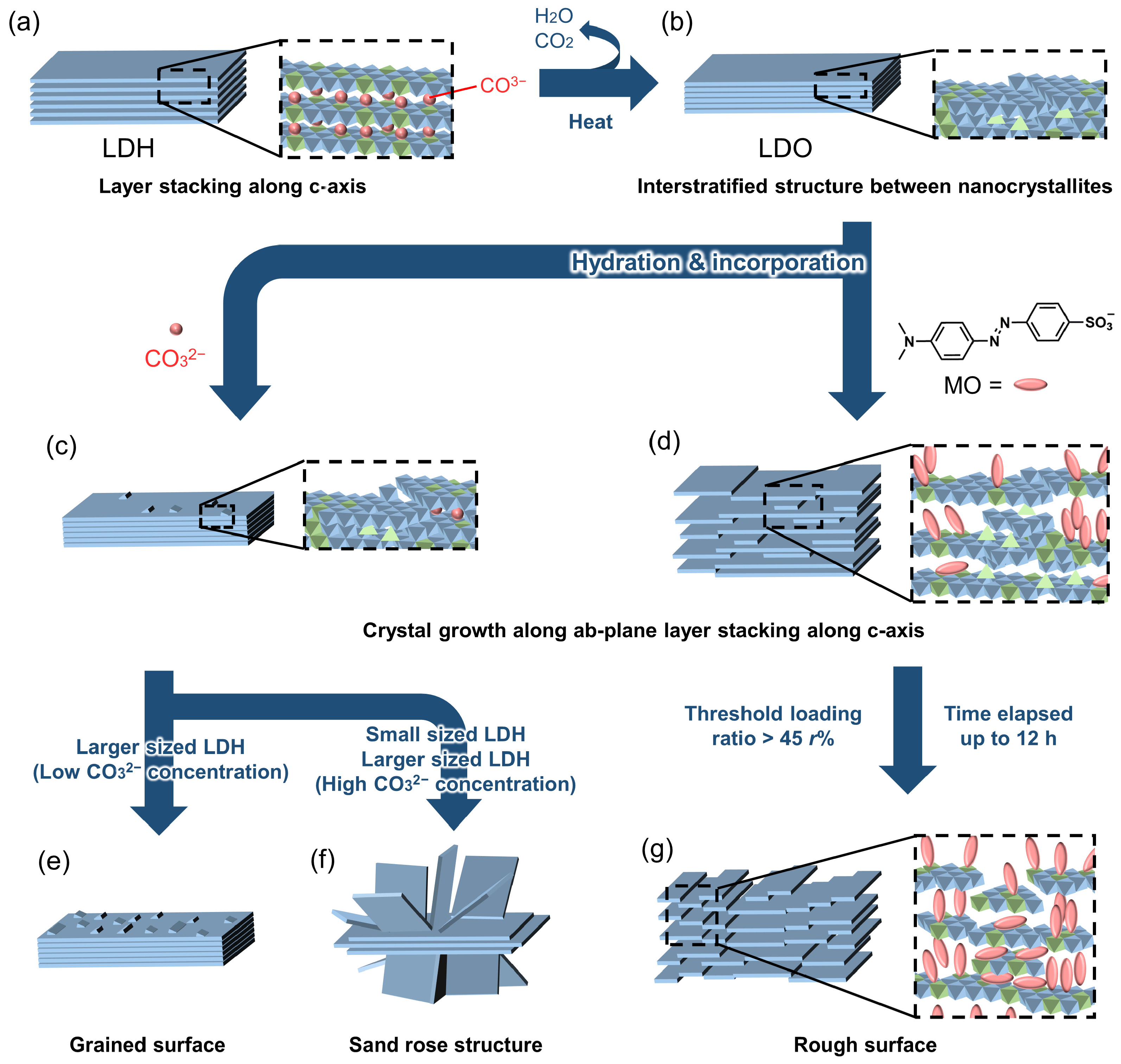
Abstract
:1. Introduction
2. Materials and Methods
2.1. Materials
2.2. Synthesis
2.2.1. MgAl-LDH with a Diameter of 2000 nm (LDH2000) [44,51]
2.2.2. MgAl-LDH with a Diameter of 350 nm (LDH350)
2.2.3. Calcination of LDH2000 and LDH350 (LDO2000 and LDO350)
2.2.4. Reconstruction of the LDHs with CO32−
2.2.5. Reconstruction of the LDHs with Various MO Concentrations and Reaction Times
2.3. Characterization
3. Results
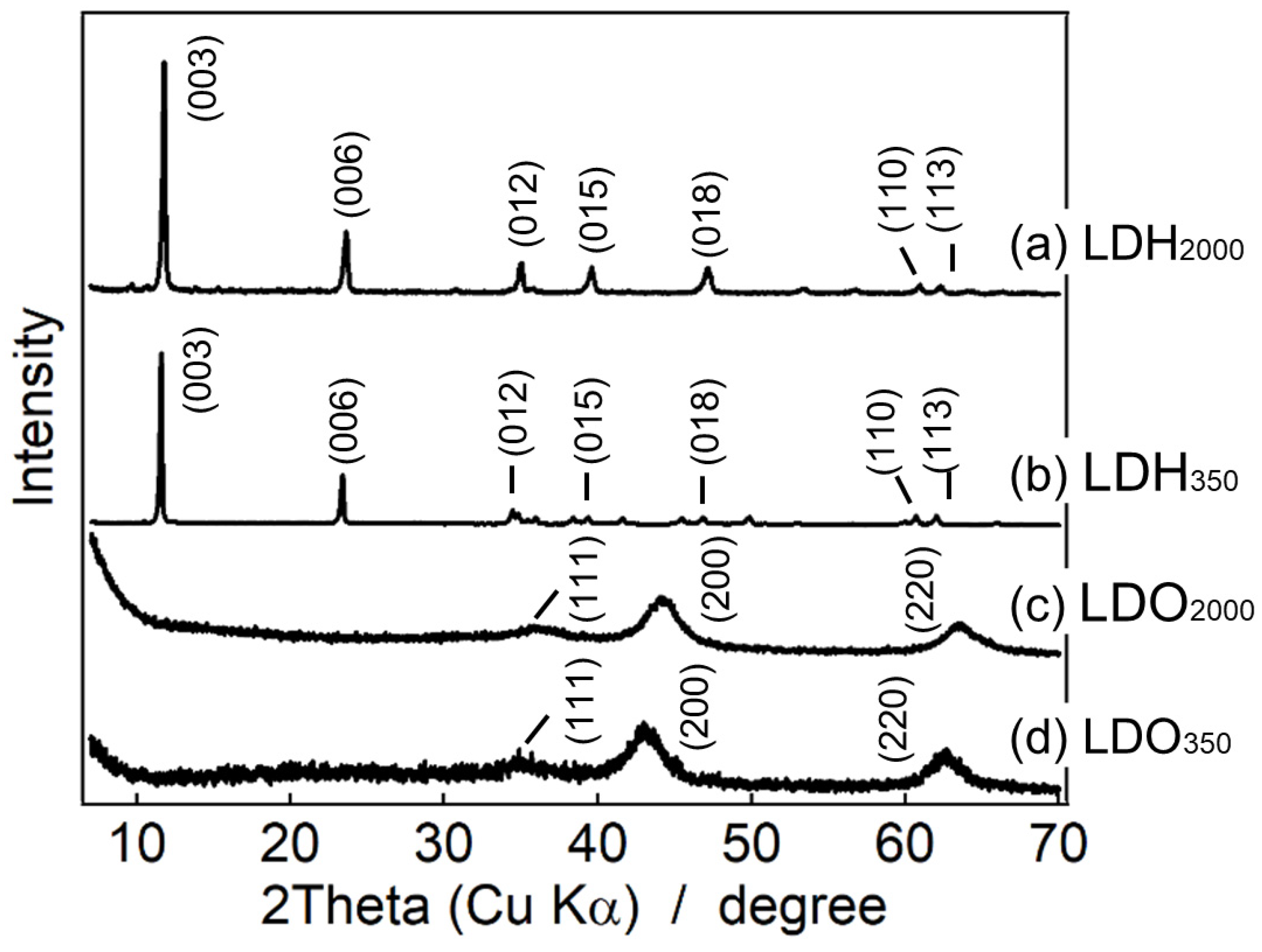
3.1. XRD Patterns of the LDHs and the LDOs
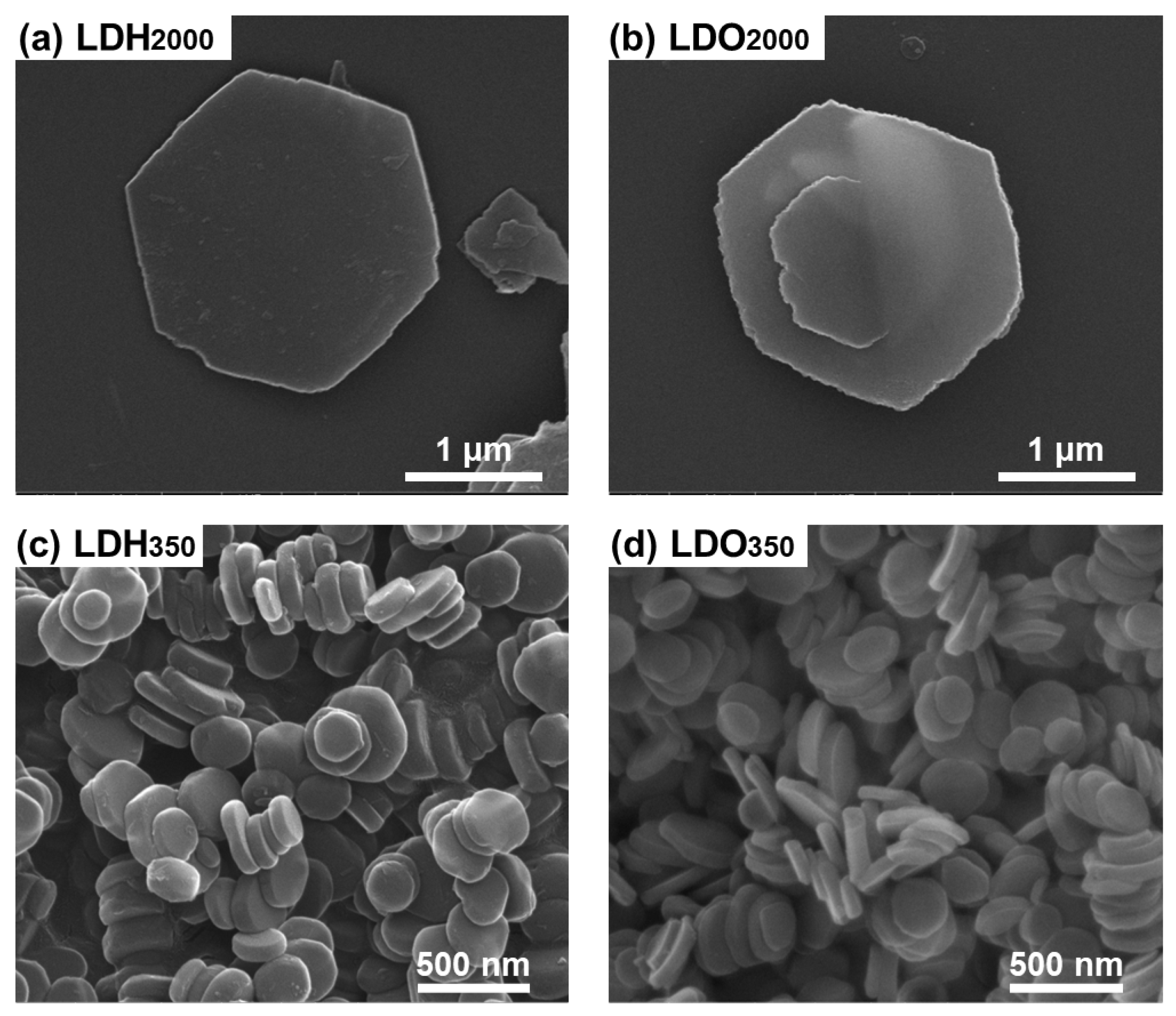
3.2. SEM Images of the LDHs and the LDOs
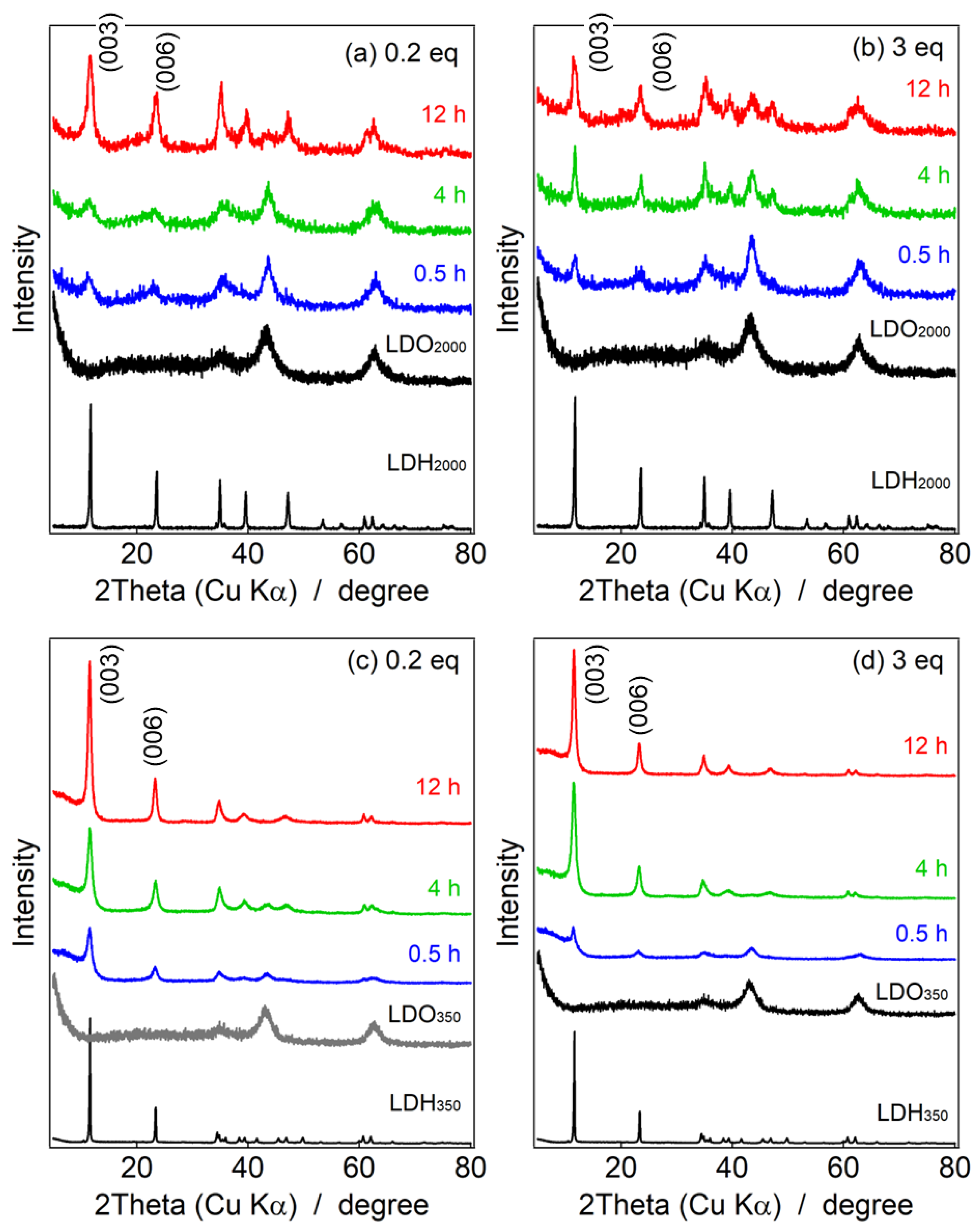
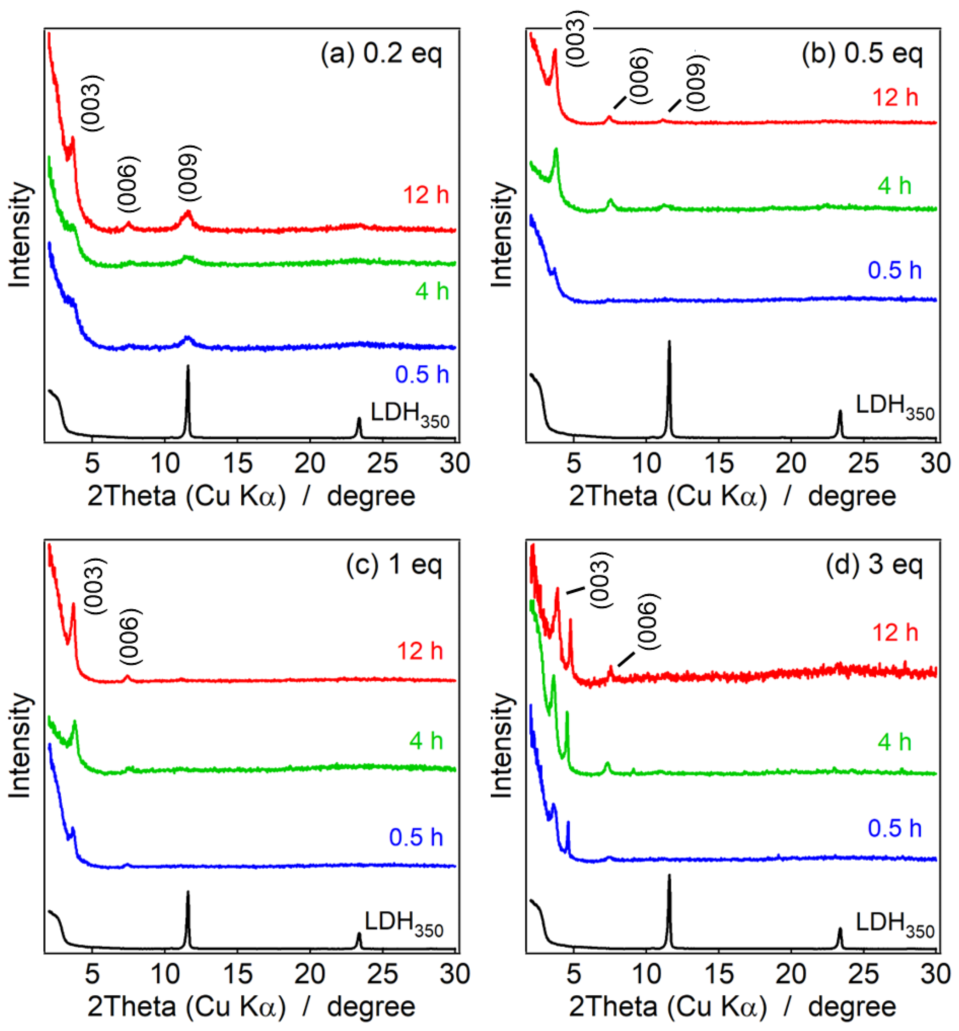
3.3. XRD Pattern of the Reconstructed LDHs
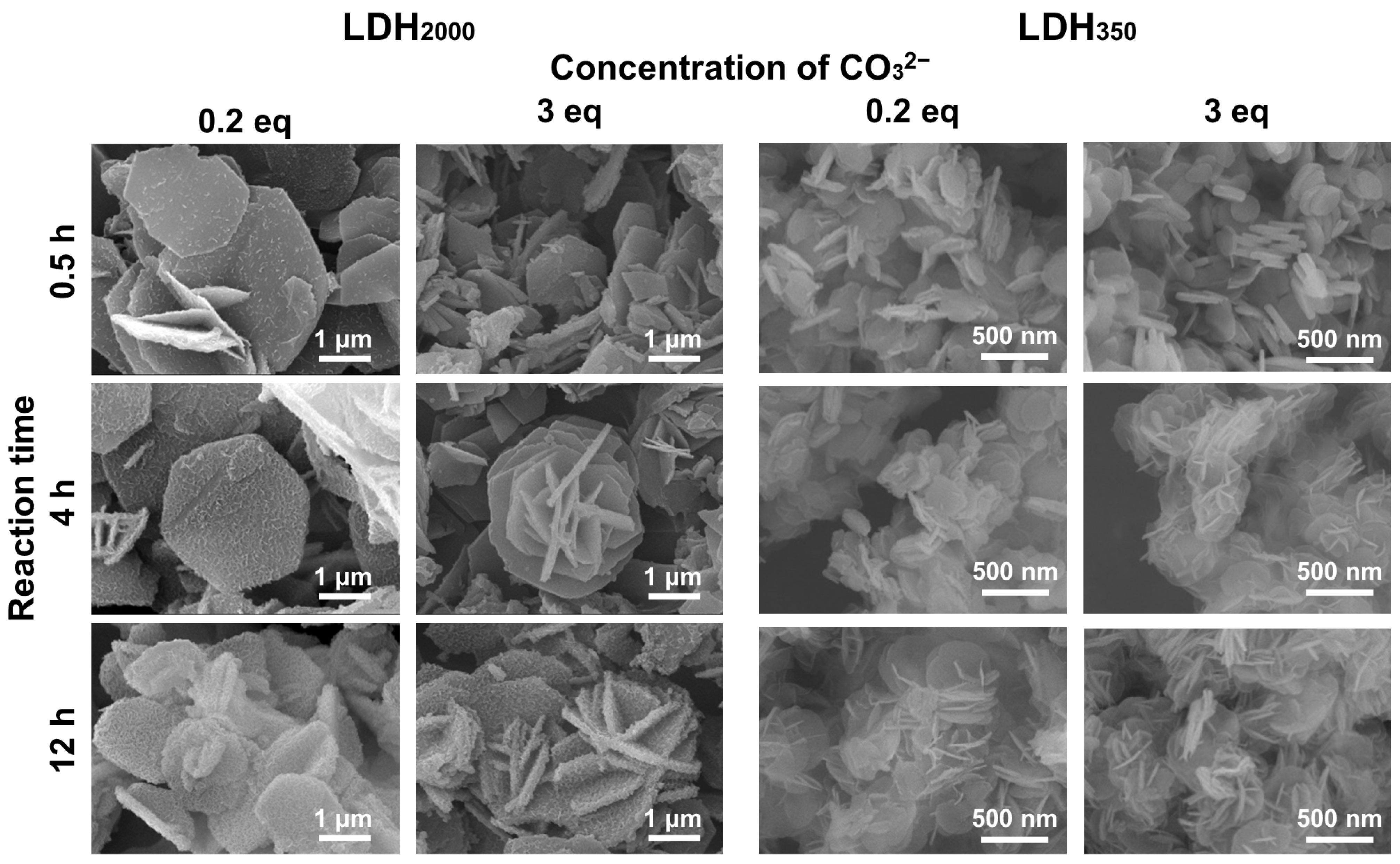
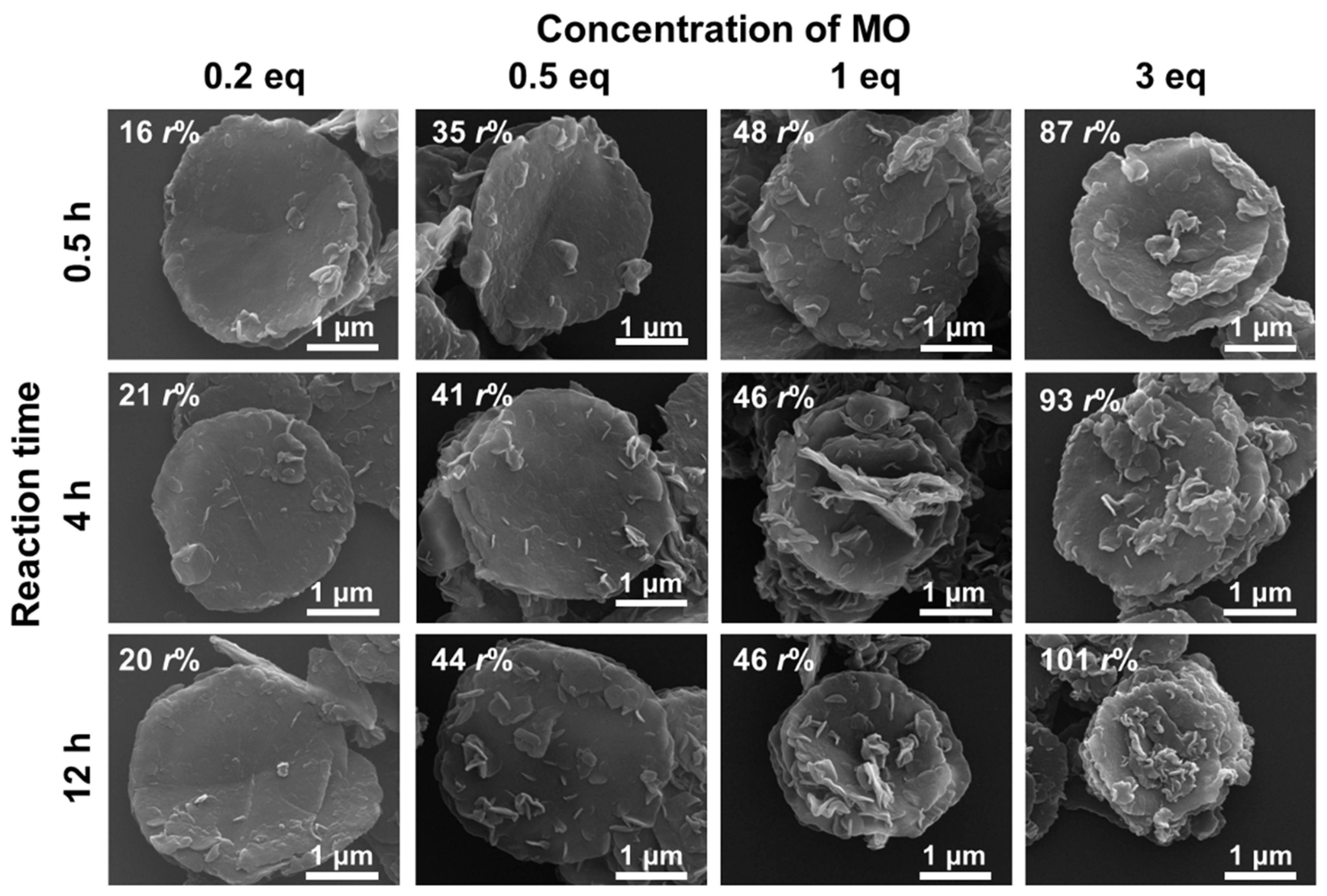
3.4. SEM Images of the Reconstructed LDHs
4. Conclusions
Supplementary Materials
Author Contributions
Funding
Data Availability Statement
Conflicts of Interest
References
- Clapham, P.B.; Huteley, M.C. Reduction of Lens Reflexion by the “Moth Eye” Principle. Nature 1973, 244, 281–282. [Google Scholar] [CrossRef]
- Wilson, S.J.; Hutley, M.C. The Optical Properties of “Moth Eye” Antireflection Surfaces. Opt. Acta 1982, 29, 993–1009. [Google Scholar] [CrossRef]
- Alimanesh, M.; Rouhi, J.; Hassan, Z. Broadband Anti-Reflective Properties of Grown ZnO Nanopyramidal Structure on Si Substrate via Low-Temperature Electrochemical Deposition. Ceram. Int. 2016, 42, 5136–5140. [Google Scholar] [CrossRef]
- Zheng, Y.; Zhou, Z.; Cao, W.; Li, C.; Wang, Q.; Huang, Y.; Shen, S. Robust, Low Haze and Graded Porous Anti-Reflective Glass by Tailoring the Nanostructures. Ceram. Int. 2020, 46, 18623–18631. [Google Scholar] [CrossRef]
- Kang, S.M.; Jang, S.; Lee, J.K.; Yoon, J.; Yoo, D.E.; Lee, J.W.; Choi, M.; Park, N.G. Moth-Eye TiO2 Layer for Improving Light Harvesting Efficiency in Perovskite Solar Cells. Small 2016, 12, 2443–2449. [Google Scholar] [CrossRef] [PubMed]
- Liu, E.; Hu, Y.; Li, H.; Tang, C.; Hu, X.; Fan, J.; Chen, Y.; Bian, J. Photoconversion of CO2 to Methanol over Plasmonic Ag/TiO2 Nano-Wire Films Enhanced by Overlapped Visible-Light-Harvesting Nanostructures. Ceram. Int. 2015, 41, 1049–1057. [Google Scholar] [CrossRef]
- Sreedhar, A.; Sreekanth, T.V.M.; Kwon, J.H.; Yi, J.; Sohn, Y.; Gwag, J.S. Ag Nanoparticles Decorated Ion-Beam-Assisted TiO2 Thin Films for Tuning the Water Splitting Activity from UV to Visible Light Harvesting. Ceram. Int. 2017, 43, 12814–12821. [Google Scholar] [CrossRef]
- Barthlott, W.; Neinhuis, C. Purity of the Sacred Lotus, or Escape from Contamination in Biological Surfaces. Planta 1997, 202, 1–8. [Google Scholar] [CrossRef]
- Feng, L.; Zhang, Y.; Xi, J.; Zhu, Y.; Wang, N.; Xia, F.; Jiang, L. Petal Effect: A Superhydrophobic State with High Adhesive Force. Langmuir 2008, 24, 4114–4119. [Google Scholar] [CrossRef]
- Uchida, K.; Nishikawa, N.; Izumi, N.; Yamazoe, S.; Mayama, H.; Kojima, Y.; Yokojima, S.; Nakamura, S.; Tsujii, K.; Irie, M. Phototunable Diarylethene Microcrystalline Surfaces: Lotus and Petal Effects upon Wetting. Angew. Chem. Int. Ed. 2010, 49, 5942–5944. [Google Scholar] [CrossRef]
- Yu, X.; Liu, X.; Shi, X.; Zhang, Z.; Wang, H.; Feng, L. SiO2 Nanoparticle-Based Superhydrophobic Spray and Multi-Functional Surfaces by a Facile and Scalable Method. Ceram. Int. 2019, 45, 15741–15744. [Google Scholar] [CrossRef]
- Zang, D.; Xun, X.; Gu, Z.; Dong, J.; Pan, T.; Liu, M. Fabrication of Superhydrophobic Self-Cleaning Manganese Dioxide Coatings on Mg Alloys Inspired by Lotus Flower. Ceram. Int. 2020, 46, 20328–20334. [Google Scholar] [CrossRef]
- Mizuno, N.; Misono, M. Heterogeneous Catalysis. Chem. Rev. 1998, 98, 199–217. [Google Scholar] [CrossRef] [PubMed]
- Liu, J.; Zhang, G. Recent Advances in Synthesis and Applications of Clay-Based Photocatalysts: A Review. Phys. Chem. Chem. Phys. 2014, 16, 8178–8192. [Google Scholar] [CrossRef] [PubMed]
- Schrade, A.; Mailänder, V.; Ritz, S.; Landfester, K.; Ziener, U. Surface Roughness and Charge Influence the Uptake of Nanoparticles: Fluorescently Labeled Pickering-Type Versus Surfactant-Stabilized Nanoparticles. Macromol. Biosci. 2012, 12, 1459–1471. [Google Scholar] [CrossRef]
- Niu, Y.; Yu, M.; Hartono, S.B.; Yang, J.; Xu, H.; Zhang, H.; Zhang, J.; Zou, J.; Dexter, A.; Gu, W.; et al. Nanoparticles Mimicking Viral Surface Topography for Enhanced Cellular Delivery. Adv. Mater. 2013, 25, 6233–6237. [Google Scholar] [CrossRef]
- Choy, J.H.; Choi, S.J.; Oh, J.M.; Park, T. Clay Minerals and Layered Double Hydroxides for Novel Biological Applications. Appl. Clay Sci. 2007, 36, 122–132. [Google Scholar] [CrossRef]
- Oh, J.M.; Biswick, T.T.; Choy, J.H. Layered Nanomaterials for Green Materials. J. Mater. Chem. 2009, 19, 2553–2563. [Google Scholar] [CrossRef]
- Yamaguchi, T.; Kim, H.; Jung, C.B.; Kim, S.Y.; Oh, J. Size and Surface Charge Effect of Layered Double Hydroxide Particles upon Blood Cells. Appl. Clay Sci. 2022, 225, 106549. [Google Scholar] [CrossRef]
- Yamaguchi, T.; Kim, H.M.; Oh, J.M. Photochemical Consideration in the Interactions between Blood Proteins and Layered Inorganic Materials. Int. J. Mol. Sci. 2022, 23, 11367. [Google Scholar] [CrossRef]
- Kuramoto, K.; Intasa-Ard, S.G.; Bureekaew, S.; Ogawa, M. Mechanochemical Synthesis of Finite Particle of Layered Double Hydroxide-Acetate Intercalation Compound: Swelling, Thin Film and Ion Exchange. J. Solid State Chem. 2017, 253, 147–153. [Google Scholar] [CrossRef]
- Wijitwongwan, R.P.; Intasa-ard, S.G.; Ogawa, M. Preparation of Layered Double Hydroxides toward Precisely Designed Hierarchical Organization. ChemEngineering 2019, 3, 68. [Google Scholar] [CrossRef]
- Ogawa, M.; Inomata, K. Preparation of Layered Double Hydroxides. Clay Sci. 2011, 15, 131–137. [Google Scholar]
- Kim, T.H.; Hong, I.T.; Oh, J.M. Size- and Surface Charge-Controlled Layered Double Hydroxides for Efficient Algal Flocculation. Environ. Sci. Nano 2018, 5, 183–190. [Google Scholar] [CrossRef]
- Sikander, U.; Sufian, S.; Salam, M.A. A Review of Hydrotalcite Based Catalysts for Hydrogen Production Systems. Int. J. Hydrogen Energy 2017, 42, 19851–19868. [Google Scholar] [CrossRef]
- Mishra, G.; Dash, B.; Pandey, S. Layered Double Hydroxides: A Brief Review from Fundamentals to Application as Evolving Biomaterials. Appl. Clay Sci. 2018, 153, 172–186. [Google Scholar] [CrossRef]
- Jeong, I.R.; Lee, J.H.; Song, J.; Oh, Y.S.; Cho, S. Control of Structural Disorder in Spinel Ceramics Derived from Layered Double Hydroxides. Ceram. Int. 2020, 46, 6594–6599. [Google Scholar] [CrossRef]
- El Hassani, K.; Bayram, O.; Anouar, A.; Mavi, A. Thin Films Derived from Zn(Al)O Mixed Metal Oxides Nanoparticles Dispersed in Polyethylene Glycol: Structural, Morphological and Optical Properties. Ceram. Int. 2020, 46, 14733–14738. [Google Scholar] [CrossRef]
- Xie, J.; Yamaguchi, T.; Oh, J.M. Synthesis of a Mesoporous Mg–Al–Mixed Metal Oxide with P123 Template for Effective Removal of Congo Red via Aggregation-Driven Adsorption. J. Solid State Chem. 2021, 293, 121758. [Google Scholar] [CrossRef]
- Xiao, T.; Tang, Y.; Jia, Z.; Li, D.; Hu, X.; Li, B.; Luo, L. Self-Assembled 3D Flower-like Ni2+-Fe3+ Layered Double Hydroxides and Their Calcined Products. Nanotechnology 2009, 20, 475603. [Google Scholar] [CrossRef]
- Han, J.; Dou, Y.; Wei, M.; Evans, D.G.; Duan, X. Erasable Nanoporous Antireflection Coatings Based on the Reconstruction Effect of Layered Double Hydroxides. Angew. Chem. Int. Ed. 2010, 49, 2171–2174. [Google Scholar] [CrossRef] [PubMed]
- Chen, T.; Xu, S.; Zhang, F.; Evans, D.G.; Duan, X. Formation of Photo- and Thermo-Stable Layered Double Hydroxide Films with Photo-Responsive Wettability by Intercalation of Functionalized Azobenzenes. Chem. Eng. Sci. 2009, 64, 4350–4357. [Google Scholar] [CrossRef]
- El Hassani, K.; Beakou, B.H.; Kalnina, D.; Oukani, E.; Anouar, A. Effect of Morphological Properties of Layered Double Hydroxides on Adsorption of Azo Dye Methyl Orange: A Comparative Study. Appl. Clay Sci. 2017, 140, 124–131. [Google Scholar] [CrossRef]
- Abderrazek, K.; Uheida, A.; Seffen, M.; Muhammed, M.; Frini Srasra, N.; Srasra, E. Photocatalytic Degradation of Indigo Carmine Using [Zn-Al] LDH Supported on PAN Nanofibres. Clay Miner. 2015, 50, 185–197. [Google Scholar] [CrossRef]
- Gu, Z.; Liu, W.; Dou, W.; Tang, F. Preparation of a Novel Heat Stabilizer for Poly(Vinyl Chloride)-Zn, Mg, Al-Layered Double Hydroxide. Polym. Compos. 2009, 31, 928–932. [Google Scholar] [CrossRef]
- Zeng, S.; Xu, X.; Wang, S.; Gong, Q.; Liu, R.; Yu, Y. Sand Flower Layered Double Hydroxides Synthesized by Co-Precipitation for CO2 Capture: Morphology Evolution Mechanism, Agitation Effect and Stability. Mater. Chem. Phys. 2013, 140, 159–167. [Google Scholar] [CrossRef]
- Zhang, P.; Qian, G.; Shi, H.; Ruan, X.; Yang, J.; Frost, R.L. Mechanism of Interaction of Hydrocalumites (Ca/Al-LDH) with Methyl Orange and Acidic Scarlet GR. J. Colloid Interface Sci. 2012, 365, 110–116. [Google Scholar] [CrossRef]
- Hibino, T.; Tsunashima, A. Characterization of Repeatedly Reconstructed Mg-Al Hydrotalcite-Like Compounds: Gradual Segregation of Aluminum from the Structure. Chem. Mater. 1998, 10, 4055–4061. [Google Scholar] [CrossRef]
- Pérez-Ramírez, J.; Abelló, S.; Van Der Pers, N.M. Memory Effect of Activated Mg-Al Hydrotalcite: In Situ XRD Studies during Decomposition and Gas-Phase Reconstruction. Chem. Eur. J. 2007, 13, 870–878. [Google Scholar] [CrossRef]
- Kowalik, P.; Konkol, M.; Kondracka, M.; Próchniak, W.; Bicki, R.; Wiercioch, P. Memory Effect of the CuZnAl-LDH Derived Catalyst Precursor—In Situ XRD Studies. Appl. Catal. A Gen. 2013, 464–465, 339–347. [Google Scholar] [CrossRef]
- Benselka-Hadj Abdelkader, N.; Bentouami, A.; Derriche, Z.; Bettahar, N.; de Ménorval, L.C. Synthesis and Characterization of Mg-Fe Layer Double Hydroxides and Its Application on Adsorption of Orange G from Aqueous Solution. Chem. Eng. J. 2011, 169, 231–238. [Google Scholar] [CrossRef]
- Extremera, R.; Pavlovic, I.; Pérez, M.R.; Barriga, C. Removal of Acid Orange 10 by Calcined Mg/Al Layered Double Hydroxides from Water and Recovery of the Adsorbed Dye. Chem. Eng. J. 2012, 213, 392–400. [Google Scholar] [CrossRef]
- Santos, R.M.M.; Tronto, J.; Briois, V.; Santilli, C.V. Thermal Decomposition and Recovery Properties of ZnAl-CO3 Layered Double Hydroxide for Anionic Dye Adsorption: Insight into the Aggregative Nucleation and Growth Mechanism of the LDH Memory Effect. J. Mater. Chem. A 2017, 5, 9998–10009. [Google Scholar] [CrossRef]
- Kim, B.K.; Gwak, G.H.; Okada, T.; Oh, J.M. Effect of Particle Size and Local Disorder on Specific Surface Area of Layered Double Hydroxides upon Calcination-Reconstruction. J. Solid State Chem. 2018, 263, 60–64. [Google Scholar] [CrossRef]
- De Martínez-Ortiz, M.J.; Lima, E.; Lara, V.; Vivar, J.M. Structural and Textural Evolution during Folding of Layers of Layered Double Hydroxides. Langmuir 2008, 24, 8904–8911. [Google Scholar] [CrossRef]
- Santos, R.M.M.; Gonçalves, R.G.L.; Constantino, V.R.L.; Santilli, C.V.; Borges, P.D.; Tronto, J.; Pinto, F.G. Adsorption of Acid Yellow 42 Dye on Calcined Layered Double Hydroxide: Effect of Time, Concentration, PH and Temperature. Appl. Clay Sci. 2017, 140, 132–139. [Google Scholar] [CrossRef]
- Yao, W.; Yu, S.; Wang, J.; Zou, Y.; Lu, S.; Ai, Y.; Alharbi, N.S.; Alsaedi, A.; Hayat, T.; Wang, X. Enhanced Removal of Methyl Orange on Calcined Glycerol-Modified Nanocrystallined Mg/Al Layered Double Hydroxides. Chem. Eng. J. 2017, 307, 476–486. [Google Scholar] [CrossRef]
- Darmograi, G.; Prelot, B.; Layrac, G.; Tichit, D.; Martin-Gassin, G.; Salles, F.; Zajac, J. Study of Adsorption and Intercalation of Orange-Type Dyes into Mg-Al Layered Double Hydroxide. J. Phys. Chem. C 2015, 119, 23388–23397. [Google Scholar] [CrossRef]
- Ko, S.-J.; Yamaguchi, T.; Salles, F.; Oh, J.-M. Systematic Utilization of Layered Double Hydroxide Nanosheets for Effective Removal of Methyl Orange from an Aqueous System by π-π Stacking-Induced Nanoconfinement. J. Environ. Manag. 2021, 277, 111455. [Google Scholar] [CrossRef]
- Sasai, R.; Sato, H.; Sugata, M.; Fujimura, T.; Ishihara, S.; Deguchi, K.; Ohki, S.; Tansho, M.; Shimizu, T.; Oita, N.; et al. Why Do Carbonate Anions Have Extremely High Stability in the Interlayer Space of Layered Double Hydroxides? Case Study of Layered Double Hydroxide Consisting of Mg and Al (Mg/Al = 2). Inorg. Chem. 2019, 58, 10928–10935. [Google Scholar] [CrossRef]
- Costantino, U.; Marmottini, F.; Nocchetti, M.; Vivani, R. New Synthetic Routes to Hydrotalcite-Like Compounds—Characterisation and Properties of the Obtained Materials. Eur. J. Inorg. Chem. 1998, 1998, 1439–1446. [Google Scholar] [CrossRef]
- Oh, J.M.; Hwang, S.H.; Choy, J.H. The Effect of Synthetic Conditions on Tailoring the Size of Hydrotalcite Particles. Solid State Ionics 2002, 151, 285–291. [Google Scholar] [CrossRef]
- Lee, J.Y.; Gwak, G.H.; Kim, H.M.; Kim, T.I.; Lee, G.J.; Oh, J.M. Synthesis of Hydrotalcite Type Layered Double Hydroxide with Various Mg/Al Ratio and Surface Charge under Controlled Reaction Condition. Appl. Clay Sci. 2016, 134, 44–49. [Google Scholar] [CrossRef]
- Rives, V. (Ed.) Layered Double Hydroxides: Present and Future, 1st ed.; Nova Science Publishers, Inc.: New York, NY, USA, 2001. [Google Scholar]
- Radha, S.; Navrotsky, A. Energetics of CO2 Adsorption on Mg-Al Layered Double Hydroxides and Related Mixed Metal Oxides. J. Phys. Chem. C 2014, 118, 29836–29844. [Google Scholar] [CrossRef]
- Bîrjega, R.; Pavel, O.D.; Costentin, G.; Che, M.; Angelescu, E. Rare-Earth Elements Modified Hydrotalcites and Corresponding Mesoporous Mixed Oxides as Basic Solid Catalysts. Appl. Catal. A Gen. 2005, 288, 185–193. [Google Scholar] [CrossRef]
- Yao, W.; Wang, X.; Liang, Y.; Yu, S.; Gu, P.; Sun, Y.; Xu, C.; Chen, J.; Hayat, T.; Alsaedi, A.; et al. Synthesis of Novel Flower-like Layered Double Oxides/Carbon Dots Nanocomposites for U(VI) and 241Am(III) Efficient Removal: Batch and EXAFS Studies. Chem. Eng. J. 2018, 332, 775–786. [Google Scholar] [CrossRef]
- Lei, C.; Zhu, X.; Zhu, B.; Jiang, C.; Le, Y.; Yu, J. Superb Adsorption Capacity of Hierarchical Calcined Ni/Mg/Al Layered Double Hydroxides for Congo Red and Cr(VI) Ions. J. Hazard. Mater. 2017, 321, 801–811. [Google Scholar] [CrossRef]
- Mokhtar, M.; Inayat, A.; Ofili, J.; Schwieger, W. Thermal Decomposition, Gas Phase Hydration and Liquid Phase Reconstruction in the System Mg/Al Hydrotalcite/Mixed Oxide: A Comparative Study. Appl. Clay Sci. 2010, 50, 176–181. [Google Scholar] [CrossRef]


Disclaimer/Publisher’s Note: The statements, opinions and data contained in all publications are solely those of the individual author(s) and contributor(s) and not of MDPI and/or the editor(s). MDPI and/or the editor(s) disclaim responsibility for any injury to people or property resulting from any ideas, methods, instructions or products referred to in the content. |
© 2023 by the authors. Licensee MDPI, Basel, Switzerland. This article is an open access article distributed under the terms and conditions of the Creative Commons Attribution (CC BY) license (https://creativecommons.org/licenses/by/4.0/).
Share and Cite
Yamaguchi, T.; Kim, H.-J.; Park, H.J.; Kim, T.; Khalid, Z.; Park, J.K.; Oh, J.-M. Controlling the Surface Morphology of Two-Dimensional Nano-Materials upon Molecule-Mediated Crystal Growth. Nanomaterials 2023, 13, 2363. https://doi.org/10.3390/nano13162363
Yamaguchi T, Kim H-J, Park HJ, Kim T, Khalid Z, Park JK, Oh J-M. Controlling the Surface Morphology of Two-Dimensional Nano-Materials upon Molecule-Mediated Crystal Growth. Nanomaterials. 2023; 13(16):2363. https://doi.org/10.3390/nano13162363
Chicago/Turabian StyleYamaguchi, Tetsuo, Hyoung-Jun Kim, Hee Jung Park, Taeho Kim, Zubair Khalid, Jin Kuen Park, and Jae-Min Oh. 2023. "Controlling the Surface Morphology of Two-Dimensional Nano-Materials upon Molecule-Mediated Crystal Growth" Nanomaterials 13, no. 16: 2363. https://doi.org/10.3390/nano13162363