
Medical and Dental Applications of Titania Nanoparticles: An Overview


 , , ,
, , , 
Abstract
:1. Introduction
2. Background of Nanotechnology
2.1. History of Nanotechnology
2.2. Significant Features of Nanotechnology
- This technology can bring changes in the original matter’s structure by converting it into a nanoscale product.
- The newly formed nanoscale structure should contain periodic repetition (i.e., the nanoparticle must periodically repeat itself in one or more than one direction) just like the parent matter.
- The novel properties, characteristics, and functions of a newly formed nanoscale product should be like those of the parent matter or be superior to the parent matter despite being nanometric.
2.3. Classification of NPs
3. The Titanium Oxide NPs
3.1. Basic Requirements for Employing TiO2 NPs in Medical and Dental Applications
3.1.1. Synthesis Protocols of TiO2 NPs
Conventional (Top-Down) Synthesis
Biological (Bottom-Up) Synthesis
Conventional and Biological Synthesis
- (i)
- Solution’s medium pH:
- (ii)
- Pressure:
- (iii)
- Time:
- (iv)
- Melting point:
- (v)
- Environment:
3.2. Characterization Techniques of TiO2 NPs
3.2.1. Scanning Electron Microscopy (SEM) and Transmission Electron Microscopy (TEM) for Size and Shape
3.2.2. X-ray Diffraction Powder Spectroscopy (XRD) for Crystalline Phases
3.2.3. UV–Vis Diffuse Reflectance Spectroscopy
3.2.4. Fourier Transform Infrared Spectroscopy (FTIR)
3.2.5. Elemental Composition Analysis (EDX)
3.2.6. Atomic Force Microscopy Analysis (AFM)
3.2.7. Characteristic Features of TiO2 NPs Dependent on Characterization Techniques
Role of Size and Shape of TiO2 NPs on Their Medical and Dental Applications
Role of Surface Area of TiO2 NPs on Their Medical and Dental Applications
Role of Crystalline Phases of TiO2 NPs on Their Medical and Dental Applications
Role of Dose of TiO2 NPs on Their Medical and Dental Applications
Role of Surface Coating of TiO2 NPs on Their Medical and Dental Applications
3.3. Significance of TiO2 NPs in Medical and Dental Platform
3.3.1. Significance of TiO2 NPs as a Medical Material
Use of TiO2-Based Drug Delivery Systems in Cancer Therapy
Prevention and Treatment of Infections through TiO2 Based Antibacterial Devices
Antibacterial Action of TiO2 in Orthopedic Implants
Antibacterial Applications of TiO2 in Hospitals and Medical Devices
TiO2 Implants Capable of Releasing Drugs
The TiO2-Based Biosensors
The TiO2 NPs in Sunscreens
The TiO2 NPs in Anticoagulants and Wound Dressings
3.3.2. Significance of TiO2 NPs as a Dental Biomaterial
TiO2 NPs in Orthodontics
The TiO2 NPs in Dental Implants
TiO2 NPs in Oral Disinfectants and Mouth Washes
TiO2 NPs in Restorative Materials
The TiO2 NPs in Whitening Agents
The (TiO2) NPs in Acrylic Resins
The TiO2 NPs in Oral Dentifrices
The TiO2 NPs in Dental Adhesives
The TiO2 NPs in Dental Prosthesis (Veneers, Crowns, and Bridges)
The TiO2 NPs in Scaffolds/Bone Grafting of Maxillofacial Regions
The TiO2 NPs in Endodontics
3.4. The TiO2 NPs: The Most Biocompatible Material for Medical and Dental Applications
4. Conclusions and Futures Prospects
Author Contributions
Funding
Data Availability Statement
Conflicts of Interest
References
- Ahmadi, E.D.; Hafeji, S.; Khurshid, Z.; Imran, E.; Zafar, M.S.; Saeinasab, M.; Sefat, F. Biophotonics in Dentistry. Appl. Sci. 2022, 12, 4254. [Google Scholar] [CrossRef]
- Hayat, F.; Sabbir, J.; Khurshid, Z.; Zafar, M.S.; Ghabbani, H.M.; Shahbazi, M.-A.; Sefat, F. Nanoparticles in endodontics. In Biomaterials in Endodontics; Woodhead Publishers: Shaston, UK, 2022; pp. 195–209. [Google Scholar] [CrossRef]
- Khurshid, Z.; Zafar, M.; Qasim, S.; Shahab, S.; Naseem, M.; AbuReqaiba, A. Advances in Nanotechnology for Restorative Dentistry. Materials 2015, 8, 717–731. [Google Scholar] [CrossRef] [PubMed]
- Zafar, M.S.; Khurshid, Z.; Najeeb, S.; Zohaib, S.; Rehman, I.U. Therapeutic applications of nanotechnology in dentistry. Nanostruct. Oral Med. 2017, 17, 833–862. [Google Scholar] [CrossRef]
- Najeeb, S.; Khurshid, Z.; Ghabbani, H.; Zafar, M.S.; Sefat, F. Nano glass ionomer cement: Modification for biodental applications. In Advanced Dental Biomaterials; Woodhead Publishers: Shaston, UK, 2019; pp. 217–227. ISBN 9780081024768. [Google Scholar]
- Sagadevan, S.; Imteyaz, S.; Murugan, B.; Anita, L.J.; Sridewi, N.; Weldegebrieal, G.; Fatimah, I.; Oh, W.A. Comprehensive review on green synthesis of titanium dioxide nanoparticles and their diverse biomedical applications. Green Process. Synth. 2022, 11, 44–63. [Google Scholar] [CrossRef]
- American Conference of Governmental Industrial Hygienists (ACGIH). Threshold Limit Values and Biological Exposure Indices for 1992; American Conference of Governmental Industrial Hygienists: Cincinnati, OH, USA, 1992. [Google Scholar]
- ILSI Risk Science Institute. The relevance of the rat lung response to particle overload for human risk assessment: A workshop consensus report. Inhal. Toxicol. 2000, 12, 1–17. [Google Scholar] [CrossRef] [PubMed]
- Maynard, A.D.; Kuempel, E.D. Airborne Nanostructured Particles and Occupational Health. J. Nanopart. Res. 2005, 7, 587–614. [Google Scholar] [CrossRef]
- Tsuji, J.S.; Maynard, A.D.; Howard, P.C.; James, J.T.; Lam, C.W.; Warheit, D.B.; Santamaria, A.B. Research strategies for safety evaluation of nanomaterials, part IV: Risk assessment of nanoparticles. Toxicol. Sci. Off. J. Soc. Toxicol. 2006, 89, 42–50. [Google Scholar] [CrossRef]
- Aslam, M.; Abdullah, A.Z.; Rafatullah, M. Recent development in the green synthesis of titanium dioxide nanoparticles using plant-based biomolecules for environmental and antimicrobial applications. J. Ind. Eng. Chem. 2021, 98, 1–16. [Google Scholar] [CrossRef]
- Ahmed, A.A.; Afzal, N.; Devarajan, M.; Subramani, S. Structural, morphological, optical and electrical properties of NiO films prepared on Si (100) and glass substrates at different thicknesses. Mater. Res. Express 2016, 3, 116405. [Google Scholar] [CrossRef]
- Medhi, R.; Marquez, M.D.; Lee, T.R. Visible-light-active doped metal oxide nanoparticles: Review of their synthesis, properties, and applications. ACS Appl. Nano Mater. 2020, 3, 6156–6185. [Google Scholar] [CrossRef]
- Garcia, M.F.; Rodriguez, J.A. Metal Oxide Nanoparticles. In Nanomaterials: Inorganic and Bioinorganic Perspectives; BNL-79479-2007-BC; Brookhaven National Laboratory: Long Island, NY, USA, 2007. [Google Scholar]
- Nabi, G.; Qurat-ul-Aain, K.N.R.; Tahir, M.B.; Rafique, M.; Rizwan, M.; Hussain, S.; Iqbal, T.; Majid, A. A Review on Novel Eco-Friendly Green Approach to Synthesis Titanium dioxide (TiO2) nanoparticles Using Different Extracts. J. Inorg. Organomet. Polym. Mater. 2018, 28, 1552–1564. [Google Scholar] [CrossRef]
- Lazar, M.A.; Varghese, S.; Nair, S.S. Photocatalytic water treatment by Titanium dioxide: Recent updates. Catalysts 2012, 2, 572–601. [Google Scholar] [CrossRef]
- Zhang, W.; Li, Y.; Su, Y.; Mao, K.; Wang, Q. Effect of water composition on TiO2 photocatalytic removal of endocrine disrupting compounds (EDCs) and estrogenic activity from secondary effluent. J. Hazard Mater. 2012, 215, 252–258. [Google Scholar] [CrossRef] [PubMed]
- Dastjerdi, R.; Montazer, M. A review on the application of inorganic nano-structured materials in the modification of textiles: Focus on anti-microbial properties. Colloids Surf. B Biointerfaces. 2010, 79, 5–18. [Google Scholar] [CrossRef] [PubMed]
- Kubacka, A.; Diez, M.; Rojo, D. Understanding the antimicrobial mechanism of TiO2-based nanocomposite films in a pathogenic bacterium. Sci. Rep. 2014, 4, 4134. [Google Scholar] [CrossRef]
- Venkatasubbu, D.; Ramasamy, G.; Ramakrishnan, S.; Janakiraman, K. Folate targeted PEGylated titanium dioxide nanoparticles as a nanocarrier for targeted paclitaxel drug delivery. Adv. Powder Technol. 2013, 24, 947–954. [Google Scholar] [CrossRef]
- Yin, Z.F.; Wu, L.; Yang, H.G.; Su, Y.H. Recent progress in biomedical applications of titanium dioxide. Phys. Chem. Chem. Phys. 2013, 15, 4844–4858. [Google Scholar] [CrossRef] [PubMed]
- Wang, R.; Hashimoto, K.; Fujishima, A.; Chikuni, M.; Kojima, E.; Kitamura, A.; Shimohigoshi, M.; Watanabe, T. Light-induced amphiphilic surfaces. Nature 1997, 388, 431–432. [Google Scholar] [CrossRef]
- Xu, P.; Wang, R.; Ouyang, J.; Chen, B. A new strategy for TiO2 whiskers mediated multi-mode cancer treatment. Nanoscale Res. Lett. 2015, 10, 94–104. [Google Scholar] [CrossRef] [PubMed]
- Comune, M.; Rai, A.; Palma, P.; Tondaturo, C.; Ferreira, L. Antimicrobial and pro-angiogenic properties of soluble and nanoparticle-immobilized LL37 peptides. Biomater. Sci. 2021, 9, 8153–8159. [Google Scholar] [CrossRef]
- Liu, E.; Zhou, Y.; Liu, Z.; Li, J.; Zhang, D.; Chen, J.; Cai, Z. Cisplatin loaded hyaluronic acid modified Titanium dioxide (TiO2) nanoparticles for neoadjuvant chemotherapy of ovarian cancer. J. Nanomater. 2015, 2015, 1–8. [Google Scholar]
- Behnam, M.A.; Emami, F.; Sobhani, Z.; Dehghanian, A.R. The application of titanium dioxide (TiO2) nanoparticles in the photo-thermal therapy of melanoma cancer model. Iran. J. Basic Med. Sci. 2018, 21, 1133–1139. [Google Scholar] [CrossRef]
- Abushowmi, T.H.; AlZaher, Z.A.; Almaskin, D.F.; Qaw, M.S.; Abualsaud, R.; Akhtar, S.; Al-Thobity, A.M.; Al-Harbi, F.A.; Gad, M.M.; Baba, N.Z. Comparative Effect of Glass Fiber and Nano-Filler Addition on Denture Repair Strength. J. Prosthodont. 2020, 29, 261–268. [Google Scholar] [CrossRef]
- Xia, Y.; Zhang, F.; Xie, H.; Gu, N. Nanoparticle-reinforced resin-based dental composites. J. Dent. 2008, 36, 450–455. [Google Scholar] [CrossRef]
- Ohkubo, C.; Hanatani, S.; Hosoi, T. Present status of titanium removable dentures—A review of the literature. J. Oral Rehabil. 2008, 35, 706–714. [Google Scholar] [CrossRef]
- Sodagar, A.; Akhoundi, M.; Bahador, A.; Jalali, Y.F.; Behzadi, Z.; Elhaminejad, F.; Mirhashemi, A.H. Effect of Titanium dioxide (TiO2) nanoparticles incorporation on antibacterial properties and shear bond strength of dental composite used in Orthodontics. Dent. Press J. Orthod. 2017, 22, 67–74. [Google Scholar] [CrossRef]
- Samiei, M.; Janani, M.; Asl-Aminabadi, N.; Ghasemi, N.; Divband, B.; Shirazi, S.; Kafili, K. Effect of the Titanium dioxide (TiO2) nanoparticles on the selected physical properties of mineral trioxide aggregate. J. Clin. Exp. Dent. 2017, 9, e191–e195. [Google Scholar]
- Bertani, R.; Bartolozzi, A.; Pontefisso, A.; Quaresimin, M.; Zappalorto, M. Improving the Antimicrobial and Mechanical Properties of Epoxy Resins via Nanomodification: An Overview. Mol. Ecules 2021, 26, 5426. [Google Scholar] [CrossRef]
- Lhotan, A.; Yates, J.; Zidan, S.; Haider, J.; Silikas, N. Assessing Fracture Toughness and Impact Strength of PMMA Reinforced with Nanoparticles and Fibre as Advanced Denture Base Materials. Materials 2021, 24, 4127. [Google Scholar] [CrossRef]
- Chambers, C.; Stewart, S.B.; Su, B.; Jankinson, H.E.; Sandy, J.; Ireland, A. Silver doped titanium dioxide nanoparticles as antimicrobial additives to dental polymers. Dent. Mater. 2017, 33, e115–e123. [Google Scholar] [CrossRef]
- Colvin, V.L.; Schlamp, M.C.; Alivisatos, A.P. Light emitting diodes made from cadmium selenide nanocrystals and a semiconducting polymer. Nature 1994, 370, 354–357. [Google Scholar] [CrossRef]
- Wang, Y.H.N. Nanometer-sized semiconductor clusters: Materials synthesis, quantum size effects, and photophysical properties. J. Phys. Chem. 1991, 95, 525–532. [Google Scholar] [CrossRef]
- Atul, R.I.; Thakare, S.R.; Khati, N.T.; Wankhade, A.V.; Burghate, D.K. Green synthesis of selenium nanoparticles under ambient condition. Chalcogenide Lett. 2010, 7, 485–489. [Google Scholar]
- Ozak, S.T.; Ozkan, P. Nanotechnology and dentistry. Eur. J. Dent. 2013, 7, 145–151. [Google Scholar]
- Saravana, K.R.; Vijayalakshmi, R. Nanotechnology in Dentistry. Ind. J. Den. Res. 2006, 17, 62–65. [Google Scholar]
- Ochekpe, A.N.; Olorunfemi, P.O.; Ngwuluka, N.C. Nanotechnology and Drug Delivery Part 1: Background and Applications. Trop. J. Pharm. Res. 2009, 8, 265–274. [Google Scholar] [CrossRef]
- Swihart, M.T. Vapor-phase synthesis of nanoparticles. Curr. Opin. Colloid. Interface Sci. 2003, 8, 127–133. [Google Scholar] [CrossRef]
- Narducci, D. An introduction to nanotechnologies: What’s in it for us? Vet. Res. Commun. 2007, 31 (Suppl. 1), 131–137. [Google Scholar] [CrossRef]
- Jayapalan, A.R.; Lee, B.Y.; Kurtis, K.E. Effect of Nano-sized Titanium Dioxide on Early Age Hydration of Portland Cement. In Nanotechnology in Construction 3; Springer: Berlin/Heidelberg, Germany, 2009; pp. 267–273. [Google Scholar]
- Srimaneepong, V.; Skallevold, H.E.; Khurshid, Z.; Zafar, M.S.; Rokaya, D.; Sapkota, J. Graphene for Antimicrobial and Coating Application. Int. J. Mol. Sci. 2022, 23, 499. [Google Scholar] [CrossRef]
- Foong, L.K.; Foroughi, M.M.; Mirhosseini, A.F.; Safaei, M.; Jahani, S.; Mostafavi, M.; Ebrahimpoor, N.; Sharifi, M.; Varma, R.S.; Khatami, M. Applications of nano-materials in diverse dentistry regimes. RSC Adv. 2020, 10, 15430–15460. [Google Scholar] [CrossRef]
- Yan, L.; Zhao, F.; Wang, J.; Zu, Y.; Gu, Z.; Zhao, Y. A Safe-by-Design Strategy towards Safer Nanomaterials in Nanomedicines. Adv. Mater. 2019, 31, 1805391. [Google Scholar] [CrossRef]
- Niinomi, M. Mechanical biocompatibilities of titanium alloys for biomedical applications. J. Mech. Behav. Biomed. Mater. 2008, 1, 30–42. [Google Scholar] [CrossRef]
- Burello, E.; Worth, A. Predicting toxicity of nanoparticles. Nat. Nanotechnol. 2011, 6, 138–139. [Google Scholar] [CrossRef]
- Tschernitschek, H.; Borchers, L.; Geurtsen, W. Nonalloyed titanium as a bioinert metal—A review. Quintessence Int. 2005, 36, 351. [Google Scholar] [CrossRef]
- Li, Y.; Fan, Y.; Chen, Y. A novel method for preparation of nanocrystalline rutile TiO2 powders by liquid hydrolysis of TiCl. J. Mater. Chem. 2002, 12, 1387–1390. [Google Scholar] [CrossRef]
- Wang, W.N.; Lenggoro, I.W.; Terashi, Y.; Kim, T.O.; Okuyama, K. One-step synthesis of titanium oxide nanoparticles by spray pyrolysis of organic precursors. Mater. Sci. Eng. 2005, 123, 194–202. [Google Scholar] [CrossRef]
- Warheit, D.B.; Webb, K.L.; Reed, S.; Frerichs and Sayes, C.M. Pulmonary toxicity study in rats with three forms of ultrafi ne-TiO2 particles: Diff erential responses related to surface properties. Toxicology 2007, 230, 90–104. [Google Scholar] [CrossRef]
- Sayes, C.M.; Wahi, R.; Kurian, P.A.; Liu, Y.; West, J.L.; Ausman, K.D.; Warheit, D.B.; Colvin, V.L. Correlating nanoscale titania structure with toxicity: A cytotoxicity and inflammatory response study with human dermal fibroblasts and human lung epithelial cells. Toxicol. Sci. 2006, 92, 174–185. [Google Scholar] [CrossRef]
- Balayeva, N.O.; Mamiyev, Z.; Dillert, R.; Zheng, N.; Bahnemann, D.W. Rh/TiO2-Photocatalyzed Acceptorless Dehydrogenation of N-Heterocycles upon Visible-Light Illumination. ACS Catal. 2020, 10, 5542–5553. [Google Scholar] [CrossRef]
- Balayeva, N.O.; Fleisch, M.; Bahnemann, D.W. Surface-grafted WO3/TiO2 photocatalysts: Enhanced visible-light activity towards indoor air purification. Catal. Today 2018, 313, 63–71. [Google Scholar] [CrossRef]
- Balayeva, N.O.; Zheng, N.; Dillert, R.; Bahnemann, D.W. Visible-Light-Mediated Photocatalytic Aerobic Dehydrogenation of N-heterocycles by Surface-Grafted TiO2 and 4-amino-TEMPO. ACS Catal. 2019, 9, 10694–10704. [Google Scholar] [CrossRef]
- Begum, S.R.; Rao, D.M.; Reddy, P.D.S. Role of Green Route Synthesized Silver Nanoparticles in Medicinal Applications with Special Reference to Cancer Therapy. Biosciences Biotechnology. Res. Asia 2018, 15, 783–790. [Google Scholar] [CrossRef]
- Roy, A.; Elzaki, A.; Tirth, V.; Kajoak, S.; Osman, H.; Algahtani, A.; Islam, S.; Faizo, N.L.; Khandaker, M.U.; Islam, M.N.; et al. Biological synthesis of nanocatalysts and their applications. Catalysts 2021, 11, 1494. [Google Scholar] [CrossRef]
- Singh, S.C.; Mishra, S.K.; Srivastava, R.K.; Gopal, R. Optical properties of selenium quantum dots produced with laser irradi- ation of water suspended Se nanoparticles. J. Phys. Chem. 2010, 114, 17374–17384. [Google Scholar]
- Burne, R.A. Oral streptococci... products of their environment. J. Dent. Res. 1998, 77, 445–452. [Google Scholar] [CrossRef]
- Ajdić, D.; McShan, W.M.; McLaughlin, R.E.; Savić, G.; Chang, J.; Carson, M.B.; Primeaux, C.; Tian, R.; Kenton, S.; Jia, S.; et al. Genome sequence of Streptococcus mutans UA159, a cariogenic dental pathogen. Proc. Natl. Acad. Sci. USA 2002, 99, 14434–14439. [Google Scholar] [CrossRef]
- Lemos, J.A.; Quivey, R.G.; Koo, H.; Abranches, J. Streptococcus mutans: A new Gram-positive paradigm? Microbiology 2013, 159 Pt 3, 436–445. [Google Scholar] [CrossRef]
- Muñoz-Sandoval, C.; Muñoz-Cifuentes, M.J.; Giacaman, R.A.; Ccahuana-Vasquez, R.A.; Cury, J.A. Effect of bovine milk on Streptococcus mutans biofilm cariogenic properties and enamel and dentin demineralization. Pediatric Dent. 2012, 34, e197–e201. [Google Scholar]
- Liu, C.; Niu, Y.; Zhou, X.; Zhang, K.; Cheng, L.; Li, M.; Li, Y.; Wang, R.; Yang, Y.; Xu, X. Hyperosmotic response of streptococcus mutans: From microscopic physiology to transcriptomic profile. BMC Microbiol. 2013, 13, 275. [Google Scholar] [CrossRef]
- Giacaman, R.A. Sugars and beyond. The role of sugars and the other nutrients and their potential impact on caries. Oral Dis. 2018, 24, 1185–1197. [Google Scholar] [CrossRef]
- Thakkar, K.N.; Mhatre, S.S.; Parikh, R.Y. Biological synthesis of metallic nanoparticles. Nanomedicine 2010, 6, 257–262. [Google Scholar] [CrossRef]
- Nadeem, M.; Tungmunnithum, D.; Hano, C.; Abbasi, B.H.; Hashmi, S.S.; Ahmad, W. The current trends in the green syntheses of titanium oxide nanoparticles and their applications. Green Chem. Lett. Rev. 2018, 11, 492–502. [Google Scholar] [CrossRef]
- Joshi, D.R.; Adhikari, N. An overview on common organic solvents and their toxicity. J. Pharm. Res. Int. 2019, 28, 1–18. [Google Scholar] [CrossRef]
- Grewal, A.S.; Kumar, K.; Redhu, S.; Bhardwaj, S. Microwave assisted synthesis: A green chemistry approach. Int. Res. J. Pharm. Appl. Sci. 2013, 3, 278–285. [Google Scholar]
- Sharma, R.; Sarkar, A.; Jha, R.; Kumar Sharma, A.; Sharma, D. Sol-gel–mediated synthesis of TiO2 nanocrystals: Structural, optical, and electrochemical properties. Int. J. Appl. Ceram. Technol. 2020, 17, 1400–1409. [Google Scholar] [CrossRef]
- Rahimi, H.-R.; Doostmohammadi, M. Nanoparticle synthesis, applications, and toxicity. In Applications of Nanobiotechnology; IntechOpen: London, UK, 2019. [Google Scholar] [CrossRef]
- Jensen, M.P.; Patterson, D.R. Hypnotic approaches for chronic pain management: Clinical implications of recent research findings. Am. Psychol. 2014, 69, 167–177. [Google Scholar] [CrossRef] [PubMed]
- Ebnalwaled, A.A.; Essai, M.H.; Hasaneen, B.M.; Mansour, H.E. Facile and surfactant-free hydrothermal synthesis of PbS nanoparticles: The role of hydrothermal reaction time. J. Mater. Sci. Mater. Electron. 2017, 28, 1958–1965. [Google Scholar] [CrossRef]
- Masuda, H.; Yamada, H.; Satoh, M.; Asoh, H.; Nakao, M.; Tamamura, T. Highly ordered nano channel-array architecture in anodic alumina. Appl. Phys. Lett. 1997, 71, 2770. [Google Scholar] [CrossRef]
- Kumar, N.; Ali, S.; Kumar, B.; Zafar, M.S.; Khurshid, Z. Hydroxyapatite and nanocomposite implant coatings. In Dental Implants: Materials, Coatings, Surf. ace Modifications and Interfaces with Oral Tissues; Woodhead Publishers: Shaston, UK, 2020; pp. 69–92. ISBN 9780128195864. [Google Scholar]
- Liu, N.; Chen, X.; Zhang, J.; Schwank, J.W. A review on TiO2-based nanotubes synthesized via hydrothermal method: Formation mechanism, structure modification, and photocatalytic applications. Catal. Today 2014, 225, 34–51. [Google Scholar] [CrossRef]
- Kumar, N.; Fareed, M.A.; Zafar, M.S.; Ghani, F.; Khurshid, Z. Influence of various specimen storage strategies on dental resin-based composite properties. Mater. Technol. 2021, 36, 54–62. [Google Scholar] [CrossRef]
- Singh, J.; Kumar, S.; Alok, A.; Upadhyay, S.K.; Rawat, M.; Tsang, D.C. The potential of green synthesized zinc oxide nanoparticles as nutrient source for plant growth. J. Clean. Prod. 2019, 214, 1061–1070. [Google Scholar] [CrossRef]
- Mansoor, A.; Khan, M.T.; Mehmood, M.; Khurshid, Z.; Ali, M.I.; Jamal, A. Synthesis and Characterization of Titanium Oxide Nanoparticles with a Novel biological Process for Dental Application. Nanomaterials 2022, 12, 1078. [Google Scholar] [CrossRef] [PubMed]
- Mansoor, A.; Khurshid, Z.; Mansoor, E.; Khan, M.T.; Ratanayake, J.; Jamal, A. Effect of Currently Available Nanoparticle Synthesis Routes on Their Biocompatibility with Fibroblast Cell Lines. Molecules 2022, 27, 6972. [Google Scholar] [CrossRef]
- Mansoor, A.; Moeen, F.; Hussain, A.; Din, S.U.; Khan, M.T.; Said, F. Age related changes in physiology of normal human tooth enamel: A review. Pak. J. Physiol. 2020, 16, 35–40. [Google Scholar]
- Ahmad, A.; Mukherjee, P.; Senapati, S. Extracellular biosynthesis of silver nanoparticles using the fungus Fusarium oxysporum. Colloids Surf. B Biointerfaces 2003, 28, 313–318. [Google Scholar] [CrossRef]
- Korbekandi, H.; Iravani, S.; Abbasi, S. Production of nanoparticles using organisms. Crit. Rev. Biotechnol. 2009, 29, 279–306. [Google Scholar] [CrossRef] [PubMed]
- Kharissova, O.V.; Dias, H.V.; Kharisov, B.I.; Pérez, B.O.; Pérez, V.M. The greener synthesis of nanoparticles. Trends Biotechnol. 2013, 31, 240–248. [Google Scholar] [CrossRef] [PubMed]
- Ramaswamy, V.; Jagtap, N.B.; Vijayanand, S.; Bhange, D.S.; Awati, P.S. Photocatalytic decomposition of methylene blue on nanocrystalline titania prepared by different methods. Mater. Res. Bull. 2008, 43, 1145–1152. [Google Scholar] [CrossRef]
- Sethy, N.K.; Arif, Z.; Mishra, P.K.; Kumar, P. Green synthesis of Titanium dioxide (TiO2) nanoparticles from Syzygium cumini extract for photo-catalytic removal of lead (Pb) in explosive industrial wastewater. Green Process. Synth. 2020, 9, 171–181. [Google Scholar] [CrossRef]
- Taran, M.; Rad, M.; Alavi, M. Biosynthesis of TiO2 and ZnO nanoparticles by Halomonas elongata IBRC-M 10214 in different conditions of medium. BioImpacts 2018, 8, 81–89. [Google Scholar] [CrossRef] [PubMed]
- Irshad, M.A.; Nawaz, R.; Rehman, M.Z.U.; Adrees, M.; Rizwan, M.; Ali, S.; Ahmad, S.; Tasleem, S. Synthesis, characterization and advanced sustainable applications of titanium dioxide nanoparticles: A review. Ecotoxicol. Environ. Saf. 2021, 212, 111978. [Google Scholar] [CrossRef] [PubMed]
- Ealias, A.M.; Saravanakumar, M.P. A review on the classification, characterisation, synthesis of nanoparticles and their application. IOP Conf. Ser. Mater. Sci. Eng. 2017, 263, 032019. [Google Scholar]
- Kawai-Nakamura, A.; Sato, T.; Sue, K.; Tanaka, S.; Saitoh, K.; Aida, K.; Hiaki, T. Rapid and continuous hydrothermal synthesis of metal and metal oxide nanoparticles with a microtube-reactor at 523 K and 30 MPa. Mater. Lett. 2008, 62, 3471–3473. [Google Scholar] [CrossRef]
- Li, H.; Duan, X.; Liu, G.; Jia, X.; Liu, X. Morphology controllable synthesis of TiO2 by a facile hydrothermal process. Mater. Lett. 2008, 62, 4035–4037. [Google Scholar] [CrossRef]
- Patra, J.K.; Baek, K.H. Green Nanobiotechnology: Factors Affecting Synthesis and Characterization Techniques. J. Nanomater. 2014, 2014, 1–12. [Google Scholar] [CrossRef]
- Jameel, M.S.; Aziz, A.A.; Dheyab, M.A. Green synthesis: Proposed mechanism and factors influencing the synthesis of platinum nanoparticles. Green Process. Synth. 2020, 9, 386–398. [Google Scholar] [CrossRef]
- Logeswari, P.; Silambarasan, S.; Abraham, J. Synthesis of silver nanoparticles using plants extract and analysis of their antimicrobial property. J. Saudi. Chem. Soc 2015, 19, 311317. [Google Scholar] [CrossRef]
- Sadeghi, B.; Gholamhoseinpoor, F. A study on the stability and green synthesis of silver nanoparticles using Ziziphora tenuior (Zt) extract at room temperature. Spectrochim. Acta A Mol. Biomol. Spectrosc. 2015, 134, 310–315. [Google Scholar] [CrossRef] [PubMed]
- Ulug, B.; Turkdemir, M.H.; Cicek, A.; Mete, A. Role of irradiation in the green synthesis of silver nanoparticles mediated by fig (Ficus carica) leaf extract. Spectrochim. Acta A Mol. Biomol. Spectrosc. 2015, 135, 153–161. [Google Scholar] [CrossRef]
- Gopinath, V.; Mubarak, A.D.; Priyadarshini, S.; Priyadharsshini, N.M.; Thajuddin, N.; Velusamy, P. Biosynthesis of silver nanoparticles from Tribulus terrestris and its antimicrobial activity: A novel biological approach. Colloids Surf. B Biointerfaces 2012, 96, 69–74. [Google Scholar] [CrossRef]
- Hodoroaba, V.; Rades, S.; Unger, W.E.S. Inspection of morphology and elemental imaging of single nanoparticles by high- resolution SEM/EDX in transmission mode. Surf. Interface Anal. 2014, 46, 945–948. [Google Scholar] [CrossRef]
- Kumar, N.; Ghani, F.; Fareed, M.A.; Riaz, S.; Khurshid, Z.; Zafar, M.S. Bi-axial flexural strength of resin based dental composites–Influence and reliability of the testing method configuration. Mater. Technol. 2021, 37, 2166–2172. [Google Scholar] [CrossRef]
- Yano, F.; Hiraoka, A.; Itoga, T.; Kojima, H.; Kanehori, K.; Mitsui, Y. Influence of ion implantation on native oxidation of Si in a clean-room atmosphere. Appl. Surf. Sci. 1996, 100–101, 138–142. [Google Scholar] [CrossRef]
- Abisharani, J.M.; Devikala, S.; Kumar, R.D.; Arthanareeswari, M.; Kamaraj, P. Green synthesis of Titanium dioxide (TiO2) nanoparticles using Cucurbita pepo seeds extract. Mater. Today Proc. 2019, 14, 302–307. [Google Scholar] [CrossRef]
- Goutam, S.P.; Saxena, G.; Singh, V.; Yadav, A.K.; Bharagava, R.N.; Thapa, K.B. Green synthesis of Titanium dioxide (TiO2) nanoparticles using leaf extract of Jatropha curcas L. for photocatalytic degradation of tannery wastewater. Chem. Eng. J. 2018, 336, 386–396. [Google Scholar] [CrossRef]
- Zafar, M.S.; Alnazzawi, A.A.; Alrahabi, M.; Fareed, M.A.; Najeeb, S.; Khurshid, Z. Nanotechnology and Nanomaterials in Dentistry; Elsevier Ltd.: Basel, Switzerland, 2019. [Google Scholar]
- Zakrzewski, W.; Dobrzyński, M.; Zawadzka-Knefel, A.; Lubojański, A.; Dobrzyński, W.; Janecki, M.; Kurek, K.; Szymonowicz, M.; Wiglusz, R.J.; Rybak, Z. Nanomaterials Application in Endodontics. Materials 2021, 14, 5296. [Google Scholar] [CrossRef]
- Solano, R.A.; Herrera, A.P.; Maestre, D.; Cremades, A. Fe-Titanium dioxide (TiO2) nanoparticles synthesized by green chemistry for potential application in waste water photocatalytic treatment. J. Nanotechnol. 2019, 2019, 1–11. [Google Scholar] [CrossRef]
- Maar, R.R.; Zhang, R.; Stephens, D.G.; Ding, Z.; Gilroy, J.B. Near-infrared photoluminescence and electrochemiluminescence from a remarkably simple boron difluoride formazanate dye. Angew. Chem. Int. 2019, 58, 1052–1056. [Google Scholar] [CrossRef]
- Yeganeh-Faal, A.; Bordbar, M.; Negahdar, N.; Nasrollahzadeh, M. Green synthesis of the Ag/ZnO nanocomposite using Valeriana officinalis L. root extract: Application as a reusable catalyst for the reduction of organic dyes in a very short time. IET Nanobiotechnol 2017, 11, 669–676. [Google Scholar] [CrossRef]
- Irshad, M.A.; Nawaz, R.; ur Rehman, M.Z.; Imran, M.; Ahmad, M.J.; Ahmad, S.; Ali, S. Synthesis and characterization of titanium dioxide nanoparticles by chemical and green methods and their antifungal activities against wheat rust. Chemosphere 2020, 258, 127352. [Google Scholar] [CrossRef]
- Jayaseelan, C.; Elango, G. Evaluation of Catharanthus roseus leaf extract mediated biosynthesis of titanium dioxide nanoparticles against Hippobosca maculata and Bovicola ovis. Parasitol. Res. 2012, 111, 2329–2337. [Google Scholar]
- Ambika, S.; Sundrarajan, M. [EMIM] BF4 ionic liquid-mediated synthesis of iO2 nanoparticles using Vitex negundo Linn extract and its antibacterial activity. J. Mol. Liq. 2016, 221, 986–992. [Google Scholar]
- Sarwar, M.S.; Huang, Q.; Ghaffar, A.; Abid, M.A.; Zafar, M.S.; Khurshid, Z.; Latif, M. A smart drug delivery system based on biodegradable chitosan/poly(Allylamine hydrochloride) blend films. Pharmaceutics 2020, 12, 131. [Google Scholar] [CrossRef]
- Sharma, V.; Rao, L.J. An overview on chemical composition, bioactivity and processing of leaves of Cinnamomum tamala. Crit. Rev. Food Sci. Nutr. 2014, 54, 433–448. [Google Scholar] [CrossRef]
- Jalili, N.; Laxminarayana, K. A review of atomic force microscopy imaging systems: Application to molecular metrology and biological sciences. Mechatronics 2004, 14, 907–945. [Google Scholar] [CrossRef]
- Butt, H.-J.; Cappella, B.; Kappl, M. Force measurements with the atomic force microscope: Technique, interpretation and applications. Surf. Sci. Rep. 2005, 59, 1–152. [Google Scholar] [CrossRef]
- Yang, H.; Wang, Y.; Lai, S.; An, H.; Li, Y.; Chen, F. Application of Atomic Force Microscopy as a Nanotechnology Tool in Food Science. J. Food Sci. 2007, 72, R65–R75. [Google Scholar] [CrossRef]
- Cao, Z.; Zhu, W.; Wang, W.; Zhang, C.; Xu, M.; Liu, J.; Feng, S.T.; Jiang, Q.; Xie, X. Stable cerasomes for simultaneous drug delivery and magnetic resonance imaging. Int. J. Nanomed. 2014, 9, 5103–5116. [Google Scholar] [CrossRef] [PubMed]
- Louro, H.; Saruga, A.; Santos, J.; Pinhão, M.; Silva, M.J. Biological impact of metal nanomaterials in relation to their physicochemical characteristics. Toxicol. Vitr. 2019, 56, 172–183. [Google Scholar] [CrossRef]
- Louro, H. Relevance of physicochemical characterization of nanomaterials for understanding nano-cellular interactions. Adv. Exp. Med. Biol. 2018, 1048, 123–142. [Google Scholar]
- Jeyaraj, M.; Gurunathan, S.; Qasim, M.; Kang, M.H.; Kim, J.H. A Comprehensive Review on the Synthesis, Characterization, and Biomedical Application of Platinum Nanoparticles. Nanomaterials 2019, 9, 1719. [Google Scholar] [CrossRef]
- Jacobs, P.W.; Wind, S.J.; Ribeiro, F.H.; Somorjai, G.A. Nanometer size platinum particle arrays: Catalytic and surface chemical properties. Surf. Sci. 1997, 372, L249–L253. [Google Scholar] [CrossRef]
- Issa, B.; Obaidat, I.M.; Albiss, B.A.; Haik, Y. Magnetic nanoparticles: Surface effects and properties related to biomedicine applications. Int. J. Mol. Sci. 2013, 14, 21266–21305. [Google Scholar] [CrossRef]
- Braun, J.H.; Baidins, A.; Marganski, R.E. TiO2 pigment technology: A review. Prog. Org. Coat. 1992, 20, 105–138. [Google Scholar] [CrossRef]
- Shand, M.; Anderson, A.J. Aqueous phase photocatalytic nitrate destruction using titania-based materials: Routes to enhanced performance and prospects for visible light activation. Catal. Sci. Technol. 2013, 3, 879–899. [Google Scholar] [CrossRef]
- Gopinath, K.P.; Madhav, N.V.; Krishnan, A.; Malolan, R.; Rangarajan, G. Present applications of titanium dioxide for the photocatalytic removal of pollutants from water: A review. J. Environ. Manag. 2020, 270, 110906. [Google Scholar] [CrossRef]
- Cihlar, J.; Navarro, L.K.T.; Kasparek, V.; Michalicka, J.; Kastyl, J.; Castkova, K.; Celko, L. Inflfluence of LA/Ti molar ratio on the complex synthesis of anatase/brookite nanoparticles and their hydrogen production. Int. J. Hydrog. Energy 2021, 46, 8578–8593. [Google Scholar] [CrossRef]
- Jing, L.; Zhou, W.; Tian, G.; Fu, H. Surface tuning for oxide-based nanomaterials as effificient photocatalysts. Chem. Soc. Rev. 2013, 42, 9509–9549. [Google Scholar] [CrossRef]
- Spampinato, V.; Ceccone, G.; Giordani, S. Surface analysis of zinc-porphyrin functionalized carbon nano-onions. Biointerphases 2015, 10, 019006. [Google Scholar] [CrossRef]
- Volokitin, Y.; Sinzig, J.; Dejongh, L.J.; Schmid, G.; Vargaftik, M.N.; Moiseev, I.I. Quantum-size effects in the thermodynamic properties of metallic nanoparticles. Nature 1996, 384, 621–623. [Google Scholar] [CrossRef]
- Ziental, D.; Czarczynska-Goslinska, B.; Mlynarczyk, D.T.; Glowacka-Sobotta, A.; Stanisz, B.; Goslinski, T.; Sobotta, L. Titanium dioxide nanoparticles: Prospects and applications in medicine. Nanomaterials 2020, 10, 387. [Google Scholar] [CrossRef]
- Hoet, P.H.; Brüske-Hohlfeld, I.; Salata, O.V. Nanoparticles-known and unknown health risks. J. Nanobiotechnol. 2004, 2, 12. [Google Scholar] [CrossRef]
- Wang, Z.; Haidry, A.A.; Xie, L.; Zavabeti, A.; Li, Z.; Yin, W.; Fomekong, R.L.; Saruhan, B. Acetone sensing applications of Ag modified TiO2 porous nanoparticles synthesized via facile hydrothermal method. Appl. Sur. Sci. 2020, 533, 147383. [Google Scholar] [CrossRef]
- Song, B.; Zhou, T.; Yang, W.L.; Liu, J.; Shao, L.Q. Contribution of oxidative stress to TiO2 nanoparticle-induced toxicity. Environ. Toxicol. Pharmacol. 2016, 48, 130–140. [Google Scholar] [CrossRef]
- Risom, L.; Møller, P.; Loft, S. Oxidative stress-induced DNA damage by particulate air pollution. Mutat. Res. 2005, 592, 119–137. [Google Scholar] [CrossRef]
- Xia, T.; Kovochich, M.; Brant, J.; Hotze, M.; Sempf, J.; Oberley, T.; Sioutas, C.; Yeh, J.I.; Wiesner, M.R.; Nel, A.E. Comparison of the abilities of ambient and manufactured nanoparticles to induce cellular toxicity according to an oxidative stress paradigm. Nano Lett. 2006, 6, 1794–1807. [Google Scholar] [CrossRef]
- Braydich-Stolle, L.K.; Schaeublin, N.M.; Murdock, R.C.; Jiang, J.; Biswas, P.; Schlager, J.J.; Hussain, S.M. Crystal structure mediates mode of cell death in TiO2 nanotoxicity. J. Nanopart. Res. 2009, 11, 1361–1374. [Google Scholar] [CrossRef]
- Gratton, S.E.; Ropp, P.A.; Pohlhaus, P.D.; Luft, J.C.; Madden, V.J.; Napier, M.E.; DeSimone, J.M. The effect of particle design on cellular internalization pathways. Proc. Natl. Acad. Sci. USA 2008, 105, 11613–11618. [Google Scholar] [CrossRef]
- Ekimov, A.I.; Efros, A.L.; Onushchenko, A.A. Quantum size effect in semiconductor microcrystals. Solid State Commun. 1993, 88, 947–950. [Google Scholar] [CrossRef]
- Buzea, C.; Pacheco, I.I.; Robbie, K. Nanomaterials and nanoparticles: Sources and toxicity. Biointerphases 2007, 2, MR17–MR71. [Google Scholar] [CrossRef]
- Fröschl, T.; Hörmann, U.; Kubiak, P.; Kučerová, G.; Pfanzelt, M.; Weiss, C.K.; Behm, R.J.; Hüsing, N.; Kaiser, U.; Landfester, K.; et al. High surface area crystalline titanium dioxide: Potential and limits in electrochemical energy storage and catalysis. Chem. Soc. Rev. 2012, 41, 5313–5360. [Google Scholar] [CrossRef]
- Shukla, R.; Sharma, V.; Pandey, A.K.; Singh, S.; Sultana, S.; Dhawan, A. ROS-mediated genotoxicity induced by titanium dioxide nanoparticles in human epidermal cells. Toxicol. Vitr. 2011, 25, 231–241. [Google Scholar] [CrossRef]
- Balachandran, K.; Mageswari, S.; Preethi, A. Photocatalytic decomposition of A549-lung cancer cells by Titanium dioxide (TiO2) nanoparticles. Mater. Today Proc. 2021, 37, 1071–1074. [Google Scholar] [CrossRef]
- Chen, X.; Mao, S.S. Titanium dioxide nanomaterials: Synthesis, properties, modififications, and applications. Chem. Rev. 2007, 107, 2891–2959. [Google Scholar] [CrossRef]
- Nasirian, M.; Mehrvar, M. Photocatalytic degradation of aqueous Methyl Orange using nitrogen-doped TiO2 photocatalyst prepared by novel method of ultravioletassisted thermal synthesis. J. Environ. Sci. 2018, 66, 81–93. [Google Scholar] [CrossRef]
- Donaldson, K.; Stone, V. Current hypotheses on the mechanisms of toxicity of ultrafine particles. Ann. Dell’istituto Super. Sanita 2003, 39, 405–410. [Google Scholar]
- Luo, X.; Aoife, M.; Anthony, J.K.; Malcolm, R.S. Application of nanoparticles in electrochemical sensors and biosensors. Electroanalysis 2006, 18, 319–326. [Google Scholar] [CrossRef]
- Zhang, H.; Huang, X.J.; Liu, X.Y.; Zhu, Q.M.; Liu, Y.; Lin, X.F. Research advances in safety of nanometer materials and their risk assessment system. Chin. J. Process. Eng. 2013, 13, 893–900. [Google Scholar]
- Bichowsky, F.V. Titanium White—A new method for its preparation. Ind. Eng. Chem. 1929, 21, 1061–1063. [Google Scholar] [CrossRef]
- Warheit, D.; Webb, T.; Reed, K. Pulmonary toxicity screening studies in male rats with TiO2 particulates substantially encapsulated with pyrogenically deposited, amorphous silica. Part. Fibre Toxicol. 2006, 3, 3. [Google Scholar] [CrossRef]
- Khorrami, S.; Zarrabi, A.; Khaleghi, M.; Danaei, M.; Mozafari, M.R. Selective cytotoxicity of green synthesized silver nanoparticles against the MCF-7 tumor cell line and their enhanced antioxidant and antimicrobial properties. Int. J. Nanomed. 2018, 13, 8013–8024. [Google Scholar] [CrossRef]
- Xue, C.; Wu, J.; Lan, F.; Liu, W.; Yang, X.; Zeng, F.; Xu, H. Nano titanium dioxide induces the generation of ROS and potential damage in HaCaT cells under UVA irradiation. J. Nanosci. Nanotechnol. 2010, 10, 8500–8507. [Google Scholar] [CrossRef]
- Petković, J.; Zegura, B.; Stevanović, M.; Drnovšek, N.; Uskoković, D.; Novak, S.; Filipič, M. DNA damage and alterations in expression of DNA damage responsive genes induced by Titanium dioxide (TiO2) nanoparticles in human hepatoma HepG2 cells. Nanotoxicology 2011, 5, 341–353. [Google Scholar] [CrossRef]
- Di Virgilio, A.L.; Reigosa, M.; de Mele, M.F. Response of UMR 106 cells exposed to titanium oxide and aluminum oxide nanoparticles. J. Biomed. Mater. Res. Part A 2010, 92, 80–86. [Google Scholar] [CrossRef]
- Martinez-Gutierrez, F.; Olive, P.L.; Banuelos, A.; Orrantia, E.; Nino, N.; Sanchez, E.M.; Ruiz, F.; Bach, H.; Av-Gay, Y. Synthesis, characterization, and evaluation of antimicrobial and cytotoxic effect of silver and titanium nanoparticles. Nanomed. Nanotechnol. Biol. Med. 2010, 6, 681–688. [Google Scholar] [CrossRef]
- Setyawati, M.I.; Khoo, P.K.; Eng, B.H.; Xiong, S.; Zhao, X.; Das, G.K.; Tan, T.T.; Loo, J.S.; Leong, D.T.; Ng, K.W. Cytotoxic and genotoxic characterization of titanium dioxide, gadolinium oxide, and poly(lactic-co-glycolic acid) nanoparticles in human fibroblasts. J. Biomed. Mater. Res. Part A 2013, 101, 633–640. [Google Scholar] [CrossRef]
- Lee, W.G.; Kim, Y.G.; Chung, B.G.; Demirci, U.; Khademhosseini, A. Nano/Microfluidics for diagnosis of infectious diseases in developing countries. Adv. Drug Deliv. Rev 2010, 62, 449–457. [Google Scholar] [CrossRef]
- Trovarelli, A. ChemInform Abstract: Catalytic Properties of Ceria and CeO2-Containing Materials. Cheminform 1997, 38, 439–520. [Google Scholar] [CrossRef]
- Rokaya, D.; Kongkiatkamon, S.; Heboyan, A.; Dam, V.V.; Amornvit, P.; Khurshid, Z.; Srimaneepong, V.; Zafar, M.S. 3D-Printed Biomaterials in Biomedical Application. In Functional Biomaterials; Springer: Singapore, 2022; pp. 319–339. [Google Scholar]
- Lison, D.; Vietti, G.; Brule, S.V.D. Paracelsus in nanotoxicology. Part Fibre Toxicol. 2014, 11, 35. [Google Scholar] [CrossRef]
- Kukia, N.R.; Rasmi, Y.; Abbasi, A.; Koshoridze, N.; Shirpoor, A.; Burjanadze, G.; Saboory, E. Bio-effffects of Titanium dioxide (TiO2) nanoparticles on human colorectal cancer and umbilical vein endothelial cell lines. Asian Pac. J. Cancer Prev. 2018, 19, 2821–2829. [Google Scholar]
- Berginc, G. Optical properties of nanostructured materials: A review. J. Nanophoton. 2011, 5, 052502. [Google Scholar]
- Chen, Y.W.; Liou, G.G.; Pan, H.B.; Tseng, H.H.; Hung, Y.T.; Chou, C.P. Specific detection of CD133-positive tumor cells with iron oxide nanoparticles labeling using noninvasive molecular magnetic resonance imaging. Int. J. Nanomed. 2015, 10, 6997–7018. [Google Scholar]
- Golasik, M.; Herman, M.; Piekoszewski, W. Toxicological aspects of soluble titanium—A review of in vitro and in vivo studies. Metallomics 2016, 8, 1227–1242. [Google Scholar] [CrossRef] [PubMed]
- Wang, Y.; Sauvat, A.; Lacrouts, C.; Lebeau, J.; Grall, R.; Hullo, M.; Nesslany, F.; Chevillard, S. TiO2 nanomaterials non-controlled contamination could be hazardous for normal cells located in the fifield of radiotherapy. Int. J. Mol. Sci. 2020, 21, 940. [Google Scholar] [CrossRef] [PubMed]
- Li, W.; Elzatahry, A.; Aldhayan, D.; Zhao, D. Core–shell structured titanium dioxide nanomaterials for solar energy utilization. Chem. Soc. Rev. 2018, 47, 8203–8237. [Google Scholar] [CrossRef] [PubMed]
- Yin, H.; Too, H.P.; Chow, G.M. The effects of particle size and surface coating on the cytotoxicity of nickel ferrite. Biomaterials 2005, 26, 5818–5826. [Google Scholar] [CrossRef] [PubMed]
- Gupta, A.K.; Gupta, M. Cytotoxicity suppression and cellular uptake enhancement of surface modified magnetic nanoparticles. Biomaterials 2005, 26, 1565–1573. [Google Scholar] [CrossRef]
- Sayes, C.M.; Fortner, J.D.; Guo, W.; Lyon, D.; Boyd, A.M.; Ausman, K.D.; Tao, Y.J.; Sitharaman, B.; Wilson, L.J.; Highes, J.B.; et al. The differential cytotoxicity of water-soluble fullerenes. Nano Lett. 2004, 4, 1881–1887. [Google Scholar] [CrossRef]
- Li, J.; Singh, V.V.; Sattayasamitsathit, S.; Orozco, J.; Kaufmann, K.; Dong, R.; Gao, W.; Jurado-Sanchez, B.; Fedorak, Y.; Wang, J. Water-driven micromotors for rapid photocatalytic degradation of biological and chemical warfare agents. ACS Nano 2014, 8, 11118–11125. [Google Scholar] [CrossRef]
- Estelrich, J.; Sanchez-Martin, M.J.; Busquets, M.A. Nanoparticles in magnetic resonance imaging: From simple to dual contrast agents. Int. J. Nanomed. 2015, 10, 1727–1741. [Google Scholar]
- Palma, P.J.; Messias, A.; Cerqueira, A.R.; Tavares, L.D.; Caramelo, F.; Roseiro, L.; Santos, J.M. Cyclic fatigue resistance of three rotary file systems in a dynamic model after immersion in sodium hypochlorite. Odontology 2019, 107, 324–332. [Google Scholar] [PubMed]
- Skopalik, J.; Polakova, K.; Havrdova, M.; Justan, I.; Magro, M.; Milde, D.; Knopfova, L.; Smarda, J.; Polakova, H.; Gabrielova, E.; et al. Mesenchymal stromal cell labeling by new uncoated superparamagnetic maghemite nanoparticles in comparison with commercial Resovist--an initial in vitro study. Int. J. Nanomed. 2014, 9, 5355–5372. [Google Scholar] [CrossRef]
- Kolahalam, L.A.; Kasi Viswanath, I.V.; Diwakar, B.S.; Govindh, B.; Reddy, V.; Murthy, Y.L.N. Review on nanomaterials: Synthesis and applications. Mater. Today Proc. 2019, 18, 2182–2190. [Google Scholar] [CrossRef]
- Li, X.; Liang, X.; Wang, Y.; Wang, D.; Teng, M.; Xu, H.; Zhao, B.; Han, L. Graphene-Based Nanomaterials for Dental Applications: Principles, Current Advances, and Future Outlook. Front. Bioeng. Biotechnol. 2022, 10, 804201. [Google Scholar] [CrossRef] [PubMed]
- Piccinno, F.; Gottschalk, F.; Seeger, S.; Nowack, B. Industrial production quantities and uses of ten engineered Nanomaterials in Europ and the word. J. Nanoparts. Res. 2012, 14, 1109. [Google Scholar] [CrossRef]
- Sharma, S.; Sharma, R.K.; Gaur, K.; Cátala Torres, J.F.; Loza-Rosas, S.A.; Torres, A.; Saxena, M.; Julin, M.; Tinoco, A.D. Fueling a hot debate on the application of Titanium dioxide (TiO2) nanoparticles in sunscreen. Materials 2019, 12, 2317. [Google Scholar] [CrossRef]
- Rokaya, D.; Bohara, S.; Srimaneepong, V.; Kongkiatkamon, S.; Khurshid, Z.; Heboyan, A.; Zafar, M.S.; Sapkota, J. Metallic Biomaterials for Medical and Dental Prosthetic Applications. In Functional Biomaterials; Springer: Singapore, 2022; pp. 503–522. [Google Scholar]
- Tomisa, A.P.; Launey, M.E.; Lee, J.S.; Mankani, M.H.; Wegst, U.G.; Saiz, E. Nanotechnology approaches to improve dental implants. Int. J. Oral Maxillofac. Implant. 2011, 26, 25–49. [Google Scholar]
- Kumar, P.; Metha, D.S.; Guvva, S. Nanotechnological impact on future clinical dental prospects: An insight. Eur. J. Gen. Dent. 2013, 2, 86–87. [Google Scholar] [CrossRef]
- Tahir, A.; Moeen, F.; Mehmood, M.; Mansoor, A.; Abbas, Z.; Hussain, A.; Kashif, M. Compressive Strength and Flexural Strength of Titanium Nano-Enriched Gic at Different Percentages an in Vitro Study. Ann. Dent. Spec. 2019, 7, 1. [Google Scholar]
- Lautenschlager, E.P.; Monaghan, P. Titanium and titanium alloys as dental materials. Int. Dent. J. 1993, 43, 245–253. [Google Scholar] [PubMed]
- Kovvuru, S.K.; Mahita, V.N.; Manjunatha, B.; Babu, B.S.; Mahita, V.N. Nanotechnology: The Emerging Science in Dentistry. J. Orofac. Res. 2019, 2, 33. [Google Scholar]
- Moghimi, S.M.; Hunter, A.C.; Murray, J.C. Nanomedicine: Current status and future prospects. FASEB J. 2005, 19, 311–330. [Google Scholar] [CrossRef] [PubMed]
- Uskoković, V.; Bertassoni, L.E. Nanotechnology in Dental Sciences: Moving towards a Finer Way of Doing Dentistry. Materials 2010, 3, 1674–1691. [Google Scholar] [CrossRef]
- Tamirat, Y. The Role of Nanotechnology in Semiconductor Industry: Review Article. J. Mater. Sci. Nanotechnol. 2017, 5, 202. [Google Scholar]
- Mantri, S.S.; Mantri, S.P. The nano era in dentistry. J. Nat. Sci. Biol. Med. 2013, 4, 39–44. [Google Scholar] [CrossRef] [PubMed]
- Bhuvaneswarri, J.; Alam, M.; Chandrasekaran, S.; Sathya, M. Future impact of nanotechnology in dentistry—A review. Int. J. Nanotechnol. Appl. (IJNA) 2013, 3, 15–20. [Google Scholar]
- Chandki, R.; Kala, M.; Kumar, K.N.; Brigit, B.; Banthia, P.; Banthia, R. ‘Nanodentistry’: Exploring the beauty of miniature. J. Clin. Exp. Dent. 2012, 4, e119–e124. [Google Scholar] [CrossRef]
- Khan, M.T.; Moeen, F.; Safi, S.Z.; Said, F.; Mansoor, A.; Khan, A. The Structural, Physical, and In Vitro Biological Performance of Freshly Mixed and Set Endodontic Sealers. Eur. Endod. J. 2021, 6, 98–109. [Google Scholar] [CrossRef] [PubMed]
- Said, F.; Moeen, F.; Khan, M.T.; Mansoor, A.; Uzbek, U.H.; Alam, M.K.; Siddiqui, A.A. Cytotoxicity, morphology and chemical composition of two luting cements: An in vitro study. Pesqui. Bras. Odontopediatria E Clin. Integr. 2020, 20, 1–8. [Google Scholar] [CrossRef]
- Naseri, N.; Janfaza, S.; Irani, R. Visible light switchable br/TiO2 nanostructured photoanodes for bio-inspired solar energy conversion. RSC Adv. 2015, 5, 18642–18646. [Google Scholar] [CrossRef]
- Molaeirad, A.; Janfaza, S.; Karimi-Fard, A.; Mahyad, B. Photocurrent generation by adsorption of two main pigments of halobacterium salinarum on TiO2 nanostructured electrode. Biotechnol Appl. Biochem. 2015, 62, 121–125. [Google Scholar] [CrossRef] [PubMed]
- McCullagh, C.; Robertson, J.M.; Bahnemann, D.W.; Robertson, P.K. The application of TiO2 photocatalysis for disinfection of water contaminated with pathogenic micro-organisms: A review. Res. Chem. Intermed. 2007, 33, 359–375. [Google Scholar] [CrossRef]
- Gao, A.; Hang, R.; Huang, X.; Zaho, L.; Zhang, X.; Wang, L.; Tang, B.; Ma, S.; Chu, P.K. The effects of titania nanotubes with embedded silver oxide nanoparticles on bacteria and osteoblasts. Biomaterials 2014, 35, 4223–4235. [Google Scholar] [CrossRef] [PubMed]
- Hang, R.; Gao, A.; Huang, X.; Zhang, X.; Qin, L.; Tang, B. Antibacterial activity and cytocompatibility of cu–ti–o nanotubes. J. Biomed. Mater. Res. A 2014, 102, 1850–1858. [Google Scholar] [CrossRef] [PubMed]
- Darvishi, M.H.; Nomani, A.; Hashemzadeh, H.; Amini, M.; Shokrgozar, M.A.; Dinarvand, R. Targeted DNA delivery to cancer cells using a biotinylated chitosan carrier. Biotechnol Appl. Biochem. 2017, 64, 423–432. [Google Scholar] [CrossRef]
- Zabarjadi, A.; Hashemzadeh, H.; Taravat, E. Telomere, chromosome end, and telomerase enzyme as a cancer biomarker. Genet Third Millenn. 2013, 11, 3018–3027. [Google Scholar]
- Hashemzadeh, H.; Javadi, H.; Darvishi, M.H. Study of structural stability and formation mechanisms in dspc and dpsm liposomes: A coarse-grained molecular dynamics simulation. Sci. Rep. 2020, 10, 1837. [Google Scholar] [CrossRef]
- Li, Q.; Wang, X.; Lu, X.; Tian, H.; Jiang, H.; Lv, G.; Guo, D.; Wu, C.; Chen, B. The incorporation of daunorubicin in cancer cells through the use of titanium dioxide whiskers. Biomaterials 2009, 30, 4708–4715. [Google Scholar] [CrossRef] [PubMed]
- Akram, M.W.; Raziq, F.; Fakhar-e-Alam, M.; Aziz, K.; Alimgeer, M.; Atif, M.; Amir, A.; Hanif, W.; Farooq, A. Tailoring of au-titanium dioxide (TiO2) nanoparticles conjugated with doxorubicin for their synergistic response and photodynamic therapy applications. J. Photochem. Photobiol. A Chem. 2019, 384, 112040. [Google Scholar] [CrossRef]
- Hashemzadeh, H.; Allahverdi, A.; Ertl, P.; Naderi-Manesh, H. Comparison between three-dimensional spheroid and two-dimensional monolayer in a549 lung cancer and pc9 normal cell lines under treatment of silver nanoparticles. Modares J. Biotechnol. 2019, 10, 573–580. [Google Scholar]
- Shea, C.M. Future management research directions in nanotechnology: A case study. J. Eng. Technol. Manag. 2005, 22, 185–200. [Google Scholar] [CrossRef]
- Nešić, M.D.; Žakula, J.; Korićanac, L.; Stepić, M.; Radoičić, M.; Popović, I.; Šaponjić, Z.V.; Petković, M. Light controlled metallo-drug delivery system based on the TiO2-nanoparticles and Ru-complex. J. Photochem. Photobiol. A-Chem. 2017, 347, 55–66. [Google Scholar] [CrossRef]
- Wang, T.; Jiang, H.; Wan, L.; Zhao, Q.; Jiang, T.; Wang, B.; Wang, S. Potential application of functional porous titanium dioxide (TiO2) nanoparticles in light-controlled drug release and targeted drug delivery. Acta Biomater. 2015, 13, 354–363. [Google Scholar] [CrossRef] [PubMed]
- Yin, M.; Ju, E.; Chen, Z.; Li, Z.; Ren, J.; Qu, X. Upconverting nanoparticles with a mesoporous TiO2 shell for near-infrared-triggered drug delivery and synergistic targeted cancer therapy. Chem. Eur. J. 2014, 20, 14012–14017. [Google Scholar] [CrossRef]
- Chu, X.; Mao, L.; Johnson, O.; Li, K.; Phan, J.; Yin, Q.; Li, L.; Zhang, J.; Chen, W.; Zhang, Y. Exploration of TiO2 nanoparticle mediated microdynamic therapy on cancer treatment. Nanomedicine 2019, 18, 272–281. [Google Scholar] [CrossRef]
- Jafari, S.; Lavasanifar, A.; Hejazi, M.S.; Maleki-Dizaji, N.; Mesgari, M.; Molavi, O. Stat3 inhibitory stattic enhances immunogenic cell death induced by chemotherapy in cancer cells. DARU J. Pharm. Sci. 2020, 28, 159–169. [Google Scholar] [CrossRef]
- Harada, A.; Ono, M.; Yuba, E.; Kono, K. Titanium dioxide nanoparticle-entrapped polyion complex micelles generate singlet oxygen in the cells by ultrasound irradiation for sonodynamic therapy. Biomater. Sci. 2013, 1, 65–73. [Google Scholar] [CrossRef]
- Wang, Q.; Huang, J.Y.; Li, H.Q.; Chen, Z.; Zhao, A.Z.; Wang, Y.; Zhang, K.Q.; Sun, H.T.; Al-Deyab, S.S.; Lai, Y.K. TiO2 nanotube platforms for smart drug delivery: A review. Int. J. Nanomed. 2016, 11, 4819–4834. [Google Scholar] [CrossRef]
- Gannon, C.J.; Patra, C.R.; Bhattacharya, R.; Mukherjee, P.; Curley, S.A. Intracellular gold nanoparticles enhance non-invasive radiofrequency thermal destruction of human gastrointestinal cancer cells. J. Nanobiotechnol. 2008, 6, 2. [Google Scholar] [CrossRef] [PubMed]
- Calamak, S.; Shahbazi, R.; Eroglu, I.; Gultekinoglu, M.; Ulubayram, K. An overview of nanofiber-based antibacterial drug design. Expert Opin. Drug Discov. 2017, 12, 391–406. [Google Scholar] [CrossRef] [PubMed]
- Kaviyarasu, K.; Geetha, N.; Kanimozhi, K.; Maria Magdalane, C.; Siveranjani, S.; Ayeshamariam, A.; Kenned, J.; Mazza, M. In vivo cytotoxicity effect and antibacterial performance of human lung epithelial cells a549 activity of zinc oxide doped TiO2 nanocrystals: Investigation of bio-medical application by chemical method. Mater. Sci. Eng. C 2017, 74, 325–333. [Google Scholar] [CrossRef] [PubMed]
- Matsunaga, T.; Tomoda, R.; Nakajima, T.; Wake, H. Photoelectrochemical sterilization of microbial cells by semiconductor powders. FEMS Microbiol. Lett. 1985, 29, 211–214. [Google Scholar] [CrossRef]
- Foster, H.A.; Ditta, I.B.; Varghese, S.; Steele, A. Photocatalytic disinfection using titanium dioxide: Spectrum and mechanism of antimicrobial activity. Appl. Microbiol. Biotechnol. 2011, 90, 1847–1868. [Google Scholar] [CrossRef]
- Parnia, F.; Yazdani, J.; Javaherzadeh, V.; Dizaj, S.M. Overview of nanoparticle coating of dental implants for enhanced osseointegration and antimicrobial purposes. J. Pharm. Pharm. Sci. 2017, 20, 148–160. [Google Scholar] [CrossRef]
- Hou, X.; Mao, D.; Ma, H.; Ai, Y.; Zhao, X.; Deng, J.; Li, D.; Liao, B. Antibacterial ability of ag–TiO2 nanotubes prepared by ion implantation and anodic oxidation. Mater. Lett. 2015, 161, 309–312. [Google Scholar] [CrossRef]
- Bakhsheshi-Rad, H.R.; Hamzah, E.; Ismail, A.F.; Aziz, M.; Daroonparvar, M.; Saebnoori, E.; Chami, A. In vitro degradation behavior, antibacterial activity and cytotoxicity of TiO2-MAO/ZnHA composite coating on Mg alloy for orthopedic implants. Surf. Coat. Technol. 2018, 334, 450–460. [Google Scholar] [CrossRef]
- Roy, A.S.; Parveen, A.; Koppalkar, A.R.; Prasad, M.A. Effect of nano-titanium dioxide with different antibiotics against methicillin-resistant staphylococcus aureus. J. Biomater. Nanobiotechnol. 2010, 1, 37. [Google Scholar] [CrossRef]
- Liu, W.; Su, P.; Chen, S.; Wang, N.; Wang, J.; Liu, Y.; Ma, Y.; Li, H.; Zhang, Z.; Webster, T.J. Antibacterial and osteogenic stem cell differentiation properties of photoinduced TiO2 nanoparticle-decorated TiO2 nanotubes. Nanomedicine 2015, 10, 713–723, retracted. [Google Scholar] [CrossRef]
- Jin, Z.; Yan, X.; Shen, K.; Fang, X.; Zhang, C.; Ming, Q.; Lai, M.; Cai, K. Tio2 nanotubes promote osteogenic differentiation of mesenchymal stem cells via regulation of lncrna ccl3-as. Colloids Surf. B Biointerfaces 2019, 181, 416–425. [Google Scholar] [CrossRef]
- Bhardwaj, G.; Webster, T.J. Reduced bacterial growth and increased osteoblast proliferation on titanium with a nanophase TiO2 surface reatment. Int. J. Nanomed. 2017, 12, 363. [Google Scholar] [CrossRef]
- Velasco, E.; Baldovino-Medrano, V.G.; Gaigneaux, E.M.; Giraldo, S.A. Development of an efficient strategy for coating tio 2 on polyester– cotton fabrics for bactericidal applications. Top Catal. 2016, 59, 378–386. [Google Scholar] [CrossRef]
- Chawengkijwanich, C.; Hayata, Y. Development of TiO2 powder-coated food packaging film and its ability to inactivate Escherichia coli in vitro and in actual tests. Int. J. Food Microbiol. 2008, 3, 288–292. [Google Scholar] [CrossRef] [PubMed]
- Sekiguchi, Y.; Yao, Y.; Ohko, Y.; Tanaka, K.; Ishido, T.; Fujishima, A.; Kubota, Y. Self-sterilizing catheters with titanium dioxide photocatalyst thin films for clean intermittent catheterization: Basis and study of clinical use. Int. J. Urol. Off. J. Jpn. Urol. Assoc. 2007, 14, 426–430. [Google Scholar] [CrossRef] [PubMed]
- Ohko, Y.; Utsumi, Y.; Niwa, C.; Tatsuma, T.; Kobayakawa, K.; Satoh, Y.; Kubota, Y.; Fujishima, A. Self-sterilizing and self-cleaning of silicone catheters coated with TiO(2) photocatalyst thin films: A preclinical work. J. Biomed. Mater. Res. 2001, 58, 97–101. [Google Scholar] [CrossRef]
- Nakamura, H.; Tanaka, M.; Shinohara, S.; Gotoh, M.; Karube, I. Development of a self-sterilizing lancet coated with a titanium dioxide photocatalytic nano-layer for self-monitoring of blood glucose. Biosens. Bioelectron. 2007, 22, 1920–1925. [Google Scholar] [CrossRef]
- Harmankaya, N.; Karlsson, J.; Palmquist, A.; Halvarsson, M.; Igawa, K.; Andersson, M.; Tengvall, P. Raloxifene and alendronate containing thin mesoporous titanium oxide films improve implant fixation to bone. Acta Biomater. 2013, 9, 7064–7073. [Google Scholar] [CrossRef]
- Park, S.W.; Lee, D.; Choi, Y.S.; Jeon, H.B.; Lee, C.H.; Moon, J.H. Mesoporous TiO2 implants for loading high dosage of antibacterial agent. Appl. Surf. Sci. 2014, 303, 140–146. [Google Scholar] [CrossRef]
- Galli, S.; Naito, Y.; Karlsson, J.; He, W.; Andersson, M.; Wennerberg, A.; Jimbo, R. Osteoconductive Potential of Mesoporous Titania Implant Surfaces Loaded with Magnesium: An Experimental Study in the Rabbit. Clin. Implant. Dent. Relat.Res. 2015, 17, 1048–1059. [Google Scholar] [CrossRef]
- Cecchinato, F.; Xue, Y.; Karlsson, J.; He, W.; Wennerberg, A.; Mustafa, K.; Andersson, M.; Jimbo, R. In vitro evaluation of human fetal osteoblast response to magnesium loaded mesoporous TiO2 coating. J. Biomed. Mater. Res. Part A 2014, 102, 3862–3871. [Google Scholar] [CrossRef]
- Kennedy, J.F.; Kay, I.M. Hydrous titanium oxides-new supports for the simple immobilisation of enzymes. J. Chem. Soc. Perkin 1976, 3, 329–335. [Google Scholar] [CrossRef]
- Kurokawa, Y.; Sano, T.; Ohta, H.; Nakagawa, Y. Immobilization of enzyme onto cellulose–titanium oxide composite fiber. Biotechnol. Bioeng. 1993, 42, 394–397. [Google Scholar] [CrossRef]
- Wang, X.; Li, G.; Liu, L.; Cheng, Y.; Zheng, W.; Wu, S.; Wu, F.; Sun, W. Application of titanium dioxide nanowires and electroreduced graphene oxide modified electrodes for the electrochemical detection of specific tlh gene sequence from vibrio parahaemolyticus. Anal. Methods 2015, 7, 2623–2629. [Google Scholar] [CrossRef]
- Atchudan, R.; Muthuchamy, N.; Edison, T.; Perumal, S.; Vinodh, R.; Park, K.H.; Lee, Y.R. An ultrasensitive photoelectrochemical biosensor for glucose based on bio-derived nitrogen-doped carbon sheets wrapped titanium dioxide nanoparticles. Biosens. Bioelectron. 2019, 126, 160–169. [Google Scholar] [CrossRef]
- Ge, L.; Liu, Q.; Hao, N.; Kun, W. Recent developments of photoelectrochemical biosensors for food analysis. J. Mater. Chem. B. 2019, 7, 7283–7300. [Google Scholar] [CrossRef]
- Zhang, X.; Peng, J.; Song, Y.; Chen, Y.; Lu, F.; Gao, W. Porous hollow carbon nanobubbles@ZnCdS multi-shelled dodecahedral cages with enhanced visible-light harvesting for ultrasensitive photoelectrochemical biosensors. Biosens. Bioelectron. 2019, 133, 125–132. [Google Scholar] [CrossRef]
- De la Calle, I.; Menta, M.; Klein, M.; Séby, F. Screening of TiO2 and Au nanoparticles in cosmetics and determination of elemental impurities by multiple techniques (DLS, SP-ICP-MS, ICP-MS and ICP-OES). Talanta 2017, 171, 291–306. [Google Scholar] [CrossRef]
- Altsitsiadis, E.; Undheim, T.; de Vries, E.; Hinrichs, B.; Stockflfleth, E.; Trakatelli, M.; on behalf of the EPIDERM Group. Health literacy, sunscreen and sunbed use: An uneasy association. Br. J. Dermatol. 2012, 167, 14–21. [Google Scholar] [CrossRef]
- Barnard, A.S. One-to-one comparison of sunscreen effificacy, aesthetics and potential nanotoxicity. Nat. Nanotechnol. 2010, 5, 271–274. [Google Scholar] [CrossRef]
- Veierød, M.B.; Weiderpass, E.; Thörn, M.; Hansson, J.; Lund, E.; Armstrong, B.; Adami, H.-O. A prospective study of pigmentation, sun exposure, and risk of cutaneous malignant melanoma in women. JNCI J. Natl. Cancer Inst. 2003, 95, 1530–1538. [Google Scholar] [CrossRef]
- Rodrigues, N.D.; Stavros, V.G. From fundamental science to product: A bottom-up approach to sunscreen development. Sci. Prog. 2018, 101, 8–31. [Google Scholar] [CrossRef]
- Chen, Z.; Han, S.; Zheng, P.; Zhang, J.; Zhou, S.; Jia, G. Landscape of lipidomic metabolites in gut-liver axis of Sprague-Dawley rats after oral exposure to titanium dioxide nanoparticles. Part Fibre Toxicol. 2022, 19, 53. [Google Scholar] [CrossRef]
- Kiss, B.; Br, T.; Czifra, G.; Tóth, B.I.; Kertész, Z.; Szikszai, Z.; Kiss, Á.Z.; Juhász, I.; Zouboulis, C.C.; Hunyadi, J. Investigation of micronized titanium dioxide penetration in human skin xenografts and its effect on cellular functions of human skin-derived cells. Exp. Dermatol. 2008, 17, 659–667. [Google Scholar] [CrossRef]
- Forestier, S. Rationale for sunscreen development. J. Am. Acad. Dermatol. 2008, 58, S133–S138. [Google Scholar] [CrossRef]
- Rodrigues, N.D.; Staniforth, M.; Stavros, V.G. Photophysics of sunscreen molecules in the gas phase: A stepwise approach towards understanding and developing next-generation sunscreens. Proc. Math. Phys. Eng. Sci. 2016, 472, 20160677. [Google Scholar] [CrossRef]
- Bocca, B.; Caimi, S.; Senofonte, O.; Alimonti, A.; Petrucci, F. ICP-MS based methods to characterize nanoparticles of TiO2 and ZnO in sunscreens with focus on regulatory and safety issues. Sci. Total Environ. 2018, 630, 922–930. [Google Scholar] [CrossRef]
- Jugan, M.L.; Barillet, S.; Simon-Deckers, A.; Sauvaigo, S.; Douki, T.; Herlin, N.; Carrière, M. Cytotoxic and genotoxic impact of TiO2 nanoparticles on A549 cells. J. Biomed. Nanotechnol. 2011, 7, 22–23. [Google Scholar] [CrossRef]
- Popov, A.P.; Priezzhev, A.V.; Lademann, J.; Myllylä, R. Titanium dioxide (TiO2) nanoparticles as an effffective UV-B radiation skin-protective compound in sunscreens. J. Phys. D Appl. Phys. 2005, 38, 2564–2570. [Google Scholar] [CrossRef]
- European Commission; Directorate General for Health & Consumers. Opinion on Titanium Dioxide (Nano Form): COLIPA n◦ S75; European Commission: Luxembourg, 2013; ISBN 978-92-79-30114-8. [Google Scholar]
- European Commission; Directorate-General for Health and Food Safety. Opinion on Titanium Dioxide (Nano Form) as UV-Filter in Sprays; European Commission: Luxembourg, 2018; ISBN 978-92-76-00201-7. [Google Scholar]
- Filipe, P.; Silva, J.N.; Silva, R.; de Castro, C.J.L.; Marques Gomes, M.; Alves, L.C.; Santus, R.; Pinheiro, T. Stratum corneum is an effffective barrier to TiO2 and ZnO nanoparticle percutaneous absorption. Ski. Pharm. Physiol. 2009, 22, 266–275. [Google Scholar] [CrossRef]
- Sadrieh, N.; Wokovich, A.M.; Gopee, N.V.; Zheng, J.; Haines, D.; Parmiter, D.; Siitonen, P.H.; Cozart, C.R.; Patri, A.K.; McNeil, S.E.; et al. Lack of signifificant dermal penetration of titanium dioxide from sunscreen formulations containing nano-and submicron-size TiO2 particles. Toxicol. Sci. 2010, 115, 156–166. [Google Scholar] [CrossRef]
- Crosera, M.; Prodi, A.; Mauro, M.; Pelin, M.; Florio, C.; Bellomo, F.; Adami, G.; Apostoli, P.; De Palma, G.; Bovenzi, M.; et al. Titanium dioxide nanoparticle penetration into the skin and effffects on HaCaT cells. Int. J. Environ. Res. Public Health 2015, 12, 9282–9297. [Google Scholar] [CrossRef]
- Xie, G.; Lu, W.; Lu, D. Penetration of titanium dioxide nanoparticles through slightly damaged skin in vitro and in vivo. J. Appl. Biomater. Funct. Mater. 2015, 13, 356–361. [Google Scholar] [CrossRef]
- Pelclova, D.; Navratil, T.; Kacerova, T.; Zamostna, B.; Fenclova, Z.; Vlckova, S.; Kacer, P. NanoTiO2 sunscreen does not prevent systemic oxidative stress caused by UV radiation and a minor amount of nanoTiO2 is absorbed in humans. Nanomaterials 2019, 9, 888. [Google Scholar] [CrossRef] [PubMed]
- Lingaraju, K.; Basavaraj, R.; Jayanna, K.; Bhavana, S.; Devaraja, S.; Swamy, H.K.; Nagaraju, G.; Nagabhushana, H.; Naika, H.R. Biocompatible fabrication of Titanium dioxide (TiO2) nanoparticles: Antimicrobial, anticoagulant, antiplatelet, direct hemolytic and cytotoxicity properties. Inorg. Chem. Commun. 2021, 127, 108505. [Google Scholar] [CrossRef]
- Babitha, S.; Korrapati, P.S. Biodegradable zein-polydopamine polymeric scaffold impregnated with Titanium dioxide (TiO2) nanoparticles for skin tissue engineering. Biomed. Mater. 2017, 12, 055008. [Google Scholar] [CrossRef] [PubMed]
- Stan, M.S.; Nica, I.C.; Dinischiotu, A.; Varzaru, E.; Iordache, O.G.; Dumitrescu, I.; Popa, M.; Chififiriuc, M.C.; Pircalabioru, G.G.; Lazar, V.; et al. Photocatalytic, antimicrobial and biocompatibility features of cotton knit coated with Fe-N- Doped titanium dioxide nanoparticles. Materials 2016, 9, 789. [Google Scholar] [CrossRef]
- Seisenbaeva, G.A.; Fromell, K.; Vinogradov, V.V.; Terekhov, A.N.; Pakhomov, A.V.; Nilsson, B.; Ekdahl, K.N.; Vinogradov, V.V.; Kessler, V.G. Dispersion of Titanium dioxide (TiO2) nanoparticles improves burn wound healing and tissue regeneration through specifific interaction with blood serum proteins. Sci. Rep. 2017, 7, 15448. [Google Scholar] [CrossRef]
- Poosti, M.; Ramazanzadeh, B.; Zebarjad, M.; Javadzadeh, P.; Naderinasab, M.; Shakeri, M.T. Shear bond strength and antibacterial effects of orthodontic composite containing Titanium dioxide (TiO2) nanoparticles. Eur. J. Orthod. 2013, 35, 676–679. [Google Scholar] [CrossRef] [PubMed]
- Tahir, A.; Moeen, F.; Mehmood, M.; Mansoor, A.; Abbas, Z.; Shahzad, M.H.; Kashif, M. Evaluation of Shear Bond Strength to Human Teeth and Microhardness of GIC Containing Titanium Nano Particles: An In-Vitro Study. Int. Med. J. 2020, 25, 1477–1488. [Google Scholar]
- Schmeidl, K.; Janiszewska-Olszowska, J.; Grocholewicz, K. Clinical Features and Physical Properties of Gummetal Orthodontic Wire in Comparison with Dissimilar Archwires: A Critical Review. BioMed Res. Int. 2021, 2021, 6611979. [Google Scholar] [CrossRef]
- Chun, M.-J.; Shim, E.; Kho, E.-H.; Park, K.-J.; Jung, J.; Kim, J.-M.; Kim, B.; Lee, K.-H.; Cho, D.-L.; Bai, D.-H.; et al. Surface Modifification of Orthodontic Wires with Photocatalytic Titanium Oxide for its Antiadherent and Antibacterial Properties. Angle Orthod. 2007, 77, 483–488. [Google Scholar] [CrossRef]
- Özyıldız, F.; Güden, M.; Uzel, A.; Karaboz, I.; Akil, O.; Bulut, H. Antimicrobial activity of TiO2-coated orthodontic ceramic brackets against Streptococcus mutans and Candida albicans. Biotechnol. Bioprocess Eng. 2010, 15, 680–685. [Google Scholar] [CrossRef]
- Amin, F.; Rahman, S.; Khurshid, Z.; Zafar, M.S.; Sefat, F.; Kumar, N. Effect of Nanostructures on the Properties of Glass Ionomer Dental Restoratives/Cements: A Comprehensive Narrative Review. Materials 2021, 14, 6260. [Google Scholar] [CrossRef] [PubMed]
- Azzawi, Z.; Hamad, T.I.; Kadhim, S.A.; Naji, G.A. Osseointegration evaluation of laser-deposited titanium dioxide nanoparticles on commercially pure titanium dental implants. J. Mater. Sci. Mater. Med. 2018, 29, 96. [Google Scholar] [CrossRef] [PubMed]
- Souza, J.; Sordi, M.B.; Kanazawa, M.; Ravindran, S.; Henriques, B.; Silva, F.S.; Aparicio, C.; Cooper, L.F. Nano-scale modification of titanium implant surfaces to enhance osseointegration. Acta Biomater. 2019, 94, 112–131. [Google Scholar] [CrossRef] [PubMed]
- Li, H.; Zhang, S.; Nie, B.; Du, Z.; Long, T.; Yue, B. The antimicrobial peptide KR-12 promotes the osteogenic differentiation of human bone marrow stem cells by stimulating BMP/SMAD signaling. RSC Adv. 2018, 8, 15547–15557. [Google Scholar] [CrossRef]
- Choi, S.H.; Jang, Y.S.; Jang, J.H.; Bae, T.S.; Lee, S.J.; Lee, M.H. Enhanced antibacterial activity of titanium by surface modification with polydopamine and silver for dental implant application. J. Appl. Biomater. Funct. Mater. 2019, 17, 2280800019847067. [Google Scholar] [CrossRef]
- Ji, M.K.; Oh, G.; Kim, J.W.; Park, S.; Yun, K.D.; Bae, J.C.; Lim, H.P. Effects on Antibacterial Activity and Osteoblast Viability of Non-Thermal Atmospheric Pressure Plasma and Heat Treatments of TiO2 Nanotubes. J. Nanosci. Nanotechnol. 2017, 17, 2312–2315. [Google Scholar] [CrossRef] [PubMed]
- Vishnu, J.; Manivasagam, V.K.; Gopal, V.; Bartomeu Garcia, C.; Hameed, P.; Manivasagam, G.; Webster, T.J. Hydrothermal treatment of etched titanium: A potential surface nano-modification technique for enhanced biocompatibility. Nanomed. Nanotechnol. Biol. Med. 2019, 20, 102016. [Google Scholar] [CrossRef] [PubMed]
- Chen, S.; Yang, J.; Li, K.; Lu, B.; Ren, L. Carboxylic acid-functionalized TiO2 nanoparticle-loaded PMMA/PEEK copolymer matrix as a dental resin for 3D complete denture manufacturing by stereolitographic technique. Int. J. Food Prop. 2018, 21, 2557–2565. [Google Scholar] [CrossRef]
- Shirai, R.; Miura, T.; Yoshida, A.; Yoshino, F.; Ito, T.; Yoshinari, M.; Yajima, Y. Antimicrobial effect of titanium dioxide after ultraviolet irradiation against periodontal pathogen. Dent. Mater. J. 2016, 35, 511–516. [Google Scholar] [CrossRef] [PubMed]
- Luttrell, T.; Halpegamage, S.; Tao, J.; Kramer, A.; Sutter, E.; Batzill, M. Why is anatase a better photocatalyst than rutile?—Model studies on epitaxial TiO2 films. Sci. Rep. 2014, 4, 4043. [Google Scholar] [CrossRef] [PubMed]
- Cheng, Y.; Yang, H.; Yang, Y.; Huang, J.; Wu, K.; Chen, Z.; Wang, X.; Lin, C.; Lai, Y. Progress in tio 2 nanotube coatings for biomedical applications: A review. J. Mater. Chem. B 2018, 6, 1862–1886. [Google Scholar] [CrossRef]
- Yuan, Q.; He, G.; Tan, Z.; Gong, P.; Li, X.Y.; Chen, Z.Q.; Ban, Y.; Tang, H.; Wang, F.F.; Man, Y.; et al. Biocompatibility of nano-TiO2/ha bioceramic coating for nerve regeneration around dental implants. In Key Engineering Materials; Trans Tech Publications Ltd.: Wollerau, Switzerland, 2007; Volume 330, pp. 1393–1396. [Google Scholar]
- Yang, W.-E.; Huang, H.-H. Multiform TiO2 nano-network enhances biological response to titanium surface for dental implant applications. Appl. Surf. Sci. 2019, 471, 1041–1052. [Google Scholar] [CrossRef]
- Li, H.; Lai, Y.; Huang, J.; Tang, Y.; Yang, L.; Chen, Z.; Zhang, K.; Wang, X.; Tan, L.P. Multifunctional wettability patterns prepared by laser processing on superhydrophobic TiO2 nanostructured surfaces. J. Mater. Chem. 2015, 3, 342–347. [Google Scholar] [CrossRef]
- Lai, Y.; Lin, L.; Pan, F.; Hujng, J.; Song, R.; Huang, Y.; Lin, C.; Fuchs, H.; Chi, L. Bioinspired patterning with extreme wettability contrast on TiO2 nanotube array surface: A versatile plat form for biomedical applications. Small 2013, 9, 2945–2953. [Google Scholar] [CrossRef]
- Zafar, M.S.; Alnazzawi, A.A.; Alrahabi, M.; Fareed, M.A.; Najeeb, S.; Khurshid, Z. 18-Nanotechnology and nanomaterials in dentistry. In Advanced Dental Biomaterials; Woodhead Publishing: Shaston, UK, 2019; pp. 477–505. ISBN 9780081024768. [Google Scholar] [CrossRef]
- Liu, W.; Su, P.; Su, C.; Wang, N.; Ma, Y.; Liu, Y.; Wang, J.; Zhang, Z.; Li, H.; Webste, T.J. Synthesis of TiO2 nanotubes with ZnO nanoparticles to achieve antibacterial properties and stem cell compatibility. Nanoscale 2014, 6, 9050–9062. [Google Scholar] [CrossRef] [PubMed]
- Li, Z.; Qiu, J.; Du, L.Q.; Jia, L.; Liu, H.; Ge, S. TiO2 nanorod arrays modified Ti substrates promote the adhesion, proliferation and osteogenic differentiation of human periodontal ligament stem cells. Mater. Sci. Eng. C Mater. Biol. Appl. 2017, 76, 684–691. [Google Scholar] [CrossRef] [PubMed]
- Jia, L.; Qiu, J.; Du, L.; Li, Z.; Liu, H.; Ge, S. TiO2 nanorod arrays as a photocatalytic coating enhanced antifungal and antibacterial efficiency of Ti substrates. Nanomedicine 2017, 12, 761–776. [Google Scholar] [CrossRef]
- Faria, H.A.M.; de Queiroz, A.A.A. A novel drug delivery of 5-fluorouracil device based on TiO2/zns nanotubes. Mater. Sci. Eng. C 2015, 56, 260–268. [Google Scholar] [CrossRef]
- Panpaliya, N.P.; Dahake, P.T.; Kale, Y.J.; Dadpe, M.V.; Kendre, S.B.; Siddiqi, A.G.; Maggavi, U.R. In vitro evaluation of antimicrobial property of silver nanoparticles and chlorhexidine against five different oral pathogenic bacteria. Saudi Dent. J. 2019, 31, 76–83. [Google Scholar] [CrossRef] [PubMed]
- Besinis, A.; De Peralta, T.; Handy, R.D. The antibacterial effects of silver, titanium dioxide and silica dioxide nanoparticles compared to the dental disinfectant chlorhexidine on Streptococcus mutans using a suite of bioassays. Nanotoxicology 2014, 8, 1–16. [Google Scholar] [CrossRef]
- Pacheco, R.R.; Vitti, R.P.; Andrada, A.C.; MenMartins, C.; Catelan, C.; Khurshid, Z. Restorative materials used in endodontics. Biomater. Endod. 2022, 2, 99–122. [Google Scholar]
- Garcia-Contreras, R.; Scougall-Vilchis, R.J.; Contreras-Bulnes, R.; Sakagami, H.; Morales-Luckie, R.A.; Nakajima, H. Mechanical, antibacterial and bond strength properties of nano-titanium-enriched glass ionomer cement. J. Appl. Oral Sci. Rev. FOB 2015, 23, 321–328. [Google Scholar] [CrossRef]
- Najeeb, S.; Khurshid, Z.; Zafar, M.S.; Khan, A.S.; Zohaib, S.; Martí, J.M.N.; Sauro, S.; Matinlinna, J.P.; Rehman, I.U. Modifications in glass ionomer cements: Nano-sized fillers and bioactive nanoceramics. Int. J. Mol. Sci. 2016, 17, 1134. [Google Scholar] [CrossRef] [PubMed]
- Bilek, O.; Fialova, T.; Otahal, A.; Adam, V.; Smerkova, K.; Fohlerova, Z. Antibacterial activity of AgNPs-TiO2 nanotubes: Influence of different nanoparticle stabilizers. RSC Adv. 2020, 10, 44601–44610. [Google Scholar] [CrossRef] [PubMed]
- Martín, J.; Vildósola, P.; Bersezio, C.; Herrera, A.; Bortolatto, J.; Saad, J.R.; Oliveira, O.B., Jr.; Fernández, E. Effectiveness of 6% hydrogen peroxide concentration for tooth bleaching—A double-blind, randomized clinical trial. J. Dent. 2015, 43, 965–972. [Google Scholar] [CrossRef] [PubMed]
- Sun, J.; Xu, Y.; Zhu, B.; Gao, G.; Ren, J.; Wang, H.; Lin, Y.; Cao, B. Synergistic effects of titanium dioxide and cellulose on the properties of glassionomer cement. Dent. Mater. J. 2019, 38, 41–51. [Google Scholar] [CrossRef] [PubMed]
- Gjorgievska, E.; Van Tendeloo, G.; Nicholson, J.W.; Coleman, N.J.; Slipper, I.J.; Booth, S. The incorporation of nanoparticles into conventional glass-ionomer dental restorative cements. Microsc. Microanal. Off. J. Microsc. Soc. Am. Microbeam Anal. Soc. Microsc. Soc. Can. 2015, 21, 392–406. [Google Scholar] [CrossRef]
- Yang, J.; Mei, S.; Ferreira, J.M.F. Hydrothermal synthesis of nanosized titania powders: Influence of tetraalkyl ammonium hydroxides on particle characteristics. J. Am. Ceram. Soc. 2004, 4, 1696–1702. [Google Scholar] [CrossRef]
- Elsaka, S.E.; Hamouda, I.M.; Swain, M.V. Titanium dioxide nanoparticles addition to a conventional glass-ionomer restorative: Inflfluence on physical and antibacterial properties. J. Dent. 2011, 39, 589–598. [Google Scholar] [CrossRef]
- Hibino, Y.; Kuramochi, K.; Harashima, A.; Honda, M.; Yamazaki, A.; Nagasawa, Y. Correlation between the strength of glass ionomer cements and their bond strength to bovine teeth. Dent. Mater. J. 2004, 23, 656–660. [Google Scholar] [CrossRef]
- Monteiro, N.R.; Basting, R.T.; Amaral, F.L.B.D.; França, F.M.G.; Turssi, C.P.; Gomes, O.P.; Lisboa Filho, P.N.; Kantovitz, K.R.; Basting, R.T. Titanium dioxide nanotubes incorporated into bleaching agents: Physicochemical characterization and enamel color change. J. Appl. Oral Sci. 2020, 28. [Google Scholar] [CrossRef]
- Komatsu, O.; Nishida, H.; Sekino, T.; Yamamoto, K. Application of titanium dioxide nanotubes to tooth whitening. Nano Biomed. 2014, 6, 63–72. [Google Scholar]
- Argueta-Figueroa, L.; Torres-Gómez, N.; Scougall-Vilchis, R.J.; García-Contreras, R. Biocompatibility and antibacterial activity of nitrogen-doped titanium dioxide nanoparticles for use in dental resin formulations. Int. J. Nanomed. 2016, 11, 6459. [Google Scholar]
- Kurzmann, C.; Verheyen, J.; Coto, M.; Kumar, R.V.; Divitini, G.; Shokoohi-Tabrizi, H.A.; Verheyen, P.; De Moor, R.J.G.; Moritz, A.; Agis, H. In vitro evaluation of experimental light activated gels for tooth bleaching. Photochem. Photobiol. Sci. 2019, 18, 1009–1019. [Google Scholar] [CrossRef]
- Endo, K.; Ueno, T.; Kondo, S.; Wakisaka, N.; Murono, S.; Ito, M.; Kataoka, K.; Kato, Y.; Yoshizaki, T. Tumor-targeted chemotherapy with the nanopolymer-based drug NC-6004 for oral squamous cell carcinoma. Cancer Sci. 2013, 104, 369–374. [Google Scholar] [CrossRef]
- Ladha, K.; Shah, D. An In-Vitro Evaluation of the Flexural Strength of Heat-Polymerized Poly (Methyl Methacrylate) Denture Resin Reinforced with Fibers. J. Indian Prosthodont. Soc. 2011, 11, 215. [Google Scholar] [CrossRef]
- Kasina, S.P.; Ajaz, T.; Attili, S.; Surapaneni, H.; Cherukuri, M.; Srinath, H.P. To evaluate and compare the porosities in the acrylic mandibular denture bases processed by two different polymerization techniques, using two different brands of commercially available denture base resins—An in vitro study. J. Int. Oral Health 2014, 6, 72–77. [Google Scholar]
- Sakaguchi, R.; Power, J. Craig’s Restorative Dental Materials, 13th ed.; Elsevier Health Sciences: Amsterdam, The Netherlands, 2012; ISBN 9780323081085. [Google Scholar]
- Ayaz, E.A.; Durkan, R.; Koroglu, A.; Bagis, B. Comparative Effect of Different Polymerization Techniques on Residual Monomer and Hardness Properties of PMMA-Based Denture Resins. J. Appl. Biomater. Funct. Mater. 2014, 12, 228–233. [Google Scholar] [CrossRef]
- Arenas-Arrocena, M.C.; Argueta-Figueroa, L.; García-Contreras, R.; Martínez-Arenas, O.; Camacho-Flores, B.; del RodriguezTorres, M.P.; de la Fuente-Hernández, J.; Acosta-Torres, L.S. New Trends for the Processing of Poly(Methyl Methacrylate) Biomaterial for Dental Prosthodontics. In Acrylic Polymers in Healthcare; InTech: London, UK, 2017. [Google Scholar]
- Moura, J.S.; Jose Da Silva, W.; Pereira, T.; Del, A.A.; Cury, B.; Cunha, R.; Rodrigues Garcia, M. Inflfluence of Acrylic Resin Polymerization Methods and Saliva on the Adherence of Four Candida Species. J. Prosthet. Dent. 2006, 96, 205–211. [Google Scholar] [CrossRef]
- Mehendale, A.V. Adherence of Candida Albicans on Polyamides in Comparison with Conventional Acrylic Surfaces—A Short Study. J. Indian Prosthodont. Soc. 2018, 18 (Suppl. 2), S71. [Google Scholar] [CrossRef]
- Aeran, H.; Kumar, V.; Uniyal, S.; Tanwer, P. Nanodentistry: Is just a Fiction or Future. J. Oral Biol. Craniofac. Res. 2015, 5, 207–211. [Google Scholar] [CrossRef]
- Mansoor, A.; Moeen, F.; Mehmood, M.; Hassan, S.M.U.; Ubaidullah, M.; Said, F. Age related changes in bleached enamel through scanning electron microscopy and atomic force microscopy. Pak. J. Physiol. 2019, 15, 43–48. [Google Scholar]
- Ali Sabri, B.; Satgunam, M.; Abreeza, N.; Abed, A. A Review on Enhancements of PMMA Denture Base Material with Different Nano-Fillers. Cogent Eng. 2021, 8, 1875968. [Google Scholar] [CrossRef]
- Alrahlah, A.; Fouad, H. Titanium Oxide (TiO2)/Polymethylmethacrylate (PMMA) Denture Base Nanocomposites: Mechanical, Viscoelastic and Antibacterial Behavior. Materials 2018, 11, 1096. [Google Scholar] [CrossRef]
- Totu, E.E.; Nechifor, A.C. Poly (methyl methacrylate) with Titanium dioxide (TiO2) nanoparticles inclusion for stereolitographic complete denture manufacturing—The fututre in dental care for elderly edentulous patients? J. Dent. 2017, 59, 68–77. [Google Scholar] [CrossRef]
- Cierech, M.; Szerszeń, M.; Wojnarowicz, J.; Łojkowski, W.; Kostrzewa-Janicka, J.; Mierzwińska-Nastalska, E. Preparation and Char acterisation of Poly(methyl metacrylate)-Titanium Dioxide Nanocomposites for Denture Bases. Polymers 2020, 12, 2655. [Google Scholar] [CrossRef]
- Shirkavand, S.; Moslehifard, E. Effect of Titanium dioxide (TiO2) nanoparticles on Tensile Strength of Dental Acrylic Resins. Dent. Clin. Dent. Prospect. 2014, 8, 197–203. [Google Scholar]
- Yoshijima, Y.; Murakami, K.; Kayama, S.; Liu, D.; Hirota, K.; Ichikawa, T.; Miyake, Y. Effect of Substrate Surface Hydrophobicity on the Adherence of Yeast and Hyphal Candida. Mycoses 2010, 53, 221–226. [Google Scholar] [CrossRef]
- Cannon, R.D.; Chaffifin, W.L. Oral Colonization by Candida Albicans. Oral Biol. Med. 1999, 10, 359–383. [Google Scholar] [CrossRef]
- De Matteis, V.; Cascione, M.; Toma, C.C.; Leporatti, S. Morphomechanical and Organelle Perturbation Induced by Silver Nanoparticle Exposure. J. Nanopart. Res. 2018, 20, 273. [Google Scholar] [CrossRef]
- Zaidi, S.J.A.; Shaikh, A.H.; Kazmi, S.; Ahmad, M.; Afaq, A.; Mansoor, A. Emergency Management Readiness of Pakistani Dentists at Public Sector Teaching Hospitals. Pak. J. Med. Dent. 2022, 11, 78–84. [Google Scholar] [CrossRef]
- Attarilar, S.; Yang, J.; Ebrahimi, M.; Wang, Q.; Liu, J.; Tang, Y.; Yang, J. The Toxicity Phenomenon and the Related Occurrence in Metal and Metal Oxide Nanoparticles: A Brief Review from the Biomedical Perspective. Front. Bioeng. Biotechnol. 2020, 8, 822. [Google Scholar] [CrossRef]
- Naz, S.; Gul, A.; Zia, M. Toxicity of Copper Oxide Nanoparticles: A Review Study. IET Nanobiotechnol. 2020, 14, 1–13. [Google Scholar] [CrossRef]
- Xu, H.H.; Moreau, J.L.; Sun, L.; Chow, L.C. Nanocomposite containing amorphous calcium phosphate nanoparticles for caries inhibition. Dent. Mater. Off. Publ. Acad. Dent. Mater. 2011, 27, 762–769. [Google Scholar] [CrossRef]
- Weir, A.; Westerhoff, P.; Fabricius, L.; Hristovski, K.; von Goetz, N. Titanium dioxide nanoparticles in food and personal care products. Environ. Sci. Technol. 2012, 46, 2242–2250. [Google Scholar] [CrossRef]
- Afonso, R.L.; Pessan, J.P.; Igreja, B.B.; Cantagallo, C.F.; Danelon, M.; Delbem, A.C. In situ protocol for the determination of dose-response effect of low-fluoride dentifrices on enamel remineralization. J. Appl. Oral Sci. Rev. FOB 2013, 21, 525–532. [Google Scholar] [CrossRef]
- Koray, M.; Oner-Iyidogan, Y.; Soyman, M.; Gürdöl, F. The effects of fluorides and/or trace elements on the solubilities of enamel and cementum. J. Trace Elem. Med. Biol. Organ Soc. Miner. Trace Elem. (GMS) 1996, 10, 255–259. [Google Scholar] [CrossRef]
- Huang, J.; Best, S.M.; Bonfield, W.; Buckland, T. Development and characterization of titanium-containing hydroxyapatite for medical applications. Acta Biomater. 2011, 6, 241–249. [Google Scholar] [CrossRef]
- Eimar, H.; Marelli, B.; Nazhat, S.N.; Abi Nader, S.; Amin, W.M.; Torres, J.; de Albuquerque, R.F., Jr.; Tamimi, F. The role of enamel crystallography on tooth shade. J. Dent. 2011, 39 (Suppl. 3), e3–e10. [Google Scholar] [CrossRef]
- Ghadimi, E.; Eimar, H.; Marelli, B.; Nazhat, S.N.; Asgharian, M.; Vali, H.; Tamimi, F. Trace elements can influence the physical properties of tooth enamel. SpringerPlus 2013, 2, 499. [Google Scholar] [CrossRef]
- FDI World Dental Federation. FDI policy statement on Minimal Intervention Dentistry (MID) for managing dental caries: Adapted by the General Assembly: September 2016, Poznan, Poland. Int. Dent. J. 2017, 67, 6–7. [Google Scholar] [CrossRef]
- Sofan, E.; Sofan, A.; Palaia, G.; Tenore, G.; Romeo, U.; Migliau, G. Classifification review of dental adhesive systems: From the IV generation to the universal type. Ann. Stomatol. 2017, 8, 1–17. [Google Scholar]
- Ferracane, J.L. Models of Caries Formation around Dental Composite Restorations. J. Dent. Res. 2017, 96, 364–371. [Google Scholar] [CrossRef]
- Amin, F.; Fareed, M.A.; Zafar, M.S.; Khurshid, Z.; Palma, P.J.; Kumar, N. Degradation and Stabilization of Resin-Dentine Interfaces in Polymeric Dental Adhesives: An Updated Review. Coatings 2022, 12, 1094. [Google Scholar] [CrossRef]
- Sun, J.; Petersen, E.J.; Watson, S.S.; Sims, C.M.; Kassman, A.; Frukhtbeyn, S.; Skrtic, D.; Ok, M.T.; Jacobs, D.S.; Reipa, V.; et al. Biophysical characterization of functionalized titania nanoparticles and their application in dental adhesives. Acta Biomater. 2017, 53, 585–597. [Google Scholar] [CrossRef]
- Esteban Florez, F.L.; Kraemer, H.; Hiers, R.D.; Sacramento, C.M.; Rondinone, A.J.; Silverio, K.G.; Khajotia, S.S. Sorption, solubility and cytotoxicity of novel antibacterial nanofifilled dental adhesive resins. Sci. Rep. 2020, 10, 13503. [Google Scholar] [CrossRef]
- Ramos-Tonello, C.M.; Lisboa-Filho, P.N.; Arruda, L.B.; Tokuhara, C.K.; Oliveira, R.C.; Furuse, A.Y.; Rubo, J.H.; Borges, A.F.S. Titanium dioxide nanotubes addition to self-adhesive resin cement: Effect on physical and biological properties. Dent. Mater. 2017, 33, 866–875. [Google Scholar] [CrossRef]
- Sturmer, M.; Garcia, I.M.; Souza, V.S.; Visioli, F.; Scholten, J.D.; Samuel, S.M.W.; Leitune, V.C.B.; Collares, F.M. Titanium dioxide nanotubes with triazine-methacrylate monomer to improve physicochemical and biological properties of adhesives. Dent. Mater. 2021, 37, 223–235. [Google Scholar]
- Ibrahim, M.A.; Meera, P.B.; Neo, J.; Fawzy, A.S. Characterization of Chitosan/TiO2 Nano-Powder Modified Glass-Ionomer Cement for Restorative Dental Applications. J. Esthet. Restor. Dent. 2017, 29, 146–156. [Google Scholar] [CrossRef]
- Magni, E.; Ferrari, M.; Hickel, R.; Ilie, N. Evaluation of the mechanical properties of dental adhesives and glass-ionomer cements. Clin Oral Investig. 2010, 14, 79–87. [Google Scholar] [CrossRef]
- Srinivasan, S.; Jayasree, R.; Chennazhi, K.P.; Nair, S.V.; Jayakumar, R. Biocompatible alginate/nano bioactive glass ceramic composite scaffolds for periodontal tissue regeneration. Carbohydr. Polym. 2012, 87, 274–283. [Google Scholar] [CrossRef]
- Huang, L.; Dai, T.; Xuan, Y.; Tegos, G.P.; Hamblin, M.R. Synergistic combination of chitosan acetate with nanoparticle silver as a topical antimicrobial: Efficacy against bacterial burn infections. Antimicrob. Agents Chemother. 2011, 55, 3432–3438. [Google Scholar] [CrossRef]
- Wang, J.; Fan, Y. Lung Injury Induced by TiO2 Nanoparticles Depends on Their Structural Features: Size, Shape, Crystal Phases, and Surface Coating. Int. J. Mol. Sci. 2014, 15, 22258–22278. [Google Scholar] [CrossRef]
- Chang, H.P.; Tseng, Y.C. Miniscrew implant applications in contemporary orthodontics. Kaohsiung J. Med. Sci. 2014, 30, 111–115. [Google Scholar] [CrossRef]
- Takahashi, N.; Nyvad, B. The role of bacteria in the caries process: Ecological perspectives. J. Dent. Res. 2011, 90, 294–303. [Google Scholar] [CrossRef] [PubMed]
- Usha, C.; Ru, S. Dental caries—A complete changeover (Part I). J. Conserv. Dent. JCD 2009, 12, 46–54. [Google Scholar] [CrossRef]
- Agnihotry, A.; Fedorowicz, Z.; Nasser, M.; Gill, K.S. Resorbable versus titanium plates for orthognathic surgery. Cochrane Database Syst. Rev. 2017, 10, CD006204. [Google Scholar] [CrossRef]
- Costa, F.W.; Bezerra, M.F.; Ribeiro, T.R.; Pouchain, E.C.; Sabóia, V.; Soares, E.C. Biomechanical analysis of titanium plate systems in mandibular condyle fractures: A systematized literature review. Acta Cir. Bras. 2012, 27, 424–429. [Google Scholar] [CrossRef]
- Hill, C.S.; Luoma, A.M.; Wilson, S.R.; Kitchen, N. Titanium cranioplasty and the prediction of complications. Br. J. Neurosurg. 2012, 26, 832–837. [Google Scholar] [CrossRef]
- Sefat, F.; Raja, T.I.; Zafar, M.S.; Khurshid, Z.; Najeeb, S.; Zohaib, S.; Ahmadi, E.D.; Rahmati, M.; Mozafari, M. Nanoengineered Biomaterials for Cartilage Repair; Wood Head Publishers: Shastion, UK, 2019; Chapter 3; ISBN 9780128133552. [Google Scholar] [CrossRef]
- Himel, V.T.; Levitan, M.E. Use of nickel titanium instruments for cleaning and shaping root canal systems. Tex. Dent. J. 2003, 120, 262–268. [Google Scholar] [PubMed]
- Tomson, P.L.; Simon, S.R. Contemporary Cleaning and Shaping of the Root Canal System. Prim. Dent. J. 2016, 5, 46–53. [Google Scholar] [CrossRef] [PubMed]
- Mansoor, A.; Moeen, F.; Mehmood, M.; Hassan, S.M.; Ullah, M.U.; Khan, M.T. Correlation between micro-hardness and mineral content in the healthy tooth enamel of humans belonging to different age groups. Pak. Armed Forces Med. J. 2019, 69, 1204–1209. Available online: https://www.pafmj.org/index.php/PAFMJ/article/view/3636 (accessed on 28 September 2022).
- Jafari, S.; Mahyad, B.; Hashemzadeh, H.; Janfaza, S.; Gholikhani, T.; Tayebi, L. Biomedical Applications of TiO2 Nanostructures: Recent Advances. Int. J. Nanomed. 2020, 15, 3447–3470. [Google Scholar]
- Liu, F.; Hong, T.; Xie, J.; Zhan, X.; Wang, Y. Application of reactive oxygen species-based nanomaterials in dentistry: A review. Crystals 2021, 11, 266. [Google Scholar] [CrossRef]
- Zafar, M.S.; Ullah, R.; Qamar, Z.; Fareed, M.A.; Amin, F.; Khurshid, Z.; Sefat, F. 2-Properties of Dental Biomaterials; Wood Head Publishers: Shastion, UK, 2019; pp. 7–35. ISBN 9780081024768. [Google Scholar] [CrossRef]
- Warheit, D.B.; Donner, E.M. Risk assessment strategies for nanoscale and fine-sized titanium dioxide particles: Recognizing hazard and exposure issues. Food Chem. Toxicol. 2015, 85, 138–147. [Google Scholar] [CrossRef] [PubMed]
- Baan, R.A. Carcinogenic hazards from inhaled carbon black, titanium dioxide, and talc not containing asbestos or asbestiform fibers: Recent evaluations by an IARC monographs working group. Inhal. Toxicol. 2007, 19 (Suppl. 1), 213–222. [Google Scholar] [CrossRef] [PubMed]
- Dabrowska, S.; Chudoba, T.; Wojnarowicz, J.; Łojkowski, W. Current trends in the development of microwave reactors for the synthesis of nanomaterials in laboratories and industries: A Review. Crystals 2018, 8, 379. [Google Scholar]
- Thakur, B.; Kumar, A.; Kumar, D. Green synthesis of titanium dioxide nanoparticles using Azadirachta indica leaf extract and evaluation of their antibacterial activity. S. Afr. J. Bot. 2019, 124, 223–227. [Google Scholar]
- Namita Ashish, S. Nanotechnology definitions, research, industry and property rights. In Nanoscience in Food and Agriculture 1, Sustainable Agriculture Reviews 20; Ranjan, S., Dasgupta, N., Lichtfouse, E., Eds.; Springer International Publishing: Basel, Switzerland, 2016; pp. 43–64. [Google Scholar]
- US Food and Drug Administration. FDA Advances New Proposed Regulation to Make Sure That Sunscreens Are Safe and Effffective. Available online: http://www.fda.gov/news-events/press-announcements/fda-advances-new-proposed-regulation-make-sure-sunscreens-are-safe-and-effffective (accessed on 6 November 2019).
- Li, W.; Zeng, T. Preparation of TiO2 anatase nanocrystals by TiCl4 hydrolysis with additive H2SO. PLoS ONE 2011, 6, e21082. [Google Scholar] [CrossRef]
- Wang, R.R.; Fenton, A. Titanium for prosthodontic applications: A review of the literature. Quintessence 1996, 27, 401–408. [Google Scholar]
- Khan, M.T.; Mansoor, A.; Hussain, A.; Hussain, M.W.; Ahmad, F.; Mansoor, E. Effectiveness of bcg vaccination against sars-cov-2. Pak. J. Physiol. 2021, 17, 40–44. [Google Scholar]
- Mueller, N.C.; Nowack, B. Exposure modeling of engineered nanoparticles in the environment. Environ. Sci Technol. 2008, 42, 4447–4453. [Google Scholar] [CrossRef]
- Gupta, K.; Singh, R.P.; Pandey, A.; Pandey, A. Photocatalytic antibacterial performance of TiO2 and Agdoped TiO2 against S. aureus. P. aeruginosa. Beilsten J. Nanotechnol. 2013, 4, 345–351. [Google Scholar] [CrossRef] [PubMed]
- Bonetta, S.; Bonetta, S.; Motta, F.; Strini, A.; Carraro, E. Photocatalytic bacterial inactivation by TiO2-coated surfaces. AMB Express 2013, 3, 59. [Google Scholar] [CrossRef] [PubMed]
- Othman, S.H.; Abd Salam, N.R.; Zainal, N.; Kadir Basha, R.; Talib, R.A. Antimicrobial activity of TiO2 nanoparticle-coated film for potential food packaging applications. Int. J. Photoenergy 2014, 2014, 945930. [Google Scholar] [CrossRef]
- Du, S.; Wu, J.; Alshareedah, O.; Shi, X. Nanotechnology in cement-based materials: A review of durability, modeling, and advanced characterization. Nanomaterials 2019, 9, 1213. [Google Scholar] [CrossRef]
- Sengul, A.B.; Asmatulu, E. Toxicity of metal and metal oxide nanoparticles: A review. Environ. Chem. Lett. 2020, 18, 1659–1683. [Google Scholar] [CrossRef]
- Ikarashi, Y.; Momma, J.; Tsuchiya, T.; Nakamura, A. Evaluation of skin sensitization potential of nickel, chromium, titanium and zirconium salts using guinea-pigs and mice. Biomaterials 1996, 17, 2103–2108. [Google Scholar] [CrossRef]
- Jones, K.; Morton, J.; Smith, I.; Jurkschat, K.; Harding, A.H.; Evans, G. Human in vivo and in vitro studies on gastrointestinal absorption of titanium dioxide nanoparticles. Toxicol. Lett. 2015, 233, 95–101. [Google Scholar] [CrossRef] [PubMed]


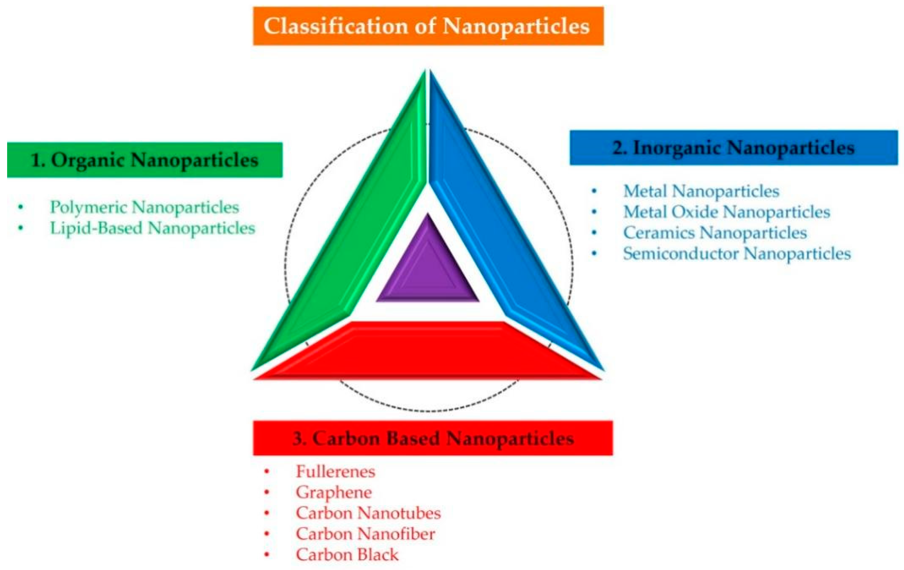
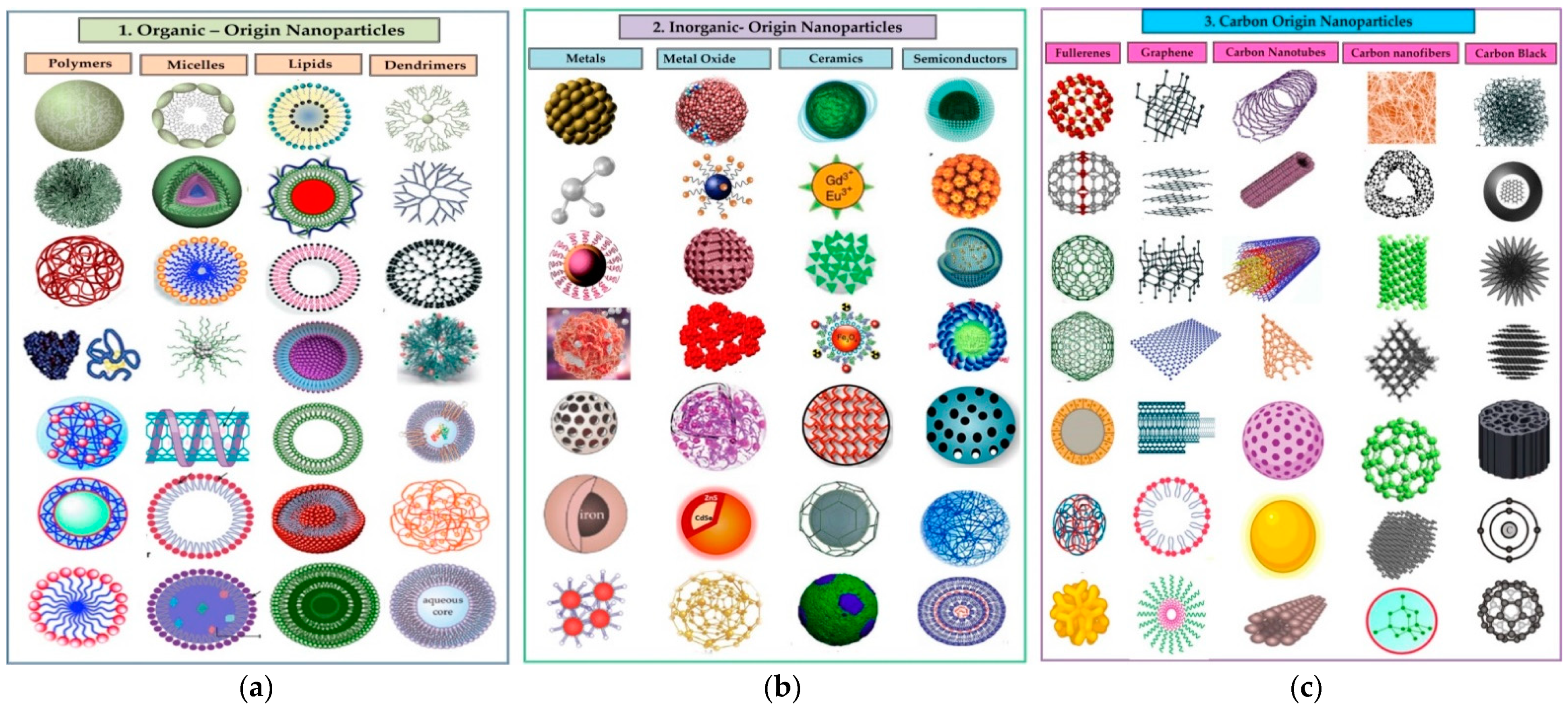
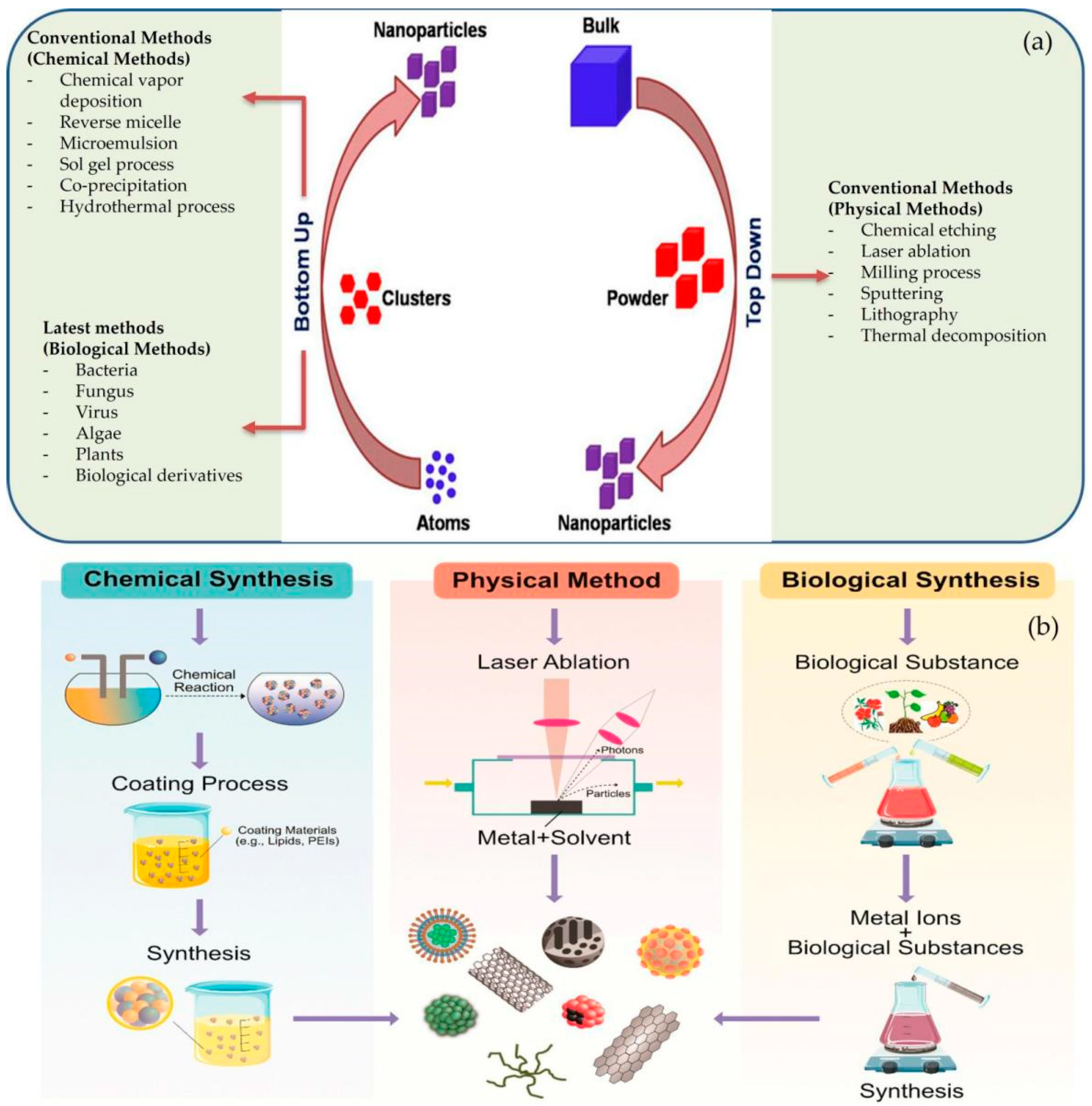
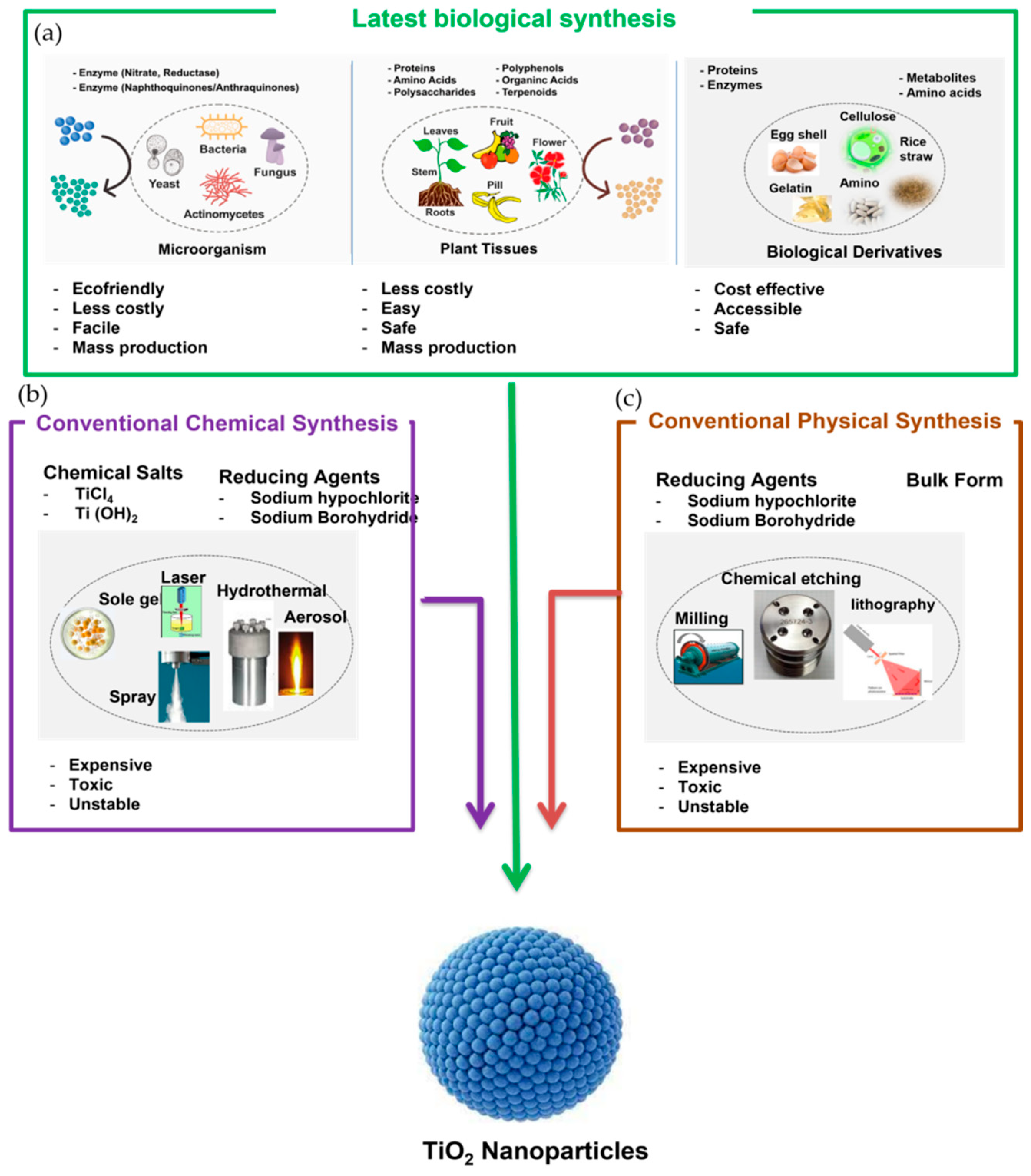
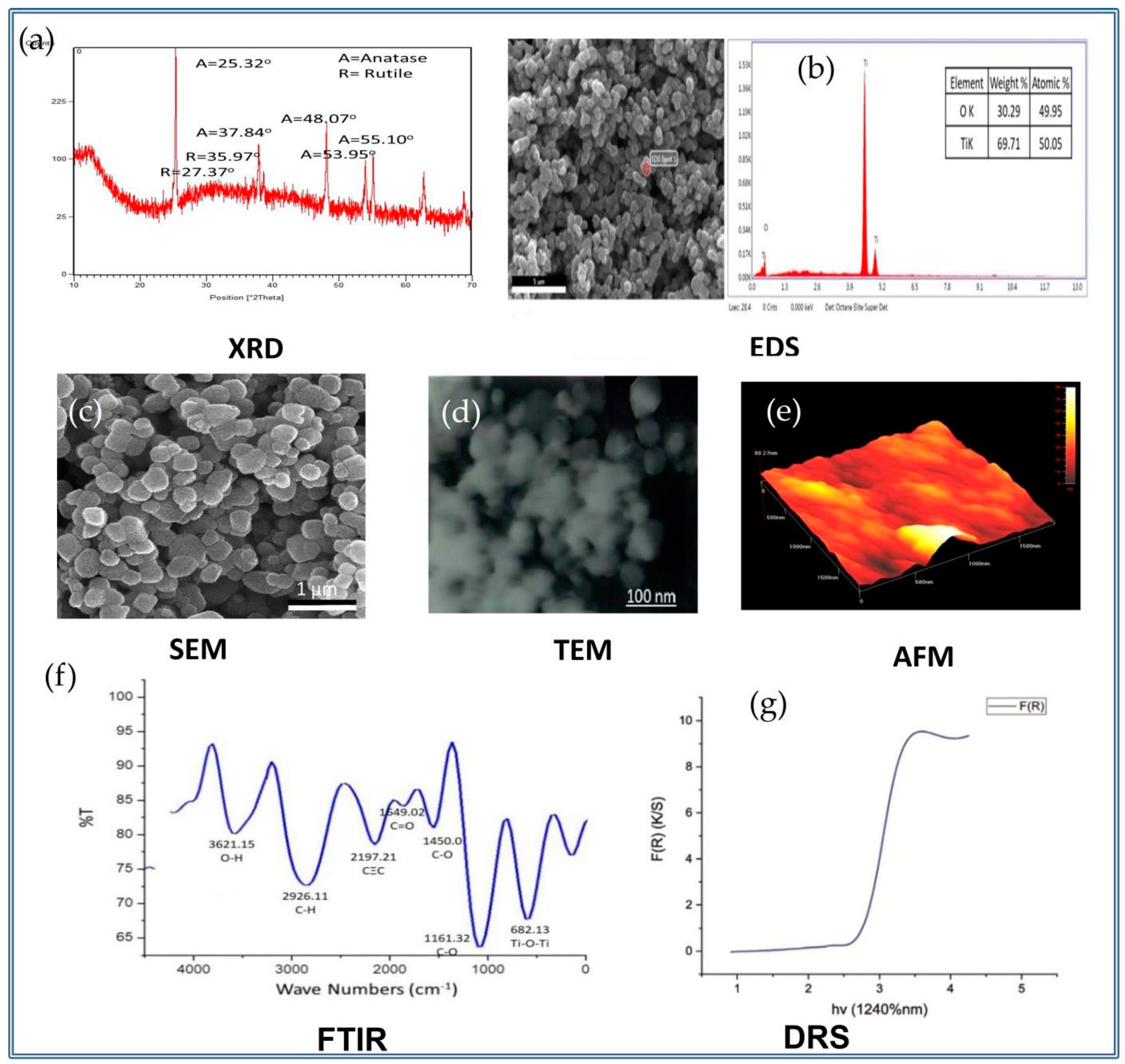
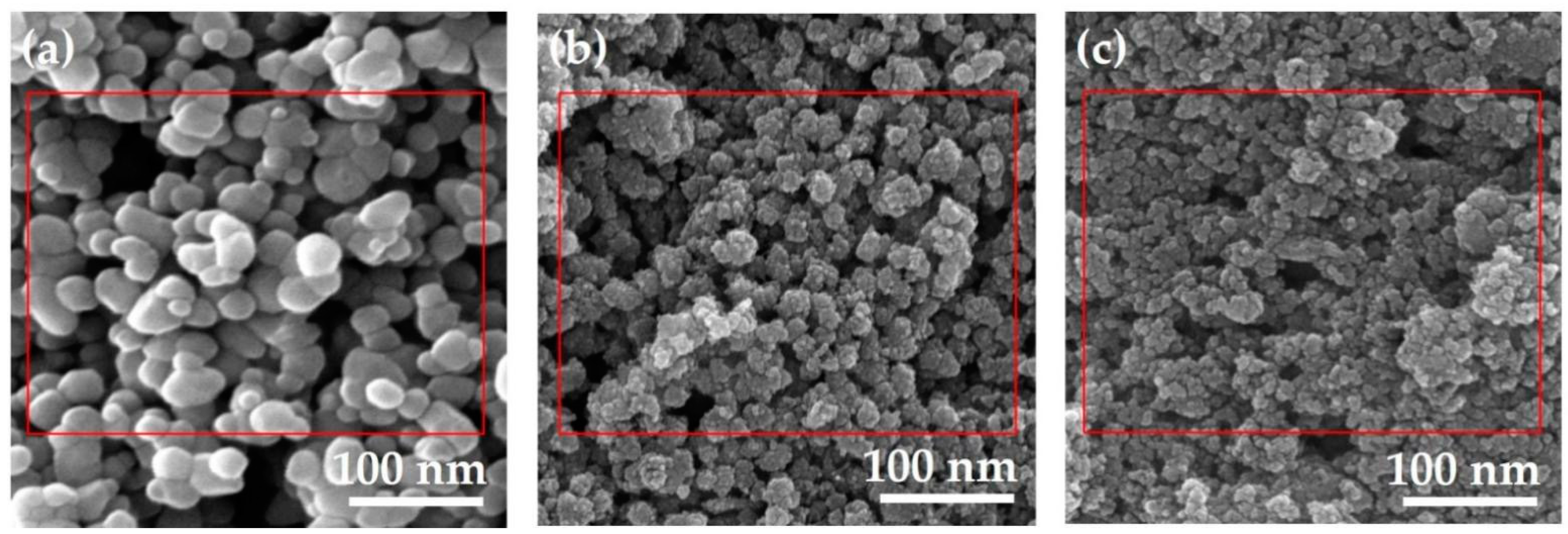
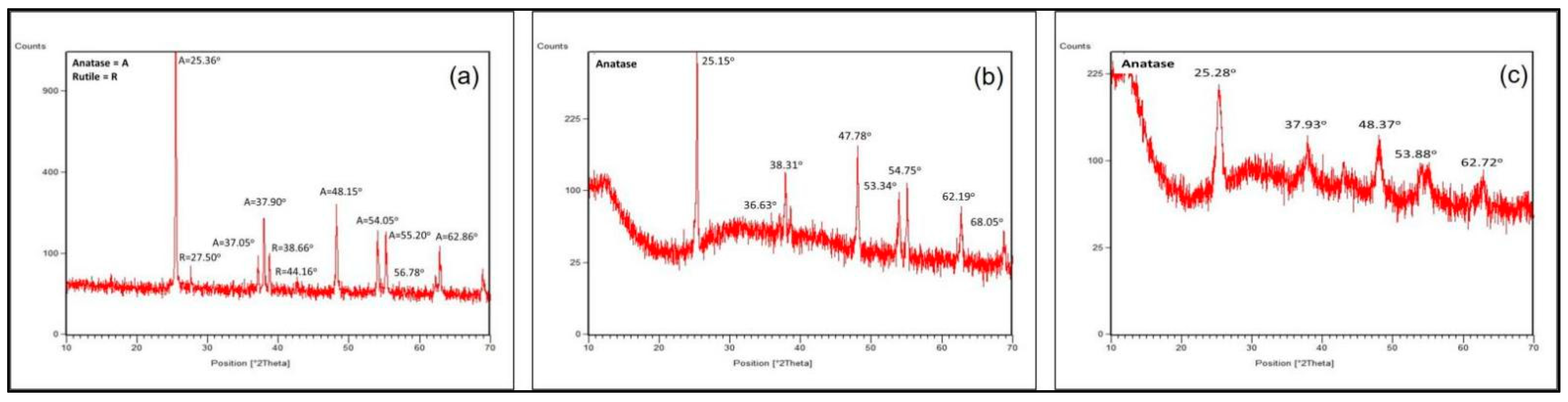
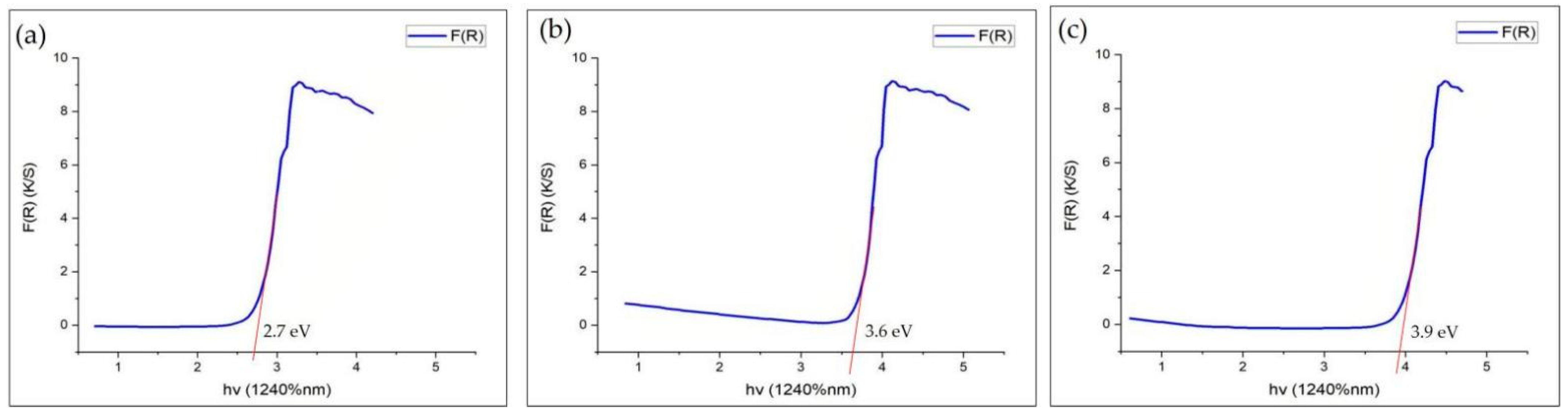
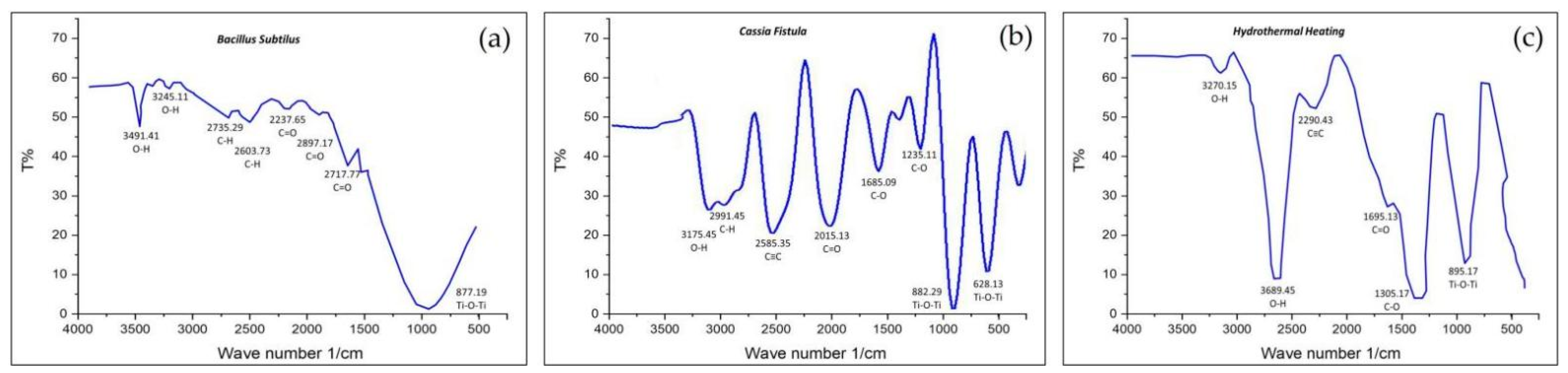
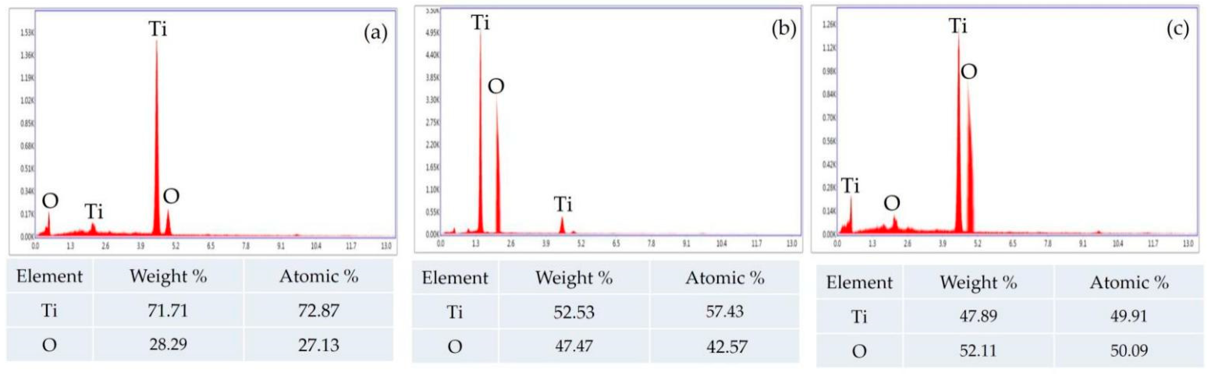
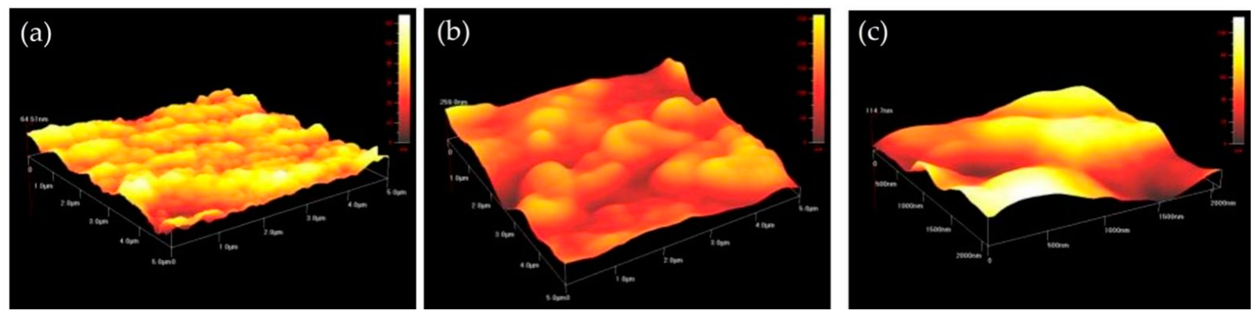
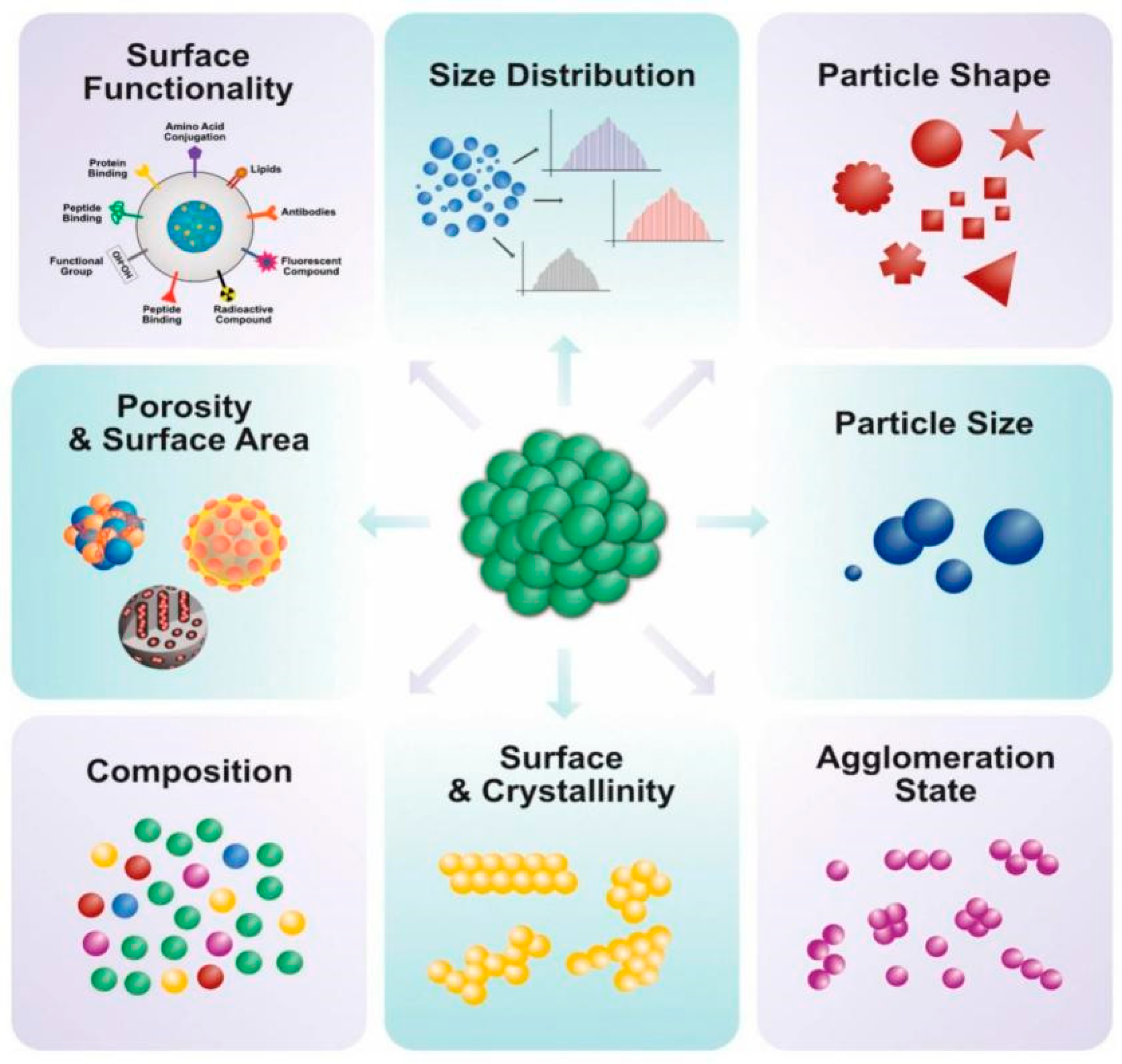
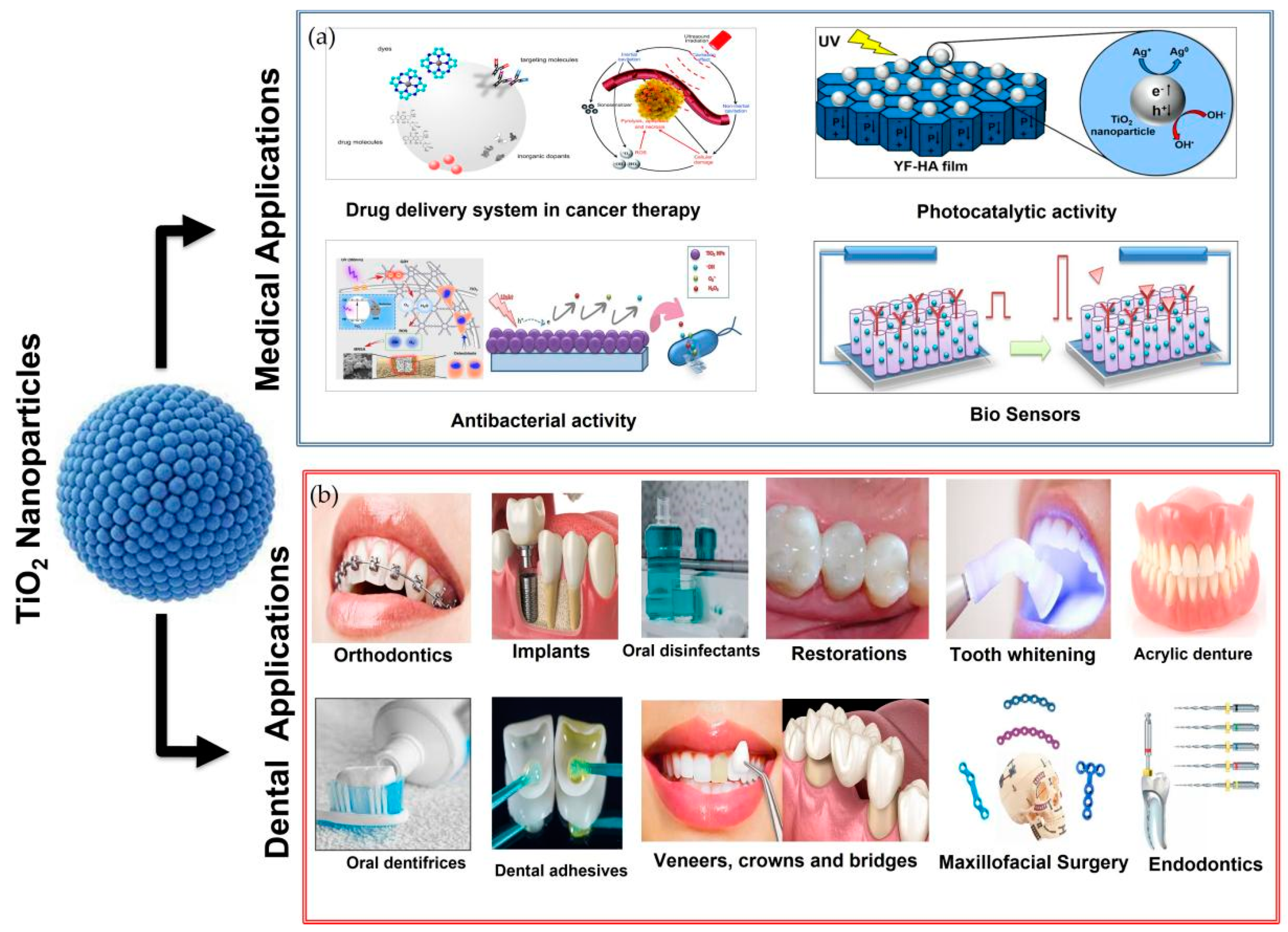
| Properties | Conventional TiO2 | Biological TiO2 | References |
|---|---|---|---|
| Nature | Expensive, toxic | Cost effective, nontoxic | [40] |
| Reducing agent | Dimethylformamide, ethylene glycol, hydrazine hydrate, sodium borohydride, polyol, sodium citrate, and N,N-dimethylformamide | Biomolecules include phenolics, polysaccharides, flavones, terpenoids, alkaloids, proteins, amino acids, enzymes, predominantly, and nitrate reductase | [51] |
| Method | Stabilizer (surfactant) is added to the first solution to prevent the agglomeration of NPs | There is no need to add a stabilizing agent | [85] |
| Environmental impact | Environmental pollution is a disadvantage of the chemical method and the chemical reduction methods are energy-intensive | Synthesis carried out in environmental conditions, and they are safe enough and consume no energy | [90] |
| Antibacterial activity | The chemically synthesized NPs show comparatively lower antimicrobial activity against pathogenic bacteria | The NPs synthesized from biological means are showing better antimicrobial activity against the pathogenic bacteria | [91] |
| Stability | NPs are not stable | NPs are highly stable | [85] |
| Byproducts formation | Harmful byproducts formed | No harmful byproducts formed | [91] |
| Biocompatible coating formation | No biocompatible coating is formed | The additional safe and effective surface area for interaction in the biological environment is produced due to the biocompatible coating on the nanoparticle’s surface | [51] |
| Process | Non-economical | Economical | [40] |
Publisher’s Note: MDPI stays neutral with regard to jurisdictional claims in published maps and institutional affiliations. |
© 2022 by the authors. Licensee MDPI, Basel, Switzerland. This article is an open access article distributed under the terms and conditions of the Creative Commons Attribution (CC BY) license (https://creativecommons.org/licenses/by/4.0/).
Share and Cite
Mansoor, A.; Khurshid, Z.; Khan, M.T.; Mansoor, E.; Butt, F.A.; Jamal, A.; Palma, P.J. Medical and Dental Applications of Titania Nanoparticles: An Overview. Nanomaterials 2022, 12, 3670. https://doi.org/10.3390/nano12203670
Mansoor A, Khurshid Z, Khan MT, Mansoor E, Butt FA, Jamal A, Palma PJ. Medical and Dental Applications of Titania Nanoparticles: An Overview. Nanomaterials. 2022; 12(20):3670. https://doi.org/10.3390/nano12203670
Chicago/Turabian StyleMansoor, Afsheen, Zohaib Khurshid, Muhammad Talal Khan, Emaan Mansoor, Faaz Ahmad Butt, Asif Jamal, and Paulo J. Palma. 2022. "Medical and Dental Applications of Titania Nanoparticles: An Overview" Nanomaterials 12, no. 20: 3670. https://doi.org/10.3390/nano12203670