Binder-Free Electrode Based on ZnO Nanorods Directly Grown on Aluminum Substrate for High Performance Supercapacitors


 , ,
, , 
Abstract
:1. Introduction
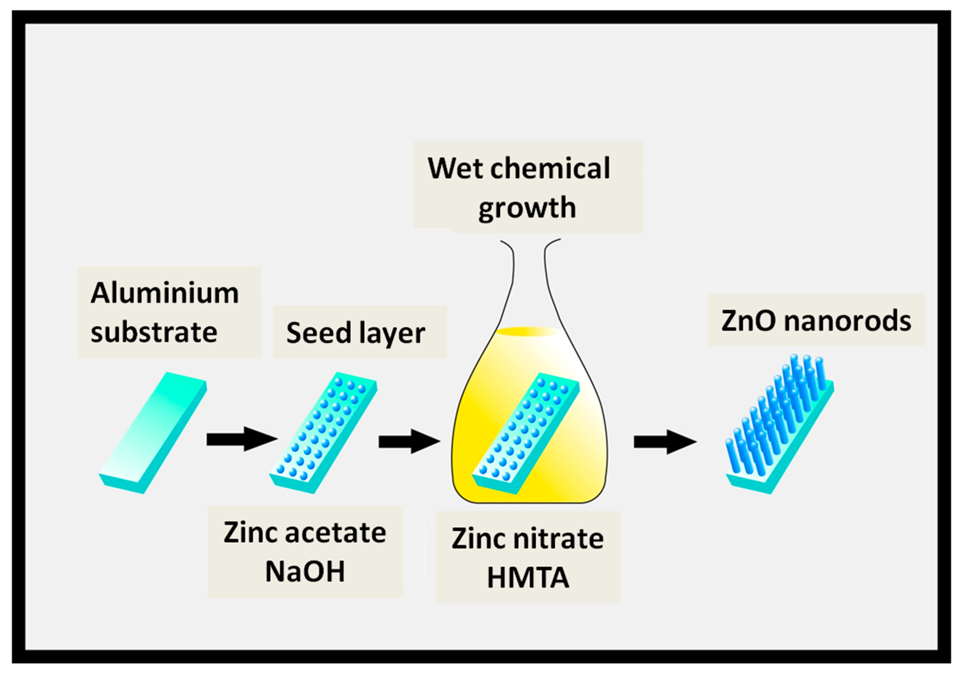
2. Experimental Details
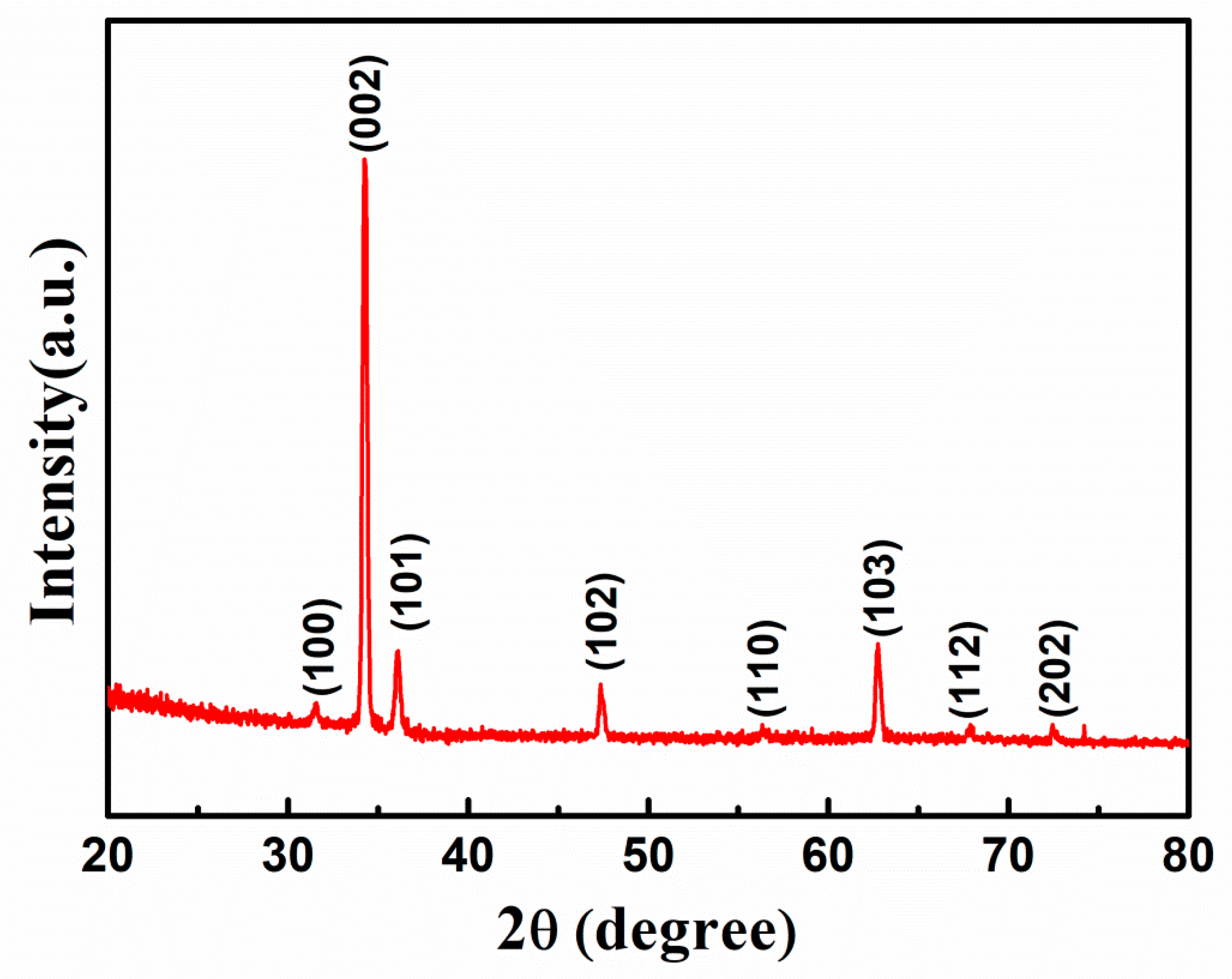
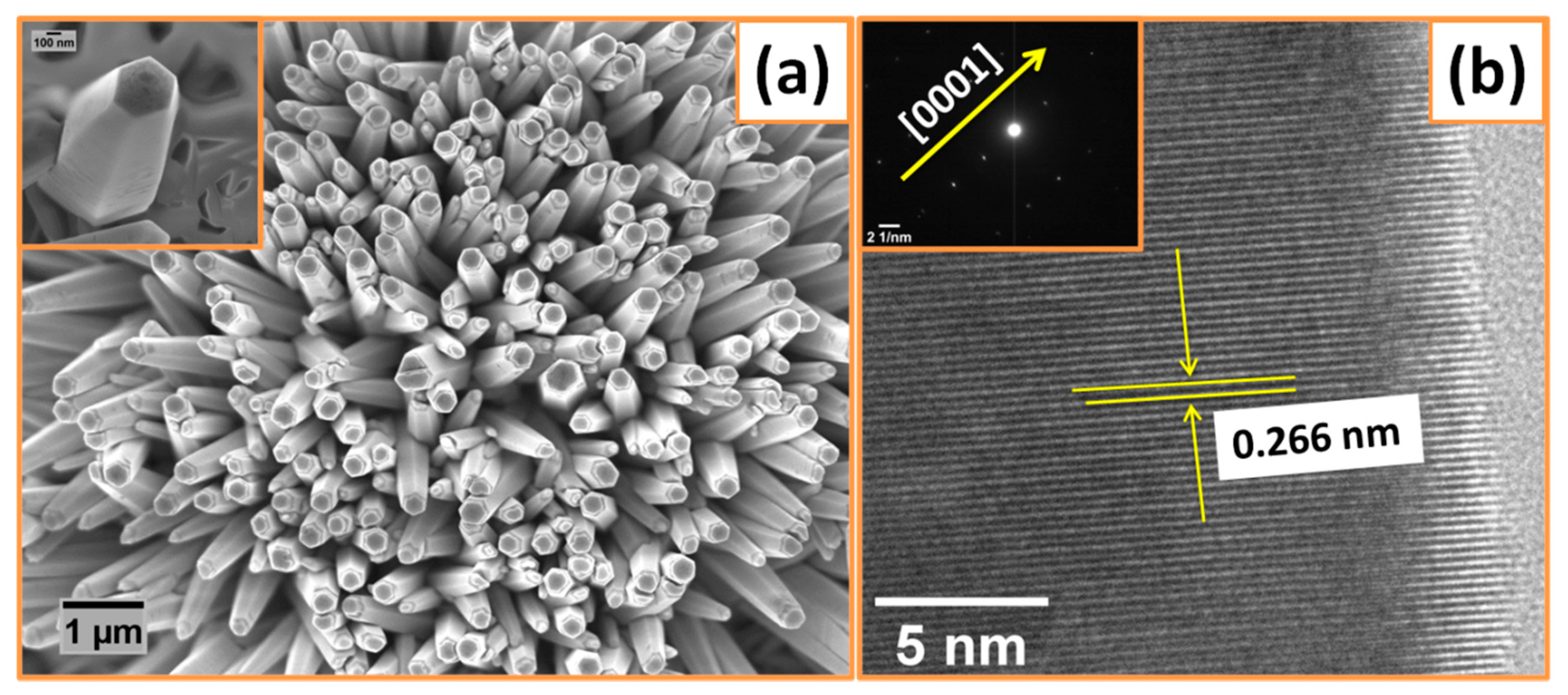
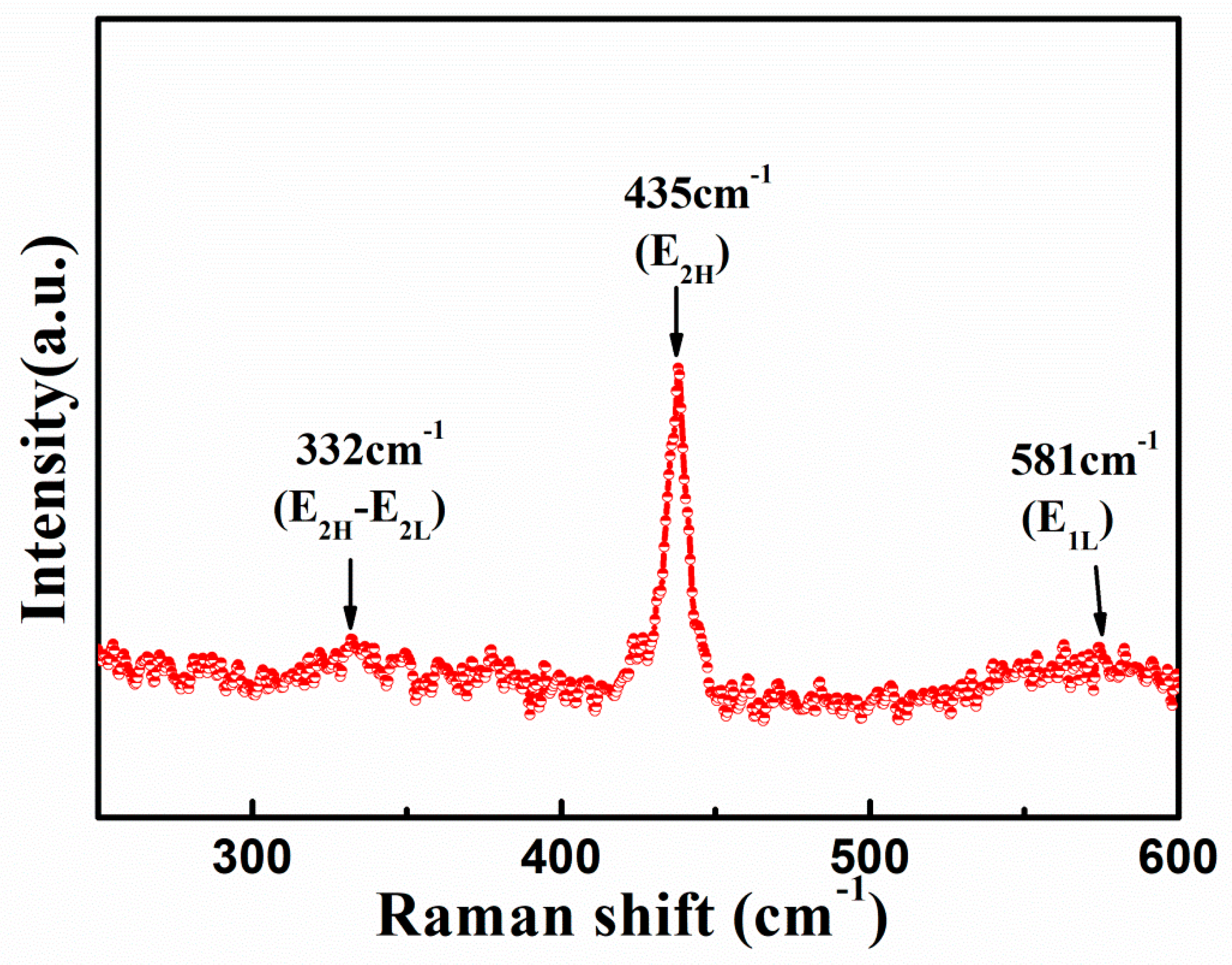
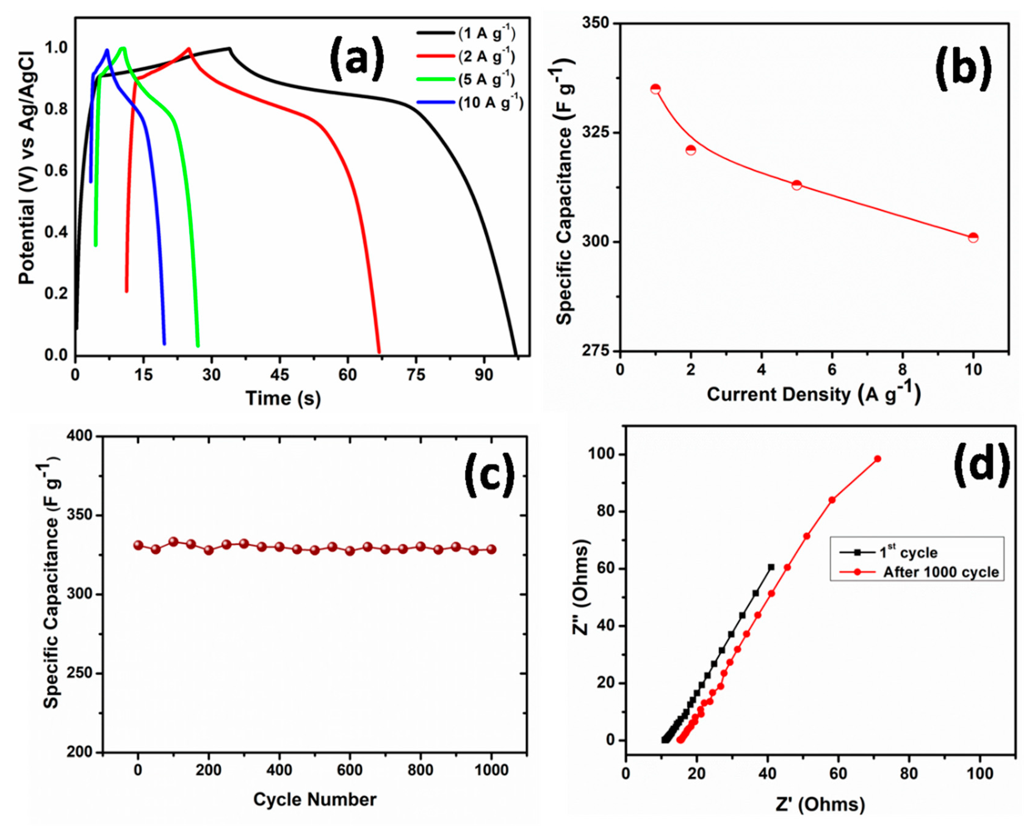
3. Results and Discussion
4. Conclusions
Supplementary Materials
Author Contributions
Funding
Acknowledgments
Conflicts of Interest
References
- Shao, Y.L.; El-Kady, M.F.; Wang, L.J.; Zhang, Q.H.; Li, Y.G.; Wang, H.Z.; Mousavi, M.F.; Kaner, R.B. Graphene-based materials for flexible supercapacitors. Chem. Soc. Rev. 2015, 44, 3639–3665. [Google Scholar] [CrossRef] [PubMed]
- Zhong, C.; Deng, Y.D.; Hu, W.B.; Qiao, J.L.; Zhang, L.; Zhang, J.J. A review of electrolyte materials and compositions for electrochemical supercapacitors. Chem. Soc. Rev. 2015, 44, 7484–7539. [Google Scholar] [CrossRef] [PubMed]
- Liang, J.; Zhu, G.; Lu, Z.; Zhao, P.; Wang, C.; Ma, Y.; Xu, Z.; Wang, Y.; Hu, Y.; Ma, L.; et al. Integrated perovskite solar capacitors with high energy conversion efficiency and fast photo-charging rate. J. Mater. Chem. A 2018, 6, 2047–2052. [Google Scholar] [CrossRef]
- Simon, P.; Gogotsi, Y. Materials for electrochemical capacitors. Nat. Mater. 2008, 7, 845–854. [Google Scholar] [CrossRef] [Green Version]
- Bagheri, N.; Aghaei, A.; Ghotbi, M.Y.; Marzbanrad, E.; Vlachopoulos, N.; Häggman, L.; Wang, M.; Boschloo, G.; Hagfeldt, A.; Skunik-Nuckowska, M.; et al. Combination of asymmetric supercapacitor utilizing activated carbon and nickel oxide with cobalt polypyridyl-based dye-sensitized solar cell. Electrochim. Acta 2014, 143, 390–397. [Google Scholar] [CrossRef]
- Navarrete-Astorga, E.; Rodr´ıguez-Moreno, J.; Dalchiele, E.A.; Schrebler, R.; Leyton, P.; Ramos-Barrado, J.R.; Martín, F. A transparent solid-state ion gel for supercapacitor device applications. J. Solid State Electrochem. 2017, 21, 1431–1444. [Google Scholar] [CrossRef]
- Rodríguez-Moreno, J.; Navarrete-Astorga, E.; Dalchiele, E.A.; Sánchez, L.; Ramos-Barrado, J.R.; Martín, F. Polyvinylpyrrolidone–LiClO4 solid polymer electrolyte and its application in transparent thin film supercapacitors. J. Power Sources 2013, 237, 270–276. [Google Scholar] [CrossRef]
- Rodríguez-Moreno, J.; Navarrete-Astorga, E.; Dalchiele, E.A.; Schrebler, R.; Ramos-Barrado, J.R.; Martín, F. Vertically aligned ZnO@CuS@PEDOTcore@shellnanorod arrays decorated with MnO2 nanoparticles for a high-performance and semi-transparent supercapacitor electrode. Chem. Commun. 2014, 5652, 5652–5655. [Google Scholar] [CrossRef]
- Xiao, N.; Tan, H.; Zhu, J.; Tan, L.; Rui, X.; Dong, X.; Yan, Q. High-Performance Supercapacitor Electrodes Based on Graphene Achieved by Thermal Treatment with the Aid of Nitric Acid. ACS Appl. Mater. Interface 2013, 5, 9656. [Google Scholar] [CrossRef]
- Hu, C.C.; Chang, K.H.; Lin, M.C.; Wu, Y.T. Design and tailoring of the nanotubular arrayed architecture of hydrous RuO2 for next generation supercapacitors. Nano Lett. 2006, 6, 2690–2695. [Google Scholar] [CrossRef]
- Hung, C.J.; Hung, J.H.; Lin, P.; Tseng, T.Y. Electrophoretic fabrication and characterizations of manganese oxide/carbon nanotube nanocomposite pseudocapacitors. J. Electrochem. Soc. 2011, 158, A942–A947. [Google Scholar] [CrossRef]
- Chen, Z.; Augustyn, V.; Wen, J.; Zhang, Y.; Shen, M.; Dunn, B.; Lu, Y. High-performance supercapacitors based on intertwined CNT/V2O5 nanowire nanocomposites. Adv. Mater. 2011, 23, 791–795. [Google Scholar] [CrossRef] [PubMed]
- Kalpana, D.; Omkumar, K.S.; Kumar, S.S.; Renganathan, N.G. A novel high power symmetric ZnO/carbon aerogel composite electrode for electrochemical supercapacitor. Electrochim. Acta 2006, 52, 1309–1315. [Google Scholar] [CrossRef]
- Kim, I.H.; Kim, K.B. Electrochemical characterization of hydrous ruthenium oxide thin-film electrodes for electrochemical capacitor applications. J. Electrochem. Soc. 2006, 153, A383–A389. [Google Scholar] [CrossRef]
- Li, X.; Wang, Z.; Qiu, Y.; Pan, Q.; Hu, P.A. 3D Graphene/ZnONanorods Composite Networks as Supercapacitor Electrodes. J. Alloys Compd. 2015, 620, 31–37. [Google Scholar] [CrossRef]
- Huang, G.; Zhang, W.; Xu, S.; Li, Y.; Yang, Y. MicrosphericalZnO Synthesized from a Metal-Organic Precursor for Supercapacitors. Ionics 2016, 22, 2169–2174. [Google Scholar] [CrossRef]
- Chen, H.C.; Lyu, Y.R.; Fang, A.; Lee, G.J.; Karuppasamy, L.; Wu, J.J.; Lin, C.K.; Anandan, S.; Chen, C.Y. The Design of ZnONanorod Arrays Coated with MnOx for High Electrochemical Stability of a Pseudocapacitor Electrode. Nanomaterials 2020, 10, 475. [Google Scholar] [CrossRef] [Green Version]
- Gao, J.W.Z.; Li, Z.; Wang, B.; Yan, Y.; Liu, Q.; Mann, T.; Zhang, M.; Jiang, Z. Green synthesis of graphene nanosheets/ZnO composites and electrochemical properties. J Solid State Chem. 2011, 184, 1421–1427. [Google Scholar]
- Lu, T.; Pan, L.; Li, H.; Zhu, G.; Lv, T.; Liu, X.; Sun, Z.; Chen, T.; Chua, D.H. Microwave-assisted synthesis of graphene-ZnO nanocomposites for electrochemical supercapacitors. J. Alloy Compd. 2011, 509, 5488–5492. [Google Scholar] [CrossRef]
- Zhang, Y.; Sun, X.; Pan, L. Carbon nanotube–ZnO nanocomposite electrodes for supercapacitors. Solid State Ion. 2009, 180, 1525–1528. [Google Scholar] [CrossRef]
- Zhang, Y.; Li, H.; Lu, T. Capacitive behavior of graphene–ZnO composite film for supercapacitors. J. Electroanal. Chem. 2009, 634, 68–71. [Google Scholar] [CrossRef]
- Jayalakshmi, M.; Palaniappa, M.; Balasubramanian, K. Single step solution combustion synthesis of ZnO/carbon composite and its electrochemical characterization for supercapacitor application. Int. J. Electrochem. Sci. 2008, 3, 96–103. [Google Scholar]
- Kim, C.H.; Kim, B.H. Zinc oxide/activated carbon nanofiber composites for high-performance supercapacitor electrodes. J. Power Sources 2015, 274, 512–520. [Google Scholar] [CrossRef]
- He, X.; Yoo, J.E.; Lee, M.H.; Bae, J. Morphology Engineering of ZnO Nanostructures for High Performance Supercapacitors: Enhanced Electrochemistry of ZnONanocones Compared to ZnO Nanowires. Nanotechnology 2017, 28, 245402–245421. [Google Scholar] [CrossRef]
- Alver, Ü.; Tanrıverdi, A.; Akgül, Ö. Hydrothermal Preparation of ZnO Electrodes Synthesized from Different Precursors for Electrochemical Supercapacitors. Synth. Met. 2016, 211, 30–34. [Google Scholar] [CrossRef]
- Luo, Q.; Xu, P.; Qiu, Y.; Cheng, Z.; Chang, X.; Fan, H. Synthesis of ZnO Tetrapods for High-Performance Supercapacitor Applications. Mater. Lett. 2017, 198, 192–195. [Google Scholar] [CrossRef]
- Fu, Z.W.; Feng, H.; Ye, Z.; Yue, C.; Qin, Q.Z. The electrochemical reaction of zinc oxide thin films with lithium. J. Electrochem. Soc. 2003, 150, A714–A720. [Google Scholar] [CrossRef]
- Woo, M.A.; Kim, T.W.; Kim, I.Y.; Hwang, S.J. Synthesis and lithium electrode application of ZnO-ZnFe2O4 nanocomposites and porously assembled ZnFe2O4 nanoparticles. Solid State Ion. 2011, 182, 91–97. [Google Scholar] [CrossRef]
- Park, K.T.; Xia, F.; Kim, S.W.; Kim, S.B.; Song, T.; Paik, U.; Park, W.I. Facile synthesis of ultrathin ZnO nanotubes with wellorganized hexagonal nanowalls and sealed layouts: Applications for lithium ion battery anodes. J. Phys. Chem. C 2013, 117, 1037–1043. [Google Scholar] [CrossRef]
- Greene, L.E.; Law, M.; Tan, D.H.; Montano, M.; Goldberger, J.; Somorjai, G.; Yang, P.D. General Route to Vertical ZnO Nanowire Arrays Using Textured ZnO Seeds. Nano Lett. 2005, 5, 1231–1236. [Google Scholar] [CrossRef]
- Wang, Y.Y.; Jiang, X.J.; Yang, L.S.; Jia, N.; Ding, Y. In situ synthesis of C/Cu/ZnO porous hybrids as anode materials for lithium ion batteries. ACS Appl. Mater. Interfaces 2014, 6, 1525–1532. [Google Scholar] [CrossRef] [PubMed]
- Chen, J.; Liu, Y.; Li, W.; Yang, H.; Xu, L. The large electrochemical capacitance of nitrogen doped mesoporous carbon derived from egg white by using a ZnO template. RSC Adv. 2015, 5, 98177. [Google Scholar] [CrossRef]
- You, J.B.; Zhang, X.W.; Dong, J.J.; Song, X.M.; Yin, Z.G.; Chen, N.F.; Yan, H. Localized-Surface-Plasmon Enhanced the 357 nm Forward Emission from ZnMgO Films Capped by Pt Nanoparticles. Nanoscale Res. Lett. 2009, 4, 1121. [Google Scholar] [CrossRef] [PubMed] [Green Version]
- Yang, W.; Wang, F.; Guan, Z.; He, P.; Liu, Z.; Hu, L.; Chen, M.; Zhang, C.; He, X.; Fu, Y. Comparative Study of ZnO Thin Films Grown on Quartz Glass and Sapphire (001) Substrates by Means of Magnetron Sputtering and High-Temperature Annealing. Appl. Sci. 2019, 9, 4509. [Google Scholar] [CrossRef] [Green Version]
- Gaddama, V.; Neellaa, N.; Nayakb, M.M.; Rajannaa, K. Al:ZnONanosheets on Flexible Stainless Steel Substrate as Impact Sensor. Mater. Today Proc. 2018, 5, 10779. [Google Scholar] [CrossRef]
- Zhao, S.H.; Guo, J.X.; Jiang, F.; Su, Q.M.; Zhang, J.; Du, G.H. Growth of hierarchal porous CoO nanowire arrays on carbon cloth as binder-free anodes for high-performance flexible lithium-ion batteries. J. Alloys Compd. 2016, 655, 372. [Google Scholar] [CrossRef]
- Arguello, C.A.; Rousseau, D.L.; Porto, S.P.S. First-order Raman effect in wurtzite-type crystals. Phys. Rev. 1969, 181, 1351. [Google Scholar] [CrossRef]
- Damen, T.C.; Porto, S.P.S.; Tell, B. Raman effect in zinc oxide. Phys. Rev. 1966, 142, 570. [Google Scholar] [CrossRef]
- Huang, M.H.; Wu, Y.; Feick, H.; Tran, N.; Weber, E.; Yang, P. Catalytic growth of zinc oxide nanowires by vapor transport. Adv. Mater. 2001, 13, 113. [Google Scholar] [CrossRef]
- Hung, C.H.; Whang, W.T. A novel low-temperature growth and characterization of single crystal ZnO nanorods. Mater. Chem. Phys. 2003, 82, 705–710. [Google Scholar] [CrossRef]
- Hwang, Y.H.; Lee, S.M.; Kim, Y.J.; Kahng, Y.H.; Lee, K. A new approach of structural and chemical modification on graphene electrodes for high-performance supercapacitors. Carbon 2016, 100, 7–15. [Google Scholar] [CrossRef]
- Arul, N.S.; Mangalaraj, D.; Ramachandran, R.; Grace, A.N.; Han, J.I. Fabrication of CeO2/Fe2O3 composite nanospindles for enchancedvisble light driven photocatalysts and supercapacitor electrodes. J. Mater. Chem. A 2015, 3, 15248. [Google Scholar] [CrossRef]
- Krishnamoorthy, K.; Veerasubramani, G.K.; Pazhamalai, P.; Kim, S.J. Designing two dimensional nanoarchitectured MoS2 sheets grown on Mo foil as a binder free electrode for supercapacitors. Electrochim. Acta 2016, 190, 305–312. [Google Scholar] [CrossRef]
- GVeerasubramani, K.; Krishnamoorthy, K.; Kim, S.J. Electrochemical performance of an asymmetric supercapacitor based on graphene and cobalt molybdate electrodes. RSC Adv. 2015, 5, 16319. [Google Scholar] [CrossRef]
- Ramachandran, R.; Saranya, M.; Kollu, P.; Raghupathy, B.P.C.; Jeong, S.K.; Gracem, A.N. Solvothermal synthesis of zinc sulfide decorated graphene (ZnS/G) nanocomposites for novel supercapacitor electrodes. Electrochim. Acta 2015, 178, 647–657. [Google Scholar] [CrossRef]
- Zhang, J.; Kong, L.B.; Cai, J.J.; Luo, Y.C.; Kang, L. Nanoflake-like cobalt hydroxide/ ordered mesoporous carbon composite for electrochemical capacitors. J. Solid State Electrochem. 2010, 14, 2065–2075. [Google Scholar] [CrossRef]
- Shi, S.; Zhuang, X.; Chen, B.; Wang, X. Solution blowig of ZnO nanoflake-encapsulated carbon nanofibers as electrodes for supercapacitors. J. Mater. Chem. A 2013, 1, 13779. [Google Scholar] [CrossRef]
- Zhang, X.; Wang, X.; Jiang, L.; Wu, H.; Wu, C.; Su, J. Effect of aqueous electrolytes on the electrochemical behaviors of supercapacitors based on hierarchically porous carbons. J. Power Sources 2012, 216, 290–296. [Google Scholar] [CrossRef]
- Niu, H.; Zhou, D.; Yang, X.; Li, X.; Wang, Q.; Qu, F.Y. Towards three-dimensional hierarchical ZnO nanofiber@Ni(OH)2nanoflake core–shell heterostructures for high performance asymmetric supercapacitors. J. Mater. Chem. A 2015, 3, 18413–18421. [Google Scholar] [CrossRef]


| Electrode Material | Scan Rate (mV s−1) | Specific Capacitance (F g−1) | Ref. |
|---|---|---|---|
| ZnO nanocones | 20 | 377.4 | [24] |
| ZnO nanostructures | 5 | 5.87 | [25] |
| ZnO tetrapods | 10 | 160.4 | [26] |
| ZnO coated with MnO2 | 25 | 222 | [16] |
| ZnO/RGO | 5 | 322.1 | [41] |
| CeO2/Fe2O3 nanospindles | 5 | 142.6 | [42] |
| MoS2 on Mo foil | 5 | 197.1 | [43] |
| CoMoO4 | 5 | 98.34 | [44] |
| ZnS/graphene | 5 | 197.1 | [45] |
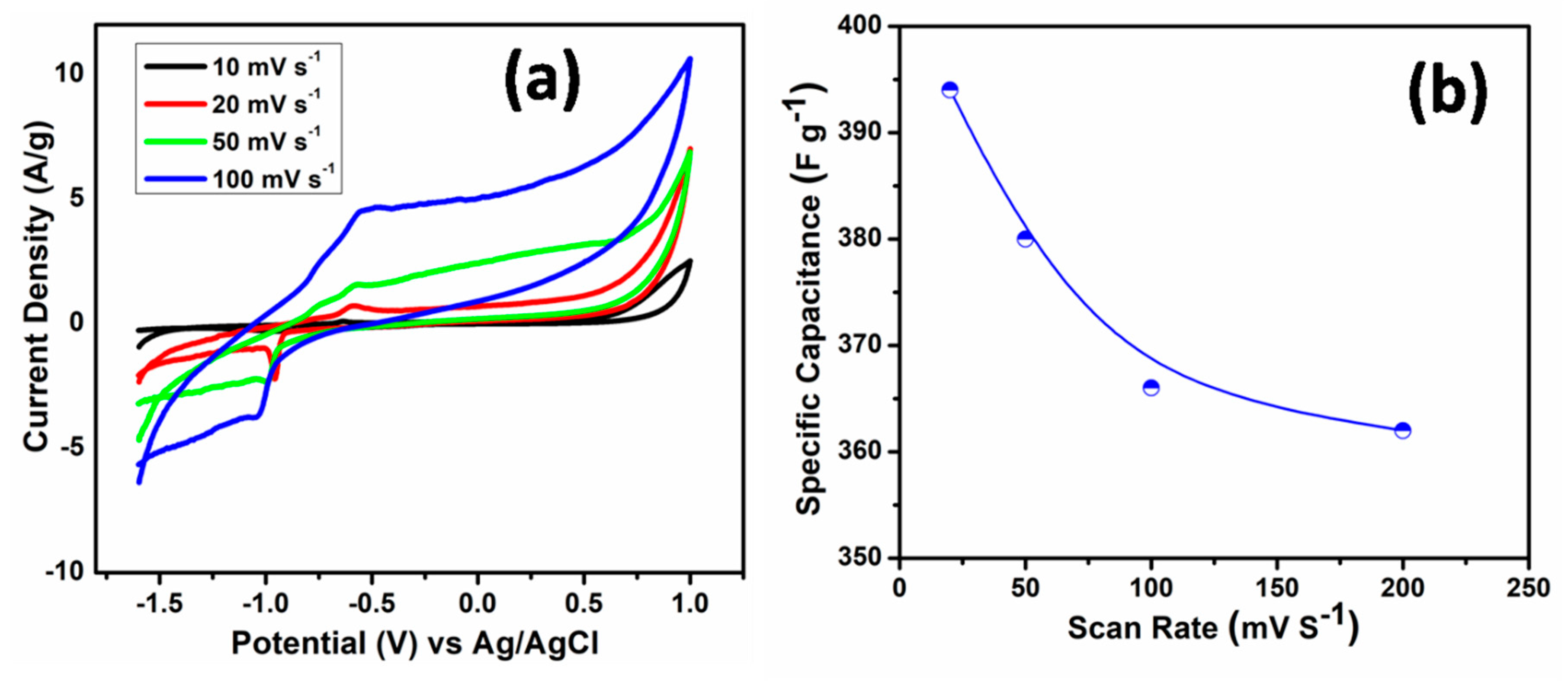
| ZnO nanorods on Al substrate | 20 | 394.1 | This work |
© 2020 by the authors. Licensee MDPI, Basel, Switzerland. This article is an open access article distributed under the terms and conditions of the Creative Commons Attribution (CC BY) license (http://creativecommons.org/licenses/by/4.0/).
Share and Cite
Ahmed, F.; Almutairi, G.; AlOtaibi, B.; Kumar, S.; Arshi, N.; Hussain, S.G.; Umar, A.; Ahmad, N.; Aljaafari, A. Binder-Free Electrode Based on ZnO Nanorods Directly Grown on Aluminum Substrate for High Performance Supercapacitors. Nanomaterials 2020, 10, 1979. https://doi.org/10.3390/nano10101979
Ahmed F, Almutairi G, AlOtaibi B, Kumar S, Arshi N, Hussain SG, Umar A, Ahmad N, Aljaafari A. Binder-Free Electrode Based on ZnO Nanorods Directly Grown on Aluminum Substrate for High Performance Supercapacitors. Nanomaterials. 2020; 10(10):1979. https://doi.org/10.3390/nano10101979
Chicago/Turabian StyleAhmed, Faheem, Ghzzai Almutairi, Bandar AlOtaibi, Shalendra Kumar, Nishat Arshi, Syed Ghazanfar Hussain, Ahmad Umar, Naushad Ahmad, and Abdullah Aljaafari. 2020. "Binder-Free Electrode Based on ZnO Nanorods Directly Grown on Aluminum Substrate for High Performance Supercapacitors" Nanomaterials 10, no. 10: 1979. https://doi.org/10.3390/nano10101979