
Three-Dimensional Impression of Biomaterials for Alveolar Graft: Scoping Review
 , ,
, ,  , , , , and
Abstract
:1. Introduction
2. Materials and Methods
2.1. Study Research and Selection Strategy
2.2. Data Extraction
2.3. Risk of Bias
3. Results
3.1. Study Selection
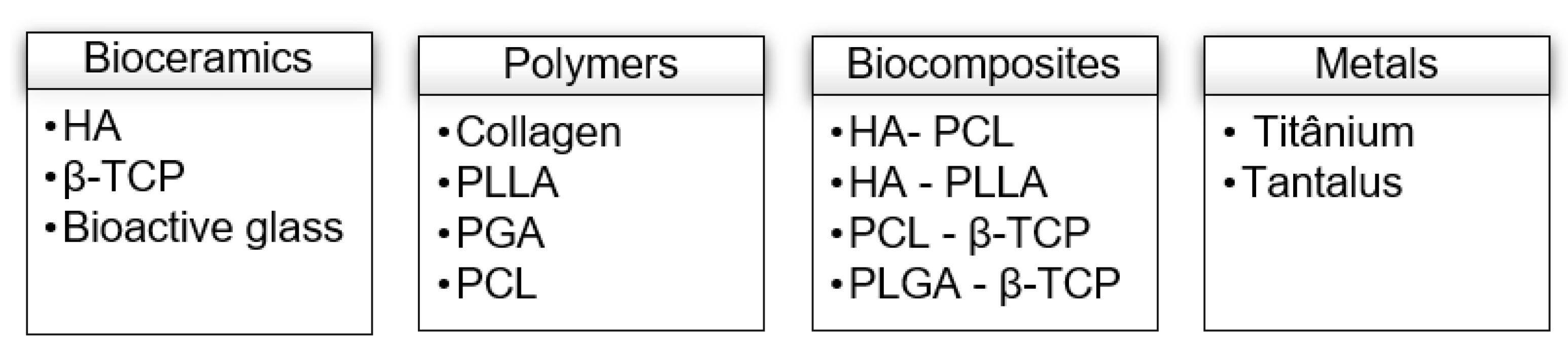
3.2. Characteristics of the Included Studies
3.2.1. In Vitro Studies
3.2.2. In Vivo Studies
3.3. Synthesis of Quantitative Evidence
3.4. Risk of Bias
4. Discussion
5. Conclusions
Author Contributions
Funding
Data Availability Statement
Conflicts of Interest
Appendix A
| Database | Search Phrase |
| Pubmed via Medline and Cochrane Library | (“Printing, Three-Dimensional” [Mesh] OR “Printing, Three Dimensional” OR “Printings, Three-Dimensional” OR “Three-Dimensional Printings” OR “3-Dimensional Printing*” OR “3 Dimensional Printing*” OR “Printing, 3-Dimensional” OR “Printings, 3-Dimensional” OR “3-D Printing*” OR “3 D Printing*” OR “Printing, 3-D” OR “Printings, 3-D” OR “Three-Dimensional Printing” OR “Three Dimensional Printing” OR “3D Printing*” OR “Printing, 3D” OR “Printings, 3D”) AND (“Bone Regeneration”[Mesh] OR “Bone Regenerations*” OR “Regeneration, Bone” OR “Regenerations, Bone” OR Osteoconduction OR “Alveolar Bone Grafting”[Mesh] OR “alveolar bone grafting*” OR “Alveolar Cleft Grafting” OR “bone graft*” OR “Bone Substitutes”[Mesh] OR “bone substitute*” OR “Replacement Material, Bone” OR “Replacement Materials, Bone” OR “Materials, Bone Replacement” OR “Substitute, Bone” OR “Substitutes, Bone” OR “Bone Replacement Material*” OR “Material, Bone Replacement” ) AND (Dentistry[Mesh] OR dentistry OR oral* OR orofacial OR dental* OR maxillofacial OR “Surgery, Oral”[Mesh] OR “surgery, oral” OR “Maxillofacial Surgery” OR “Surgery, Maxillofacial” OR “Oral Surgery” OR “Cleft Palate”[Mesh] OR “cleft palate*” OR “Palate, Cleft” OR “Palates, Cleft” OR “Cleft Palate, Isolated”) |
| Web of Science Core Collection (WOS) | TS = (“Print*, Three Dimensional” OR “Three-Dimensional Print*” OR “3-Dimensional Print*” OR “3 Dimensional Print*” OR “Print*, 3-Dimensional” OR “3-D Print*” OR “3D Print*” OR “Print*, 3-D” OR “ Print*, 3D”) AND TS = ( “Regenerati*, Bone” OR “Bone Regenerati*” OR osteoconduction OR “Alveolar Bone Graft*” OR “alveolar cleft grafting“ OR “bone graft*” OR “Replacement Material*, Bone” OR “Material*, Bone Replacement” OR “Substitute*, Bone” OR “Bone Replacement Material*” OR “ Material, Bone Replacement” OR “bone substitute*”) AND TS = (dent* OR oral* OR orofacial OR maxillofacial OR “Surgery, Oral” OR “oral surgery”) |
| EMBASE | (‘printing, three dimensional’/exp OR ‘printing, three dimensional’ OR ‘printings, three-dimensional’ OR ‘three-dimensional printings’ OR ‘3-dimensional printing*’ OR ‘3 dimensional printing*’ OR ‘printing, 3-dimensional’ OR ‘printings, 3-dimensional’ OR ‘3-d printing*’ OR ‘3 d printing*’ OR ‘printing, 3-d’ OR ‘printings, 3-d’ OR ‘three-dimensional printing’/exp OR ‘three-dimensional printing’ OR ‘three dimensional printing’/exp OR ‘three dimensional printing’ OR ‘3d printing*’ OR ‘printing, 3d’ OR ‘printings, 3d’) AND (‘bone regeneration’/exp OR ‘bone regeneration’ OR ‘regeneration, bone’/exp OR ‘regeneration, bone’ OR ‘regenerations, bone’ OR ’osteoconduction’/exp OR osteoconduction OR ‘alveolar bone grafting’/exp OR ‘alveolar bone grafting’ OR ‘alveolar cleft grafting’ OR ‘bone graft*’ OR ‘bone graft’/exp OR ‘bone graft’ OR ‘bone transplantation’/exp OR ‘bone transplantation’ OR ‘bone prosthesis’/exp OR ‘bone prosthesis’ OR ‘bone substitute*’ OR ‘replacement material, bone’ OR ‘replacement materials, bone’ OR ‘materials, bone replacement’ OR ‘substitute, bone’ OR ‘substitutes, bone’ OR ‘bone replacement material*’ OR ‘material, bone replacement’) AND (dentistry OR ‘dentistry’/exp OR ‘dentistry’ OR oral OR orofacial OR ‘dental’/exp OR dental OR maxillofacial OR ‘oral surgery’/exp OR ‘oral surgery’) |
References
- Robey, P.G. Cell Sources for Bone Regeneration: The Good, the Bad, and the Ugly (But Promising). Tissue Eng. Part B Rev. 2011, 17, 423–430. [Google Scholar] [CrossRef] [PubMed] [Green Version]
- Bhumiratana, S.; Vunjak-Novakovic, G. Concise Review: Personalized Human Bone Grafts for Reconstructing Head and Face. Stem Cells Transl. Med. 2012, 1, 64–69. [Google Scholar] [CrossRef]
- Weir, M.D.; Xu, H.H.K. Culture Human Mesenchymal Stem Cells with Calcium Phosphate Cement Scaffolds for Bone Repair. J. Biomed. Mater. Res. 2010, 93B, 93–105. [Google Scholar] [CrossRef] [PubMed] [Green Version]
- Illich, D.J.; Demir, N.; Stojković, M.; Scheer, M.; Rothamel, D.; Neugebauer, J.; Hescheler, J.; Zöller, J.E. Concise Review: Induced Pluripotent Stem Cells and Lineage Reprogramming: Prospects for Bone Regeneration. Stem Cells 2011, 29, 555–563. [Google Scholar] [CrossRef] [PubMed]
- Pagni, G.; Kaigler, D.; Rasperini, G.; Avila-Ortiz, G.; Bartel, R.; Giannobile, W.V. Bone Repair Cells for Craniofacial Regeneration. Adv. Drug Deliv. Rev. 2012, 64, 1310–1319. [Google Scholar] [CrossRef] [Green Version]
- He, X.; Dziak, R.; Mao, K.; Genco, R.; Swihart, M.; Li, C.; Yang, S. Integration of a Novel Injectable Nano Calcium Sulfate/Alginate Scaffold and BMP2 Gene-Modified Mesenchymal Stem Cells for Bone Regeneration. Tissue Eng. Part A 2013, 19, 508–518. [Google Scholar] [CrossRef] [Green Version]
- Raposo-Amaral, C.E.; Kobayashi, G.S.; Almeida, A.B.; Bueno, D.F.; de Souza e Freitas, F.R.; Vulcano, L.C.; Passos-Bueno, M.R.; Alonso, N. Alveolar Osseous Defect in Rat for Cell Therapy: Preliminary Report. Acta Cir. Bras. 2010, 25, 313–317. [Google Scholar] [CrossRef] [Green Version]
- Yoshioka, M.; Tanimoto, K.; Tanne, Y.; Sumi, K.; Awada, T.; Oki, N.; Sugiyama, M.; Kato, Y.; Tanne, K. Bone Regeneration in Artificial Jaw Cleft by Use of Carbonated Hydroxyapatite Particles and Mesenchymal Stem Cells Derived from Iliac Bone. Int. J. Dent. 2012, 2012, 352510. [Google Scholar] [CrossRef]
- Garland, M.A.; Reynolds, K.; Zhou, C.J. Environmental Mechanisms of Orofacial Clefts. Birth Defects Res. 2020, 112, 1660–1698. [Google Scholar] [CrossRef]
- Calzolari, E.; Bianchi, F.; Rubini, M.; Ritvanen, A.; Neville, A.J. Epidemiology of Cleft Palate in Europe: Implications for Genetic Research. Cleft Palate-Craniofac. J. 2004, 41, 244–249. [Google Scholar] [CrossRef]
- Taib, B.G.; Taib, A.G.; Swift, A.C.; van Eeden, S. Cleft Lip and Palate: Diagnosis and Management. Br. J. Hosp. Med. 2015, 76, 584–591. [Google Scholar] [CrossRef] [PubMed]
- Stal, S.; Chebret, L.; McElroy, C. The Team Approach in the Management of Congenital and Acquired Deformities. Clin. Plast. Surg. 1998, 25, 485–491. [Google Scholar] [CrossRef]
- Francisco, I.N. Overview of Care in Cleft Lip and Palate for Orthodontic Treatment. Ph.D. Thesis, Universidade do Porto, Porto, Portugal, 2021. [Google Scholar]
- Graber, L.; Vanarsdall, R.L.; Vig, K.W.L. Orthodontics Current Principles & Techniques, 4th ed.; Elsevier Mosby: St. Louis, MO, USA, 2005. [Google Scholar]
- Rawashdeh, M.A.; Telfah, H. Secondary Alveolar Bone Grafting: The Dilemma of Donor Site Selection and Morbidity. Br. J. Oral Maxillofac. Surg. 2008, 46, 665–670. [Google Scholar] [CrossRef] [PubMed]
- Francisco, I.; Fernandes, M.H.; Vale, F. Platelet-Rich Fibrin in Bone Regenerative Strategies in Orthodontics: A Systematic Review. Materials 2020, 13, 1866. [Google Scholar] [CrossRef]
- Valentini, V.; Cassoni, A.; Marianetti, T.M.; Romano, F.; Terenzi, V.; Iannetti, G. Reconstruction of Craniofacial Bony Defects Using Autogenous Bone Grafts: A Retrospective Study on 233 Patients. J. Craniofac. Surg. 2007, 18, 953–958. [Google Scholar] [CrossRef] [PubMed] [Green Version]
- Ahn, G.; Lee, J.-S.; Yun, W.-S.; Shim, J.-H.; Lee, U.-L. Cleft Alveolus Reconstruction Using a Three-Dimensional Printed Bioresorbable Scaffold with Human Bone Marrow Cells. J. Craniofac. Surg. 2018, 29, 1880–1883. [Google Scholar] [CrossRef] [PubMed]
- Pradel, W.; Lauer, G. Tissue-Engineered Bone Grafts for Osteoplasty in Patients with Cleft Alveolus. Ann. Anat.-Anat. Anz. 2012, 194, 545–548. [Google Scholar] [CrossRef] [PubMed]
- Luaces-Rey, R.; Arenaz-Bua, J.; Lopez-Cedrun-Cembranos, J.; Herrero-Patino, S.; Sironvalle-Soliva, S.; Iglesias-Candal, E.; Pombo-Castro, M. Is PRP Useful in Alveolar Cleft Reconstruction? Platelet-Rich Plasma in Secondary Alveoloplasty. Med. Oral 2010, 15, e619–e623. [Google Scholar] [CrossRef] [Green Version]
- Morad, G.; Kheiri, L.; Khojasteh, A. Dental Pulp Stem Cells for in Vivo Bone Regeneration: A Systematic Review of Literature. Arch. Oral Biol. 2013, 58, 1818–1827. [Google Scholar] [CrossRef]
- Zumarán, C.; Parra, M.; Olate, S.; Fernández, E.; Muñoz, F.; Haidar, Z. The 3 R’s for Platelet-Rich Fibrin: A “Super” Tri-Dimensional Biomaterial for Contemporary Naturally-Guided Oro-Maxillo-Facial Soft and Hard Tissue Repair, Reconstruction and Regeneration. Materials 2018, 11, 1293. [Google Scholar] [CrossRef]
- Albanese, M.; Zotti, F.; Lanaro, L.; Trojan, D.; Paolin, A.; Montagner, G.; Iannielli, A.; Rodella, L.F.; Nocini, P.F. Fresh-Frozen Homologous Bone in Sinus Lifting: Histological and Radiological Analysis. Minerva Stomatol. 2019, 68, 226–235. [Google Scholar] [CrossRef] [PubMed]
- Chisci, G.; Fredianelli, L. Therapeutic Efficacy of Bromelain in Alveolar Ridge Preservation. Antibiotics 2022, 11, 1542. [Google Scholar] [CrossRef] [PubMed]
- Wu, C.; Pan, W.; Feng, C.; Su, Z.; Duan, Z.; Zheng, Q.; Hua, C.; Li, C. Grafting Materials for Alveolar Cleft Reconstruction: A Systematic Review and Best-Evidence Synthesis. Int. J. Oral Maxillofac. Surg. 2018, 47, 345–356. [Google Scholar] [CrossRef] [PubMed]
- Francisco, I.; Paula, A.B.; Oliveiros, B.; Fernandes, M.H.; Carrilho, E.; Marto, C.M.; Vale, F. Regenerative Strategies in Cleft Palate: An Umbrella Review. Bioengineering 2021, 8, 76. [Google Scholar] [CrossRef] [PubMed]
- Thrivikraman, G.; Athirasala, A.; Twohig, C.; Boda, S.K.; Bertassoni, L.E. Biomaterials for Craniofacial Bone Regeneration. Dent. Clin. N. Am. 2017, 61, 835–856. [Google Scholar] [CrossRef]
- Zhang, L.; Yang, G.; Johnson, B.N.; Jia, X. Three-Dimensional (3D) Printed Scaffold and Material Selection for Bone Repair. Acta Biomater. 2019, 84, 16–33. [Google Scholar] [CrossRef]
- Seitz, H.; Rieder, W.; Irsen, S.; Leukers, B.; Tille, C. Three-Dimensional Printing of Porous Ceramic Scaffolds for Bone Tissue Engineering. J. Biomed. Mater. Res. 2005, 74B, 782–788. [Google Scholar] [CrossRef]
- Faggion, C.M. Guidelines for Reporting Pre-Clinical In Vitro Studies on Dental Materials. J. Evid. Based Dent. Pract. 2012, 12, 182–189. [Google Scholar] [CrossRef]
- Weinand, C.; Pomerantseva, I.; Neville, C.M.; Gupta, R.; Weinberg, E.; Madisch, I.; Shapiro, F.; Abukawa, H.; Troulis, M.J.; Vacanti, J.P. Hydrogel-β-TCP Scaffolds and Stem Cells for Tissue Engineering Bone. Bone 2006, 38, 555–563. [Google Scholar] [CrossRef]
- Park, J.Y.; Shim, J.-H.; Choi, S.-A.; Jang, J.; Kim, M.; Lee, S.H.; Cho, D.-W. 3D Printing Technology to Control BMP-2 and VEGF Delivery Spatially and Temporally to Promote Large-Volume Bone Regeneration. J. Mater. Chem. B 2015, 3, 5415–5425. [Google Scholar] [CrossRef]
- Zhang, Z.; Li, Y.; He, P.; Liu, F.; Li, L.; Zhang, H.; Ji, P.; Yang, S. Nanotube-Decorated Hierarchical Tantalum Scaffold Promoted Early Osseointegration. Nanomed. Nanotechnol. Biol. Med. 2021, 35, 102390. [Google Scholar] [CrossRef] [PubMed]
- Alksne, M.; Kalvaityte, M.; Simoliunas, E.; Rinkunaite, I.; Gendviliene, I.; Locs, J.; Rutkunas, V.; Bukelskiene, V. In Vitro Comparison of 3D Printed Polylactic Acid/Hydroxyapatite and Polylactic Acid/Bioglass Composite Scaffolds: Insights into Materials for Bone Regeneration. J. Mech. Behav. Biomed. Mater. 2020, 104, 103641. [Google Scholar] [CrossRef] [PubMed]
- Bae, E.-B.; Park, K.-H.; Shim, J.-H.; Chung, H.-Y.; Choi, J.-W.; Lee, J.-J.; Kim, C.-H.; Jeon, H.-J.; Kang, S.-S.; Huh, J.-B. Efficacy of RhBMP-2 Loaded PCL/β -TCP/BdECM Scaffold Fabricated by 3D Printing Technology on Bone Regeneration. BioMed Res. Int. 2018, 2018, 2876135. [Google Scholar] [CrossRef] [PubMed] [Green Version]
- Cao, Y.; Xiao, L.; Cao, Y.; Nanda, A.; Xu, C.; Ye, Q. 3D Printed β-TCP Scaffold with Sphingosine 1-Phosphate Coating Promotes Osteogenesis and Inhibits Inflammation. Biochem. Biophys. Res. Commun. 2019, 512, 889–895. [Google Scholar] [CrossRef] [PubMed]
- Chen, Y.-W.; Shen, Y.-F.; Ho, C.-C.; Yu, J.; Wu, Y.-H.A.; Wang, K.; Shih, C.-T.; Shie, M.-Y. Osteogenic and Angiogenic Potentials of the Cell-Laden Hydrogel/Mussel-Inspired Calcium Silicate Complex Hierarchical Porous Scaffold Fabricated by 3D Bioprinting. Mater. Sci. Eng. C 2018, 91, 679–687. [Google Scholar] [CrossRef]
- Chiu, Y.-C.; Shie, M.-Y.; Lin, Y.-H.; Lee, A.K.-X.; Chen, Y.-W. Effect of Strontium Substitution on the Physicochemical Properties and Bone Regeneration Potential of 3D Printed Calcium Silicate Scaffolds. IJMS 2019, 20, 2729. [Google Scholar] [CrossRef] [Green Version]
- Cooke, M.E.; Ramirez-GarciaLuna, J.L.; Rangel-Berridi, K.; Park, H.; Nazhat, S.N.; Weber, M.H.; Henderson, J.E.; Rosenzweig, D.H. 3D Printed Polyurethane Scaffolds for the Repair of Bone Defects. Front. Bioeng. Biotechnol. 2020, 8, 557215. [Google Scholar] [CrossRef]
- Dai, Q.; Li, Q.; Gao, H.; Yao, L.; Lin, Z.; Li, D.; Zhu, S.; Liu, C.; Yang, Z.; Wang, G.; et al. 3D Printing of Cu-Doped Bioactive Glass Composite Scaffolds Promotes Bone Regeneration through Activating the HIF-1α and TNF-α Pathway of HUVECs. Biomater. Sci. 2021, 9, 5519–5532. [Google Scholar] [CrossRef]
- Dubey, N.; Ferreira, J.A.; Malda, J.; Bhaduri, S.B.; Bottino, M.C. Extracellular Matrix/Amorphous Magnesium Phosphate Bioink for 3D Bioprinting of Craniomaxillofacial Bone Tissue. ACS Appl. Mater. Interfaces 2020, 12, 23752–23763. [Google Scholar] [CrossRef]
- Fahimipour, F.; Dashtimoghadam, E.; Mahdi Hasani-Sadrabadi, M.; Vargas, J.; Vashaee, D.; Lobner, D.C.; Jafarzadeh Kashi, T.S.; Ghasemzadeh, B.; Tayebi, L. Enhancing Cell Seeding and Osteogenesis of MSCs on 3D Printed Scaffolds through Injectable BMP2 Immobilized ECM-Mimetic Gel. Dent. Mater. 2019, 35, 990–1006. [Google Scholar] [CrossRef]
- Gómez-Cerezo, M.N.; Lozano, D.; Arcos, D.; Vallet-Regí, M.; Vaquette, C. The Effect of Biomimetic Mineralization of 3D-Printed Mesoporous Bioglass Scaffolds on Physical Properties and in Vitro Osteogenicity. Mater. Sci. Eng. C 2020, 109, 110572. [Google Scholar] [CrossRef] [PubMed]
- Han, L.; Guo, Y.; Jia, L.; Zhang, Q.; Sun, L.; Yang, Z.; Dai, Y.; Lou, Z.; Xia, Y. 3D Magnetic Nanocomposite Scaffolds Enhanced the Osteogenic Capacities of Rat Bone Mesenchymal Stem Cells in Vitro and in a Rat Calvarial Bone Defect Model by Promoting Cell Adhesion. J. Biomed. Mater. Res. 2021, 109, 1670–1680. [Google Scholar] [CrossRef]
- Huang, K.-H.; Wang, C.-Y.; Chen, C.-Y.; Hsu, T.-T.; Lin, C.-P. Incorporation of Calcium Sulfate Dihydrate into a Mesoporous Calcium Silicate/Poly-ε-Caprolactone Scaffold to Regulate the Release of Bone Morphogenetic Protein-2 and Accelerate Bone Regeneration. Biomedicines 2021, 9, 128. [Google Scholar] [CrossRef] [PubMed]
- Jeong, J.E.; Park, S.Y.; Shin, J.Y.; Seok, J.M.; Byun, J.H.; Oh, S.H.; Kim, W.D.; Lee, J.H.; Park, W.H.; Park, S.A. 3D Printing of Bone-Mimetic Scaffold Composed of Gelatin/Β-Tri-Calcium Phosphate for Bone Tissue Engineering. Macromol. Biosci. 2020, 20, 2000256. [Google Scholar] [CrossRef] [PubMed]
- Kao, C.-T.; Lin, C.-C.; Chen, Y.-W.; Yeh, C.-H.; Fang, H.-Y.; Shie, M.-Y. Poly(Dopamine) Coating of 3D Printed Poly(Lactic Acid) Scaffolds for Bone Tissue Engineering. Mater. Sci. Eng. C 2015, 56, 165–173. [Google Scholar] [CrossRef] [PubMed]
- Ke, D.; Tarafder, S.; Vahabzadeh, S.; Bose, S. Effects of MgO, ZnO, SrO, and SiO2 in Tricalcium Phosphate Scaffolds on in Vitro Gene Expression and in Vivo Osteogenesis. Mater. Sci. Eng. C 2019, 96, 10–19. [Google Scholar] [CrossRef] [PubMed]
- Kim, B.-S.; Yang, S.-S.; Kim, C.S. Incorporation of BMP-2 Nanoparticles on the Surface of a 3D-Printed Hydroxyapatite Scaffold Using an ε-Polycaprolactone Polymer Emulsion Coating Method for Bone Tissue Engineering. Colloids Surf. B Biointerfaces 2018, 170, 421–429. [Google Scholar] [CrossRef] [PubMed]
- Kim, J.A.; Yun, H.; Choi, Y.-A.; Kim, J.-E.; Choi, S.-Y.; Kwon, T.-G.; Kim, Y.K.; Kwon, T.-Y.; Bae, M.A.; Kim, N.J.; et al. Magnesium Phosphate Ceramics Incorporating a Novel Indene Compound Promote Osteoblast Differentiation in Vitro and Bone Regeneration in Vivo. Biomaterials 2018, 157, 51–61. [Google Scholar] [CrossRef] [PubMed]
- Lee, S.J.; Won, J.-E.; Han, C.; Yin, X.Y.; Kim, H.K.; Nah, H.; Kwon, I.K.; Min, B.-H.; Kim, C.-H.; Shin, Y.S.; et al. Development of a Three-Dimensionally Printed Scaffold Grafted with Bone Forming Peptide-1 for Enhanced Bone Regeneration with in Vitro and in Vivo Evaluations. J. Colloid Interface Sci. 2019, 539, 468–480. [Google Scholar] [CrossRef] [PubMed]
- Li, J.; Chen, M.; Wei, X.; Hao, Y.; Wang, J. Evaluation of 3D-Printed Polycaprolactone Scaffolds Coated with Freeze-Dried Platelet-Rich Plasma for Bone Regeneration. Materials 2017, 10, 831. [Google Scholar] [CrossRef]
- Li, Y.; Li, Q.; Li, H.; Xu, X.; Fu, X.; Pan, J.; Wang, H.; Fuh, J.Y.H.; Bai, Y.; Wei, S. An Effective Dual-factor Modified 3D-printed PCL Scaffold for Bone Defect Repair. J. Biomed. Mater. Res. 2020, 108, 2167–2179. [Google Scholar] [CrossRef] [PubMed]
- Lin, Y.; Umebayashi, M.; Abdallah, M.-N.; Dong, G.; Roskies, M.G.; Zhao, Y.F.; Murshed, M.; Zhang, Z.; Tran, S.D. Combination of Polyetherketoneketone Scaffold and Human Mesenchymal Stem Cells from Temporomandibular Joint Synovial Fluid Enhances Bone Regeneration. Sci. Rep. 2019, 9, 472. [Google Scholar] [CrossRef] [PubMed] [Green Version]
- Lin, Y.-H.; Chiu, Y.-C.; Shen, Y.-F.; Wu, Y.-H.A.; Shie, M.-Y. Bioactive Calcium Silicate/Poly-ε-Caprolactone Composite Scaffolds 3D Printed under Mild Conditions for Bone Tissue Engineering. J. Mater. Sci. Mater. Med. 2018, 29, 11. [Google Scholar] [CrossRef] [PubMed]
- Lin, Y.-H.; Chuang, T.-Y.; Chiang, W.-H.; Chen, I.-W.P.; Wang, K.; Shie, M.-Y.; Chen, Y.-W. The Synergistic Effects of Graphene-Contained 3D-Printed Calcium Silicate/Poly-ε-Caprolactone Scaffolds Promote FGFR-Induced Osteogenic/Angiogenic Differentiation of Mesenchymal Stem Cells. Mater. Sci. Eng. C 2019, 104, 109887. [Google Scholar] [CrossRef] [PubMed]
- Martin, V.; Ribeiro, I.A.; Alves, M.M.; Gonçalves, L.; Claudio, R.A.; Grenho, L.; Fernandes, M.H.; Gomes, P.; Santos, C.F.; Bettencourt, A.F. Engineering a Multifunctional 3D-Printed PLA-Collagen-Minocycline-NanoHydroxyapatite Scaffold with Combined Antimicrobial and Osteogenic Effects for Bone Regeneration. Mater. Sci. Eng. C 2019, 101, 15–26. [Google Scholar] [CrossRef] [PubMed]
- Mi, X.; Su, Z.; Fu, Y.; Li, S.; Mo, A. 3D Printing of Ti3C2-MXene-Incorporated Composite Scaffolds for Accelerated Bone Regeneration. Biomed. Mater. 2022, 17, 035002. [Google Scholar] [CrossRef]
- Miao, Y.; Shi, X.; Li, Q.; Hao, L.; Liu, L.; Liu, X.; Chen, Y.; Wang, Y. Engineering Natural Matrices with Black Phosphorus Nanosheets to Generate Multi-Functional Therapeutic Nanocomposite Hydrogels. Biomater. Sci. 2019, 7, 4046–4059. [Google Scholar] [CrossRef]
- Midha, S.; Kumar, S.; Sharma, A.; Kaur, K.; Shi, X.; Naruphontjirakul, P.; Jones, J.R.; Ghosh, S. Silk Fibroin-Bioactive Glass Based Advanced Biomaterials: Towards Patient-Specific Bone Grafts. Biomed. Mater. 2018, 13, 055012. [Google Scholar] [CrossRef]
- Pan, T.; Song, W.; Xin, H.; Yu, H.; Wang, H.; Ma, D.; Cao, X.; Wang, Y. MicroRNA-Activated Hydrogel Scaffold Generated by 3D Printing Accelerates Bone Regeneration. Bioact. Mater. 2022, 10, 1–14. [Google Scholar] [CrossRef]
- Park, S.A.; Lee, H.; Kim, S.; Kim, K.; Jo, D.; Park, S. Three-dimensionally Printed Polycaprolactone/Beta-tricalcium Phosphate Scaffold Was More Effective as an rhBMP-2 Carrier for New Bone Formation than Polycaprolactone Alone. J. Biomed. Mater. Res. 2021, 109, 840–848. [Google Scholar] [CrossRef]
- Ratheesh, G.; Shi, M.; Lau, P.; Xiao, Y.; Vaquette, C. Effect of Dual Pore Size Architecture on In Vitro Osteogenic Differentiation in Additively Manufactured Hierarchical Scaffolds. ACS Biomater. Sci. Eng. 2021, 7, 2615–2626. [Google Scholar] [CrossRef]
- Remy, M.T.; Akkouch, A.; He, L.; Eliason, S.; Sweat, M.E.; Krongbaramee, T.; Fei, F.; Qian, F.; Amendt, B.A.; Song, X.; et al. Rat Calvarial Bone Regeneration by 3D-Printed β-Tricalcium Phosphate Incorporating MicroRNA-200c. ACS Biomater. Sci. Eng. 2021, 7, 4521–4534. [Google Scholar] [CrossRef]
- Roh, H.-S.; Lee, C.-M.; Hwang, Y.-H.; Kook, M.-S.; Yang, S.-W.; Lee, D.; Kim, B.-H. Addition of MgO Nanoparticles and Plasma Surface Treatment of Three-Dimensional Printed Polycaprolactone/Hydroxyapatite Scaffolds for Improving Bone Regeneration. Mater. Sci. Eng. C 2017, 74, 525–535. [Google Scholar] [CrossRef]
- Shim, J.-H.; Won, J.-Y.; Park, J.-H.; Bae, J.-H.; Ahn, G.; Kim, C.-H.; Lim, D.-H.; Cho, D.-W.; Yun, W.-S.; Bae, E.-B.; et al. Effects of 3D-Printed Polycaprolactone/β-Tricalcium Phosphate Membranes on Guided Bone Regeneration. IJMS 2017, 18, 899. [Google Scholar] [CrossRef] [Green Version]
- Shuai, C.; Yang, W.; Feng, P.; Peng, S.; Pan, H. Accelerated Degradation of HAP/PLLA Bone Scaffold by PGA Blending Facilitates Bioactivity and Osteoconductivity. Bioact. Mater. 2021, 6, 490–502. [Google Scholar] [CrossRef]
- Tcacencu, I.; Rodrigues, N.; Alharbi, N.; Benning, M.; Toumpaniari, S.; Mancuso, E.; Marshall, M.; Bretcanu, O.; Birch, M.; McCaskie, A.; et al. Osseointegration of Porous Apatite-Wollastonite and Poly(Lactic Acid) Composite Structures Created Using 3D Printing Techniques. Mater. Sci. Eng. C 2018, 90, 1–7. [Google Scholar] [CrossRef]
- Tsai, C.-H.; Hung, C.-H.; Kuo, C.-N.; Chen, C.-Y.; Peng, Y.-N.; Shie, M.-Y. Improved Bioactivity of 3D Printed Porous Titanium Alloy Scaffold with Chitosan/Magnesium-Calcium Silicate Composite for Orthopaedic Applications. Materials 2019, 12, 203. [Google Scholar] [CrossRef] [Green Version]
- Umeyama, R.; Yamawaki, T.; Liu, D.; Kanazawa, S.; Takato, T.; Hoshi, K.; Hikita, A. Optimization of Culture Duration of Bone Marrow Cells before Transplantation with a β-Tricalcium Phosphate/Recombinant Collagen Peptide Hybrid Scaffold. Regen. Ther. 2020, 14, 284–295. [Google Scholar] [CrossRef] [PubMed]
- Wang, P.; Yin, H.-M.; Li, X.; Liu, W.; Chu, Y.-X.; Wang, Y.; Wang, Y.; Xu, J.-Z.; Li, Z.-M.; Li, J.-H. Simultaneously Constructing Nanotopographical and Chemical Cues in 3D-Printed Polylactic Acid Scaffolds to Promote Bone Regeneration. Mater. Sci. Eng. C 2021, 118, 111457. [Google Scholar] [CrossRef] [PubMed]
- Wang, S.; Li, R.; Xu, Y.; Xia, D.; Zhu, Y.; Yoon, J.; Gu, R.; Liu, X.; Zhao, W.; Zhao, X.; et al. Fabrication and Application of a 3D-Printed Poly-ε-Caprolactone Cage Scaffold for Bone Tissue Engineering. BioMed Res. Int. 2020, 2020, 1–12. [Google Scholar] [CrossRef]
- Wu, Y.-H.; Chiu, Y.-C.; Lin, Y.-H.; Ho, C.-C.; Shie, M.-Y.; Chen, Y.-W. 3D-Printed Bioactive Calcium Silicate/Poly-ε-Caprolactone Bioscaffolds Modified with Biomimetic Extracellular Matrices for Bone Regeneration. IJMS 2019, 20, 942. [Google Scholar] [CrossRef] [PubMed] [Green Version]
- Xia, D.; Qin, Y.; Guo, H.; Wen, P.; Lin, H.; Voshage, M.; Schleifenbaum, J.H.; Cheng, Y.; Zheng, Y. Additively Manufactured Pure Zinc Porous Scaffolds for Critical-Sized Bone Defects of Rabbit Femur. Bioact. Mater. 2023, 19, 12–23. [Google Scholar] [CrossRef] [PubMed]
- Xu, Z.; Wang, N.; Liu, P.; Sun, Y.; Wang, Y.; Fei, F.; Zhang, S.; Zheng, J.; Han, B. Poly(Dopamine) Coating on 3D-Printed Poly-Lactic-Co-Glycolic Acid/β-Tricalcium Phosphate Scaffolds for Bone Tissue Engineering. Molecules 2019, 24, 4397. [Google Scholar] [CrossRef] [PubMed] [Green Version]
- Xu, Z.; Wang, N.; Ma, Y.; Dai, H.; Han, B. Preparation and Study of 3D Printed Dipyridamole/β-Tricalcium Phosphate/ Polyvinyl Alcohol Composite Scaffolds in Bone Tissue Engineering. J. Drug Deliv. Sci. Technol. 2022, 68, 103053. [Google Scholar] [CrossRef]
- Yun, S.; Choi, D.; Choi, D.-J.; Jin, S.; Yun, W.-S.; Huh, J.-B.; Shim, J.-H. Bone Fracture-Treatment Method: Fixing 3D-Printed Polycaprolactone Scaffolds with Hydrogel Type Bone-Derived Extracellular Matrix and β-Tricalcium Phosphate as an Osteogenic Promoter. IJMS 2021, 22, 9084. [Google Scholar] [CrossRef] [PubMed]
- Zamani, Y.; Amoabediny, G.; Mohammadi, J.; Zandieh-Doulabi, B.; Klein-Nulend, J.; Helder, M.N. Increased Osteogenic Potential of Pre-Osteoblasts on Three-Dimensional Printed Scaffolds Compared to Porous Scaffolds for Bone Regeneration. Iran. Biomed. J. 2021, 25, 78–87. [Google Scholar] [CrossRef] [PubMed]
- Zhang, Y.; Wang, C.; Fu, L.; Ye, S.; Wang, M.; Zhou, Y. Fabrication and Application of Novel Porous Scaffold in Situ-Loaded Graphene Oxide and Osteogenic Peptide by Cryogenic 3D Printing for Repairing Critical-Sized Bone Defect. Molecules 2019, 24, 1669. [Google Scholar] [CrossRef] [Green Version]
- Zhao, N.; Wang, Y.; Qin, L.; Guo, Z.; Li, D. Effect of Composition and Macropore Percentage on Mechanical and in Vitro Cell Proliferation and Differentiation Properties of 3D Printed HA/β-TCP Scaffolds. RSC Adv. 2017, 7, 43186–43196. [Google Scholar] [CrossRef] [Green Version]
- Zhong, L.; Chen, J.; Ma, Z.; Feng, H.; Chen, S.; Cai, H.; Xue, Y.; Pei, X.; Wang, J.; Wan, Q. 3D Printing of Metal–Organic Framework Incorporated Porous Scaffolds to Promote Osteogenic Differentiation and Bone Regeneration. Nanoscale 2020, 12, 24437–24449. [Google Scholar] [CrossRef]
- van Hout, W.M.M.T.; Mink van der Molen, A.B.; Breugem, C.C.; Koole, R.; Van Cann, E.M. Reconstruction of the Alveolar Cleft: Can Growth Factor-Aided Tissue Engineering Replace Autologous Bone Grafting? A Literature Review and Systematic Review of Results Obtained with Bone Morphogenetic Protein-2. Clin. Oral. Investig. 2011, 15, 297–303. [Google Scholar] [CrossRef]
- Khojasteh, A.; Kheiri, L.; Motamedian, S.R.; Nadjmi, N. Regenerative Medicine in the Treatment of Alveolar Cleft Defect: A Systematic Review of the Literature. J. Cranio-Maxillofac. Surg. 2015, 43, 1608–1613. [Google Scholar] [CrossRef] [PubMed]
- Kamal, M.; Ziyab, A.H.; Bartella, A.; Mitchell, D.; Al-Asfour, A.; Hölzle, F.; Kessler, P.; Lethaus, B. Volumetric Comparison of Autogenous Bone and Tissue-Engineered Bone Replacement Materials in Alveolar Cleft Repair: A Systematic Review and Meta-Analysis. Br. J. Oral Maxillofac. Surg. 2018, 56, 453–462. [Google Scholar] [CrossRef] [PubMed]
- Bose, S.; Sarkar, N.; Banerjee, D. Effects of PCL, PEG and PLGA Polymers on Curcumin Release from Calcium Phosphate Matrix for in Vitro and in Vivo Bone Regeneration. Mater. Today Chem. 2018, 8, 110–120. [Google Scholar] [CrossRef] [PubMed]
- Lee, J.S.; Park, T.H.; Ryu, J.Y.; Kim, D.K.; Oh, E.J.; Kim, H.M.; Shim, J.-H.; Yun, W.-S.; Huh, J.B.; Moon, S.H.; et al. Osteogenesis of 3D-Printed PCL/TCP/BdECM Scaffold Using Adipose-Derived Stem Cells Aggregates; An Experimental Study in the Canine Mandible. IJMS 2021, 22, 5409. [Google Scholar] [CrossRef] [PubMed]
- Lee, S.-H.; Lee, K.-G.; Hwang, J.-H.; Cho, Y.S.; Lee, K.-S.; Jeong, H.-J.; Park, S.-H.; Park, Y.; Cho, Y.-S.; Lee, B.-K. Evaluation of Mechanical Strength and Bone Regeneration Ability of 3D Printed Kagome-Structure Scaffold Using Rabbit Calvarial Defect Model. Mater. Sci. Eng. C 2019, 98, 949–959. [Google Scholar] [CrossRef]
- Ishack, S.; Mediero, A.; Wilder, T.; Ricci, J.L.; Cronstein, B.N. Bone Regeneration in Critical Bone Defects Using Three-Dimensionally Printed β-Tricalcium Phosphate/Hydroxyapatite Scaffolds Is Enhanced by Coating Scaffolds with Either Dipyridamole or BMP-2: AGENTS THAT STIMULATE A2A RECEPTORS FURTHER ENHANCE HA/Β-TCP SCAFFOLDS BONE REGENERATION. J. Biomed. Mater. Res. 2017, 105, 366–375. [Google Scholar] [CrossRef] [Green Version]
- Yun, J.; Heo, S.; Lee, M.; Lee, H. Evaluation of a Poly(Lactic-Acid) Scaffold Filled with Poly(Lactide-Co-Glycolide)/Hydroxyapatite Nanofibres for Reconstruction of a Segmental Bone Defect in a Canine Model. Vet. Med. 2019, 64, 531–538. [Google Scholar] [CrossRef] [Green Version]
- Le Guéhennec, L.; Van Hede, D.; Plougonven, E.; Nolens, G.; Verlée, B.; De Pauw, M.; Lambert, F. In Vitro and in Vivo Biocompatibility of Calcium-phosphate Scaffolds Three-dimensional Printed by Stereolithography for Bone Regeneration. J. Biomed. Mater. Res. 2020, 108, 412–425. [Google Scholar] [CrossRef]
- Ryu, J.-I.; Yang, B.-E.; Yi, S.-M.; Choi, H.-G.; On, S.-W.; Hong, S.-J.; Lim, H.-K.; Byun, S.-H. Bone Regeneration of a 3D-Printed Alloplastic and Particulate Xenogenic Graft with RhBMP-2. IJMS 2021, 22, 12518. [Google Scholar] [CrossRef]
- Won, J.-Y.; Park, C.-Y.; Bae, J.-H.; Ahn, G.; Kim, C.; Lim, D.-H.; Cho, D.-W.; Yun, W.-S.; Shim, J.-H.; Huh, J.-B. Evaluation of 3D Printed PCL/PLGA/ β -TCP versus Collagen Membranes for Guided Bone Regeneration in a Beagle Implant Model. Biomed. Mater. 2016, 11, 055013. [Google Scholar] [CrossRef]
- Bekisz, J.M.; Flores, R.L.; Witek, L.; Lopez, C.D.; Runyan, C.M.; Torroni, A.; Cronstein, B.N.; Coelho, P.G. Dipyridamole Enhances Osteogenesis of Three-Dimensionally Printed Bioactive Ceramic Scaffolds in Calvarial Defects. J. Cranio-Maxillofac. Surg. 2018, 46, 237–244. [Google Scholar] [CrossRef] [PubMed]
- Chang, P.-C.; Lin, Z.-J.; Luo, H.-T.; Tu, C.-C.; Tai, W.-C.; Chang, C.-H.; Chang, Y.-C. Degradable RGD-Functionalized 3D-Printed Scaffold Promotes Osteogenesis. J. Dent. Res. 2021, 100, 1109–1117. [Google Scholar] [CrossRef] [PubMed]
- Chen, M.-C.; Chiu, H.-C.; Kuo, P.-J.; Chiang, C.-Y.; Fu, M.M.; Fu, E. Bone Formation with Functionalized 3D Printed Poly-ε-Caprolactone Scaffold with Plasma-Rich-Fibrin Implanted in Critical-Sized Calvaria Defect of Rat. J. Dent. Sci. 2021, 16, 1214–1221. [Google Scholar] [CrossRef] [PubMed]
- Diomede, F.; Gugliandolo, A.; Cardelli, P.; Merciaro, I.; Ettorre, V.; Traini, T.; Bedini, R.; Scionti, D.; Bramanti, A.; Nanci, A.; et al. Three-Dimensional Printed PLA Scaffold and Human Gingival Stem Cell-Derived Extracellular Vesicles: A New Tool for Bone Defect Repair. Stem Cell Res. Ther. 2018, 9, 104. [Google Scholar] [CrossRef] [PubMed] [Green Version]
- El-Habashy, S.E.; El-Kamel, A.H.; Essawy, M.M.; Abdelfattah, E.-Z.A.; Eltaher, H.M. Engineering 3D-Printed Core–Shell Hydrogel Scaffolds Reinforced with Hybrid Hydroxyapatite/Polycaprolactone Nanoparticles for in Vivo Bone Regeneration. Biomater. Sci. 2021, 9, 4019–4039. [Google Scholar] [CrossRef] [PubMed]
- Fama, C.; Kaye, G.J.; Flores, R.; Lopez, C.D.; Bekisz, J.M.; Torroni, A.; Tovar, N.; Coelho, P.G.; Witek, L. Three-Dimensionally-Printed Bioactive Ceramic Scaffolds: Construct Effects on Bone Regeneration. J. Craniofac. Surg. 2021, 32, 1177–1181. [Google Scholar] [CrossRef]
- He, M.; Hou, Y.; Zhu, C.; He, M.; Jiang, Y.; Feng, G.; Liu, L.; Li, Y.; Chen, C.; Zhang, L. 3D-Printing Biodegradable PU/PAAM/Gel Hydrogel Scaffold with High Flexibility and Self-Adaptibility to Irregular Defects for Nonload-Bearing Bone Regeneration. Bioconjug. Chem. 2021, 32, 1915–1925. [Google Scholar] [CrossRef]
- Jia, L.; Yang, Z.; Sun, L.; Zhang, Q.; Guo, Y.; Chen, Y.; Dai, Y.; Xia, Y. A Three-Dimensional-Printed SPION/PLGA Scaffold for Enhanced Palate-Bone Regeneration and Concurrent Alteration of the Oral Microbiota in Rats. Mater. Sci. Eng. C 2021, 126, 112173. [Google Scholar] [CrossRef]
- Johnson, Z.M.; Yuan, Y.; Li, X.; Jashashvili, T.; Jamieson, M.; Urata, M.; Chen, Y.; Chai, Y. Mesenchymal Stem Cells and Three-Dimensional-Osteoconductive Scaffold Regenerate Calvarial Bone in Critical Size Defects in Swine. Stem Cells Transl. Med. 2021, 10, 1170–1183. [Google Scholar] [CrossRef]
- Kim, J.-W.; Yang, B.-E.; Hong, S.-J.; Choi, H.-G.; Byeon, S.-J.; Lim, H.-K.; Chung, S.-M.; Lee, J.-H.; Byun, S.-H. Bone Regeneration Capability of 3D Printed Ceramic Scaffolds. IJMS 2020, 21, 4837. [Google Scholar] [CrossRef]
- Lee, D.J.; Kwon, J.; Kim, Y.; Wang, X.; Wu, T.; Lee, Y.; Kim, S.; Miguez, P.; Ko, C. Effect of Pore Size in Bone Regeneration Using Polydopamine-laced Hydroxyapatite Collagen Calcium Silicate Scaffolds Fabricated by 3D Mould Printing Technology. Orthod. Craniofac. Res. 2019, 22, 127–133. [Google Scholar] [CrossRef] [PubMed]
- Liang, T.; Wu, J.; Li, F.; Huang, Z.; Pi, Y.; Miao, G.; Ren, W.; Liu, T.; Jiang, Q.; Guo, L. Drug-loading Three-dimensional Scaffolds Based on Hydroxyapatite-sodium Alginate for Bone Regeneration. J. Biomed. Mater. Res. 2021, 109, 219–231. [Google Scholar] [CrossRef] [PubMed]
- Lim, H.-K.; Hong, S.-J.; Byeon, S.-J.; Chung, S.-M.; On, S.-W.; Yang, B.-E.; Lee, J.-H.; Byun, S.-H. 3D-Printed Ceramic Bone Scaffolds with Variable Pore Architectures. IJMS 2020, 21, 6942. [Google Scholar] [CrossRef]
- Liu, A.; Sun, M.; Yang, X.; Ma, C.; Liu, Y.; Yang, X.; Yan, S.; Gou, Z. Three-Dimensional Printing Akermanite Porous Scaffolds for Load-Bearing Bone Defect Repair: An Investigation of Osteogenic Capability and Mechanical Evolution. J. Biomater. Appl. 2016, 31, 650–660. [Google Scholar] [CrossRef]
- Lopez, C.D.; Diaz-Siso, J.R.; Witek, L.; Bekisz, J.M.; Gil, L.F.; Cronstein, B.N.; Flores, R.L.; Torroni, A.; Rodriguez, E.D.; Coelho, P.G. Dipyridamole Augments Three-Dimensionally Printed Bioactive Ceramic Scaffolds to Regenerate Craniofacial Bone: Plast. Reconstr. Surg. 2019, 143, 1408–1419. [Google Scholar] [CrossRef]
- Naudot, M.; Garcia Garcia, A.; Jankovsky, N.; Barre, A.; Zabijak, L.; Azdad, S.Z.; Collet, L.; Bedoui, F.; Hébraud, A.; Schlatter, G.; et al. The Combination of a Poly-caprolactone/Nano-hydroxyapatite Honeycomb Scaffold and Mesenchymal Stem Cells Promotes Bone Regeneration in Rat Calvarial Defects. J. Tissue Eng. Regen. Med. 2020, 14, 1570–1580. [Google Scholar] [CrossRef]
- Pae, H.; Kang, J.; Cha, J.; Lee, J.; Paik, J.; Jung, U.; Kim, B.; Choi, S. 3D-printed Polycaprolactone Scaffold Mixed with Β-tricalcium Phosphate as a Bone Regenerative Material in Rabbit Calvarial Defects. J. Biomed. Mater. Res. 2019, 107, 1254–1263. [Google Scholar] [CrossRef]
- Qiao, S.; Wu, D.; Li, Z.; Zhu, Y.; Zhan, F.; Lai, H.; Gu, Y. The Combination of Multi-Functional Ingredients-Loaded Hydrogels and Three-Dimensional Printed Porous Titanium Alloys for Infective Bone Defect Treatment. J. Tissue Eng. 2020, 11, 204173142096579. [Google Scholar] [CrossRef]
- Qin, H.; Wei, Y.; Han, J.; Jiang, X.; Yang, X.; Wu, Y.; Gou, Z.; Chen, L. 3D Printed Bioceramic Scaffolds: Adjusting Pore Dimension Is Beneficial for Mandibular Bone Defects Repair. J. Tissue Eng. Regen. Med. 2022, 16, 409–421. [Google Scholar] [CrossRef]
- Qin, Y.; Liu, A.; Guo, H.; Shen, Y.; Wen, P.; Lin, H.; Xia, D.; Voshage, M.; Tian, Y.; Zheng, Y. Additive Manufacturing of Zn-Mg Alloy Porous Scaffolds with Enhanced Osseointegration: In Vitro and in Vivo Studies. Acta Biomater. 2022, 145, 403–415. [Google Scholar] [CrossRef]
- Rogowska-Tylman, J.; Locs, J.; Salma, I.; Woźniak, B.; Pilmane, M.; Zalite, V.; Wojnarowicz, J.; Kędzierska-Sar, A.; Chudoba, T.; Szlązak, K.; et al. In Vivo and in Vitro Study of a Novel Nanohydroxyapatite Sonocoated Scaffolds for Enhanced Bone Regeneration. Mater. Sci. Eng. C 2019, 99, 669–684. [Google Scholar] [CrossRef] [PubMed]
- Seo, Y.-W.; Park, J.-Y.; Lee, D.-N.; Jin, X.; Cha, J.-K.; Paik, J.-W.; Choi, S.-H. Three-Dimensionally Printed Biphasic Calcium Phosphate Blocks with Different Pore Diameters for Regeneration in Rabbit Calvarial Defects. Biomater. Res. 2022, 26, 25. [Google Scholar] [CrossRef] [PubMed]
- Shim, J.-H.; Jeong, J.; Won, J.-Y.; Bae, J.-H.; Ahn, G.; Jeon, H.; Yun, W.-S.; Bae, E.-B.; Choi, J.-W.; Lee, S.-H.; et al. Porosity Effect of 3D-Printed Polycaprolactone Membranes on Calvarial Defect Model for Guided Bone Regeneration. Biomed. Mater. 2017, 13, 015014. [Google Scholar] [CrossRef] [PubMed]
- Tovar, N.; Witek, L.; Atria, P.; Sobieraj, M.; Bowers, M.; Lopez, C.D.; Cronstein, B.N.; Coelho, P.G. Form and Functional Repair of Long Bone Using 3D-printed Bioactive Scaffolds. J. Tissue Eng Regen. Med. 2018, 12, 1986–1999. [Google Scholar] [CrossRef]
- Tulyaganov, D.U.; Fiume, E.; Akbarov, A.; Ziyadullaeva, N.; Murtazaev, S.; Rahdar, A.; Massera, J.; Verné, E.; Baino, F. In Vivo Evaluation of 3D-Printed Silica-Based Bioactive Glass Scaffolds for Bone Regeneration. JFB 2022, 13, 74. [Google Scholar] [CrossRef]
- Ulbrich, L.M.; de Souza Balbinot, G.; Brotto, G.L.; Leitune, V.C.B.; Soares, R.M.D.; Collares, F.M.; Ponzoni, D. 3D Printing of Poly(Butylene Adipate-co-terephthalate) (PBAT)/Niobium Containing Bioactive Glasses (BAGNb) Scaffolds: Characterization of Composites, in Vitro Bioactivity, and in Vivo Bone Repair. J. Tissue Eng. Regen. Med. 2022, 16, 267–278. [Google Scholar] [CrossRef]
- Van Hede, D.; Liang, B.; Anania, S.; Barzegari, M.; Verlée, B.; Nolens, G.; Pirson, J.; Geris, L.; Lambert, F. 3D-Printed Synthetic Hydroxyapatite Scaffold With In Silico Optimized Macrostructure Enhances Bone Formation In Vivo. Adv. Funct Materials 2022, 32, 2105002. [Google Scholar] [CrossRef]
- Wang, M.M.; Flores, R.L.; Witek, L.; Torroni, A.; Ibrahim, A.; Wang, Z.; Liss, H.A.; Cronstein, B.N.; Lopez, C.D.; Maliha, S.G.; et al. Dipyridamole-Loaded 3D-Printed Bioceramic Scaffolds Stimulate Pediatric Bone Regeneration in Vivo without Disruption of Craniofacial Growth through Facial Maturity. Sci. Rep. 2019, 9, 18439. [Google Scholar] [CrossRef] [Green Version]
- Yu, L.; Yang, Y.; Zhang, B.; Bai, X.; Fei, Q.; Zhang, L. Rapid Human-derived IPSC Osteogenesis Combined with Three-dimensionally Printed Ti6Al4V Scaffolds for the Repair of Bone Defects. J. Cell. Physiol. 2020, 235, 9763–9772. [Google Scholar] [CrossRef]
- Zhang, W.; Feng, C.; Yang, G.; Li, G.; Ding, X.; Wang, S.; Dou, Y.; Zhang, Z.; Chang, J.; Wu, C.; et al. 3D-Printed Scaffolds with Synergistic Effect of Hollow-Pipe Structure and Bioactive Ions for Vascularized Bone Regeneration. Biomaterials 2017, 135, 85–95. [Google Scholar] [CrossRef]
- Bekisz, J.M.; Fryml, E.; Flores, R.L. A Review of Randomized Controlled Trials in Cleft and Craniofacial Surgery. J. Craniofac. Surg. 2018, 29, 293–301. [Google Scholar] [CrossRef] [PubMed]
- Ghayor, C.; Chen, T.-H.; Bhattacharya, I.; Özcan, M.; Weber, F.E. Microporosities in 3D-Printed Tricalcium-Phosphate-Based Bone Substitutes Enhance Osteoconduction and Affect Osteoclastic Resorption. IJMS 2020, 21, 9270. [Google Scholar] [CrossRef] [PubMed]


| Authors, Year | Control Group | Experimental Group | Cell Culture | Evaluation Time | Bone Regeneration Evaluation Method | Printing Technique | Results | Conclusion |
|---|---|---|---|---|---|---|---|---|
| Alksne M. et al., 2020 [34] | PLLA scaffold | -PLLA scaffold + HA 10% -PLLA scaffold + BG | Rat dental pulp stem cells DPSCs | 1, 7, 10 days | ALP activity evaluated by p-nitrophenol assay and osteogenesis-related gene expression quantified with qPCR | Extrusion-based bioprinting | The scaffold with BG shows better osteoinductive properties than that with HA | PLLA+BG scaffold is promising in bone regeneration |
| Bae E. et al., 2018 [35] | PCL/ β TCP scaffold | -dECM/PCL/ β TCP scaffold -dECM/PCL/β TCP/ rhBMP-2 scaffold | MC3T3-E1 cells (mouse preosteoblasts) | 1, 3, 5, 7, 14, 21 e 28 days | ALP activity evaluated by p-nitro phenol assay | Extrusion-based bioprinting | The dECM/PCL/β TCP/rhBMP-2 scafffold showed higher FA expression than the other scaffolds | dECM can be combined with rhBMP-2 to enhance bone regeneration |
| Cao Y. et al., 2019 [36] | β TCP scaffold | S1P coated β -TCP scaffold | RAW264.7 cells (macrophage cells) + BMSC cells (Rat bone marrow stromal cells) | 3 days | Osteogenic-related gene expression quantified by qRT-PCR | 3D-Bioplotter | S1P-coated β-TCP scaffold increased the expression of osteogenesis-related genes | S1P-coated β-TCP scaffold promotes bone regeneration |
| Chen Y. et al., 2018 [37] | Cells cultured on the tissue culture plate without scaffold | -PDASC/PCL scaffold -PDASC/PCL/hydrogel scaffold | RFP-HUVEC cells + Wharton’s jelly mesenchymal stem cells (WJMSCs) | 1, 3, 7 days | Osteogenic-related protein secretion determined by an ELISA | Inkjet-based bioprinting | PDASC/PCL/hydrogel scaffold showed higher expression of osteogenesis-related proteins | PDASC/PCL/hydrogel scaffold can be applied in bone regeneration |
| Chiu Y. et al., 2019 [38] | SC scaffold | SrSC scaffold | Mouse fibroblasts L929 cell line | 1, 3, 7 days | Expression levels of osteogenic-related proteins via western blot | 3D printing | Increased mineralization in the SrSC scaffold | SrSC scaffold is promising in bone regeneration |
| Cooke M. et al., 2020 [39] | DPSCs without dexamethasone and β-glycerol-2-phosphate in a LayFomm scaffold | DPSCs with dexamethasone and β-glycerol-2-phosphate in a LayFomm scaffold | Dental Pulp Stem Cells (DPSCs) | 21 days | Histological evaluation of the calcified matrix formed | Fused deposition modeling | DPSCs with dexamethasone and β-glycerol-2-phosphate in a LayFomm scaffold are able to form mineralized matrix | LayFomm is a promising scaffold for craniofacial bone regeneration |
| Dai Q. et al., 2021 [40] | 0Cu-BG | -2Cu-BG -5Cu-BG -10Cu-BG | Mouse bone mesenchymal stem cells (BMSCs) | 1, 3, 7 days | Osteogenesis-related gene expression quantified by qRT-PCR | Extrusion-based hydrogel 3D printing | -In the presence of Cu there is increased differentiation of stem cells -The highest osteogenesis-related gene expression occurred in the group with 2Cu | Bioactive glass containing Cu promotes stem cell proliferation and regenerated bone tissue quality |
| Dubey N. et al., 2020 [41] | Hydrogel scaffold | Hydrogel scaffold with MgP | Dental pulp stem cells (DPSCs) | 7, 14 days | Osteogenesis-related gene expression quantified by qRT-PCR | Microvalve Bioprinting | The scaffold with MP increased the expression of osteogenesis-related genes | The presence of MP in the scaffold can increase bone formation |
| Fahimipour F. et al., 2019 [42] | βTCP/collagen/heparin scaffold | βTCP/collagen/heparin/ BMP-2 scaffold | Mesenchymal stem cells (MSCs) | 7, 14 days | Osteogenesis-related gene expression quantified by qRT-PCR | Extrusion-based bioprinting | The presence of BMP-2 led to an increased expression of osteogenesis-related genes | The β TCP/collagen/heparin/ BMP-2 scaffold is effective and should be explored for other bioactive molecules |
| Gómez-Cerezo M. et al., 2020 [43] | BG/ PVA scaffold | -BG/PVA-2d -BG/PVA-30d | rBMSCs (femora marrow rats) | 3, 7 days | Osteogenesis-related gene expression quantified by qRT-PCR | Extrusion-based additive manufacturing method | The BG/PVA-2d scaffold showed higher expression of genes related to osteogenesis | Immersion of the BG/PVA scaffold in PBS improves the osteogenic properties of the scaffolf |
| Han L. et al., 2021 [44] | PLGA scaffold without Fe coating | Fe-coated PLGA scaffold | rBMSCs | 1, 2, 3, 7 e 14 days | Osteogenesis-related gene expression quantified by qRT-PCR | 3D printing | Fe-coated PLGA scaffold increased expression of osteogenesis-related genes | 3D scaffolds with nanocomposites enhance osteogenic differentiation of mesenchymal stem cells |
| Huang K. et al., 2021 [45] | SC/ CS scaffold | SC/CS/BMP-2 scaffold | Human dental pulp stem cells (hDPSCs) | 3 days | ALP activity via western blot | Extrusion-based bioprinting | The SC/CS/BMP-2 scaffold showed higher levels of osteogenic ALP activity | SC/CS/BMP-2 scaffold is promising for bone regeneration |
| Jeong J. et al., 2020 [46] | 100% gelatin scaffold | Gelatin and β-TCP scaffold | MC3T3-E1 preosteoblast cells | 7 days | ALP activity evaluated by p-nitro phenol assay | Extrusion-based bioprinting | Scaffolds with 60% β-TCP and 40% gelatin show the best cellular activity | Scaffolds with 60% β-TCP and 40% gelatin are a bone substitute with potential |
| Kao C. et al., 2015 [47] | PLLA scaffold | PLLA/PDA scaffold | Human adipose-derived stem cells (hADSCs) | 3, 7 days | ALP activity evaluated by p-nitro phenol assay | Stereolithography | ALP activity was higher in the PLLA/PDA scaffold | PDA is a promising tool in bone regeneration |
| Ke, D. et al., 2018 [48] | β TCP scaffold | β-TCP, SrO, SiO2, MgO and ZnO scaffold | Human preosteoblast cell line (hFOB 1.19) | 3, 9 days | Osteogenesis-related gene expression quantified by qRT-PCR | Fused deposition modeling | The β TCP/MgO and β TCP/SiO2 scaffolds demonstrated the highest expression of osteogenesis-related genes | The β TCP/MgO and β TCP/SiO2 scaffolds are promising for bone regeneration |
| Kim B. et al., 2018 [49] | PCL scaffold | PCL + BMP-2 + HA scaffold | Human bone marrow-derived mesenchymal stem cells (hMSCs) | 7 days | ALP activity | 3D printing | The PCL+ BMP-2 + HA scaffold increased the activity of FA | Osteogenic properties are superior in the PCL + BMP-2 + HA scaffold |
| Kim J. et al., 2017 [50] | MgP ceramic scaffold | MgP/KR-34893 scaffold | Human bone marrow-derived mesenchymal stem cells (hMSCs) | 1, 3, 5, 7 days | Osteogenesis-related gene expression quantified by qRT-PCR | Extrusion-based bioprinting | MgP/ KR-34893 scaffold increased the expression of osteogenesis-related genes | Addition of KR-34893 promotes greater osteogenic differentiation |
| Lee S. et al., 2018 [51] | PCL scaffold | PCL/BFP-1 scaffold | Human tonsil-derived mesenchymal stem cells (hTMSCs) | 7, 14 days | ALP activity evaluated by p-nitro phenol assay | Fused deposition modeling | The PCL/BFP-1 scaffold was shown to have the highest osteogenic efficacy | The PCL/BFP-1 scaffold is promising is efficient in bone regeneration |
| Li J. et al., 2017 [52] | PCL scaffold | -PCL and traditional PRP scaffold -PCL/PRP scaffold freeze-dried | Human dental pulps DPSCs | 7, 14 days | Osteogenesis-related gene expression quantified by qRT-PCR | Fused deposition modeling | The freeze-dried PCL/PRP scaffold increased the expression of osteogenesis-related genes | The freeze-dried PCL/PRP scaffold promotes greater bone formation |
| Li Y. et al., 2019 [53] | PCL scaffold | PCL/Asp@Lipo/BFP-1 scaffold | Human mesenchymal stem cells (hMSCs) | 7, 14, 21 days | ALP activity quantified by AKP assay kit | 3D printing, method not described | The 3:7 Asp@Lipo/BFP-1 ratio was shown to have the highest osteogenic efficacy | This is a promising scaffold for craniofacial bone regeneration |
| Lin Y. et al., 2019 [54] | Culture of hSF-MSCs | PEEK scaffold with hSF-MSCs | Human mesenchymal stem cells (MSCs) | 1, 4, 7, 14, 21 days | Osteogenesis-related gene expression quantified by qRT-PCR | Laser sintering technique | hSF-MSCs proliferate in the PEEK scaffold | PEEK/ hSF-MSCs is a promising scaffold in bone regeneration |
| Lin YH. et al., 2017 [55] | PCL scaffold | -PCL/10%SC scaffold -PCL/30%SC scaffold -PCL/50%SC scaffold | Wharton’s Jelly mesenchymal stem cells (WJMSCs) | 7 days | Osteogenesis-related gene expression quantified by qRT-PCR | Extrusion-based bioprinting | PCL/50% scaffold induced higher expression of osteogenesis-related genes | PCL/SC scaffold shows favorable osteoconductive properties and is a promising biomaterial for bone regeneration |
| Lin YH. et al., 2019 [56] | Neat graphene | GCP scaffold | Human Wharton’s Jelly mesenchymal stem cells (WJMSCs) | 3, 7 days | Osteogenesis-related gene expression via western blot | Extrusion-based bioprinting | GCP scaffold induced higher expression of osteogenesis-related proteins | GCP scaffold promotes osteogenesis |
| Martin V. et al., 2019 [57] | PLLA/col scaffold | -PLLA/col/MH scaffold -PLLA/col/MH/HA scaffold | Human bone marrow-derived mesenchymal stem cells (hMSCs) | 5, 10, 15 days | Osteogenesis-related gene expression quantified by qRT-PCR | Extrusion-based bioprinting | -Incorporation of HA increased the expression of osteogenesis-related genes -The combination of HA and MH resulted in increased osteogenic activity | PLLA/col/MH/HA scaffolds stimulates osteogenesis and has a therapeutic action against Staphylococcus aureus, which makes it promising in bone regeneration |
| Mi X. et al., 2022 [58] | HA/Sodium alginate scaffold | HA/Sodium alginate/Ti3C2 MXene | Bone mesenchymal stem cells (BMSCs) | 7, 14 days | Osteogenesis-related gene expression quantified by qRT-PCR | Extrusion-based bioprinting | The experimental scaffold exhibited excellent biocompatibility, promoted cell proliferation and upregulated osteogenic gene expression | Ti3C2 MXene composite 3D-printed scaffolds are promising for clinical bone defect treatment |
| Miao Y. et al., 2019 [59] | Hidrogel scaffold | Hidrogel scaffold with FP | Mesenchymal stem cells hMSCs | 7, 14 days | Osteogenesis-related gene expression quantified by qRT-PCR | Nanosheets via liquid phase stripping method | The addition of FP increased the osteogenesis-related gene expression | Hydrogel and FP scaffold may constitute a good strategy for bone regeneration |
| Midha S. et al., 2018 [60] | Bioactive glass 45S5 | -Bioactive Silk Fibrin Glass with Strontium -Strontium-free fibrin silk bioactive glass | TVA-BMSC cell line | 21 days | Osteogenesis-related gene expression quantified by qRT-PCR | Extrusion-based bioprinting | The strontium group showed higher expression of osteogenesis-related genes | Silk fibrin bioactive glass promising for bone formation |
| Pan T. et al., 2022 [61] | Hydrogel scaffold combined with miRNA | Hydrogel scaffold with miRNA and 0.25;1;2.5% GTA | Mesenchymal stem cells hMSCs | 7, 14, 21, 28, 42 days | Osteogenesis-related gene expression quantified by qRT-PCR | Extrusion-based bioprinting | The scaffold with 1% GTA presented the best characteristics for bone regeneration | The hydrogel/miRNA/1%GTA scaffold is promising for bone regeneration |
| Park J. et al., 2015 [32] | PCL scaffold | PCL/VEGF/BMP-2 scaffold | Human dental pulp stem cells (DPSCs) | 7, 14 days | Growth Factor Release Rate | Extrusion-based bioprinting | Bone regeneration was superior in the scaffold with growth factors | Scaffolds with growth factors are a promising alternative |
| Park S. et al., 2020 [62] | PCL scaffold | PCL/ β TCP scaffold | Mouse preosteoblast cell line MC3T3-E1 | 7 days | ALP activity quantified by AKP assay kit | Selective laser sintering | The PCL/ β TCP scaffold showed higher ALP | The addition of β TCP to the PCL scaffold is advantageous for bone regeneration |
| Ratheesh G. et al., 2021 [63] | FDM-manufactured PCL scaffold | PCL scaffold by FDM and MEW | Human joint tissue explant cells | 3, 7, 21 days | Osteogenesis-related gene expression quantified by qRT-PCR | MEW and FDM | The PCL scaffold by FDM/MEW showed higher expression of genes related to osteogenesis | MEW membrane promotes a more favorable environment for osteogenic differentiation |
| Remy M. et al., 2021 [64] | β TCP/miRNA scaffold | βTCP/miRNA/collagen scaffold | Primary human BMSCs (hBMSCs) | 7 days | Osteogenesis-related gene expression quantified by qRT-PCR | Stereolithography | The β TCP/miRNA/collagen scaffold showed higher expression of osteogenesis-related genes | The β TCP/miRNA/collagen scaffold is promising in the treatment of bone defects |
| Roh H. et al., 2016 [65] | PCL/HA scaffold | PCL/HA and MgO scaffold | Pre-osteoblast (MC3T3-E1) cells | 1, 3 e 5 days | Osteogenesis-related gene expression quantified by qRT-PCR | Extrusion-based bioprinting | The addition of MgO increased the osteogenesis-related gene expression | PCL/HA/MgO scaffold is promising for bone formation |
| Shim J. et al., 2017 [66] | Collagen membrane | -PCL scaffold -PCL/ β-TCP scaffold | NIH3T3 (mouse fibroblasts) + MC3T3-E1 (mouse preosteoblasts | 1, 4, 7, 14 days | Proliferation rates of fibroblasts | Multilayer membrane 3D printing | Osteogenic differentiation was higher in the PCL/ β-TCP scaffold | The PCL/ β-TCP scaffold shows good results in bone regeneration |
| Shuai C. et al., 2020 [67] | HA/PLLA scaffold | HA/PLLA e PGA scaffold | MG-63 human osteoblast-like cells | 8 weeks | Formation of mineralized matrix | Laser-assisted bioprinting | The HA/PLLA/PGA scaffold has proven to be a suitable environment for cell culture | The HA/PLLA/PGA scaffold is capable of bone and vascular formation |
| Tcacencu I. et al., 2018 [68] | -SW ceramic glass-ceramic scaffold -PLLA scaffold | AW/PLLA scaffold | Bone marrow-derived stromal cells (BMSCs) | 7, 14 days | ALP activity evaluated by p-nitro phenol assay | Indirect 3D printing/fused filament fabrication | The AW scaffold showed higher activity of ALP | AW scaffold has good osteoconductive properties |
| Tsai C. et al., 2019 [69] | Ti scaffold | Ti scaffold with Mg- SC and CH | Human Wharton’s Jelly mesenchymal stem cells (WJMSCs) | 3, 7 days | ALP activity quantified by AKP assay kit | Selective laser melting | The Ti/Mg-CS/CH scaffold increased the activity of ALP | Ti/Mg-CS/CH scaffold increases osteogenesis |
| Umeyama R. et al., 2020 [70] | β TCP scaffold | β TCP/RCP scaffold | Bone marrow cells isolated from C57BL/6J mice | 4, 7, 14 days | Osteogenesis-related gene expression quantified by qRT-PCR | 3D printing | The β TCP/RCP showed higher Osteogenesis-related gene expression | The addition of RCP is efficient in bone regeneration |
| Wang P. et al., 2021 [71] | PLLA scaffold | -Sodium hydroxide conditioned PLLA scaffold -PlA scaffold with PDA conditioned with NaOH | Bone marrow stromal cells (BMSCs) | 7, 14 days | ALP activity evaluated by p-nitro phenol assay | Fused deposition modelling | The PLLA scaffold with PDA conditioned with sodium hydroxide showed higher activity of ALP | PLLA scaffold with PDA conditioned with sodium hydroxide is promising for bone formation |
| Wang S. et al., 2020 [72] | PCL e Bio-Oss scaffold | PCL/ Bio-Os/NaOH scaffold | Human bone marrow-derived mesenchymal stem cells (hBMMSCs) | 7, 14 days | ALP activity quantified by AKP assay kit | Fused deposition modeling | The PCL/ Bio-Oss/NaOH scaffold increased ALP | PCL/ Bio-Oss/NaOH scaffold is promising for bone formation |
| Weinand C. et al., 2006 [31] | β TCP scaffold | β TCP/type I collagen in hydrogel scaffold | Bone-marrow-derived differentiated mesenchymal stem cells (MSCs) | 6 weeks | Osteogenesis-related gene expression quantified by qRT-PCR | Inkjet-based bioprinting | Osteogenesis-related gene expression was higher in β TCP/type I collagen scaffold | The β TCP/type I collagen scaffold is promising for bone formation |
| Wu Y. et al., 2019 [73] | SC and PCL scaffold | dECM/SC/PCL scaffold | Human Wharton’s Jelly mesenchymal stem cells (WJMSCs) | 6 h, 1 and 7 days | Osteogenesis-related gene expression quantified by qRT-PCR | Extrusion-based bioprinting | The dECM/SC/PCL scaffold increased the expression of osteogenesis-related genes | dECM/SC/PCL scaffold is promising for bone regeneration |
| Xia D. et al., [74] | Zinc scaffold | Pure zinc porous scaffold | Mouse pre-osteogenic cells (MC3T3-E1 cell line) | 7, 14 days | Osteogenesis-related gene expression quantified by qRT-PCR | Laser powder bed fusion technology | Pure zinc porous scaffold showed higher expression of osteogenesis-related genes | Pure Zn porous scaffolds with customized structures represent a promising biodegradable solution for treating large bone defect |
| Xu Z. et al., 2019 [75] | β TCP/PLGA scaffold | β TCP/PLGA/PDA scaffold | Mouse pre-osteogenic cells (MC3T3-E1 cell line) | 7, 14 days | ALP activity quantified by AKP assay kit | Extrusion-based bioprinting | β TCP/PLGA/PDA scaffold increased ALP activity | The addition of PDA promotes osteogenesis |
| Xu Z. et al., 2022 [76] | β TCP/PVA scaffold | β TCP/ PVA/ dipyridamole scaffold | Mouse pre-osteogenic cells (MC3T3-E1 cell line) | 7, 14 days | ALP activity quantified by ALP assay kit | Extrusion-based bioprinting | The β TCP/ PVA/ dipyridamole scaffold increased ALP | β TCP/PVA/dipyridamole composite scaffolds have brilliant potential in new bone formation as a suitable alternative |
| Yun S. et al., 2021 [77] | PCL scaffold | dECM/β TCP/PCL scaffold | MG63 cells | 1, 3, 5, 7, 14 days | ALP activity quantified by AKP assay kit | Extrusion-based bioprinting | The dECM/ β TCP/PCL scaffold increased ALP | The dECM/β TCP/PCL scaffold was shown to have superior osteogenic potential |
| Zamani Y. et al., 2021 [78] | β TCP/PLGA scaffold by solvent/leach technique | 3D printed β TCP/ PLGA scaffold | MC3T3-E1 pre-osteoblasts | 14 days | ALP activity evaluated by p-nitro phenol assay | Extrusion-based bioprinting | The β TCP/ PLGA 3D scaffold showed higher ALP activity | The β TCP/ PLGA 3D scaffold is more favorable for bone formation |
| Zhang Y. et al., 2019 [79] | β TCP/PLGA scaffold | β TCP/PLGA/OG/BMP-2 scaffold | rMSCs | 1, 4, 7 days | ALP activity evaluated by p-nitro phenol assay | Extrusion-based bioprinting | β TCP/ PLGA/ OG/ BMP-2 scaffold increased ALP activity | β TCP/PLGA/OG/BMP-2 is a promising scaffold for bone regeneration |
| Zhang Z. et al., 2021 [33] | p-Ta scaffold | p-Ta-nt scaffold | MC3T3-E1 preosteoblasts | 7 days | Osteogenesis-related gene expression quantified by qRT-PCR | 3D printing laser melting system | Tantalum scaffold with nanotubes showed higher expression of osteogenesis-related genes | Tantalum scaffold with nanotubes holds promise for bone formation |
| Zhao N. et al., 2017 [80] | β TCP scaffold e HÁ scaffold | HA/β TCP scaffold with different HA compositions (0.20, 0.40, 0.60, 0.80 and 1.00) | Bone mesenchymal stem cells (BMSCs) | 1, 4, 7 days | Osteogenesis-related gene expression quantified by qRT-PCR | 3D printing | 40% HA scaffold showed higher osteogenic capacity | HA / β TCP scaffold is promising for bone formation |
| Zhong L. et al., 2020 [81] | PCL scafold | -PCL/DCPD scaffold -PCL/DCPD and nanoZIF-8 scaffold | Bone mesenchymal stem cells (BMSCs) | 25 days | Osteogenesis-related gene expression quantified by qRT-PCR | Extrusion-based bioprinting | PCL/DCPD/nanoZIF-8 scaffold increased osteogenesis-related gene expression | The PCL/DCPD/ nanoZIF-8 scaffold is a bone substitute with potential |
| Authors, Year | Sample Size (n)/Animal Model | Control Group | Experimental Group | Evaluation Time | Bone Regeneration Evaluation Method | Printing Technique | Results | Conclusion |
|---|---|---|---|---|---|---|---|---|
| Bae E. et al., 2018 [35] | n = 28 male SD rats | Group without scaffold (n = 7) | -Group with scaffold PCL/β-TCP (n = 7) -Group with scaffold dECM/ PCL/β-TCP (n = 7) -Group with scaffold dECM/ PCL/β-TCP/rhBMP-2 (n = 7) | 4 weeks | μ-CT, histology | Extrusion-based 3D printing | Bone formation was significantly higher in the group with the dECM/ PCL/β-TCP/rhBMP-2 scaffold (43.32% ± 7.63) | The dECM/PCL/β-TCP/rhBMP-2 scaffold promotes bone regeneration |
| Bekisz J. et al., 2018 [93] | n = 10 defects in 5 Finn Dorset sheeps | Group with HA/ β-TCP/collagen scaffold (n = 5) | Group with HA/ β-TCP/collagen/dipyridamole 100 μM scaffold (n = 5) | 3, 6 weeks | μ-CT, histology | Extrusion-based 3D printing | Osteogenesis was higher in the experimental group at 3 and 6 weeks | Dipyridamole significantly increases the capacity for bone regeneration |
| Bose S. et al., 2018 [85] | Male SD rats | Group with β-TCP scaffold | Group with β-TCP/curcumin/PCL/PEG scaffold | 6 weeks | Histology | Binder jetting | The formation of mineralized bone, after 6 weeks, was higher in the experimental group (44.9%) | The β-TCP/curcumin/PCL/PEG scaffold is an excellent candidate for bone regeneration |
| Chang P. et al., 2021 [94] | n = male SD rats | Group without scaffold (n = 6) | -Group with HA scaffold (n = 6) -Group with HA and nonoxidized RGD peptide with lower stiffness (n = 6) -Group with HA scaffold and nonoxidized RGD peptide with osteoid-like stiffness (n = 6) -Group with HA scaffold and oxidized RGD peptide with osteoid-like stiffness (n = 6) | 7, 28 days | μ-CT, Histology | Extrusion-based 3D printing | -Limited bone regeneration was observed in the group with HA scaffold and nonoxidized RGD peptide with osteoid-like stiffness -There was greater bone formation at both time points in the group with HA scaffold and oxidized RGD peptide with osteoid-like stiffness | The combination of HA with oxidized RGD peptide in a osteoid-like stiffness scaffold may be beneficial for maxillofacial regeneration |
| Chen M. et al., 2021 [95] | n = 32 male SD rats | Group without scaffold (n = 8) | -Group with PRF (n = 8) -Group with PCL scaffold (n = 8) -Group with PRF/PCL scaffold (n = 8) | 4, 8 weeks | μ-CT, histology | Fused deposition modeling | -More mineralization was observed in the groups with scaffold at 4 and 8 weeks -The presence of PRF did not influence bone formation | The use of PCL scaffolds enhances bone formation |
| Chiu Y. et al., 2019 [38] | New Zealand rabbits | Group with SC scaffold | Group with SrSC scaffold | 4, 8 weeks | μ-CT, histology | 3D printing | There is more bone and vascular formation in the experimental group at 4 (26.3 ± 1.9%) and 8 weeks (45.7 ± 6.2%) | SrSC scaffold enhances bone regeneration |
| Cooke M. et al., 2020 [39] | n = 12 male SD rats | Group without LayFomm scaffold (n = 6) | Group with LayFomm scaffold (n = 6) | 6 weeks | μ-CT | Fused deposition modeling | -The mechanical properties of the scaffold are a limitation in large defects -There is greater production of mineralized tissue in the group with LayFomm scaffold | LayFomm scaffold is promising in craniofacial regeneration |
| Dai Q. et al., 2021 [40] | n = 40 defects in 20 male SD rats | Defects without scaffold | -Defects with Gel/SF scaffold -Defect with Gel/SF/0Cu-BG scaffold -Defect with Gel/SF/2Cu-BG, Gel/SF/5Cu-BG and Gel/SF/10Cu-BG scaffold | 4, 8 weeks | μ-CT, histology | Extrusion-based hydrogel 3D printing | -The group with the Gel/SF/2Cu-BG scaffold produced the largest number of blood vessels -At 4 weeks, the Gel/SF/5Cu-BG scaffold presented the highest bone formation -At 8 weeks, the Gel/SF/2Cu-BG scaffold presented the highest bone formation | The most effective scaffold for bone regeneration was Gel/SF/5Cu-BG |
| Diomede F. et al., 2018 [96] | n = 24 male Wistar rats | Group with PLLA scaffold (n = 4) | -Group with PLLA scaffold and hGMSCs (n = 4) -Group with PLLA/EV scaffold (n = 4) -Group with PLLA/hGMSCs/EVs scaffold (n = 4) -Group with PLLA/PEI-EVs scaffold (n = 4) -Group with PLLA/EIP-EVs/hGMSCs scaffold (n = 4) | 6 weeks | μ-CT | Fused deposition modeling | The groups with the PLLA/PEI-EVs and PLLA/PEI-EVs/ hGMSCs scaffolds demonstrated greater bone regeneration and better osteogenic properties with 12.27% and 9.71% new bone formation, respectively | PLLA scaffolds conjugated with PEI-EVs are promising in bone regeneration |
| Dubey N. et al., 2020 [41] | n = 16 male Fisher 344 rats | Group without scaffold (n = 4) | -Group with PTFE (n = 4) -Group with ECM scaffold (n = 4) -Group with ECM/MgP scaffold (n = 4) | 4, 8 weeks | μ-CT, histology | Microvalve 3D printing | -The control group and the PTFE membrane group showed little bone formation -In the group with the ECM/AMP scaffold, a greater bone density was observed at 4 and 8 weeks than in the other groups | The presence of MgP enhances bone regeneration and is promising for bone defect repair |
| El-Habashy S. et al., 2021 [97] | n = 24 New Zealand rabbits | Grupo without scaffold (n = 6) | -Group with polyvinyl acid scaffold (n = 6) -Group with HA scaffold (n = 6) -Group with HA/PCL scaffold (n = 6) | 2, 6 weeks | μ-CT | Extrusion-based 3D printing | The HA/PCL scaffold showed better biocompatibility, osteoconduction and osteogenic properties at both time points | HA/PCL scaffold is promising in bone defect repair |
| Fahimipour F. et al., 2019 [42] | n = 15 male Fisher 344 rats | -Group with β-TCP/collagen/heparin scaffold (n = 5) -Group with β-TCP/collagen/BMP-2 scaffold (n = 5) | Group with β TCP/collagen/heparin/BMP-2 scaffold (n = 5) | 6 weeks | Histology, qPCR | Inkjet-based 3D printing | The experimental group showed superior osteogenic differentiation and increased bone formation | The bioactive molecule BMP-2 increases scaffold efficiency in bone regeneration |
| Fama C. et al., 2020 [98] | n = 14 defects in 7 rats | ------ | -Group with porous β-TCP scaffold (n = 7) -Group with non-porous β-TCP scaffold (n = 7) | 8 weeks | μ-CT, histology | 3D printed scaffolds | -In the groups with the non-porous scaffold, greater bone formation was observed -The porous scaffold exhibited greater soft tissue volume | Non-porous scaffold enhances bone regeneration |
| Guéhennec L. et al., 2019 [90] | n = 12 male SD rats | Group with HA scaffold (n = 6) | Group with HA:60- β TCP:40 scaffold (n = 6) | 3, 6 months | μ-CT, histology | Stereolithography | The groups showed similar amount of bone formed 3 and 6 months after intervention | Calcium phosphate scaffolds have good osseointegration and biocompatibility and should be studied to achieve the ideal level of bone regeneration |
| Han L. et al., 2021 [44] | n = 14 male SD rats | Group without scaffold (n = 6) | -Group with Fe-coated PLGA scaffold (n = 4) -Group with PLGA scaffold without Fe coating (n = 4) | 8 weeks | μ-CT | 3D printing | The amount of bone formed was higher in the Fe-coated scafold, followed by the uncoated scaffold | Magnetic scaffold promotes bone regeneration |
| He M. et al., 2021 [99] | n = 12 female SD rats | Group without scaffold (n = 4) | Group with hydrogel scaffold with PPG-1.5 (n = 4) | 4 weeks | Histology | Extrusion-based 3D printing | In the group with the PPG-1.5 scaffold, bone formation was higher | PPG-1.5 scaffold provides good mechanical support for bone growth |
| Huang K. et al., 2021 [45] | n = 6 male New Zealand rabbits | Group with SC/CS scaffold (n = 3) | Group with SC/CS/BMP-2 scaffold (n = 3) | 4 weeks | μ-CT, histology | Extrusion-based 3D printing | The MS/CS/BMP-2 scaffold promoted greater vascular and bone growth | The MS/CS scaffold can act as a carrier for BMP-2 and is an ideal biomaterial for bone regeneration |
| Ishack S. et al., 2017 [88] | n = 15 murine rats | Group with HA/β-TCP scaffold (n = 5) | -Group with HA/ β-TCP/dipyridamole scaffold (n = 5) -Group with HA/ β-TCP/BMP-2 scaffold (n = 5) | 2, 4, and 8 weeks | μ-CT, histology | Extrusion-based 3D printing | The experimental groups demonstrated greater bone formation at 2, 4 and 8 (47.5 ± 5% for dipyridamole and 48.3 ± 4% for BMP-2) weeks compared to the control group | Addition of dipyridamole and BMP-2 to HA/ β-TCP scaffold promotes bone formation |
| Jeong J. et al., 2020 [47] | n = 20 male SD rats | Group with 100% gelatin scaffold (n = 4) | Group with gelatin scaffold (40%) and β-TCP (60%) | 4 weeks | μ-CT | Extrusion-based 3D printing | The scaffold with β-TCP induced significantly more bone formation | The presence of β-TCP provides a more favorable environment for bone formation |
| Jia L. et al., 2021 [100] | n = 18 male SD rats | Group without scaffold (n = 6) | -Group with PLLA scaffold (n = 6) -Group with PLLA scaffold and iron oxide (n = 6) | 4 weeks | μ-CT | Direct ink writing technique | Iron oxide scaffold promoted bone formation and altered the composition of the oral microbiom | Iron oxide scaffold can be used to treat bone defects of the palate |
| Johnson Z. et al., 2021 [101] | n = 6 yorkshire farm pigs | Group without scaffold (n = 3) | Group with HA/ β-TCP scaffold (n = 3) | 8 weeks | μ-CT, histology | Stereolithography | Bone regeneration was superior in the group with the HA/ β-TCP scaffold | HA/ β-TCP scaffold seems to be effective in bone regeneration |
| Ke D. et al., 2018 [48] | n=12 rat distal femoral defects | Group with β TCP scaffold | Group with β TCP, SiO2, and MgO scaffold | 8, 12, 16 weeks | Histology | Fused deposition modeling | -At week 8, both groups had similar amounts of mineralized bone -The experimental group presented greater bone formation at 12 and 16 weeks | The β TCP/Si/Mg scaffold significantly increased osteogenesis compared to the control group matrix, making it promising for bone regeneration |
| Kim J. et al., 2020 [102] | n = 12 adult male beagles | Group without scaffold (n = 4) | -Group with β-TCP/ HA scaffold without synthetic polymer (n = 4) -Group with β-TCP/ HA scaffold with synthetic polymer (n = 4) | 4, 8 weeks | Histology, imagiologia | Stereolithography | The group with the β-TCP/ HA scaffold without the synthetic polymer showed greater bone regeneration in both moments | The β-TCP/ HA scaffold without the synthetic polymer can be used for bone regeneration |
| Kim J. et al., 2017 [50] | n = 24 male SD rats | Group without scaffold (n = 6) | -Group with MgP scaffold (n = 6) -Group with MgP scaffold and 5 μM of KR-34893 (n = 6) -Group with MgP scaffold and 25 μM KR-34893 (n = 6) | 4, 8 weeks | μ-CT, histology | Extrusion-based 3D printing | -The number of osteoclasts decreases in the presence of KR-34893 - Bone formation is higher in groups with scaffold containing KR-34893 | The compound KR-34893 is gradually released from the scaffold, increasing bone volume |
| Lee D. et al., 2018 [103] | n = 12 male SD rats | ------ | Group with HCCS-PDA scaffold and 250 μm pore size (n = 6) Group with HCCS-PDA scaffold and 500 μm pores (n = 6) | 8 weeks | μ-CT, histology | Digital light processing-type 3D printing system | -Limited bone growth was observed in the group with the 250 μm pore scaffold -The group with the 500 μm pore scaffold showed greater bone regeneration | The pore size of the HCCS-PDA scaffold that induces the most effective bone regeneration is 500 μm |
| Lee J. et al., 2021 [86] | n = 10 beagles | Group with PCL/ β-TCP/dECM scaffold (n = 5) | Group with PCL/ β-TCP/bdECM scaffold + ADSC injection (n = 5) | 8 weeks | μ-CT, histology, qPCR | Fused deposition modeling | The experimental group demonstrated greater expression of genes related to osteogenesis and osteoblasts | Injection of stem cells derived from adipose tissue enhances ossification |
| Lee S. et al., 2019 [51] | n = 12 Male New Zealand white rabbits | Group with PCL scaffold (n = 3) | -Group with PCLD scaffold (n = 3) -Group with PCLDB100 scaffold (n = 3) -Group with PCLDB1000 scaffold (n = 3) | 8 weeks | Histology, imagiologia | Fused deposition modeling | In the group treated with PCLDB1000 scaffold, a higher rate of bone formation and number of blood vessels was observed | PCLDB1000 scaffold is promising for bone regeneration |
| Lee SH. et al., 2019 [87] | New Zealand rabbits | Group with PCL scaffold | Group with PCL kagome-structure scaffold | 4, 16 weeks | μ-CT, histology, immunohistochemistry | Extrusion-based 3D printing | The experimental group demonstrated bone formation at 4 and 16 weeks | The scaffold with kagome-structure can be applied in bone defect reconstruction |
| Liang T. et al., 2021 [104] | n = 9 beagles | Group without scaffold | -Group with HA/SA scaffold -Group with HA/SA/NG scaffold -Group with HA/SA/CGRP scaffold | 1, 2, and 3 months | μ-CT | Micro extrusion 3D printing | -Greater bone growth was observed in the experimental groups at months 1, 2, and 3 -The groups with HA/SA/NG and HA/SA/CGRP scaffolds demonstrated greater osteogenic potential | -HA/SA scaffold is promising for bone regeneration -NG and CGRP may lead to increased bone proliferation |
| Li J. et al., 2017 [52] | n = 24 ratos machos SD | Grupo com matriz PCL (n = 8) | -Group with PCL matrix and traditional PRP (n = 8) -PCL matrix/PRP freeze-dried (n= 8) | 2, 4, 8, 12 weeks | μ-CT, histology | Fused deposition modeling | -Addition of freeze-dried PRP to the PCL matrix promotes greater bone regeneration | Addition of freeze-dried PRP to the PCL matrix promotes greater bone regeneration |
| Li Y. et al., 2019 [53] | Male New Zealand rabbits | Group without scaffold | -Group with PCL/Asp@Lipo/BFP-1 scaffold -Group with PCL/Asp@Lipo scaffold -Group with PCL/BFP-1 scaffold | 8 weeks | Histology | 3D printing, method not described | The group treated with PCL/Asp@Lipo/BFP-1 scaffold showed greater bone formation, followed by the group treated with PCL/BFP-1 | The hybrid scaffold PCL/Asp@Lipo/BFP-1 showed good osteogenic properties |
| Lim H. et al., 2020 [105] | n = 12 male New Zealand rabbits | ----- | Group with HA/TCP scaffols with pores 0.8; 1.0; 1.2; 1.4 mm | 4, 8 weeks | μ-CT | Digital light processing | -At week 4, larger pores result in greater bone formation -At week 8, there was no correlation between % bone formation and pore size | Pore size only influences bone regeneration in the initial phase |
| Lin YH. et al., 2019 [54] | n = 10 female New Zealand rabbits | Group without PEEK scaffold | -Group with PEEK scaffold and hSF-MSCs in standard culture medium -Group with PEEK scaffold + hSF-MSCs in osteogenic culture medium -Group with PEEK scaffold | 4, 12 weeks | μ-CT, histology | Laser sintering technique | The largest volume of bone formed was observed in the group with PEEK scaffold + hSF-MSCs) in a standard culture medium at 4 and 12 weeks | The combination of PEEK scaffold + hSF-MSCs is effective in regenerating bone defects |
| Lin YH. et al., 2017 [56] | n = 12 New Zealand rabbits | Group with SC/PCL scaffold (n = 6) | Group with graphene/SC/PCL scaffold in a 10/40/50 ratio (n = 6) | 4, 8 weeks | μ-CT, histology | Extrusion-based 3D printing | In the experimental group, the volume of bone formed was significantly higher at 4 and 8 weeks | PCL scaffolds containing graphene and calcium silicate are promising in bone regeneration |
| Liu A. et al., 2016 [106] | n = 20 male New Zealand rabbits | Group with β TCP scaffold (n = 10) | Group with akermanite scaffold (n = 10) | 6, 12 weeks | μ-CT, histology | Extrusion-based 3D printing | -The percentage of bone formed at 6 and 12 weeks was significantly higher in the experimental group -The βTCP scaffold exhibited low mechanical properties | Akermanite scaffold is promising in bone regeneration |
| Lopez C. et al., 2019 [107] | n = 15 New Zealand rabbits | Group with β TCP scaffold (n = 5) | -Group with β-TCP and collagen scaffold (n = 5) -Group with β-TCP and collagen and dipyridamole scaffold (n = 5) | 8 weeks | μ-CT, histology | Extrusion-based 3D printing | In the groups without dipyridamole, less bone growth and more residual scaffold was observed than in the group with dipyridamole | Dipyridamole significantly increased the bone regenerative capacity of the bioceramic scaffold |
| Mi X. et al., 2022 [58] | n = 36 male SD rats | Group without scaffold (n = 12) | -Group with HA/sodium alginate scaffold (n = 12) -Group with HA/sodium alginate/Ti3C2 MXene scaffold (n = 12) | 4, 8 weeks | μ-CT, histology | Extrusion-based 3D printing | The group with the scaffold with Ti3C2 MXene promoted bone healing to a significantly greater degree than the other groups | The Ti3C2 MXene composite 3D-printed scaffolds are promising for clinical bone defect treatment |
| Miao Y. et al., 2019 [59] | Male Wistar rats | -Group without scaffold -Hydrogel scaffold group | Group with hydrogel scaffold and FP nanoparticles | 3, 6, and 9 weeks | μ-CT, histology | Nanosheets via liquid phase stripping method | -The incorporation of FP promoted mineralization and reinforced the mechanical properties of the scaffold -Bone regeneration in the experimental group was superior at 3, 6, and 9 weeks | The hydrogel/FP scaffold can be applied in bone regeneration |
| Naudot M. et al., 2020 [108] | n = 22 male SD rats | Group with PCL scaffold (n = 11) | Group with PCL/HA/ BM-MSCs scaffold (n = 11) | 2 months | μ-CT, histology | Electrospinning and electrospraying | The experimental group showed significantly higher bone formation over the two months | The combination of PCL scaffold with HA and BM-MSCs is promising for bone defect regeneration |
| Pan T. et al., 2022 [61] | n = 20 BALB/c rats | -Group without scaffold (n = 4) -Group with hydrogel scaffold combined with miRNA (n = 4) | -Group with hydrogel scaffold with miRNA and 0.25 GTA (n = 4) -Group with hydrogel scaffold with miRNA and 1 GTA (n = 4) -Group with hydrogel scaffold with miRNA and 2.5 GTA (n = 4) | 2, 4, 8 weeks | μ-CT, histology | Extrusion-based 3D printing | Bone regeneration was significantly higher in the groups with 1GTA and 2.5GTA at 2, 4 and 8 weeks | The presence of miRNA and GTA induces osteogenesis, making this scaffold promising for the area of bone regeneration |
| Park S. et al., 2020 [62] | n = 8 defects in 4 male beagles | Defects in a PCL scaffold (n = 2) | -Defects with PCL/T50 scaffold (n = 2) -Defects with PCL/T0/B2 scaffold (n = 2) -Defects with PCL/T50/B2 scaffold (n = 2) | 3 months | μ-CT | Selective laser sintering | -The volume of bone formed in defects with the PCL/T50 scaffold was significantly higher than with the PCL scaffols -In the scaffolds with rhBMP-2, bone regeneration was significantly higher | PCL/T50 scaffold is beneficial for transporting rhBMP-2 and regenerating bone in mandibular defects |
| Park J. et al., 2015 [32] | n = 30 BALB/c-nu/nu | Group with PCL scaffold (n = 10) | -Group with PCL/BMP-2 scaffold (n = 10) -Group with PCL/BMP-2/VEGF scaffold (n = 10) | 4 weeks | Quantification of osteogenic genes in dental pulp stem cells | Extrusion-based 3D printing | Bone regeneration was faster in the vascularized scaffold | Vascularized scaffold is promising in bone regeneration |
| Pae H. et al., 2018 [109] | n = 10 male New Zealand rabbits | Group without scaffold | -Group with PCL scaffold -Group with PCL/10% β-TCP scaffold -Group with PCL/10% β-TCP and collagen membrane | 2, 8 weeks | μ-CT | 3D printing | Bone formation was only observed in the scaffolds containing β-TCP | Addition of β-TCP to the PCL scaffold increases osteoconductivity |
| Qiao S. et al., 2020 [110] | n = 30 female New Zealand rabbits | Group with Ti scaffold (n = 15) | Group with Ti scaffold modified by hydrogel with medium concentrations of silver nanoparticles (n = 15) | 6, 12 weeks | μ-CT, histology | 3D printing | The experimental group showed significantly higher bone regeneration at 6 and 12 weeks | Hydrogel-modified Ti scaffold with medium concentrations of silver nanoparticles is promising for treating bone defects |
| Qin H. et al., 2022 [111] | n = 24 male New Zealand white rabbits | ---- | -Group with magnesium-substituted calcium scaffold with 480 μm pore size -Group with magnesium-substituted calcium scaffold with 600 μm pore size -Group with magnesium-substituted calcium scaffold with 720 μm pore size | 2,4,8, 12 weeks | μ-CT, histology | Digital light processing | There was a higher new bone ingrowth rate in the 600 μm group than the other two groups at 4–12 weeks post-implantation | The magnesium-substituted calcium scaffold with 600 μm pore size is promising to guide new bone ingrowth |
| Qin Y. et al., 2022 [112] | n = 10 male New Zealand rabbits | Group with pure Zn scaffolds (n = 10) | Group with Zn-1Mg porous scaffolds (n = 10) | 6, 12 weeks | histology | Laser powder bed fusion | The experimental group showed enhanced bone formation compared with pure Zn counterparts | Zn-1Mg porous scaffolds presented promising results to fulfill customized requirements of biodegradable bone implants. |
| Remy M. et al., 2021 [64] | n = 30 male SD rats | -Group with β TCP scaffold (n = 5) -Group with β TCP scaffold and collagen (n = 5) | -Group with β-TCP/collagen/empty vector (n = 5) -Group with β-TCP/pDNA 5 μg miRNA 200c (n = 5) -Group with β-TCP/collagen/pDNA 1 μg miRNA-200c (n = 5) -Group with β-TCP/collagen/pDNA 5 μg miRNA-200c (n = 5) | 4 weeks | μ-CT, histology | Stereolithography | -The groups that contained miR-200c demonstrated greater bone formation -Bone formation was higher in the scaffold containing βTCP/collagen/pDNA 5 μg miR-200c | Incorporation of miR increases scaffold efficacy in bone regeneration |
| Rogowska-Tylman J. et al., 2019 [113] | n = 15 male rabbits | -Group with β TCP scaffold -Group with PCL scaffold | -Group with β-TCP/ HA scaffold -Group with PCL/HA scaffold | 3 months | μ-CT, histology, immunohistochemistry | Foaming process/3D printing | The highest bone growth occurred in the group that had the β-TCP/ HA scaffold, followed by the group with the β TCP | The addition of HA particles increases bone regeneration |
| Ryu J. et al., 2021 [91] | n = 32 mandibular defects in male beagle dogs | -Group without scaffold (n = 8) -Group with Bio-Oss and rhBMP-2 (n = 12) | Group with HA scaffold/ β-TCP/ rhBMP-2 (n = 12) | 6, 12 weeks | Histology, imagiology | Stereolithography | There was no significant difference between the Bio-Oss group and the experimental group | Bone formation is not significantly different with HA scaffold/ β- TCP/ rhBMP-2 or with Bio-Oss particles and rhBMP-2 |
| Seo Y. et al., 2022 [114] | n = 40 bone defects in New Zealand White rabbit | Group without scaffold (n = 10) | -Group with β-TCP/ HA scaffold with 0.8 mm pore diameter (n = 10) -Group with β-TCP/ HA scaffold with 1 mm pore diameter (n = 10) -Group with β-TCP/ HA scaffold with 1.2 mm pore diameter (n = 10) | 2, 8 weeks | μ-CT, histology | Stereolithography | Among the experimental groups, the 1.0- and 1.2-mm groups exhibited signifcantly larger areas of new bone compared with the 0.8-mm group | β-TCP/ HA block substitutes with different pore diameter promoted faster bone regeneration than that in the natural healing group |
| Shim J. et al., 2017 [66] | n = 3 male beagle dogs | Group with collagen membrane (n = 1) | -Group with PCL scaffold (n = 1) -Group with PCL scaffold/ β-TCP (n = 1) | 8 weeks | μ-CT, histology | Multilayer membrane 3D printing | PCL/ β-TCP scaffold is more effective than PCL and than collagen membrane in terms of bone regeneration | PCL/ β-TCP scaffold appears to be a more effective alternative to collagen membrane in bone regeneration |
| Shim J. et al., 2017 [115] | n = 8 New Zealand rabbits | Group without scaffold | -Group with 30% porous PCL membrane -Group with 50% porous PCL membrane -Group with 70% porosity PCL membrane | 4 weeks | μ-CT, Histometric Analysis | Extrusion-based 3D printing | -The group with the 30% porosity scaffold showed a higher level of bone formation compared to the experimental groups -The control group obtained more bone formation than the scaffold with 50% porosity | -Bone formation was significantly higher in PCL membranes with low porosity -The PCL membrane with 30% porosity is the most favorable for bone regeneration |
| Shuai C. et al., 2021 [67] | n = 18 New Zealand rabbits | Group without scaffold (n = 6) | -Group with PLLA/PGA/HA scaffold (n = 6) -Group with PLLA/HA scaffold (n = 6) | 4, 8 weeks | μ-CT | Laser-assisted 3D printing | The PLLA/PGA/HA scaffold showed greater osteogenesis and vascularization | PLLA/PGA/HA scaffold is promising for bone regeneration |
| Tcacencu I. et al., 2018 [68] | n = 15 male SD rats | Group with PLLA scaffold (n = 3) | -Group with glass-ceramic scaffold AW (n = 3) -PLLA/AW scaffold Group (n = 6) | 12 weeks | Histology | Indirect 3D printing/fused filament fabrication | -No bone formation was observed in the control group -The highest bone formation occurred in the group with the PLLA/AW scaffold | PLLA/AW scaffold is effective in bone regeneration |
| Tovar N. et al., 2018 [116] | n = 14 New Zealand rabbits | Group without scaffold (n = 4) | Group with β-TCP scaffold (n = 10) | 8, 12, 24 weeks | μ-CT, histology | Extrusion-based 3D printing | -The control group showed limited bone growth -In the experimental group, the amount of bone formed was greater at 12 and 24 weeks | The β-TCP scaffolds are biocompatible, resorbable and can regenerate bone |
| Tsai C. et al., 2019 [69] | n = 12 New Zealand rabbits | Group with titanium scaffold (n = 6) | Group with titanium/Mg- CS and CH scaffold | 6 weeks | Histology | Selective laser melting | Less bone regeneration was observed in the control group | Mineralization was higher in the experimental scaffold, which makes it promising for bone defect regeneration |
| Tulyaganov D. et al., 2022 [117] | n = 16 male Chinchilla rabbits | Group with glass powder (n = 8) | Group with robocast glass scaffold (n = 8) | 3, 6 months | Histology | Extrusion-based 3D printing | The scaffolds exhibited a clear osteogenic effect upon implantation and underwent gradual resorption followed by ossification | The scaffold is promising in bone tissue engineering and show promise for potential translation to clinical assessment |
| Ulbrich L. et al., 2021 [118] | n = 120 male Wistar rats | -Group with empty bone defects -Group with autogenous bone -Group with Bio-Oss scaffold -Group with PBAT scaffold | Group with PBAT/BG scaffold | 15, 30, 60 days | μ-CT | Fused deposition modeling | PBAT/ BAGNb presented new bone formation comparable to controls | The combination of PBAT and BAGNb may be an alternative to produce bioactive materials with controllable shapes and properties for bone regeneration treatments |
| Umeyama R. et al., 2020 [70] | C57BL/6J male rats | Group with β-TCP/RCP scaffold | Group with β-TCP/RCP scaffold and bone marrow cells cultured in an osteogenic environment for 4, 7, and 14 days | 8 weeks | Histology | 3D printing | The group with the scaffold whose cells had been cultured in an osteogenic environment for 7 days showed the highest osteogenic potential | Bone marrow cells should be cultured in osteogenic medium for 7 days before integrating β-TCP/RCP scaffold |
Van hede D. et al., 2021 [119] | n = 16 Wistar male rats | -CaP matrix with orthogonal geometry -CAP matrix + Bio-Oss | Group with CaP matrix with gyroid geometry | 4, 8 weeks | μ-CT | Stereolithography | In the group with the gyroid scaffold, greater bone formation was observed at 4 and 8 weeks | Gyroid geometry is promising for bone regeneration |
| Wang M. et al., 2019 [120] | n = 16 New Zealand rabbits | Group with autologous bone graft (n = 8) | Group with β-TCP scaffold and dipyridamole (n = 8) | 24 weeks | Histology | 3D printing | The group with the experimental scaffold demonstrated greater bone regeneration | The β-TCP and dipyridamole scaffold is promising in bone defect regeneration |
| Wang P. et al., 2021 [71] | n = 72 SD female rats | Group with PLLA scaffold (n = 8) | -Group with PLLA scaffold conditioned with sodium hydroxide (n = 8) -Group with PLLA scaffold with PDA conditioned with sodium hydroxide (n = 8) | 4, 8 weeks | μ-CT, histology | Fused deposition modeling | Bone formation at weeks 4 and 8 was higher in the group with the scaffold with PDA, followed by the PLLA scaffold conditioned with sodium hydroxide | The presence of PDA increases osteogenesis in the scaffold |
| Wang S. et al., 2020 [72] | n = 12 female BALB/c mice | Group with PCL/Bio-Oss scaffold (n = 6) | Group with PCL/Bio-Oss/NaOH scaffold (n = 6) | 8 weeks | Histology | Fused deposition modeling | In the group with the PCL/Bio-Oss/NaOH scaffold, a greater bone formation was observed | NaOH treatment increased the hydrophilicity of the scaffold by increasing the osteogenic properties |
| Won J. et al., 2016 [92] | n = 3 male beagle dogs | Group with collagen membrane | Group with PCL/PLGA/β-TCP and Bio-Oss scaffold | 8 weeks | μ-CT, histology | Extrusion-based 3D printing | -Bone formation was similar in both groups -The scaffold of the experimental group showed better mechanical properties | The PCL/PLGA/β-TCP scaffold promotes bone regeneration levels similar to collagen membrane, but has better mechanical properties |
| Wu Y. et al., 2019 [73] | Wistar rats | Group with SC/PCL scaffold | Group with dECM/SC/PCL scaffold | 4 weeks | μ-CT | Extrusion-based 3D printing | Bone regeneration was superior in the dECM/SC/PCL group | Decellularization combined with 3D scaffolds can be applied in bone regeneration |
| Xia D. et al., 2022 [74] | n = 15 New Zealand rabbits | Group with zinc scaffold | Group with pure zinc porous scaffold | 4, 12, 24 weeks | μ-CT | Laser powder bed fusion technology | Bone regeneration was superior in the group with pure zinc porous scaffold | Pure Zn porous scaffolds with customized structures represent a promising biodegradable solution for treating large bone defect |
| Xu Z. et al., 2019 [75] | n = 6 BALB/c mice | Group without scaffold | -Group with PLGA/ β -TCP scaffold -Group with PLGA scaffold/ β -TCP/1 mg polydopamine -Group with PLGA scaffold / β -TCP/2 mg polydopamine | 2, 6 weeks | μ-CT, histology | Extrusion-based 3D printing | The higher the PDA concentration, the greater the bone regeneration at 2 and 6 weeks | The addition of PDA allows for good results, and has a lot of potential in bone regeneration |
| Yu L. et al., 2020 [121] | n = 18 SD rats | Group with Ti scaffold | -Group with Ti and MSC scaffold -Group with Ti scaffold and RA | 8 weeks | μ-CT, histology | 3D printing | -In the control group, bone formation was almost null -The greatest bone regeneration occurred in the group with RA | The combination of pluripotent stem cells and Ti scaffolds with RA can be used to repair bone defects |
| Yun J. et al., 2019 [89] | n = 12 beagles | Group without scaffold | -Group with PLLA/PLGA/HA scaffold -Group with PLLA/PLGA/HA/BMP-2 scaffold | 20 weeks | μ-CT, histology, imagiology | Extrusion-based 3D printing | -The PLLA/PLGA/HA scaffold is biodegradable and was replaced by bone -Bone regeneration was significantly higher in the group with BMP-2 | Bone defects can be successfully treated with PLLA/PLGA/HA/BMP-2 |
| Yun S. et al., 2021 [77] | n = 27 SD rats | Group without scaffold (n = 3) | -Group with dECM scaffold (n = 8) -Group with β TCP scaffold (n = 8) -Group with dECM/ β TCP scaffold (n = 8) | 4 weeks | μ-CT, histology | Extrusion-based 3D printing | The group with the dECM/ β TCP scaffold showed greater bone formation | The dECM/ β TCP scaffold has ideal osteogenic potential to treat bone defects |
| Zhang W. et al., 2017 [122] | n = 38 male New Zealand rabbits | Group with β-TCP scaffold (n = 12) | -Group with BRT scaffold (n = 12) -Group with BRT-H scaffold (n = 14) | 4, 12 weeks | μ-CT | Extrusion-based 3D printing | The group with the BRT-H scaffold promoted significantly more bone regeneration | BRT-H scaffold is promising in the repair of large bone defects |
| Zhang Y. et al., 2019 [79] | n = 24 male Wistar rats | Group without scaffold (n = 6) | -Group with β TCP/ PLGA/ OG /BMP- 2 (n = 6) -Group with β TCP/ PLGA/OG (n = 6) -Group with β TCP/ PLGA (n = 6) | 4, 12 weeks | μ-CT, histology | Extrusion-based 3D printing | In the group with β TCP/ PLGA/OG/BMP- 2 the highest bone formation was observed, followed by the group with β TCP/PLGA/OG and β TCP/PLGA | BMP-2 peptide and OG are favorable for bone growth and enhance bone regeneration, making PTG/P scaffold promising in the repair of bone defects |
| Zhang Z. et al., 2021 [33] | n = 12 New Zealand rabbits | Group with p-Ta scaffold (n = 6) | Group with p-Ta-nt scaffold (n = 6) | 2 weeks | histology | 3D printing laser melting system | Bone formation was significantly higher in the experimental group | Tantalum matrices with nanotubes show promise in bone regeneration |
| Zhong L. et al., 2020 [81] | n = 24 male SD rats | Group without scaffold (n = 6) | -Group with PCL scaffold (n = 6) -Group with PCL/DCPD scaffold (n = 6) -Group with PCL/DCPD scaffold/ nanoZIF-8 (n = 6) | 12 weeks | μ-CT | Extrusion-based 3D printing | The group with the PCL/DCPD/nanoZIF-8 scaffold induced significantly more bone formation | NanoZIF-8 has great potential in treating bone defects |
| In Vitro Studies | In Vivo Studies | ||
|---|---|---|---|
| Biomaterials | β-TCP | 16 | 27 |
| PCL | 16 | 20 | |
| HA | 7 | 16 | |
| PLLA | 7 | 6 | |
| CS | 4 | 6 | |
| Collagen | 4 | 5 | |
| PLGA | 4 | 5 | |
| dECM | 3 | 5 | |
| Hydrogel | 5 | 3 | |
| MgP | 2 | 2 | |
| Zn-1Mg | 0 | 4 | |
| BG | 3 | 1 | |
| PDA | 3 | 0 | |
| MgO | 2 | 1 | |
| HCCS-PDA | 1 | 2 | |
| Ti | 1 | 1 | |
| PVA | 2 | 0 | |
| OG | 1 | 1 | |
| p-Ta-nt | 1 | 1 | |
| nanoZIF-8 | 1 | 1 | |
| DCPD | 1 | 1 | |
| Layform | 1 | 1 | |
| Sodium alginate | 1 | 1 | |
| Gelatin | 1 | 1 | |
| SiO | 1 | 1 | |
| PEEK | 1 | 1 | |
| PGA | 1 | 1 | |
| AW | 1 | 1 | |
| Gel/SF | 0 | 1 | |
| CaP matrix | 0 | 1 | |
| Robocast glass | 0 | 1 | |
| PEI-EVs | 0 | 1 | |
| PTFE | 0 | 1 | |
| Polyvinyl acid | 0 | 1 | |
| PEG | 0 | 1 | |
| PCLD | 0 | 1 | |
| SA | 0 | 1 | |
| Graphene | 0 | 1 | |
| Akermanite | 0 | 1 | |
| Ti3C2 MXene | 1 | 1 | |
| FP nanoparticles | 0 | 1 | |
| PBAT | 0 | 1 | |
| Polydopamine | 0 | 1 | |
| BRT | 0 | 1 | |
| GCP | 1 | 0 | |
| Bioactive Silk Fibrin Glass | 1 | 0 | |
| Biomolecules | RhBMP-2 | 6 | 7 |
| Dipyridamole | 1 | 4 | |
| PRF | 0 | 3 | |
| hSF-MSCs | 1 | 2 | |
| miRNA | 2 | 1 | |
| NaOH | 2 | 1 | |
| Curcumin | 0 | 1 | |
| RGD | 0 | 1 | |
| Asp@Lipo | 1 | 1 | |
| BFP-1 | 1 | 1 | |
| RCP | 1 | 1 | |
| VEGF | 1 | 1 | |
| Heparin | 0 | 1 | |
| ADSCs | 0 | 1 | |
| NG | 0 | 1 | |
| CGRP | 0 | 1 | |
| BM-MSCs | 0 | 1 | |
| pDNA | 0 | 1 | |
| DPSCs | 1 | 0 | |
| Dexamethasona | 1 | 0 | |
| Glycerol | 1 | 0 | |
| KR-34893 | 1 | 0 | |
| PRP | 1 | 0 | |
| Study Type | µ-CT | Histology | qRT-PCR (Osteogenesis-Related Gene Expression) | p-Nitrophenol Assay (ALP Activity) | AKT Assay Kit (ALP Activity) | Imagiology | Western-Blot (Expression Levels of Osteogenic-Related Proteins) | Imunohistochemistry | Western-Blot (ALP Activity) | ELISA (Osteogenic-Related Protein Secretion) |
|---|---|---|---|---|---|---|---|---|---|---|
| In vitro | 0 | 1 | 27 | 9 | 7 | 0 | 2 | 0 | 1 | 1 |
| In vivo | 57 | 56 | 3 | 0 | 0 | 4 | 0 | 2 | 0 | 0 |
| Study Type | Extrusion Based Bioprinting | Fused Deposition Modeling | 3D Printing (No Specific Method) | Stereolithograhy | Laser Sintering Technique | Digital Light Processing Type 3D Printing System | Selective Laser melting | Laser Powder Bed Fusion | Inkjet-Based Bioprinting | Microvalve Bioprinting | Extrusion-Based Hydrogel | Nanosheets via Liquid Phase Stripping Method | Multilayer Membrane 3D Printing | Indirect 3D Printing/Fused Filament Fabrication | Binder Jetting | Direct Ink Writing Technique | Micro Extrusion | Foaming Process/3D Printing | Electrospinning and Electrospraying | 3D Printed Scaffolds |
|---|---|---|---|---|---|---|---|---|---|---|---|---|---|---|---|---|---|---|---|---|
| In vitro | 23 | 6 | 6 | 2 | 3 | 1 | 2 | 1 | 2 | 1 | 1 | 1 | 1 | 1 | 0 | 0 | 0 | 0 | 0 | 0 |
| In vivo | 27 | 10 | 8 | 7 | 3 | 3 | 2 | 2 | 1 | 1 | 1 | 1 | 1 | 1 | 1 | 1 | 1 | 1 | 1 | 1 |
| Structured Summary | Scientific Background and Explanation of Rationale | Specific Objectives and/or Hypotheses | Intervention for Each Group | Outcome | Sample Size | Random Allocation | Allocation Concealment Mechanism | Implementation | Blinding | Statistical Methods | Outcomes and Estimation | Limitations | Funding | Protocol | |
|---|---|---|---|---|---|---|---|---|---|---|---|---|---|---|---|
| Alksne M. et al., 2020 [34] | Y | Y | Y | Y | Y | Y | N | N | N | N | Y | Y | Y | Y | Y |
| Bae E. et al., 2018 [35] | Y | Y | Y | Y | Y | N | Y | Y | N | Y | Y | Y | Y | Y | N |
| Cao Y. et al., 2019 [36] | Y | Y | Y | Y | Y | N | N | N | N | N | Y | Y | N | Y | N |
| Chen Y. et al., 2018 [37] | Y | Y | Y | Y | Y | N | N | N | N | N | Y | Y | N | Y | N |
| Chiu Y. et al., 2019 [38] | Y | Y | Y | Y | Y | N | N | N | N | N | Y | Y | N | Y | N |
| Cooke M. et al., 2020 [39] | Y | Y | Y | Y | Y | N | N | N | N | N | Y | Y | Y | Y | N |
| Dai Q. et al., 2021 [40] | Y | Y | Y | Y | Y | N | N | N | N | N | Y | Y | N | Y | N |
| Dubey N. et al., 2020 [41] | Y | Y | Y | Y | Y | N | Y | N | N | N | Y | Y | Y | N | N |
| Fahimipour F. et al., 2019 [42] | Y | Y | Y | Y | Y | N | N | N | N | N | Y | Y | Y | Y | N |
| Gómez-Cerezo M. et al., 2020 [43] | Y | Y | Y | Y | Y | N | N | N | N | N | Y | Y | N | Y | N |
| Han L. et al., 2021 [44] | Y | Y | Y | Y | Y | N | Y | Y | N | N | Y | Y | Y | Y | N |
| Huang K. et al., 2021 [45] | Y | Y | Y | Y | Y | N | N | N | N | N | Y | Y | Y | Y | Y |
| Jeong J. et al., 2020 [46] | Y | Y | Y | Y | Y | N | N | N | N | N | Y | Y | N | Y | N |
| Kao C. et al., 2015 [47] | Y | Y | Y | Y | Y | N | N | N | N | N | Y | Y | N | Y | N |
| Ke, D. et al., 2018 [48] | Y | Y | Y | Y | Y | N | N | N | N | N | Y | Y | N | Y | N |
| Kim B. et al., 2018 [49] | Y | Y | Y | Y | Y | N | N | N | N | Y | Y | Y | N | Y | N |
| Kim J. et al., 2017 [50] | Y | Y | Y | Y | Y | N | N | N | N | N | Y | Y | N | Y | N |
| Lee S. et al., 2018 [51] | Y | Y | Y | Y | Y | Y | Y | Y | N | N | Y | Y | N | Y | N |
| Li J. et al., 2017 [52] | Y | Y | Y | Y | Y | N | Y | N | N | N | Y | Y | Y | Y | Y |
| Li Y. et al., 2019 [53] | Y | Y | Y | Y | Y | N | N | N | N | N | Y | Y | N | Y | Y |
| Lin Y. et al., 2019 [54] | Y | Y | Y | Y | Y | N | N | N | N | N | Y | Y | N | N | Y |
| Lin YH. et al., 2017 [55] | Y | Y | Y | Y | Y | N | N | N | N | N | Y | Y | N | S | N |
| Lin YH. et al., 2019 [56] | Y | Y | Y | Y | Y | N | N | N | N | N | Y | Y | N | Y | Y |
| Martin V. et al., 2019 [57] | Y | Y | N | Y | Y | N | N | N | N | N | Y | Y | N | Y | N |
| Mi X. et al., 2022 [58] | Y | Y | Y | Y | Y | N | Y | N | N | N | Y | Y | Y | Y | N |
| Miao Y. et al., 2019 [59] | Y | Y | Y | Y | Y | N | N | N | N | N | Y | Y | N | Y | N |
| Midha S. et al., 2018 [60] | Y | Y | Y | Y | Y | N | N | N | N | N | Y | Y | Y | Y | N |
| Pan T. et al., 2022 [61] | Y | Y | Y | Y | Y | N | Y | N | N | N | Y | Y | Y | Y | N |
| Park J. et al., 2015 [32] | Y | Y | Y | Y | Y | N | Y | N | N | N | Y | Y | N | Y | N |
| Park S. et al., 2020 [62] | Y | Y | Y | Y | Y | N | N | N | N | N | Y | Y | N | Y | Y |
| Ratheesh. G. et al., 2021 [63] | Y | Y | Y | Y | Y | N | N | N | N | N | Y | Y | Y | Y | N |
| Remy M. et al., 2021 [64] | Y | Y | Y | Y | Y | N | N | N | N | N | Y | Y | Y | S | N |
| Roh H. et al., 2016 [65] | Y | Y | Y | Y | Y | N | Y | N | N | N | Y | Y | N | Y | N |
| Shim J. et al 2017 [115] | Y | Y | Y | Y | Y | N | Y | N | N | N | Y | N | N | Y | N |
| Shuai C. et al., 2020 [67] | Y | Y | Y | Y | Y | N | N | N | N | N | Y | Y | N | Y | N |
| Tcacencu I. et al., 2018 [68] | Y | Y | Y | Y | Y | N | Y | N | N | N | Y | Y | N | Y | N |
| Tsai C. et al., 2019 [69] | Y | Y | Y | Y | Y | N | N | N | N | N | Y | Y | N | Y | N |
| Umeyama R. et al., 2020 [70] | Y | Y | Y | Y | Y | N | N | N | N | N | Y | Y | N | Y | N |
| Wang P. et al., 2021 [71] | Y | Y | Y | Y | Y | N | Y | N | N | N | Y | Y | N | Y | N |
| Wang S. et al., 2020 [72] | Y | Y | Y | Y | Y | N | N | N | N | N | Y | Y | Y | Y | N |
| Weinand C. et al., 2006 [31] | Y | Y | Y | Y | Y | N | N | N | N | N | Y | N | Y | N | N |
| Wu Y. et al., 2019 [73] | Y | Y | Y | Y | Y | N | N | N | N | N | Y | N | N | Y | N |
| Xia D. et al., 2022 [74] | Y | Y | Y | Y | Y | N | N | N | N | N | Y | Y | Y | Y | Y |
| Xu Z. et al., 2019 [75] | Y | Y | Y | Y | Y | N | N | N | N | N | Y | N | N | Y | N |
| Xu Z. et al., 2022 [76] | Y | Y | Y | Y | Y | N | Y | N | N | N | Y | Y | N | Y | N |
| Yun S. et al., 2021 [77] | Y | Y | Y | Y | Y | N | Y | N | N | N | Y | N | Y | Y | N |
| Zamani Y. et al., 2021 [78] | Y | Y | Y | Y | Y | N | Y | N | N | N | Y | Y | N | Y | N |
| Zhang Y. et al., 2019 [79] | Y | Y | Y | Y | Y | N | Y | N | N | N | Y | Y | N | Y | N |
| Zhang Z. et al., 2021 [33] | Y | Y | Y | Y | Y | N | Y | N | N | N | Y | Y | Y | Y | N |
| Zhong L. et al., 2020 [81] | Y | Y | Y | Y | Y | N | Y | N | N | N | Y | Y | Y | Y | N |
| Zhao N. et al., 2017 [80] | Y | Y | Y | Y | Y | N | N | N | N | N | Y | N | Y | Y | N |
| Sequence Generation | Baseline Characteristics | Allocation Concealment | Random Housing | Blinding | Random Outcome Assessment | Blinding | Incomplete Outcome Data | Selective Outcome Reporting | Other Sources of Bias | |
|---|---|---|---|---|---|---|---|---|---|---|
| Bae E. et al., 2018 [35] | N | Y | N | Y | N | Y | Y | Y | Y | Y |
| Bekisz J. et al., 2018 [93] | N | Y | N | N | N | Y | N | Y | Y | Y |
| Bose S. et al., 2018 [85] | N | Y | N | U | N | Y | N | Y | Y | Y |
| Chang P. et al., 2021 [94] | N | Y | N | N | N | Y | N | Y | Y | Y |
| Chen M. et al., 2021 [95] | N | Y | N | N | N | Y | N | Y | Y | Y |
| Chiu Y. et al., 2019 [38] | N | Y | N | N | N | Y | N | Y | Y | Y |
| Cooke M. et al., 2020 [39] | N | Y | N | U | N | Y | N | Y | Y | Y |
| Dai Q. et al., 2021 [40] | N | Y | N | N | N | Y | N | Y | Y | Y |
| Diomede F. et al., 2018 [96] | N | Y | N | Y | N | Y | N | Y | Y | Y |
| Dubey N. et al., 2020 [41] | N | Y | N | N | N | Y | N | Y | Y | Y |
| El-Habashy S. et al., 2021 [97] | N | Y | N | N | N | Y | N | Y | Y | Y |
| Fahimipour F. et al., 2019 [42] | N | Y | N | Y | N | Y | N | Y | Y | Y |
| Fama C. et al., 2020 [98] | U | N | N | N | N | N | N | N | N | Y |
| Guéhennec L. et al., 2019 [90] | N | Y | N | N | N | Y | N | Y | Y | N |
| Han L. et al., 2021 [44] | N | Y | N | N | N | Y | N | Y | Y | Y |
| He M. et al., 2021 [99] | N | Y | N | N | N | Y | N | Y | Y | Y |
| Huang K. et al., 2021 [45] | N | Y | N | N | N | Y | N | Y | Y | Y |
| Ishack S. et al., 2017 [88] | N | Y | N | N | N | Y | N | Y | Y | Y |
| Jeong J. et al., 2020 [46] | N | Y | N | N | N | Y | N | Y | Y | Y |
| Jia L. et al., 2021 [100] | N | Y | N | Y | N | Y | N | Y | Y | Y |
| Johnson Z. et al., 2021 [101] | N | Y | N | N | N | Y | N | Y | Y | Y |
| Ke D. et al., 2018 [48] | N | Y | N | Y | N | Y | N | Y | Y | Y |
| Kim J. et al., 2020 [102] | N | Y | N | N | N | Y | N | Y | Y | Y |
| Kim J. et al., 2017 [50] | N | Y | N | N | N | Y | N | Y | Y | Y |
| Lee D. et al., 2018 [103] | N | Y | N | N | N | Y | N | Y | Y | Y |
| Lee J. et al., 2021 [86] | N | Y | N | N | N | Y | N | Y | Y | Y |
| Lee S. et al., 2019 [51] | N | Y | N | N | N | Y | N | Y | Y | Y |
| Lee SH. et al., 2019 [87] | N | Y | N | N | N | Y | N | Y | Y | Y |
| Liang T. et al., 2021 [104] | N | Y | N | Y | N | Y | N | Y | Y | Y |
| Li J. et al., 2017 [52] | N | Y | N | N | N | Y | N | Y | Y | Y |
| Li Y. et al., 2019 [53] | N | Y | N | N | N | Y | N | Y | Y | Y |
| Lim H. et al., 2020 [105] | N | Y | N | N | N | Y | N | Y | Y | Y |
| Lin YH. et al., 2019 [56] | N | Y | N | N | N | Y | N | Y | Y | Y |
| Lin YH. et al., 2017 [55] | N | Y | N | N | N | Y | N | Y | Y | Y |
| Liu A. et al., 2016 [106] | N | Y | N | N | N | Y | N | Y | Y | Y |
| Lopez C. et al., 2019 [107] | N | Y | N | N | N | Y | Y | Y | Y | Y |
| Mi X. et al., 2022 [58] | N | Y | N | N | N | Y | N | Y | Y | Y |
| Miao Y. et al., 2019 [59] | N | N | N | N | N | Y | N | Y | Y | Y |
| Naudot M. et al., 2020 [108] | N | Y | N | U | N | Y | N | Y | Y | Y |
| Pan T. et al., 2022 [61] | N | Y | N | N | N | Y | N | Y | Y | Y |
| Park S. et al., 2020 [62] | N | Y | N | U | N | Y | N | Y | Y | Y |
| Park J. et al., 2015 [32] | N | Y | N | N | N | Y | N | Y | Y | Y |
| Pae H. et al., 2018 [109] | N | Y | N | Y | N | Y | N | Y | Y | Y |
| Qiao S. et al., 2020 [110] | N | Y | N | N | N | Y | N | Y | Y | Y |
| Qin H. et al., 2022 [111] | N | Y | N | Y | N | Y | N | Y | Y | N |
| Qin Y. et al., 2022 [112] | N | Y | N | Y | N | Y | N | Y | Y | Y |
| Remy M. et al., 2021 [64] | N | Y | N | N | N | Y | N | Y | Y | Y |
| Rogowska-Tylman J. et al., 2019 [113] | N | Y | N | N | N | N | N | N | Y | Y |
| Ryu J. et al., 2021 [91] | N | Y | N | N | N | Y | Y | Y | Y | N |
| Seo Y. et al., 2022 [114] | N | Y | N | Y | N | Y | N | Y | Y | Y |
| Shim J. et al., 2017 [115] | N | Y | N | N | N | Y | N | Y | Y | Y |
| Shim J. et al., 2018 [66] | N | Y | N | Y | N | Y | Y | Y | Y | N |
| Shuai C. et al., 2021 [67] | N | Y | N | N | N | Y | N | Y | Y | Y |
| Tcacencu I. et al., 2018 [68] | N | Y | N | N | N | Y | N | Y | Y | Y |
| Tovar N. et al., 2018 [116] | N | Y | N | N | N | Y | N | Y | Y | N |
| Tsai C. et al., 2019 [69] | N | N | N | N | N | Y | N | Y | Y | Y |
| Tulyaganov D. et al., 2022 [117] | N | Y | N | Y | N | Y | N | Y | Y | Y |
| Ulbrich L. et al., 2021 [118] | Y | Y | N | Y | N | Y | N | Y | Y | Y |
| Umeyama R. et al., 2020 [70] | N | U | N | N | N | Y | N | Y | Y | N |
| Van hede D. et al., 2021 [119] | N | N | N | N | N | Y | N | Y | Y | N |
| Wang M. et al., 2019 [120] | N | Y | N | N | N | Y | Y | Y | Y | Y |
| Wang P. et al., 2021 [71] | N | Y | N | N | N | Y | N | Y | Y | Y |
| Wang S. et al., 2020 [72] | N | Y | N | U | N | Y | N | Y | Y | Y |
| Won J. et al., 2016 [92] | N | Y | N | N | N | Y | Y | Y | Y | Y |
| Wu Y. et al., 2019 [73] | N | N | N | Y | N | Y | N | Y | Y | Y |
| Xia D. et al., 2022 [74] | N | Y | N | Y | N | Y | N | Y | Y | Y |
| Xu Z. et al., 2019 [76] | N | N | N | N | N | Y | N | Y | Y | Y |
| Yu L. et al., 2020 [121] | N | N | N | N | N | Y | N | Y | Y | Y |
| Yun J. et al., 2019 [89] | N | Y | N | N | N | Y | N | Y | Y | Y |
| Yun S. et al., 2021 [77] | N | N | N | Y | N | Y | N | Y | Y | Y |
| Zhang W. et al., 2017 [122] | N | Y | N | N | N | Y | N | Y | Y | Y |
| Zhang Y. et al., 2019 [79] | N | Y | N | N | N | Y | N | Y | Y | Y |
| Zhang Z. et al., 2021 [33] | N | N | N | N | N | Y | N | Y | Y | Y |
| Zhong L. et al., 2020 [81] | N | Y | N | N | N | Y | N | Y | Y | Y |
Disclaimer/Publisher’s Note: The statements, opinions and data contained in all publications are solely those of the individual author(s) and contributor(s) and not of MDPI and/or the editor(s). MDPI and/or the editor(s) disclaim responsibility for any injury to people or property resulting from any ideas, methods, instructions or products referred to in the content. |
© 2023 by the authors. Licensee MDPI, Basel, Switzerland. This article is an open access article distributed under the terms and conditions of the Creative Commons Attribution (CC BY) license (https://creativecommons.org/licenses/by/4.0/).
Share and Cite
Francisco, I.; Basílio, Â.; Ribeiro, M.P.; Nunes, C.; Travassos, R.; Marques, F.; Pereira, F.; Paula, A.B.; Carrilho, E.; Marto, C.M.; et al. Three-Dimensional Impression of Biomaterials for Alveolar Graft: Scoping Review. J. Funct. Biomater. 2023, 14, 76. https://doi.org/10.3390/jfb14020076
Francisco I, Basílio Â, Ribeiro MP, Nunes C, Travassos R, Marques F, Pereira F, Paula AB, Carrilho E, Marto CM, et al. Three-Dimensional Impression of Biomaterials for Alveolar Graft: Scoping Review. Journal of Functional Biomaterials. 2023; 14(2):76. https://doi.org/10.3390/jfb14020076
Chicago/Turabian StyleFrancisco, Inês, Ângela Basílio, Madalena Prata Ribeiro, Catarina Nunes, Raquel Travassos, Filipa Marques, Flávia Pereira, Anabela Baptista Paula, Eunice Carrilho, Carlos Miguel Marto, and et al. 2023. "Three-Dimensional Impression of Biomaterials for Alveolar Graft: Scoping Review" Journal of Functional Biomaterials 14, no. 2: 76. https://doi.org/10.3390/jfb14020076