
In Vivo Evaluation of the Effects of B-Doped Strontium Apatite Nanoparticles Produced by Hydrothermal Method on Bone Repair

 , ,
, , 
Abstract
:1. Introduction
2. Material and Method
2.1. Fabrication of Nanoparticles
2.2. Characterization of Nanoparticles
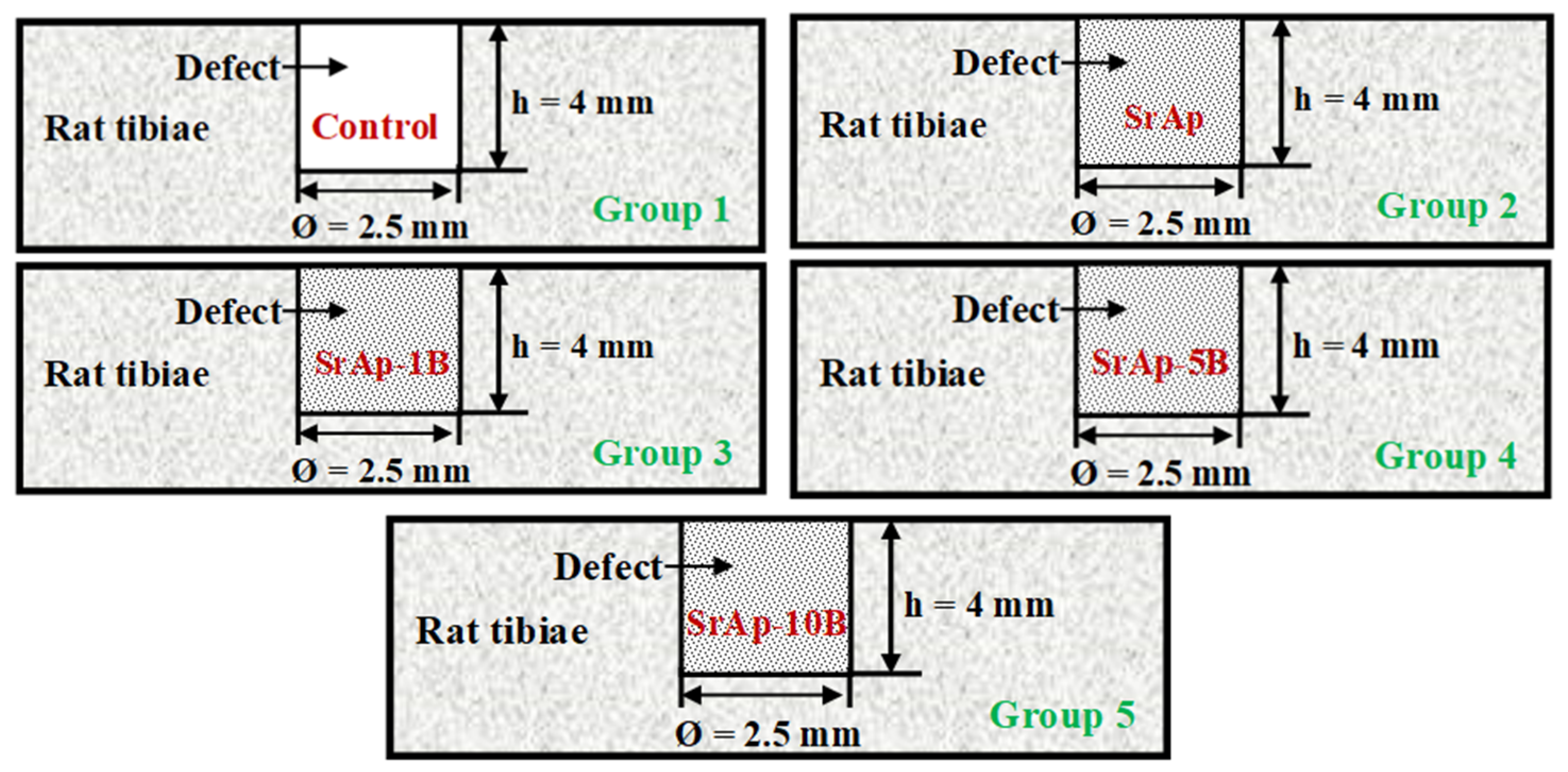
2.3. Animals and Study Design
2.4. Surgical Procedures
2.5. Histopathological Procedures
2.6. Statistical Analysis
3. Results
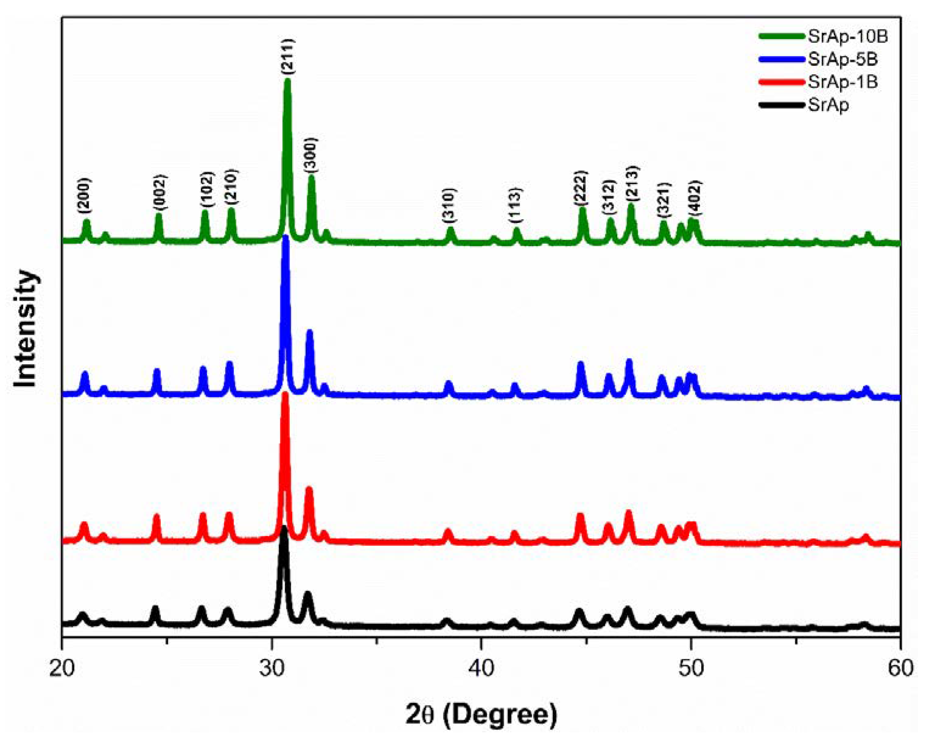
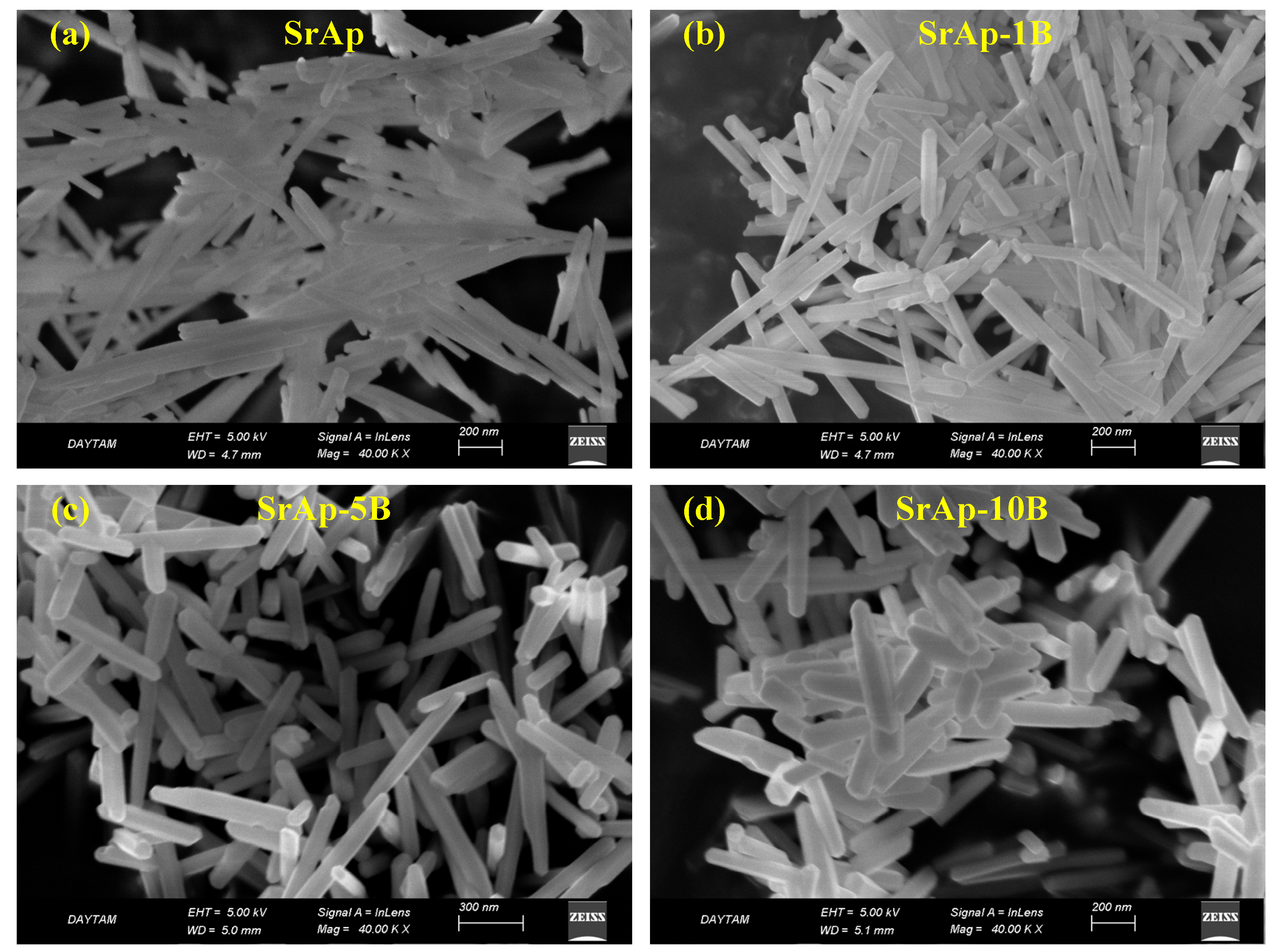
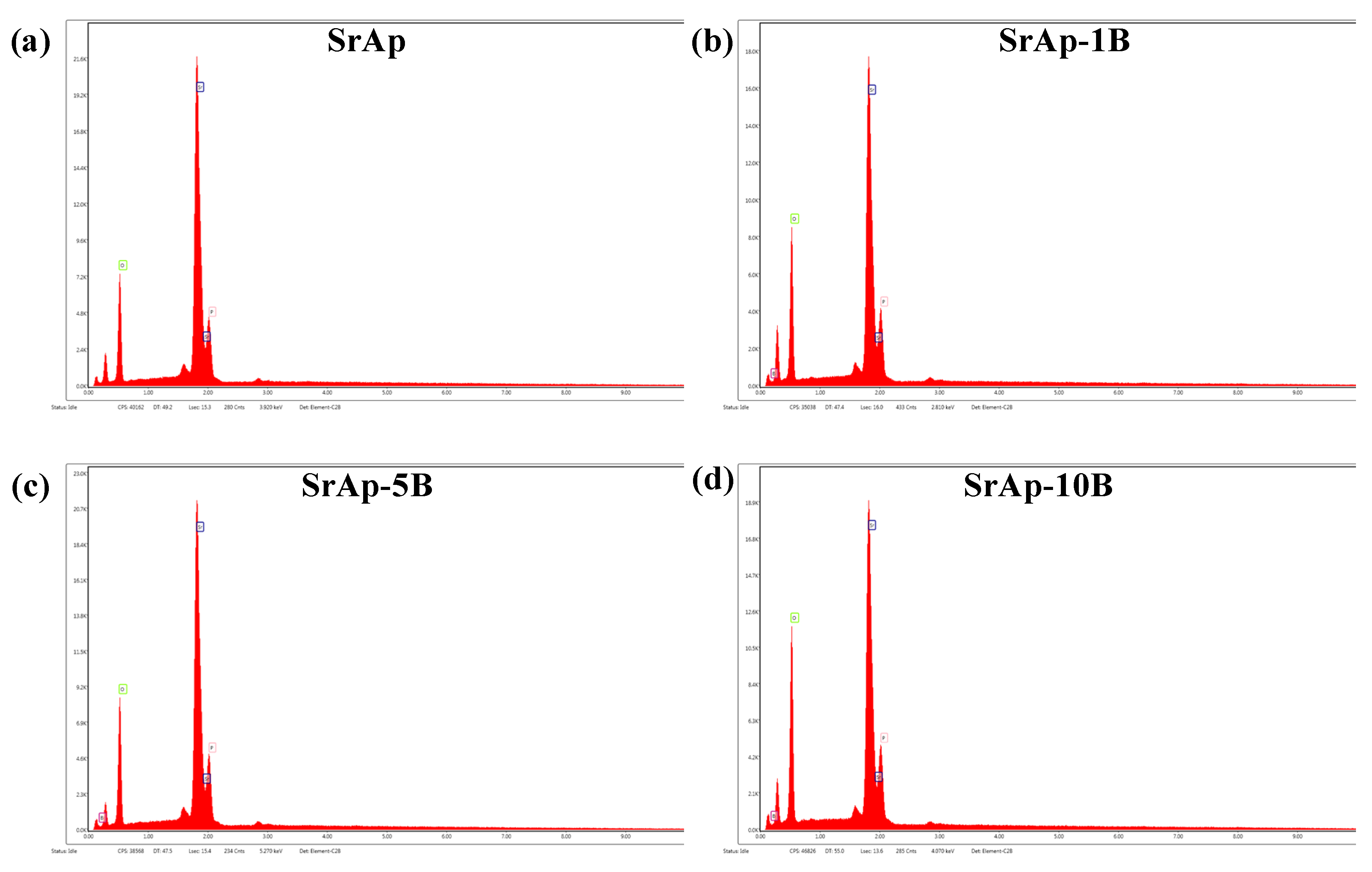
3.1. Structural and Morphological Characterizations of Fabricated Nanoparticles
3.2. In Vivo Biocompatibility Characterizations
4. Discussion
5. Conclusions
Supplementary Materials
Author Contributions
Funding
Institutional Review Board Statement
Informed Consent Statement
Data Availability Statement
Conflicts of Interest
References
- Figdor, D. Apical periodontitis: A very prevalent problem. Oral Surg. Oral Med. Oral Pathol. 2002, 94, 651–652. [Google Scholar] [CrossRef] [PubMed] [Green Version]
- Segura-Egea, J.J.; Castellanos-Cosano, L.; Machuca, G.; López-López, J.; Martín-González, J.; Velasco-Ortega, E.; Sánchez-Domínguez, B.; López-Frías, F.J. Diabetes mellitus, periapical inflammation and endodontic treatment outcome. Med. Oral Patol. Oral Cir. Bucal. 2012, 17, e356. [Google Scholar] [CrossRef] [PubMed] [Green Version]
- Lazarski, M.P.; Walker, W.A., III; Flores, C.M.; Schindler, W.G.; Hargreaves, K.M. Epidemiological evaluation of the outcomes of nonsurgical root canal treatment in a large cohort of insured dental patients. J. Endod. 2001, 27, 791–796. [Google Scholar] [CrossRef] [PubMed] [Green Version]
- Rahbaran, S.; Gilthorpe, M.S.; Harrison, S.D.; Gulabivala, K. Comparison of clinical outcome of periapical surgery in endodontic and oral surgery units of a teaching dental hospital: A retrospective study. Oral Surg. Oral Med. Oral Pathol. Oral Radiol. Endod. 2001, 91, 700–709. [Google Scholar] [CrossRef]
- Nair, P.R. Light and electron microscopic studies of root canal flora and periapical lesions. J. Endod. 1987, 13, 29–39. [Google Scholar] [CrossRef]
- Nair, P.R. Pathogenesis of apical periodontitis and the causes of endodontic failures. Crit. Rev. Oral Biol. Med. 2004, 15, 348–381. [Google Scholar] [CrossRef] [Green Version]
- Ricucci, D.; Siqueira, J.F., Jr. Fate of the tissue in lateral canals and apical ramifications in response to pathologic conditions and treatment procedures. J. Endod. 2010, 36, 1–15. [Google Scholar] [CrossRef]
- Persoon, I.; Özok, A. Definitions and epidemiology of endodontic infections. Curr. Oral Health Rep. 2017, 4, 278–285. [Google Scholar] [CrossRef] [Green Version]
- Smadi, L. Apical periodontitis and endodontic treatment in patients with type II diabetes mellitus: Comparative cross-sectional survey. J. Contemp. Dent. Pract. 2017, 18, 358–362. [Google Scholar]
- Liljestrand, J.; Mäntylä, P.; Paju, S.; Buhlin, K.; Kopra, K.; Persson, G.; Hernandez, M.; Nieminen, M.; Sinisalo, J.; Tjäderhane, L. Association of endodontic lesions with coronary artery disease. J. Dent. Res. 2016, 95, 1358–1365. [Google Scholar] [CrossRef] [Green Version]
- López-López, J.; Castellanos-Cosano, L.; Estrugo-Devesa, A.; Gómez-Vaquero, C.; Velasco-Ortega, E.; Segura-Egea, J.J. Radiolucent periapical lesions and bone mineral density in post-menopausal women. Gerodontology 2015, 32, 195–201. [Google Scholar] [CrossRef]
- Von Arx, T.; AlSaeed, M. The use of regenerative techniques in apical surgery: A literature review. Saudi Dent. J. 2011, 23, 113–127. [Google Scholar] [CrossRef] [Green Version]
- Christiansen, R.; Kirkevang, L.L.; Hørsted-Bindslev, P.; Wenzel, A. Randomized clinical trial of root-end resection followed by root-end filling with mineral trioxide aggregate or smoothing of the orthograde gutta-percha root filling–1-year follow-up. Int. Endod. J. 2009, 42, 105–114. [Google Scholar] [CrossRef]
- Uğur Aydın, Z.; Toptaş, O.; Göller Bulut, D.; Akay, N.; Kara, T.; Akbulut, N. Effects of root-end filling on the fractal dimension of the periapical bone after periapical surgery: Retrospective study. Clin. Oral Investig. 2019, 23, 3645–3651. [Google Scholar] [CrossRef]
- Von Arx, T. Apical surgery: A review of current techniques and outcome. Saudi Dent. J. 2011, 23, 9–15. [Google Scholar] [CrossRef] [Green Version]
- Nasker, P.; Mukherjee, M.; Kant, S.; Tripathy, S.; Sinha, A.; Das, M. Fluorine substituted nano hydroxyapatite: Synthesis, bio-activity and antibacterial response study. Ceram. Int. 2018, 44, 22008–22013. [Google Scholar] [CrossRef]
- Farzadi, A.; Bakhshi, F.; Solati-Hashjin, M.; Asadi-Eydivand, M.; abu Osman, N.A. Magnesium incorporated hydroxyapatite: Synthesis and structural properties characterization. Ceram. Int. 2014, 40, 6021–6029. [Google Scholar] [CrossRef] [Green Version]
- Wierichs, R.J.; Wolf, T.G.; Campus, G.; Carvalho, T.S. Efficacy of nano-hydroxyapatite on caries prevention—A systematic review and meta-analysis. Clin. Oral Investig. 2022, 26, 3373–3381. [Google Scholar] [CrossRef]
- Ge, M.; Ge, K.; Gao, F.; Yan, W.; Liu, H.; Xue, L.; Jin, Y.; Ma, H.; Zhang, J. Biomimetic mineralized strontium-doped hydroxyapatite on porous poly (l-lactic acid) scaffolds for bone defect repair. Int. J. Nanomed. 2018, 13, 1707. [Google Scholar] [CrossRef] [Green Version]
- Huang, J.; Huang, Z.; Yao, D.; Wu, C.; Cheng, Y.; Wu, F. Crystallization process and growth mechanism for hexagonal prism of strontium hydroxyapatite by urea hydrolysis. J. Cryst. Growth 2019, 512, 105–111. [Google Scholar] [CrossRef]
- Ma, X.; Liu, Y.; Zhu, B. Synthesis and characterization of pure strontium apatite particles and nanoporous scaffold prepared by dextrose-templated method. Mater. Res. Express 2018, 5, 025002. [Google Scholar] [CrossRef]
- Gurgenc, T.; Biryan, F. Production, thermal and dielectrical properties of Ag-doped nano-strontium apatite and nano h-BN filled poly (4-(3-(2, 3, 4-trimethoxyphenyl) acryloyl) phenyl acrylate) composites. J. Polym. Res. 2020, 27, 194. [Google Scholar] [CrossRef]
- Raucci, M.G.; Giugliano, D.; Alvarez-Perez, M.; Ambrosio, L. Effects on growth and osteogenic differentiation of mesenchymal stem cells by the strontium-added sol–gel hydroxyapatite gel materials. J. Mater. Sci. Mater. Med. 2015, 26, 90. [Google Scholar] [CrossRef] [PubMed]
- Feng, Q.L.; Kim, T.N.; Wu, J.; Park, E.S.; Kim, J.O.; Lim, D.Y.; Cui, F.Z. Antibacterial effects of Ag-HAp thin films on alumina substrates. Thin Solid Films 1998, 335, 214–219. [Google Scholar] [CrossRef]
- Anwar, A.; Asghar, M.N.; Kanwal, Q.; Kazmi, M.; Sadiqa, A. Low temperature synthesis and characterization of carbonated hydroxyapatite nanocrystals. J. Mol. Struct. 2016, 1117, 283–286. [Google Scholar] [CrossRef]
- Aroni, M.A.T.; de Oliveira, G.J.P.L.; Spolidório, L.C.; Andersen, O.Z.; Foss, M.; Marcantonio, R.A.C.; Stavropoulos, A. Loading deproteinized bovine bone with strontium enhances bone regeneration in rat calvarial critical size defects. Clin. Oral Investig. 2019, 23, 1605–1614. [Google Scholar] [CrossRef] [Green Version]
- Gurgenc, T. Structural characterization and dielectrical properties of Ag-doped nano-strontium apatite particles produced by hydrothermal method. J. Mol. Struct. 2021, 1223, 128990. [Google Scholar] [CrossRef]
- Pazarçeviren, A.E.; Tezcaner, A.; Keskin, D.; Kolukısa, S.T.; Sürdem, S.; Evis, Z. Boron-doped biphasic hydroxyapatite/β-tricalcium phosphate for bone tissue engineering. Biol. Trace Elem. Res. 2021, 199, 968–980. [Google Scholar] [CrossRef]
- Atila, D.; Karataş, A.; Evcin, A.; Keskin, D.; Tezcaner, A. Bacterial cellulose-reinforced boron-doped hydroxyapatite/gelatin scaffolds for bone tissue engineering. Cellulose 2019, 26, 9765–9785. [Google Scholar] [CrossRef]
- Gopi, D.; Ramya, S.; Rajeswari, D.; Karthikeyan, P.; Kavitha, L. Strontium, cerium co-substituted hydroxyapatite nanoparticles: Synthesis, characterization, antibacterial activity towards prokaryotic strains and in vitro studies. Colloids Surf. Physicochem. Eng. Asp. 2014, 451, 172–180. [Google Scholar] [CrossRef]
- Esmaeilkhanian, A.; Sharifianjazi, F.; Abouchenari, A.; Rouhani, A.; Parvin, N.; Irani, M. Synthesis and characterization of natural nano-hydroxyapatite derived from turkey femur-bone waste. Appl. Biochem. Biotechnol. 2019, 189, 919–932. [Google Scholar] [CrossRef]
- Furukawa, A. The formation of strontium apatites through alkaline hydrolysis of strontium hydrogen phosphate and their crystallographic characterization. Ceram. Int. 2021, 47, 21848–21861. [Google Scholar] [CrossRef]
- Slimen, J.B.; Mehnaoui, M.; Jebahi, S.; Boughzala, K.; Hidouri, M. Thermal and structural properties of sodium, potassium and carbonate doped strontium hydroxyfluorapatite. J. Indian Chem. Soc. 2022, 99, 100475. [Google Scholar] [CrossRef]
- Tunçay, E.Ö.; Demirtaş, T.T.; Gümüşderelioğlu, M. Microwave-induced production of boron-doped HAp (B-HAp) and B-HAp coated composite scaffolds. J. Trace Elem. Med. Biol. 2017, 40, 72–81. [Google Scholar] [CrossRef]
- Jodati, H.; Tezcaner, A.; Alshemary, A.Z.; Şahin, V.; Evis, Z. Effects of the doping concentration of boron on physicochemical, mechanical, and biological properties of hydroxyapatite. Ceram. Int. 2022, 48, 22743–22758. [Google Scholar] [CrossRef]
- Yedekçi, B.; Tezcaner, A.; Alshemary, A.Z.; Yılmaz, B.; Demir, T.; Evis, Z. Synthesis and sintering of B, Sr, Mg multi-doped hydroxyapatites: Structural, mechanical and biological characterization. J. Mech. Behav. Biomed. Mater. 2021, 115, 104230. [Google Scholar] [CrossRef]
- Chen, X.B.; Nisbet, D.R.; Li, R.W.; Smith, P.; Abbott, T.B.; Easton, M.A.; Zhang, D.-H.; Birbilis, N. Controlling initial biodegradation of magnesium by a biocompatible strontium phosphate conversion coating. Acta Biomater. 2014, 10, 1463–1474. [Google Scholar] [CrossRef]
- Maia, M.T.; Luz, É.P.C.G.; Andrade, F.K.; Rosa, M.d.F.; Borges, M.d.F.; Arcanjo, M.R.A.; Vieira, R.S. Advances in bacterial cellulose/strontium apatite composites for bone applications. Polym. Rev. 2021, 61, 736–764. [Google Scholar] [CrossRef]
- Kolmas, J.; Velard, F.; Jaguszewska, A.; Lemaire, F.; Kerdjoudj, H.; Gangloff, S.C.; Kaflak, A. Substitution of strontium and boron into hydroxyapatite crystals: Effect on physicochemical properties and biocompatibility with human Wharton-Jelly stem cells. Mater. Sci. Eng. C 2017, 79, 638–646. [Google Scholar] [CrossRef]
- Cullity, B. Elements of X-ray Diffraction; Addison-Wesely: Read, MA, USA, 1978. [Google Scholar]
- Badran, H.; Yahia, I.; Hamdy, M.S.; Awwad, N. Lithium-doped hydroxyapatite nano-composites: Synthesis, characterization, gamma attenuation coefficient and dielectric properties. Radiat. Phys. Chem. 2017, 130, 85–91. [Google Scholar] [CrossRef]
- Akbulut, Y.; Gul, M.; Dundar, S.; Ozcan, E.C.; Ozercan, I.H.; Bozoglan, A.; Karasu, N.; Acikan, I.; Bingül, M.B. Evaluation of Effects of Systemic Zoledronic Acid Application on Bone Maturation in the Consolidation Period in Distraction Osteogenesis. J. Craniofac. Surg. 2021, 32, 2901–2905. [Google Scholar] [CrossRef]
- Nimmy, E.; Wilson, P. Investigations on sonofragmentation of hydroxyapatite crystals as a function of strontium incorporation. Ultrason. Sonochem. 2019, 50, 188–199. [Google Scholar]
- Stanić, V.; Janaćković, D.; Dimitrijević, S.; Tanasković, S.B.; Mitrić, M.; Pavlović, M.S.; Krstić, A.; Jovanović, D.; Raičević, S. Synthesis of antimicrobial monophase silver-doped hydroxyapatite nanopowders for bone tissue engineering. Appl. Surface Sci. 2011, 257, 4510–4518. [Google Scholar] [CrossRef]
- Ravi, N.D.; Balu, R.; Sampath Kumar, T. Strontium-substituted calcium deficient hydroxyapatite nanoparticles: Synthesis, characterization, and antibacterial properties. J. Am. Ceram. Soc. 2012, 95, 2700–2708. [Google Scholar] [CrossRef]
- Kavitha, R.; Ravichandran, K.; Narayanan, T.S. Deposition of strontium phosphate coatings on magnesium by hydrothermal treatment: Characteristics, corrosion resistance and bioactivity. J. Alloys Compd. 2018, 745, 725–743. [Google Scholar] [CrossRef]
- Zhu, X.; Eibl, O.; Scheideler, L.; Geis-Gerstorfer, J. Characterization of nano hydroxyapatite/collagen surfaces and cellular behaviors. J. Biomed. Mater. Res. Part A Off. J. Soc. Biomater. Jpn. Soc. Biomater. Aust. Soc. Biomater. Korean Soc. Biomater. 2006, 79, 114–127. [Google Scholar] [CrossRef] [PubMed]
- Corno, M.; Busco, C.; Civalleri, B.; Ugliengo, P. Periodic ab initio study of structural and vibrational features of hexagonal hydroxyapatite Ca10(PO4)6(OH)2. Phys. Chem. Chem. Phys. 2006, 8, 2464–2472. [Google Scholar] [CrossRef] [PubMed]
- Aksoy, M.; Aksakal, B.; Aslan, N.; Dikici, B. Boron-Doped Hydroxyapatite Coatings on NiTi Alloys Using the Electrophoretic Deposition Method: Enhanced Corrosion and Adhesion Performances. J. Mater. Eng. Perform. 2021, 30, 7365–7375. [Google Scholar] [CrossRef]
- Geng, Z.; Cui, Z.; Li, Z.; Zhu, S.; Liang, Y.; Liu, Y.; Li, X.; He, X.; Yu, X.; Wang, R. Strontium incorporation to optimize the antibacterial and biological characteristics of silver-substituted hydroxyapatite coating. Mater. Sci. Eng. C 2016, 58, 467–477. [Google Scholar] [CrossRef]
- Kim, H.; Mondal, S.; Bharathiraja, S.; Manivasagan, P.; Moorthy, M.S.; Oh, J. Optimized Zn-doped hydroxyapatite/doxorubicin bioceramics system for efficient drug delivery and tissue engineering application. Ceram. Int. 2018, 44, 6062–6071. [Google Scholar] [CrossRef]
- Bose, S.; Mandal, S.; Barua, A.K.; Mukhopadhyay, S. Properties of boron doped ZnO films prepared by reactive sputtering method: Application to amorphous silicon thin film solar cells. J. Mater. Sci. Technol. 2020, 55, 136–143. [Google Scholar] [CrossRef]
- Riaz, M.; Zia, R.; Ijaz, A.; Hussain, T.; Mohsin, M.; Malik, A. Synthesis of monophasic Ag doped hydroxyapatite and evaluation of antibacterial activity. Mater. Sci. Eng. C 2018, 90, 308–313. [Google Scholar] [CrossRef]
- Shi, C.; Gao, J.; Wang, M.; Fu, J.; Wang, D.; Zhu, Y. Ultra-trace silver-doped hydroxyapatite with non-cytotoxicity and effective antibacterial activity. Mater. Sci. Eng. C 2015, 55, 497–505. [Google Scholar] [CrossRef]
- Ogo, S.; Onda, A.; Yanagisawa, K. Selective synthesis of 1-butanol from ethanol over strontium phosphate hydroxyapatite catalysts. Appl. Catal. A Gen. 2011, 402, 188–195. [Google Scholar] [CrossRef]
- Prekajski, M.; Mirković, M.; Todorović, B.; Matković, A.; Marinović-Cincović, M.; Luković, J.; Matović, B. Ouzo effect—New simple nanoemulsion method for synthesis of strontium hydroxyapatite nanospheres. J. Eur. Ceram. Soc. 2016, 36, 1293–1298. [Google Scholar] [CrossRef]
- Reem, A.-W.; Jafer, R.; Yahia, I.; Al-Ghamdi, A.A.; Al-ghamdi, M.A.; El-Naggar, A. Fast and easy synthesis of novel Strontium apatite nanostructured phase: Structure, spectroscopy, and dielectric analysis. Ceram. Int. 2017, 43, 17153–17159. [Google Scholar]
- Radovanović, Ž.; Jokić, B.; Veljović, D.; Dimitrijević, S.; Kojić, V.; Petrović, R.; Janaćković, D. Antimicrobial activity and biocompatibility of Ag+-and Cu2+-doped biphasic hydroxyapatite/α-tricalcium phosphate obtained from hydrothermally synthesized Ag+-and Cu2+-doped hydroxyapatite. Appl. Surface Sci. 2014, 307, 513–519. [Google Scholar] [CrossRef]
- Wang, C.; Feng, J.; Zhou, J.; Huang, X.; Wang, L.; Liu, G.; Cheng, J. Microstructure, mechanical properties and in vitro biocompatibilities of a novel bionic hydroxyapatite bone scaffold prepared by the addition of boron nitride. J. Mater. Sci. 2020, 55, 14501–14515. [Google Scholar] [CrossRef]
- O’Donnell, M.; Fredholm, Y.; De Rouffignac, A.; Hill, R. Structural analysis of a series of strontium-substituted apatites. Acta Biomater. 2008, 4, 1455–1464. [Google Scholar] [CrossRef]
- Boughzala, K.; Salem, E.B.; Chrifa, A.B.; Gaudin, E.; Bouzouita, K. Synthesis and characterization of strontium–lanthanum apatites. Mater. Res. Bull. 2007, 42, 1221–1229. [Google Scholar] [CrossRef]
- Pogosova, M.; González, L. Influence of anion substitution on the crystal structure and color properties of copper-doped strontium hydroxyapatite. Ceram. Int. 2018, 44, 20140–20147. [Google Scholar] [CrossRef]
- Frasnelli, M.; Cristofaro, F.; Sglavo, V.M.; Dirè, S.; Callone, E.; Ceccato, R.; Bruni, G.; Cornaglia, A.I.; Visai, L. Synthesis and characterization of strontium-substituted hydroxyapatite nanoparticles for bone regeneration. Mater. Sci. Eng. C 2017, 71, 653–662. [Google Scholar] [CrossRef] [PubMed]
- Korkmaz, M.; Turkmen, R.; Demirel, H.H.; Saritas, Z.K. Effect of boron on the repair of osteochondral defect and oxidative stress in rats: An experimental study. Biol. Trace Elem. Res. 2019, 187, 425–433. [Google Scholar] [CrossRef] [PubMed]
- Fredholm, Y.C.; Karpukhina, N.; Brauer, D.S.; Jones, J.R.; Law, R.V.; Hill, R.G. Influence of strontium for calcium substitution in bioactive glasses on degradation, ion release and apatite formation. J. R. Soc. Interface 2012, 9, 880–889. [Google Scholar] [CrossRef] [Green Version]
- Cassino, P.C.; Rosseti, L.S.; Ayala, O.I.; Martines, M.A.U.; Portugual, L.C.; Oliveira, C.G.d.; Silva, I.S.; Caldas, R.d.A. Potencial of different hydroxyapatites as biomaterials in the bone remodeling. Acta Cir. Bras. 2018, 33, 816–823. [Google Scholar] [CrossRef]
- Machado, C.P.G.; Pintor, A.V.B.; Gress, M.A.K.d.A.; Rossi, A.M.; Granjeiro, J.M.; Maia, M.D.C. Avaliação da hidroxiapatita contendo estrôncio como substituto ósseo em tíbias de ovelhas. Innov. Implant J. 2010, 5, 9–14. [Google Scholar]
- Isaac, J.; Nohra, J.; Lao, J.; Jallot, E.; Nedelec, J.-M.; Berdal, A.; Sautier, J.-M. Effects of strontium-doped bioactive glass on the differentiation of cultured osteogenic cells. Eur. Cells Mater. 2011, 21, e43. [Google Scholar] [CrossRef]
- Resende, A.C.d.; Cunha, L.R.d.; Saska, S.; Balducci-Roslindo, E.; Minarelli-Gaspar, A.M. Análise biológica da hidroxiapatita após implantação em tíbias de ratos. Rev. Bras. Ortop. 2006, 41, 132–136. [Google Scholar]
- Ciftci, E.; Köse, S.; Korkusuz, P.; Timuçin, M.; Korkusuz, F. Boron containing nano hydroxyapatites (Bn-HAp) stimulate mesenchymal stem cell adhesion, proliferation and differentiation. Key Eng. Mater. 2015, 631, 373–378. [Google Scholar] [CrossRef]
- Arslan, A.; Çakmak, S.; Gümüşderelioğlu, M. Enhanced osteogenic activity with boron-doped nanohydroxyapatite-loaded poly (butylene adipate-co-terephthalate) fibrous 3D matrix. Artif. Cells Nanomed. Biotechnol. 2018, 46, 790–799. [Google Scholar] [CrossRef] [Green Version]
- Gültan, T.; Yurtsever, M.Ç.; Gümüşderelioğlu, M. NaOH-etched/boron-doped nanohydroxyapatite-coated PEEK implants enhance the proliferation and differentiation of osteogenic cells. Biomed. Mater. 2020, 15, 035019. [Google Scholar] [CrossRef]
- Gölge, U.H.; Kaymaz, B.; Arpaci, R.; Kömürcü, E.; Göksel, F.; Güven, M.; Güzel, Y.; Cevizci, S. Effects of boric acid on fracture healing: An experimental study. Biol. Trace Elem. Res. 2015, 167, 264–271. [Google Scholar] [CrossRef]
- Gizer, M.; Köse, S.; Karaosmanoglu, B.; Taskiran, E.Z.; Berkkan, A.; Timuçin, M.; Korkusuz, F.; Korkusuz, P. The effect of boron-containing nano-hydroxyapatite on bone cells. Biol. Trace Elem. Res. 2020, 193, 364–376. [Google Scholar] [CrossRef]
- Çiftci Dede, E.; Korkusuz, P.; Bilgiç, E.; Çetinkaya, M.A.; Korkusuz, F. Boron Nano-hydroxyapatite composite increases the bone regeneration of Ovariectomized rabbit femurs. Biol. Trace Elem. Res. 2022, 200, 183–196. [Google Scholar] [CrossRef]
- Gentleman, E.; Fredholm, Y.C.; Jell, G.; Lotfibakhshaiesh, N.; O’Donnell, M.D.; Hill, R.G.; Stevens, M.M. The effects of strontium-substituted bioactive glasses on osteoblasts and osteoclasts in vitro. Biomaterials 2010, 31, 3949–3956. [Google Scholar] [CrossRef] [Green Version]


| Sample | a = b (Å) | c (Å) | V (Å3) | D002 (nm) | D300 (nm) | Dave. (nm) | η (nm−2) |
|---|---|---|---|---|---|---|---|
| SrAp | 9.7701 | 7.2726 | 601.176 | 51.67 | 18.21 | 34.94 | 8.191 × 10⁻⁴ |
| SrAp-1B | 9.7461 | 7.2580 | 597.027 | 49.01 | 30.38 | 39.70 | 6.345 × 10⁻⁴ |
| SrAp-5B | 9.7282 | 7.2492 | 594.123 | 52.61 | 37.24 | 44.93 | 4.954 × 10⁻⁴ |
| SrAp-10B | 9.7074 | 7.2347 | 590.402 | 56.34 | 40.11 | 48.23 | 4.299 × 10⁻⁴ |
| Sample | Sr | P | O | B | Sr/P |
|---|---|---|---|---|---|
| SrAp | 25.19 | 14.32 | 60.49 | - | 1.76 |
| SrAp-1B | 11.50 | 6.55 | 37.79 | 44.16 | 1.76 |
| SrAp-5B | 17.57 | 10.47 | 48.83 | 23.13 | 1.68 |
| SrAp-10B | 11.22 | 7.11 | 45.56 | 36.11 | 1.58 |
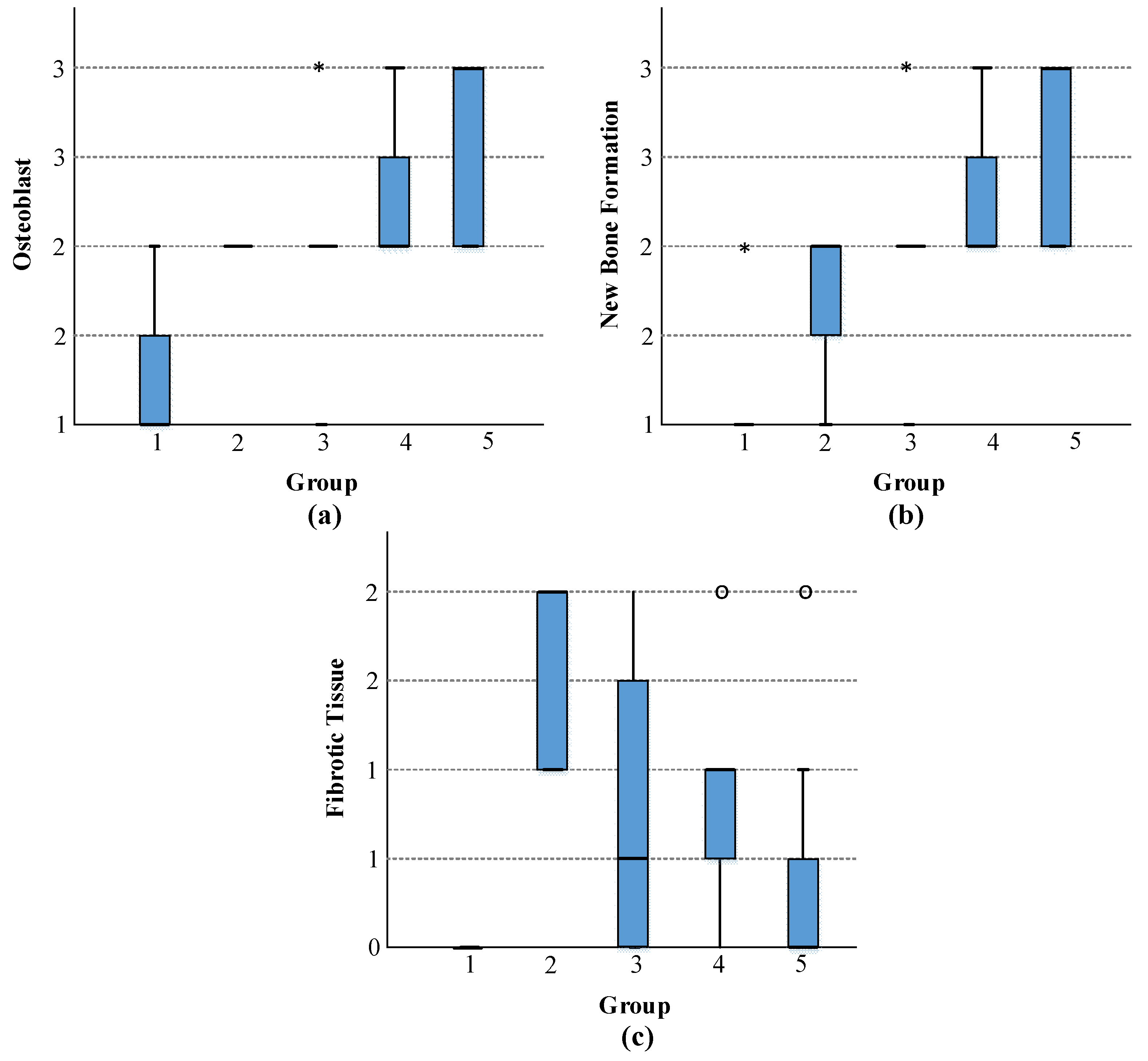
| Osteoblast Median (Min–Max) (n = 8) | New Bone Formation Median (Min–Max) (n = 8) | Fibrotic Tissue Median (Min–Max) (n = 8) | |
|---|---|---|---|
| Control (1) | 1 (1–2) | 1 (1–2) | 0 (0–0) |
| Strontium (2) | 2 (2–2) | 2 (1–2) | 2 (1–2) |
| Strontium + 1% Boron (3) | 2 (1–3) | 2 (1–3) | 0.5 (0–2) |
| Strontium + 5% Boron (4) | 2 (2–3) | 2 (2–3) | 1 (0–2) |
| Strontium + 10% Boron (5) | 3 (2–3) | 3 (2–3) | 0 (0–2) |
| p | <0.001 | <0.001 | <0.001 |
| p * | 1–4: p = 0.014 1–5: p < 0.001 Others: p > 0.05 | 1–4: p = 0.007 1–5: p < 0.001 Others: p > 0.05 | 1–2: p = 0.001 2–5: p = 0.020 Others: p > 0.05 |
Publisher’s Note: MDPI stays neutral with regard to jurisdictional claims in published maps and institutional affiliations. |
© 2022 by the authors. Licensee MDPI, Basel, Switzerland. This article is an open access article distributed under the terms and conditions of the Creative Commons Attribution (CC BY) license (https://creativecommons.org/licenses/by/4.0/).
Share and Cite
Oztekin, F.; Gurgenc, T.; Dundar, S.; Ozercan, I.H.; Yildirim, T.T.; Eskibaglar, M.; Ozcan, E.C.; Macit, C.K. In Vivo Evaluation of the Effects of B-Doped Strontium Apatite Nanoparticles Produced by Hydrothermal Method on Bone Repair. J. Funct. Biomater. 2022, 13, 110. https://doi.org/10.3390/jfb13030110
Oztekin F, Gurgenc T, Dundar S, Ozercan IH, Yildirim TT, Eskibaglar M, Ozcan EC, Macit CK. In Vivo Evaluation of the Effects of B-Doped Strontium Apatite Nanoparticles Produced by Hydrothermal Method on Bone Repair. Journal of Functional Biomaterials. 2022; 13(3):110. https://doi.org/10.3390/jfb13030110
Chicago/Turabian StyleOztekin, Faruk, Turan Gurgenc, Serkan Dundar, Ibrahim Hanifi Ozercan, Tuba Talo Yildirim, Mehmet Eskibaglar, Erhan Cahit Ozcan, and Cevher Kursat Macit. 2022. "In Vivo Evaluation of the Effects of B-Doped Strontium Apatite Nanoparticles Produced by Hydrothermal Method on Bone Repair" Journal of Functional Biomaterials 13, no. 3: 110. https://doi.org/10.3390/jfb13030110