Article

Endophytic Fungi of Emersed Halophytes in River Deltas and Tidal Flats of the Korean Ramsar Wetlands

1
Water Quality Research Institute, Waterworks Headquarters Incheon Metropolitan City, Incheon 21316, Korea
2
Incheon Metropolitan City Institute of Public Health and Environment, Incheon 22320, Korea
3
Department of Hydrogen and Renewable Energy, Kyungpook National University, Daegu 41566, Korea
4
Microorganism Resources Division, National Institute of Biological Resources, Incheon 22689, Korea
5
School of Life Sciences, Kyungpook National University, Daegu 41566, Korea
*
Authors to whom correspondence should be addressed.
J. Mar. Sci. Eng. 2021, 9(4), 430; https://doi.org/10.3390/jmse9040430
Submission received: 10 March 2021 / Revised: 7 April 2021 / Accepted: 11 April 2021 / Published: 15 April 2021
(This article belongs to the Special Issue Coastal Wetlands)

Abstract

:
This study aimed to obtain information on the diversity and distribution of the endophytic fungi in Ramsar wetlands. Vast salt marshes in Suncheon Bay, Korea, are formed by two types of ecotones (tidal flats and deltas) that are supported by the emersed halophytes Phragmites australis and Suaeda japonica. Overall, 324 endophytes were isolated from P. australis (six sampling points in the delta and five in the tidal flats) and S. japonica (six in tidal flats). Margalef’s, Menhinick’s, Shannon’s, and Simpson’s diversity indices significantly varied among the ecotones. In particular, higher variance in diversity value and unevenness was observed in the delta marsh compared with the tidal flat marsh. Further, morphological diversity in the delta salt marsh was 1.8 times higher than that of the tidal flat. Comprising several dominant genera (Aspergillus, Cladosporium, and Penicillium), Epicoccum, Paraconiothyrium, Septoriella, and Talaromyces were widely distributed regardless of the aquatic conditions or halophyte species. This study highlights the distinct distribution and diversity of marine endophytes in various ecotones that can physically protect the coastal areas. This data is of much value to secure a national culture collection for future restoration of the coastal wetlands and their ecosystems.

1. Introduction

Salt marshes (coastal wetlands), transitional zones between land and ocean, are areas in which water is permanently collected, thereby constituting a biodiversity repository [1,2]. One of the origins of salt marshes is tidal flats in coastal areas, a type of ecotone between land and ocean. They develop well on coasts protected by bays or islands, being exposed during low tide and submerged below sea level during high tide [2]. Salt marshes also develop in estuaries at river mouths where freshwater, seawater, and land meet [1]. As the water flow rate slows down in estuaries, inorganic and organic matter form sedimentary sites. In turn, condensed flourishing of emersed halophytes in those sites creates structurally stable salt marshes or coastal wetlands [2,3].
However, because salt marshes are structurally stable, they can be easily depleted by the construction of seawalls. For example, salt marshes on the Korean Peninsula have quickly disappeared [4,5], with 25% of tidal flats and coastal wetlands having disappeared within 50 years, while the destruction of the remaining region is ongoing [4,5]. After the 21st century, owing to preservation efforts, 23 Ramsar wetlands were secured [4,5], with the most representative salt marsh being Suncheon Bay. Because tidal flats surrounding the bay have developed and deltas have formed in the estuary, these two types of ecotones are a valuable repository of vast animal and microbial biodiversity. In addition to the environmental effect of tidal ranges unique to tidal flats, severe freshwater flow from the large river represents an additional physical stress, leading to the dominance of certain emersed halophytes that can withstand the water flow rate. Due to the advantage of these emersed halophytes, huge deltas and tidal flat salt marshes are physically supported and are still expanding in Suncheon Bay [4,5].
The emersed plants Phragmites australis and Suaeda japonica form a highly dominant colony with high density, a complex environmental feature that differs from other salt marshes in the Korean Peninsula. In addition, the emersed halophyte species, which survive and thrive in this complex and ever-changing environment, have a differentiated ecology from the coastal plants growing on simple coastal sandy beaches. The salt marshes developed in river estuaries and tidal flats, as well as their P. australis and S. japonica populations native to Suncheon Bay, serve as a natural purification system, as the potential habitat of various higher life forms, and also as a supporter or extender of the natural salt marshes. Therefore, the ecology of these two emersed halophyte species hold important academic and practical value for future restoration of other disappearing coastal wetlands.
Endophytic microorganisms play key roles in plant growth and reproduction, and can also induce resistance to harsh environments, such as extreme marine conditions [6,7,8]. Therefore, securing the culture collection of endophytic fungi native to emersed halophyte species and identifying their ecological diversity at well-preserved sites are essential for restoring salt marshes in the future. Such a culture collection representative of the biodiversity of these areas would be of great value in light of the Nagoya Protocol and to secure the availability of biological resources [9,10].
In this study, molecular identification of the endophytic fungi within P. australis and S. japonica species was conducted using the signature sequences of 324 fungi isolated based on their morphological features. Their distribution and diversity per salt marsh and per plant species was analyzed, and their phylogenetic relationships were derived. Overall, this study aimed to assess the differences among salt marshes developed in deltas and tidal flats, and how these affect the characteristics of endophytic clusters in the well-conserved Ramsar wetlands in the Korean Republic.

2. Materials and Methods

2.1. Site Description

The salt marshes of Suncheon Bay reflect the vast, complex, and diverse aquatic conditions of the Ramsar wetlands, located on the southern coast of the Korean Peninsula (Figure 1).
The designated area of the Ramsar wetlands covers salt marshes formed around the deltas of river estuaries (Figure 2A), where freshwater and seawater meet (a transitional zone) [12,13], and in tidal flats of the long beach, developed on the left and right of the inner side of the bay (Figure 2B,C). These ecotones not only contain species common to the communities on both sides, but also include several highly adaptable species that tend to colonize such transitional areas [13]. These two types of salt marshes that have developed in Suncheon Bay show saturated natural soil conditions without water drainage and with the underground water level approaching the surface [14]. According to soil texture classification, salt marshes have a fine clay layer of more than 25 m above the ground, and the area of the tidal flat is approximately 264,462 m2. A total of 25 genera and 33 halophyte or coastal plant species have been identified; however, shrubby sea blite (S. japonica) and common reed (P. australis) are the two dominant species in this area (Figure 1), due to the strong selective pressure of the ecotones. Other coastal or halophyte plants species inhabit the sand on the beaches from the tidal flats and deltas, but only P. australis and S. japonica serve to preserve the tidal flats and river estuaries.

2.2. Plant Sampling

Two emersed halophytic plant species, namely P. australis and S. japonica, were sampled from the tidal flats that are formed in two large areas south of the main river estuary (Figure 2B,C), and the estuary delta marsh that is formed by the accumulation of organic minerals in the opposite direction of the river bend (Figure 2A).
These three areas (Figure 2) represent the salt marsh geology that is strongly maintained by P. australis and S. japonica colonies for long time periods. Moreover, these three areas are continuously expanding due to this vegetation. Therefore, two emersed halophyte vegetations supporting the wetland terrain were sampled from the delta salt marsh (six sampling sites for P. australis) and tidal flats (five and six sampling sites for P. australis and S. japonica, respectively) (Table 1 and Figure 2). The total number of sampled sites was 17 across three regions. All of these colonies were collected from sites approximately 100 m apart from each other to ensure ecological segregation (Table 1). The roots of the halophyte species were sampled, followed by the harvest of well-flourished and conserved communities (15 individuals per each species), along with the collection of proximal root layer beach soils from their habitats to minimize physiological changes.

2.3. Isolation of Endophytic Fungi

This study adopted culture-dependent methods, in accordance with previous endophyte studies in coastal terrain [15,16] or other environments [17,18,19,20]. The samples were quickly transported at 4 °C to the laboratory. Sterile distilled water and sterilized 0.1% Tween-80 solution (Sigma-Aldrich, St. Louis, MO, USA) were sprayed on to the surface of the samples to eliminate suspended solids or normal microflora from the root surfaces. The plants were submerged in 1.0% perchloric acid for 10 min twice and were subsequently washed with sterile distilled water 3–4 times [21]. Residual water was eliminated using dry sterile gauze and 50 pieces of root from one plant sample were cut to a length of 3–4 cm. The pretreated samples were cultured in Hagem minimal medium containing 80 mg/L streptomycin (Sigma-Aldrich) to exclude root bacteria and incubated at 25 °C for 15 days [22,23]. Endophytic fungi were sub-cultured in the same media and under the same conditions. Finally, pure fungi isolates were incubated on potato dextrose agar (Difco, Detroit, MI, USA) and selected based on their morphological differences (Table 1).

2.4. Extraction of Genomic DNA and Target Gene Amplification

All of the 324 endophytic fungi collected from the 17 colonies of the two halophyte species native to the three salt marsh regions in Suncheon Bay were inoculated into potato dextrose broth (Difco) and incubated at 25 °C for 7 days at 120 rpm using a rotary shaker. Filtered mycobionts were lyophilized for 2 days. The DNeasy Plant Mini Kit (Qiagen, Hilden, Germany) was used to extract the genomic DNA from the lyophilized mycobionts and primers targeting the universal sequences internal transcribed spacer (ITS) 1, partial sequence; 5.8S ribosomal RNA and ITS2, complete sequence; and 28S ribosomal RNA, partial sequence), ITS1 (5′–TCC GTA GGT GAA CCT GCG G–3′), and ITS4 (5′–TCC TCC GCT TAT TGATAT GC–3′) were used for amplification [24]. The polymerase chain reaction (PCR) amplification conditions were as follows: pre-denaturation (94 °C, 4 min), denaturation (94 °C, 1 min), annealing (55–58 °C, 1 min), and extension (72 °C, 2 min) for a total of 35 cycles, followed by a final extension (72 °C, 2 min) [24]. The PCR products were confirmed by gel electrophoresis (1.5% agarose gel, stained with ethidium bromide), and the resulting band pattern was observed under a UV transilluminator. The AccuPrep PCR and Gel Extraction Kit (Bioneer, Daejeon, Korea) was used to purify the PCR products, and the ABI 3730XL DNA analyzer (Applied Biosystems, Waltham, MA, USA) was used for ITS sequencing.

2.5. Identification and Phylogeny

A total of 324 fungal isolates were partially identified based on signature sequence homology with data collected from the GenBank database [25]. The sequences of ITS1 and 28S (partial sequences), and 5.8S and IST2 (complete sequences) obtained from the fungal isolates were compared with the data deposited in the GenBank database using the BLASTn tool to determine their taxonomical relationships. Furthermore, evolutionary analyses were conducted using Molecular Evolutionary Genetics Analysis (MEGA) version X (64-bit for Windows) [26,27] with sequence alignments prepared using Clustal W version 2.0.10 [28,29]. The evolutionary history was inferred using the minimum evolution method to assess the evolutionary relationships between taxa [30], with ‘associated’ being taxa clustered together in a percentage of the replicate trees in the bootstrap test (1000 replicates) [31]. Evolutionary distances were computed using the Tajima–Nei method [32] and expressed according to the number of base substitutions per site. The minimum evolution tree was explored using the close-neighbor interchange algorithm [33] at a search level of 1. The neighbor-joining algorithm [34] was used to generate the initial tree. All ambiguous positions were removed for each sequence pair (pairwise deletion option). The 324 fungal signature sequences obtained in this study were deposited in GenBank with assigned accession numbers (Supplementary Tables S1 and S2).

2.6. Biodiversity Assessment

The diversity of the fungal flora was compared between each halophyte species (Table 2) [35] and the diversity at the genus level was determined using Margalef’s richness [35,36,37], Menhinick’s diversity [38,39], Shannon’s diversity [40], and Simpson’s indices (1-D) [37,41].

3. Results and Discussion

3.1. Taxonomy and Phylogeny

Among the P. australis samples, three phyla (Ascomycota (n = 178), Basidiomycota (n = 1), and Zygomycota (n = 1)), seven classes, 13 orders, 25 families, and 41 genera were identified (Table 2). Within S. japonica, two phyla (Ascomycota (n = 137) and Basidiomycota (n = 7)), six classes, 12 orders, 20 families, and 35 genera were identified (Table 2). The phylogeny of the endophyte clusters from each site was deduced (Figure S1), showing the phylogenetic relationship between the collected isolates. In total, 58 endophytic genera were secured as a national culture collection in the two species of emersed vegetation from the Ramsar wetlands of Suncheon Bay. All cultures were safely deposited in the National Institute of Biological Resources of Korea. According to the plant taxonomy system, P. australis belongs to the kingdom Viridiplantae, phylum Streptophyta, class Magnoliopsida, order Poales, family Poaceae, and genus Phragmite. P. australis is a herbaceous, perennial, monocotyledonous plant. S. japonica belongs to the kingdom Viridiplantae, phylum Streptophyta, class Magnoliopsida, Caryophyllales, family Chenopodiaceae, and genus Suaeda, and is a herbaceous, annual, dicotyledon plant [42].

3.2. Endophytic Distribution in Each Emersed Halophyte Species

Characteristically, the fungal phylum Ascomycota was highly dominant in the two emersed halophyte species; however, Basidiomycota and Zygomycota, which were widely and densely distributed in the surrounding environment, showed very low ratios. No significant dominance or distribution changes between plant species were observed at the class level; however, a more characteristic distribution was observed at the order, family, and genus levels. Depending on the fungal classification system, the class distribution was Dothideomycetes (dominance: 46.53%) > Sordariomycetes (dominance: 33.33%) > Eurotiomycetes (dominance: 13.19%) in the halophyte species S. japonica (Tables S1 and S2). Similarly, dominance and distributions were identified as Dothideomycetes (dominance: 43.33%) > Sordariomycetes (dominance: 38.89%) > Eurotiomycetes (dominance: 13.89%) in P. australis (Tables S1 and S2). However, dominance patterns began to show slightly changes when each halophyte species was compared at the order level: with Hypocreales (dominance: 28.89%) > Pleosporales (dominance: 27.22%) > Capnodiales (dominance: 16.11%) in P. australis (Tables S1 and S2); and Pleosporales (dominance: 31.25%) > Hypocreales (dominance: 22.92%) > Eurotiales (dominance: 13.19%) in S. japonica. At the family level, dominance was the following: Nectriaceae (26.67%) > Davidiellaceae (15.56%) > Trichocomaceae (13.89%) in P. australis (Tables S1 and S2); and Nectriaceae (22.92%) > Pleosporaceae (16.67%) > Trichocomaceae (13.19%) in S. japonica (Tables S1 and S2). Moreover, several genera commonly showed high dominance, but the ratio and dominance patterns were different: Cladosporium (dominance: 15.56%) > Fusarium (dominance: 13.33%) > Penicillium (dominance: 11.11%) in P. australis; and Penicillium (dominance: 13.89%) > Aspergillus (dominance: 11.81%) > Cladosporium (dominance: 11.11%) in S. japonica.

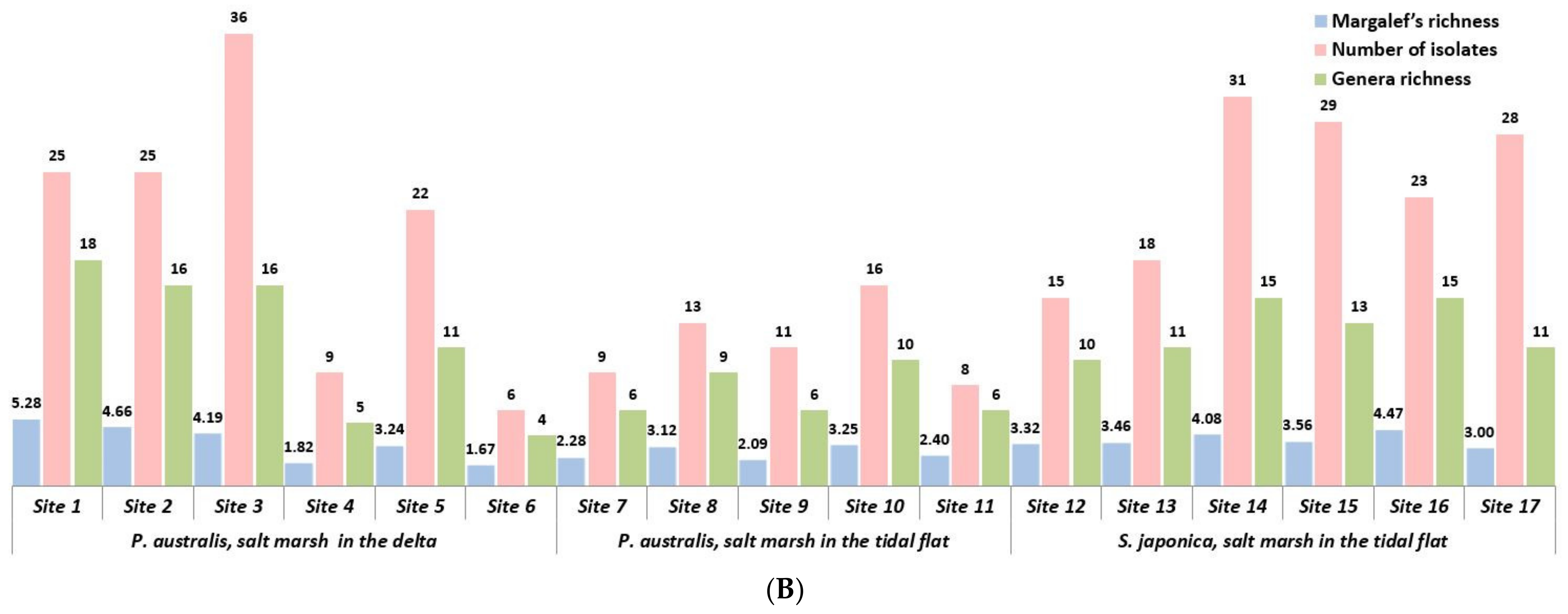
3.3. Morphological and Alpha Diversity

There are several purposes for securing diversity information. There are always different forms of geographical and ecological tensions in these various transitional zones, and comparative analysis studies on how some of these tensions have a discriminatory effect on the dominant halophyte and their endophytic clusters are scarce.
Identifying how the ecological tension of the two different ecotones affects the diversity of endophytic fungi sheds light on this purely scientific question. Furthermore, regarding application, information on the diversity of healthy salt marshes can be an indicator for restoring rapidly disappearing coastal wetlands. In addition, the two aquatic plants evaluated herein play an important role as natural purificators and structural supporters of the aquatic terrain in coastal and freshwater environments, and information on endophytic diversity has clear industrial value. Margalef’s, Menhinick’s, Shannon’s, and Simpson’s indices varied within P. australis from the delta salt marsh (5.281–1.674, 3.600–1.633, 2.080–1.434, and 0.139–0.027, respectively) and the tidal flat salt marsh (3.246–2.085, 2.500–1.809, 1.838–1.507, and 0.145–0.064, respectively), as well as within S. japonica from the tidal flat salt marsh (4.465–3.001, 3.128–2.079, 2.011–1.293, and 0.118–0.047, respectively) (Table 2). In particular, a P. australis colony from the delta showed a wide range of genera richness (abundance of genera) of 5.281–1.820 (gap between max. and min.: 3.461), whereas a colony from the tidal flat salt marsh showed a narrower range of 2.276–3.246 (max. and min gap: 0.970). Similarly, the genera richness ranges for S. japonica flourishing in the tidal flat salt marsh also showed relatively low gaps (3.001–4.456 (max. and min gap: 1.455)), with a distribution similar to that of P. australis in tidal flats. In general, the morphological diversity of the isolates and Shannon’s index, genera richness, and Margalef’s richness tends to be proportional (Shannon’s index encompasses the evasiveness and species diversity of a particular cluster). In particular, sites 4 and 6, which had low Shannon’s index or morphological diversity (Table 2), also showed low levels of richness and dominance of certain genera. Menhinick’s richness and Shannon’s indices were roughly proportional among the samples (Figure 3A), with the exceptions of sites 4 and 6, which are inland areas of the delta (Figure 2), owing to their low degree of morphological diversity (i.e., low number of isolates due to morphological differences); in addition, certain dominances were also low. Furthermore, the Simpson’s index, which reflects the dominance of a specific species, tended to be inversely proportional to the other indices (Figure 3A). Morphological diversity and genera richness were generally proportional (Figure 3B), with exception of the values at sampling site 16, as the endophytes collected from this site included several genera with higher dominances. In the case of site 15, for S. japonica in the tidal flat salt marsh, there was low genera richness; however, the existence of certain genera of Penicillium with higher dominance resulted in the observed inversely proportional values between Menhinick’s index and Shannon’s diversity.

3.4. Endophytic Distribution and Diversity within Salt Marshes in Estuaries and Tidal Flats

Even with colonies of the same plant species, the pattern of variation in the distribution and diversity of endophytes differed depending on the type of the transitional zone (Table S3). In the case of P. australis colonies that flourished in the delta salt marsh of a river estuary, 3 fungal phyla (namely Ascomycota, Basidiomycota, and Zygomycota), 6 classes, 10 orders, 18 families, and 34 fungal genera were identified, whereas in the P. australis colonies that flourished in the tidal flat salt marsh, only 1 phylum (namely Ascomycota), 4 classes, 6 orders, 11 families, and 19 genera were identified (Tables S1 and S2). Morphological fungal diversity in the wetlands in the tidal flat salt marshes was estimated to be less diverse than that in the wetlands formed in the delta of the estuary, which was clearly revealed via diversity analysis.
For P. australis native to the six sites that flourished in the delta salt marsh in the river estuary, four sites showed higher diversity compared with the P. australis colonies from the tidal flat salt marsh. Morphological diversity and the change patterns of genera richness, Margalef’s richness, and Shannon’s index were almost identical (Figure 3A). The algebraic diversity of P. australis colonies formed at the delta of the river estuary was also much higher than that of those formed at the tidal flat salt marsh (Figure 3A). However, some sites in the delta showed very low endophytic diversity, which widens the gap in diversity indices between the sampling sites in the delta salt marsh. It is reasonable to assume that sites 4 and 6 suffer differentiated ecological selective pressures caused by harsh environments. Moreover, they showed the highest Simpson’s index among all survey points (0.139 and 0.133, respectively; Figure 3A); therefore, the strong selective pressure may have affected the dominance of certain fungi since the area surveyed has a dense P. australis colony and the distance between the sites is more than 100 m, making ecological exchange between colonies difficult. Hence, it is possible that the widened gap of the biodiversity values between each site in the delta salt marsh in the estuary compared with those between the sites in the tidal flat could be due to the fact that the river estuary is a different type of ecotone (transitional zone), with extreme environmental conditions changes and thus more diverse ecological tension imposed on the colonies.
In the case of tidal flats, the key characteristic of the transitional zone is simply the space where the ground meets sea level. The stress that vegetation must endure is a marine environmental factor, such as tide ranges in a constant cycle or salt accumulation [43]. In contrast, the most important variable characteristics of river estuary water are the concentration of dissolved oxygen, salinity, and sediment load. There is extreme spatial variability in salinity, with a range of near zero at the tidal limit of tributary rivers up to 3.4% in the delta. Hence, salinity will vary considerably over time and through the seasons at each point, making it a harsh environment for organisms. Sediment often settles in flats, which are extremely difficult to colonize. No points of attachment exist for algae; therefore, a vegetation-based habitat cannot be established. Sediments can also clog the feeding and respiratory structures of species, and special adaptations exist within mud flat species to cope with this problem. Lastly, the variation in dissolved oxygen can cause problems for life forms. Nutrient-rich sediment from man-made sources can promote primary production life cycles, possibly leading to eventual decay removing the dissolved oxygen from the water; therefore, hypoxic or anoxic zones may develop. Thus, river estuaries are incredibly dynamic systems where temperature, salinity, turbidity, depth, and flow change daily in response to the tides. This dynamic environment makes estuaries highly productive habitats, but also make them difficult for many species to survive in [43].
Therefore, characteristics of the delta established in the river estuary are more diverse than those of the general tidal flats. It is an area where the ground meets sea level, and where freshwater and seawater meet. In addition, there is a flow of water due to tidal ranges; however, the flow rate is not constant, depending on the weather and climate changes, and sometimes depending on flooding. Therefore, the concentrations of salt and organic/inorganic matter are not always constant. This drastic change will widen the gap in environmental conditions between sampling points.
In contrast, relatively fewer dynamic variations were observed in the endophytic diversity in P. australis or S. japonica colonies formed in the marshes of tidal flats, which is estimated to be due to the constant ecological selective pressure originating from the tidal range. This is not a place where the massive freshwater flows of large rivers occur; it only experiences tidal ranges or consequent salt intrusion. From these results, it can be inferred that delta marshes formed in large river estuaries are much more exposed to environmental (aquatic geography) stress than marshes formed in tidal flats, and that the endophytic clusters of the dominant emersed halophyte communities flourishing in deltas are also greatly affected. P. australis colonies and their endophytes, which thrive in overcoming these dynamic environmental changes, may provide important clues to the future restoration of the salt marshes that are currently disappearing. Once the vegetation in coastal sand dunes or salt wetlands becomes damaged, the environment changes at a rapid pace as it is exposed to the strong stress introduced by the impaired protection provided by the damaged coastal vegetation.
In some of the few studies available on endophyte microorganisms of wetlands located in the coastal ecotone, bacterial communities are reported to possess variable characteristics depending on the salt concentration in the super-tidal zone [44]. In particular, the establishment of P. australis as a symbiotic halophyte plant reported in a previous study was also observed in the present study. In addition, herein, we found that the endophytic fungal diversity distinctively contrasted between the delta and the tidal flat areas. Furthermore, from the perspective of securing biological resources, a variety of novel species of endophytic microorganisms were reported in halophytes that are thriving in coastal ecotones [45,46,47,48]. This may represent the existence of a new microbial resource that has evolved and adapted to the particular selective pressures in the coastal ecotone zone, where changes are always extreme. In addition, considering the unique diversity variation of endophytic fungi in the coastal ecotone, further investigations on and culture collections of the microbial diversity of these unique coastal areas are warranted. Of note, one purpose of securing a culture collection of unique environments is to secure the new genetic resources of novel microorganisms [9]; in addition, nowadays, novel strains are also related to the competitiveness of the value of biological resources [10,49].

3.5. Wide Distribution of Certain Fungal Genera

Unexpectedly, despite the ecological tension and selective pressures at the river estuary, some sites may have more diverse genera than the tidal flats, which have relatively stable terrain conditions. This means that despite the short intervals (100 m), changes in terrain condition are dramatic, which also significantly affect the distribution of endophytic fungi in ecologically isolated native halophyte colonies. However, there were commonly eight genera distributed among the three regions that overcame such dramatic variations in terrain conditions, namely Aspergillus, Cladosporium, Epicoccum, Fusarium, Paraconiothyrium, Penicillium, Septoriella, and Talaromyces. In the case of Aspergillus and Cladosporium, the dominance ratio in each site (vegetation) was very high, whereas other genera, namely Epicoccum, Paraconiothyrium, Septoriella, Talaromyces, did not show dominance, being widely distributed in host vegetation regardless of the site. Previous reports described Epicoccum as a marine terrain-distributing fungus [50] and Talaromyces as a transitional zone fungus [51]. Aspergillus, Penicillium, and Talaromyces were reported as marine terrain-distributing fungi, with higher functions or a rock solubilizing function in marine terrains [52,53,54,55,56,57]. In the present study, 11 fungal genera were found to be commonly shared within the two tidal flats regardless of the host species (26.8% of the total genera secured in this study), namely Alternaria, Aspergillus, Cladosporium, Epicoccum, Eserohilum, Fusarium, Paraconiothyrium, Penicillium, Phialemoniopsis, Septoriella, and Talaromyces. Recently, Eserohilum was reported as an endophytic fungus [58,59]. Herein, most of these fungi were identified as endophytes in the salt marshes that developed in tidal flats and the river estuary. Taken together, Aspergillus, Cladosporium, Epicoccum, Paraconiothyrium, Penicillium, Septoriella, and Talaromyces are endogenized in a wide range regardless of the ecotone condition. In particular, Aspergillus, Cladosporium, and Penicillium are generally dominant in wide vegetation, and because the secured culture collection within these genera has morphological diversity, it is predicted that there will be a variety of species within them. In other words, the common characteristics of these genera are believed to help endogenize the species within them, thereby overcoming environmental stress and dramatic variation in terrain conditions derived from a specific ecotone, such as a transitional zone with ecological tension.
Further, this phenomenon provides another useful clue for culture collection strategy. Figure S2 shows the taxonomic information of endophytic fungi that are commonly distributed, thereby overcoming host plant species variation and diverse ecotone environments. Hence, these fungal species are likely to be resistant to the extreme, rapidly changing environment, holding potential as culture collections for the future restoration of destroyed coastal wetlands.

4. Conclusions

This is the first study to compare and analyze how different ecological tensions affect the diversity and distribution of endophytes by comparing delta and tidal flat salt marshes in the Ramsar wetlands of Suncheon Bay on the Korean Peninsula. One purpose of this study was to secure a national culture collection (in addition to the pure ecological study). In summary, this study demonstrated that the potential symbiotic endophyte fungal community shows wider biological diversity variation in relatively extreme coastal ecotones (more in the delta than in tidal flats). Furthermore, despite the extreme ecological selective pressures, 11 fungal genera found in both deltas and tidal flats were identified. Therefore, these particular taxa that can overcome a variety of ecological pressures may be used in the future restoration of destroyed coastal environments. Additional studies addressing the host halophyte–microbe relationship are needed.

Supplementary Materials

The following are available online at https://www.mdpi.com/article/10.3390/jmse9040430/s1, Figure S1: Phylogenic tree of endophyte fungi; Figure S2: Distribution of fungal endophyte symbiosis with each halophyte species and colony; Table S1: Fungal endophyte isolates from host Phragmites australis species clusters; Table S2: Fungal endophyte isolates from host Suaeda japonica species clusters; Table S3: Distribution of the endophytic genera from each site for each halophyte species.

Author Contributions

Conceptualization, Y.-H.Y.; Data curation, J.-M.P.; Formal analysis, J.-M.P.; Funding acquisition, Y.-H.Y.; Project administration, J.-G.K.; Software, J.-M.P.; Validation, J.-W.H.; Writing—Original draft, J.-M.P.; Writing—Review and editing, J.-W.H. All authors have read and agreed to the published version of the manuscript.

Funding

This work was supported by a grant from the National Institute of Biological Resources (NIBR), funded by the Ministry of Environment (MOE) of the Korea (NIBR202002104 and NIBR202102107).

Institutional Review Board Statement

Not applicable

Informed Consent Statement

Not applicable.

Data Availability Statement

All the raw sequences obtained from this study were deposited at the GenBank of the National Center for Biotechnology Information (NCBI) with assigned accession number JX040644-JX040714 (71 sequences); JX040716-JX040796 (81 sequences); JX040798-JX040819 (22 sequences); JX040821-JX040970 (150 sequences).

Conflicts of Interest

The authors declare no conflict of interest.

References

  1. Pritchard, D.W. What Is an Estuary: Physical Viewpoint. In Estuaries; Lauf, G.H., Ed.; American Association for the Advancement of Science Publishing 83: Washington, DC, USA, 1967; pp. 3–5. [Google Scholar]
  2. Adam, P. Saltmarsh Ecology; Cambridge University Press: New York, NY, USA, 1990. [Google Scholar]
  3. Watson, G.E. Big thicket plant ecology: An introduction. In Temple Big Thicket Series #5, 3rd ed.; University of North Texas Press: Denton, TX, USA, 2006. [Google Scholar]
  4. National Institute of Environmental (NIE). Research on Coastal Landscape and the Conservational Strategy (II); Case of the West coast, Korea; Ministry of Environment: Sejong, Korea, 2008. [Google Scholar]
  5. National Institute of Environmental (NIE). Research on Coastal Landscape and the Conservational Strategy (2007–2009); Ministry of Environment: Sejong, Korea, 2009. [Google Scholar]
  6. Santoyo, G.; Moreno-Hagelsieb, G.; Orozco-Mosqueda Mdel, C.; Glick, B.R. Plant growth-promoting bacterial endophytes. Microbiol. Res. 2016, 183, 92–99. [Google Scholar] [CrossRef]
  7. Bloemberg, G.V.; Lugtenberg, B.J. Molecular basis of plant growth promotion and biocontrol by rhizobacteria. Curr. Opin. Plant Biol. 2001, 4, 343–350. [Google Scholar] [CrossRef]
  8. Orozco-Mosqueda, M.D.C.; Rocha-Granados, M.D.C.; Glick, B.R.; Santoyo, G. Microbiome engineering to improve biocontrol and plant growth-promoting mechanisms. Microbiol. Res. 2018, 208, 25–31. [Google Scholar] [CrossRef] [PubMed]
  9. Park, J.M.; Hong, J.W.; Son, J.S.; Hwang, Y.J.; Cho, H.M.; You, Y.H.; Ghim, S.Y. A strategy for securing unique microbial resources—Focusing on Dokdo islands-derived microbial resources. Israel J. Ecol. Evol. 2018, 64, 1–15. [Google Scholar] [CrossRef]
  10. Ryan, M.J.; McCluskey, K.; Verkleij, G.; Robert, V.; Smith, D. Fungal biological resources to support international development: Challenges and opportunities. World J. Microbiol. Biotechnol. 2019, 35, 139. [Google Scholar] [CrossRef] [Green Version]
  11. Cultural Heritage Administration of Korea Website. Available online: http://www.heritage.go.kr/heri/idx/index.do (accessed on 21 October 2020).
  12. Amanda, S. Species Diversity Patterns at Ecotones. Master’s Thesis, University of North Carolina, Chapel Hill, NC, USA, 2009. [Google Scholar]
  13. Smith, R.L. Ecology and Field Biology, 2nd ed; Harper & Row: New York, NY, USA, 1974; p. 251. [Google Scholar]
  14. McLusky, D.S.; Elliott, M. The Estuarine Ecosystem: Ecology, Threats and Management; Oxford University Press: New York, NY, USA, 2004. [Google Scholar]
  15. Du, W.; Yao, Z.; Li, J.; Sun, C.; Xia, J.; Wang, B.; Shi, D.; Ren, L. Diversity and antimicrobial activity of endophytic fungi isolated from Securinega suffruticosa in the Yellow River Delta. PLoS ONE 2020, 15, e0229589. [Google Scholar] [CrossRef] [PubMed]
  16. Seena, S.; Sridhar, K.R. Endophytic fungal diversity of 2 sand dune wild legumes from the southwest coast of India. Can. J. Microbiol. 2004, 50, 1015–1021. [Google Scholar] [CrossRef] [PubMed]
  17. Chauhan, N.M.; Gutama, A.D.; Aysa, A. Endophytic fungal diversity isolated from different agro-ecosystem of Enset (Ensete ventericosum) in Gedeo zone, SNNPRS, Ethiopia. BMC Microbiol. 2019, 19, 172. [Google Scholar] [CrossRef] [PubMed]
  18. Chen, X.; Luo, X.; Fan, M.; Zeng, W.; Yang, C.; Wu, J.; Zhao, C.; Zhang, Y.; Zhao, P. Endophytic fungi from the ranches of Camellia taliensis (W. W. Smith) Melchior, a widely distributed wild tea plant. World J. Microbiol. Biotechnol. 2019, 35, 113. [Google Scholar] [CrossRef] [PubMed]
  19. Cheng, X.; Wei, Z.; Pu, S.; Xiang, M.; Yan, A.; Zhang, Y.; Wang, X. Diversity of endophytic fungi of Paeonialactiflora pallas and screening for fungal paeoniflorin producers. FEMS Microbiol. Lett. 2018, 365. [Google Scholar] [CrossRef]
  20. Fan, M.; Chen, X.; Luo, X.; Zhang, H.; Liu, Y.; Zhang, Y.; Wu, J.; Zhao, C.; Zhao, P. Diversity of endophytic fungi from the leaves of Vaccinium dunalianum. Lett. Appl. Microbiol. 2020, 71, 479–489. [Google Scholar] [CrossRef] [PubMed]
  21. Yamada, A.; Ogura, T.; Degawa, Y.; Ohmasa, M. Isolation of Tricholoma matsutake and T. bakamatsutake cultures from field-collected ectomycorrhizas. Mycoscience 2001, 42, 43–50. [Google Scholar] [CrossRef]
  22. Hasan, H.A. Gibberellin and auxin production plant root fungi and their biosynthesis under salinity-calcium interaction. Rostl. Vyroba. 2002, 48, 101–106. [Google Scholar] [CrossRef]
  23. Khan, S.A.; Hamayun, M.; Yoon, H.; Kim, H.Y.; Suh, S.J.; Hwang, S.K.; Kim, J.M.; Lee, I.J.; Choo, Y.S.; Yoon, U.H.; et al. Plant growth promotion and Penicillium citrinum. BMC Microbiol. 2008, 8, 231. [Google Scholar] [CrossRef] [Green Version]
  24. White, T.J.; Bruns, T.D.; Lee, S.B.; Taylor, J.W. Amplification and Direct Sequencing of Fungal Ribosomal RNA Genes for Phylogenetics. In PCR Protocols: A Guide to Methods and Applications; Innis, M.A., Gelfand, D.H., Sninsky, J.J., Eds.; Academic Press: San Diego, CA, USA, 1990; pp. 315–322. [Google Scholar]
  25. National Center for Biotechnology Information (NCBI) U.S. National Library of Medicine. GenBank Database. Available online: https://www.ncbi.nlm.nih.gov/genbank/ (accessed on 7 October 2020).
  26. Kumar, S.; Stecher, G.; Li, M.; Knyaz, C.; Tamura, K. MEGA X: Molecular Evolutionary Genetics Analysis across computing platforms. Mol. Biol. Evol. 2018, 35, 1547–1549. [Google Scholar] [CrossRef]
  27. Chen, Y.; Ye, W.; Zhang, Y.; Xu, Y. High speed BLASTN: An accelerated Mega BLAST search tool. Nucleic Acids Res. 2015, 18, 7762–7768. [Google Scholar] [CrossRef] [PubMed] [Green Version]
  28. Aiyar, A. The use of CLUSTAL W and CLUSTAL X for multiple sequence alignment. Methods Mol. Biol. 2000, 132, 221–241. [Google Scholar] [CrossRef]
  29. Larkin, M.A.; Blackshields, G.; Brown, N.P.; Chenna, R.; McGettigan, P.A.; McWilliam, H.; Valentin, F.; Wallace, I.M.; Wilm, A.; Lopez, R.; et al. Clustal W and Clustal X version 2.0. Bioinformatics 2007, 23, 2947–2948. [Google Scholar] [CrossRef] [Green Version]
  30. Rzhetsky, A.; Nei, M. A simple method for estimating and testing minimum evolution trees. Mol. Biol. Evol. 1992, 9, 945–967. [Google Scholar] [CrossRef]
  31. Felsenstein, J. Confidence limits on phylogenies: An approach using the bootstrap. Evolution 1985, 39, 783–791. [Google Scholar] [CrossRef] [PubMed]
  32. Tajima, F.; Nei, M. Estimation of evolutionary distance between nucleotide sequences. Mol. Biol. Evol. 1984, 1, 269–285. [Google Scholar] [CrossRef] [Green Version]
  33. Nei, M.; Kumar, S. Molecular Evolution and Phylogenetics; Oxford University Press: New York, NY, USA, 2000. [Google Scholar]
  34. Saitou, N.; Nei, M. The neighbor-joining method: A new method for reconstructing phylogenetic trees. Mol. Biol. Evol. 1987, 4, 406–425. [Google Scholar] [CrossRef] [PubMed]
  35. Fierer, N.; Jackson, R.B. The diversity and biogeography of soil bacterial communities. Proc. Natl. Acad. Sci. USA 2006, 103, 626–631. [Google Scholar] [CrossRef] [Green Version]
  36. Margalef, R. Information theory in ecology. Gen. Syst. 1958, 3, 36–71. [Google Scholar]
  37. Mendes, R.S.; Evangelista, L.R.; Thomaz, S.M.; Agostinho, A.A.; Gomes, L.C. Unified index to measure ecological diversity and species rarity. Ecography 2008, 31, 450–456. [Google Scholar] [CrossRef]
  38. Mendes, S.M.; Santos, J.; Freitas, H.; Sousa, J.P. Assessing the impact of understory vegetation cut on soil epigeic macrofauna from a cork-oak Montado in South Portugal. Agroforest. Syst. 2011, 82, 139–148. [Google Scholar] [CrossRef]
  39. Whittaker, R.H. Evolution of species diversity in land communities. Evol. Biol. 1972, 10, 1–67. [Google Scholar]
  40. Magurran, A.E. Measuring Biological Diversity; Blackwell Science: Oxford, NJ, USA, 2004. [Google Scholar]
  41. Lambshead, P.J.D.; Platt, H.M.; Shaw, K.M. The detection of differences among assemblages of marine benthic species based on an assessment of dominance and diversity. J. Nat. Hist. 1983, 17, 859–874. [Google Scholar] [CrossRef]
  42. International Plant Names Index. Available online: http://www.ipni.org (accessed on 7 August 2020).
  43. Kaiser, M.J.; Attrill, M.J.; Jennings, S.; Thomas, D.N. Marine Ecology: Processes, Systems and Impacts; Oxford University Press: New York, NY, USA, 2005. [Google Scholar]
  44. Ma, B.; Lv, X.; Warren, A.; Gong, J. Shifts in diversity and community structure of endophytic bacteria and archaea across root, stem and leaf tissues in the common reed, Phragmites australis, along a salinity gradient in a marine tidal wetland of northern China. Antonie Van Leeuwenhoek 2013, 104, 759–768. [Google Scholar] [CrossRef] [PubMed] [Green Version]
  45. Li, Y.R.; Zhu, Z.N.; Li, Y.Q.; Xiao, M.; Han, M.X.; Wadaan, M.A.M.; Hozzein, W.N.; An, D.D.; Li, W.J. Microbacterium halophytorum sp. nov., a novel endophytic actinobacterium isolated from halophytes. Int. J. Syst. Evol. Microbiol. 2018, 68, 3928–3934. [Google Scholar] [CrossRef] [PubMed]
  46. Liu, Y.Y.; Liu, F.; Li, Y.Q.; Lei, R.F.; Ma, Q.; Narsing Rao, M.P.; Abudourousuli, D.; Rouzi, Z.; Aosiman, M.; An, D.D.; et al. Sphingobacterium endophyticum sp. nov., a novel endophyte isolated from halophyte. Arch. Microbiol. 2020, 202, 2771–2778. [Google Scholar] [CrossRef]
  47. Zhang, Y.J.; Zhang, W.D.; Qin, S.; Bian, G.K.; Xing, K.; Li, Y.F.; Cao, C.L.; Jiang, J.H. Saccharopolyspora dendranthemae sp. nov. a halotolerant endophytic actinomycete isolated from a coastal salt marsh plant in Jiangsu, China. Antonie Van Leeuwenhoek 2013, 103, 1369–1376. [Google Scholar] [CrossRef] [PubMed]
  48. Zhu, Z.N.; Li, Y.R.; Li, Y.Q.; Xiao, M.; Han, M.X.; Wadaan, M.A.M.; Hozzein, W.N.; An, D.D.; Li, W.J. Microbacterium suaedae sp. nov., isolated from Suaeda aralocaspica. Int. J. Syst. Evol. Microbiol. 2019, 69, 411–416. [Google Scholar] [CrossRef] [PubMed]
  49. MOE (Ministry of Environment. Korea). The Fifth National Report to the Convention on Biological Diversity. 2014. Available online: https://www.cbd.int/nr5/ (accessed on 23 April 2014).
  50. Chi, L.P.; Li, X.M.; Li, L.; Li, X.; Wang, B.G. Cytotoxic thiodiketopiperazine derivatives from the deep sea-derived fungus Epicoccum nigrum SD-388. Mar. Drugs 2020, 18, 160. [Google Scholar] [CrossRef] [PubMed] [Green Version]
  51. Liu, F.; Li, Q.; Yang, H.; Cai, X.L.; Xia, X.K.; Chen, S.P.; Li, M.F.; She, Z.G.; Lin, Y.C. Structure elucidation of three diphenyl ether derivatives from the mangrove endophytic fungus SBE-14 from the South China Sea. Magn. Reson. Chem. 2009, 247, 453–455. [Google Scholar] [CrossRef] [PubMed]
  52. Afiyatullov, S.S.; Leshchenko, E.V.; Berdyshev, D.V.; Sobolevskaya, M.P.; Antonov, A.S.; Denisenko, V.A.; Popov, R.S.; Pivkin, M.V.; Udovenko, A.A.; Pislyagin, E.A.; et al. Zosteropenillines: Polyketides from the marine derived fungus Penicillium thomii. Mar. Drugs 2017, 15, 46. [Google Scholar] [CrossRef] [PubMed] [Green Version]
  53. Cabello, M.; Irrazabal, G.; Bucsinszky, A.M.; Saparrat, M.; Schalamuk, S. Effect of an arbuscular mycorrhizal fungus, Glomus mosseae, and a rock-phosphate-solubilizing fungus, Penicillium thomii, on Mentha piperita growth in a soilless medium. J. Basic Microbiol. 2005, 45, 182–189. [Google Scholar] [CrossRef]
  54. Cheng, Z.; Liu, W.; Fan, R.; Han, S.; Li, Y.; Cui, X.; Zhang, J.; Wu, Y.; Lv, X.; Zhang, Y.; et al. Terpenoids from the deep-sea-derived fungus Penicillium thomii YPGA3 and their bioactivities. Mar. Drugs 2020, 18, 164. [Google Scholar] [CrossRef] [PubMed] [Green Version]
  55. Nicoletti, R.; Vinale, F. Bioactive compounds from marine-derived Aspergillus, Penicillium, Talaromyces and Trichoderma species. Mar. Drugs 2018, 16, 408. [Google Scholar] [CrossRef] [Green Version]
  56. Park, M.S.; Oh, S.Y.; Fong, J.J.; Houbraken, J.; Lim, Y.W. The diversity and ecological roles of Penicillium in intertidal zones. Sci. Rep. 2019, 9, 13540. [Google Scholar] [CrossRef] [Green Version]
  57. Zhuravleva, O.I.; Sobolevskaya, M.P.; Leshchenko, E.V.; Kirichuk, N.N.; Denisenko, V.A.; Dmitrenok, P.S.; Dyshlovoy, S.A.; Zakharenko, A.M.; Kim, N.Y.; Afiyatullov, S.S. Meroterpenoids from the alga-derived fungi Penicillium thomii Maire and Penicillium lividum Westling. J. Nat. Prod. 2014, 77, 1390–1395. [Google Scholar] [CrossRef] [PubMed]
  58. Ek-Ramos, M.J.; Zhou, W.; Valencia, C.U.; Antwi, J.B.; Kalns, L.L.; Morgan, G.D.; Kerns, D.L.; Sword, G.A. Spatial and temporal variation in fungal endophyte communities isolated from cultivated cotton (Gossypium hirsutum). PLoS ONE 2013, 8, e66049. [Google Scholar] [CrossRef] [PubMed]
  59. Pan, F.; Liu, Z.Q.; Chen, Q.; Xu, Y.W.; Hou, K.; Wu, W. Endophytic fungus strain 28 isolated from Houttuynia cordata possesses wide-spectrum antifungal activity. Braz. J. Microbiol. 2016, 47, 480–488. [Google Scholar] [CrossRef] [PubMed] [Green Version]
Figure 1. Location of Suncheon Bay and detailed view of the highly dominant status of two emersed halophyte species: Phragmites australis (green colony), Suaeda japonica (reddish-purple colony). Map was adapted from the Cultural Heritage Administration of Korea website [11], with permission from the Cultural Heritage Administration of Korea, 2020.
Jmse 09 00430 g001
Figure 2. Aerial view of each sampling site (indicated in yellow). Salt marshes developed in (A) the delta of Suncheon Bay (Phragmites australis colonies at six sampling sites), (B) tidal flats (five sites of P. australis colonies), and (C) tidal flats (six sites of Suaeda japonica colonies). The aerial images were collected using Google Earth (version 7.3.3) and modified for this study.
Jmse 09 00430 g002
Figure 3. (A) Variation of Menhinick’s, Shannon’s, and Simpson’s indices at each sampling site. (B) Variation of morphological diversity and richness (genera and Margalef’s index) at each sampling site.
Jmse 09 00430 g003aJmse 09 00430 g003b
Table 1. Description of the sampling sites at each salt marsh and endophytic culture collection.
Host
Species
Sampling SitesGPS CoordinatesFungal Isolates and Taxon
Total Isolate NumberDivisionClassOrderFamilyGenus
Phragmites
australis
Delta 1N 34°53′00.20″/E 127°30′49.60″251481418
2N 34°52′55.78″/E 127°30′50.43″251471117
3N 34°52′54.36″/E 127°30′53.09″363681316
4N 34°52′53.69″/E 127°30′55.98″913445
5N 34°52′55.63″/E 127°30′52.68″22136811
6N 34°52′59.46″/E 127°30′52.38″612344
Tidal flat7N 34°52′38.90″/E 127°30′47.23″913566
8N 34°52′34.81″/E 127°30′27.08″1312499
9N 34°52′27.65″/E 127°30′14.77″1113456
10N 34°52′27.46″/E 127°29′55.06″16135710
11N 34°52′27.51″/E 127°29′46.53″813456
Suaeda
japonica
Tidal flat1N 34°51′45.94″/E 127°31′10.12″15257810
2N 34°51′46.41″/E 127°31′09.65″18137811
3N 34°51′49.84″/E 127°31′09.49″312581215
4N 34°51′56.41″/E 127°31′06.25″292581013
5N 34°51′59.83″/E 127°31′06.25″232581115
6N 34°51′59.73″/E 127°31′06.36″28136711
Table 2. Diversity of the endophytes from each dominant species flourishing at sampling each site.
Host
Species
Sampling Site
Character
Site
Number
Number of
Isolates
Genera RichnessMargalef’s
Richness
Menhinick’s
Index (Dmn)
Shannon’s Index (H’)Simpson’s Index (D)
Phragmites
australis
Delta125185.2813.6002.0800.027
225164.6601.9191.9190.037
336164.1862.6671.4340.081
4951.8201.6671.9710.139
522113.2352.3451.9400.082
6641.6741.6331.6630.133
Tidal flat7962.2762.0001.7390.111
81393.1192.4961.6230.064
91162.0851.8091.8380.145
106103.2462.5001.5950.083
11862.4042.1211.5070.107
Suaeda
japonica
Tidal flat1215103.3232.5822.0110.086
1318113.4602.5931.5720.118
1431154.0772.6941.2930.075
1529133.5642.4141.7240.076
1623154.4653.1281.5020.047
1728113.0012.0791.4580.106
Publisher’s Note: MDPI stays neutral with regard to jurisdictional claims in published maps and institutional affiliations.

