Article

Arthrospira platensis Variants: A Comparative Study Based on C-phycocyanin Gene and Protein, Habitat, and Growth Conditions

by
Nawal Abd El-Baky
*,
Neama Mahmoud Fattouh Rezk
and
Amro A. Amara
*
Protein Research Department, Genetic Engineering and Biotechnology Research Institute (GEBRI), City of Scientific Research and Technological Applications (SRTA-City), New Borg El-Arab City, Alexandria P.O. Box 21934, Egypt
*
Authors to whom correspondence should be addressed.
J. Mar. Sci. Eng. 2023, 11(3), 663; https://doi.org/10.3390/jmse11030663
Submission received: 31 January 2023 / Revised: 8 March 2023 / Accepted: 19 March 2023 / Published: 21 March 2023
(This article belongs to the Section Marine Biology)

Abstract

:
This study aimed to map the differences between Arthrospira sp. and Arthrospira platensis strains and variants from the order Oscillatoriales at the gene and protein levels of C-phycocyanin alpha chain via multiple alignment, phylogenetic trees of species, and analysis of the nucleotide and amino acid composition of the studied sequences. The links between gene/protein and environmental features of the habitat or source of isolation were also investigated. Phycocyanin was extracted from three A. platensis strains: an Egyptian isolate cultivated in the laboratory under static conditions in a highly saline medium and two commercial products. The French commercial strain showed the highest extraction yield but the lowest C-phycocyanin purity, and the color intensity of the extracted pigment from the Egyptian isolate was significantly weaker than those of the two commercial strains. All the analyzed species and strains had GC content of more than 54.5% in C-phycocyanin alpha chain gene and showed high abundance of alanine, an amino acid encoded exclusively by GC-biased codons, in their protein. The frequencies of the acidic amino acids aspartic acid and glutamic acid were 5.2% and 5.0% on average, respectively, which were slightly higher than those of the basic residues (4.3% arginine, 0.6% histidine, and 5.0% lysine). Data relating to the isolation source of most of the analyzed species revealed harsh conditions, such as high alkalinity, salinity, CO2 saturation, and/or temperature. These findings may link the gene/protein of C-phycocyanin, which is one of the most important bioactive proteins of A. platensis, to the adaptation of this organism to harsh environmental conditions and associate the color of the pigment to cultivation conditions and/or isolation source.

1. Introduction

From very early on, humans have benefited from their observation of birds utilizing algae floating on the surface of water. From ancient times in Egypt up to now, farmers have collected floating algae to feed their birds. Among these floating algae, one species that has attracted considerable attention of scientists worldwide is the microalga or cyanobacterium Arthrospira platensis [1,2].
Arthrospira platensis has been extensively studied since it was rediscovered as being an edible microorganism (nontoxic nutritious food) that is consumed in large quantities in Chad. People have observed that migratory birds and animals are interested in feeding on Arthrospira platensis floating on the surface of different lakes in Africa and South America. After a long migratory trip, the tired birds can recover their power through the consumption of this floating microalga. Even though the water is alkaline and salty, farm animals also drink this green water. In Ethiopia, farmers and herdsmen living in areas around soda lakes make their cattle drink water containing Arthrospira about once a month. They believe that this microalga has therapeutic effects and supplement nutrients lacking in dietary food [3].
In 1827, Turpin identified and described Arthrospira as spiral cyanobacteria using a light microscope [4]. Rich (1931) reported that Arthrospira is a dominant phytoplankton in a number of lakes in the Rift Valley of East Africa [5]. Dangeard (1940) reintroduced A. platensis to the world from a sample collected by a pharmacist from a local market in Chad [1,6].
A. platensis occupies unique marine habitats, such as alkaline lakes [3]. A. platensis producers mimic this natural phenomenon and increase the alkalinity to reduce the number of other algal species [7]. In 2013, a study proved that in addition to alkalinity, salinity is an essential factor that can suppress the growth of other algae and cyanobacteria, thus leading to A. platensis dominating the environment and benefiting from the surrounding nutrients [2].
A. platensis can survive in both sunny and dark conditions. It can utilize sunlight and CO2 to grow autotrophically. In the dark, it can utilize organic compounds and grow auxotrophically. It can benefit from an appropriate day/night environment where organic compounds exist and grow either autotrophically or auxotrophically or as a combination of the two (autoauxotrophic growth) [8,9].
Amara and Steinbüchel (2013) suggested that salinity can play a significant role in Arthrospira enrichment. They proposed that the rain/evaporation cycle on the lake and the lake’s surrounding area would lead to salt accumulation [2]. Temperature can change the amount of dissolved oxygen (pO2) and the pH value either by direct effect or by inducing chemical or physical changes [2,10]. Salinity, alkalinity, temperature, organic compounds, absence of other species due to these stresses, and other factors have been analyzed and reported to affect the growth and cultivation of the genus Spirulina [10].
In addition to providing a good supply of bioactive ingredients in the diet, such as essential amino acids and fatty acids, high protein synthesis capacity (60–70% of the cell mass), minerals, and vitamins [11,12,13], A. platensis can synthetize phycobiliproteins including C-phycocyanin (a blue natural pigment) [14]. In addition to its application as natural colorants for food additives [15], C-phycocyanin also has anti-inflammatory, antioxidant, and anticarcinogenic activities [16]. Unfortunately, this natural pigment has been poorly explored by the food industry. The limitations for its application include the extraction methods resulting in low-purity products and its low stability under storage and during processing of foods. This natural pigment is preferably extracted at neutral pH of 5–8 and temperatures below 50 °C [14].
Regions in the phycocyanin gene (e.g., intergenic spacer region) are highly conserved and used in the molecular identification and characterization of the producing microorganisms, including A. platensis [17]. It has previously been observed that the extracted phycocyanin from strains that are phenotypically close to each other but were isolated from different habitats (habitats with different light wavelengths and photoperiods) may significantly vary in color [18,19]. Therefore, the extraction of phycocyanin from each strain needs optimization [20,21].
This work aimed to map the differences between A. platensis strains and variants based on gene and protein of the phycocyanin subunit alpha and determine any relationship between the pigment structure, isolation source, cultivation conditions, and adaptation of these microalgae to harsh environments. The frequencies of nucleotides in the phycocyanin subunit alpha of nine cyanobacterial species and its amino acid composition in 25 cyanobacterial species, including Arthrospira platensis strains and variants, were calculated and linked to their source of isolation. C-phycocyanin was also comparatively extracted from dried biomass of three microalgae strains: an Egyptian A. platensis isolate cultivated in our lab and French and Chinese commercial A. platensis products.

2. Materials and Methods

2.1. Microalgae Strains

An extreme alkalophilic A. platensis previously isolated from the brackish Lake Mariout southwest of Alexandria city in Egypt was used in this study. This Egyptian isolate was authenticated (Accession CBA13040, phycocyanin alpha subunit, partial from Limnospira fusiformis LM) by Sharaf et al. [17]. The French commercial A. platensis product (spirulina tablets, VITAMINEXT, France) was purchased from a pharmacy in Casablanca, Morocco. The Chinese commercial A. platensis strain (spirulina powder organic, Starwest Botanicals, China) was obtained from a local Egyptian pharmacy.

2.2. Cultivation Medium

Amara and Steinbuchel (A–St) medium at concentration of 1.5× (highly saline medium) consisted of part A: 13.82 g/L NaHCO3, 10.71 g/L NaCO3, and 0.75 g/L K2HPO4; part B: 2.25 g/L NaNO3, 0.85 g/L K2SO4, 1.5 g/L NaCl, 0.22 g/L MgSO4·7H2O, 0.011 g/L CaCl2·2H2O, 0.012 g/L FeSO4·2H2O, and 0.1 g/L EDTA-Na2·2H2O; part C: 0.02 g/L ferric citrate; and part D: 0.1 g/L peptone and 0.01 g/L yeast extract [2]. Parts A–C of this medium were dissolved in a minimal amount of sterile distilled water and filter-sterilized through a syringe filter (pore size of 0.22 μm) from TPP (St. Louis, MO, USA). Part D was dissolved in distilled water and sterilized separately by autoclaving and then used as a broth. Finally, a 20 L glass bottle was used to grow Egyptian A. platensis under static conditions.

2.3. Cultivation Conditions

Egyptian A. platensis strain was cultivated in the laboratory under static cultivation condition in a 20 L sterile glass bottle and kept at the regular day/night cycle at temperatures ranging from 24 °C (day) to 18 °C (night). The cultivation bottle was kept closed during the cultivation process (for 27 days).

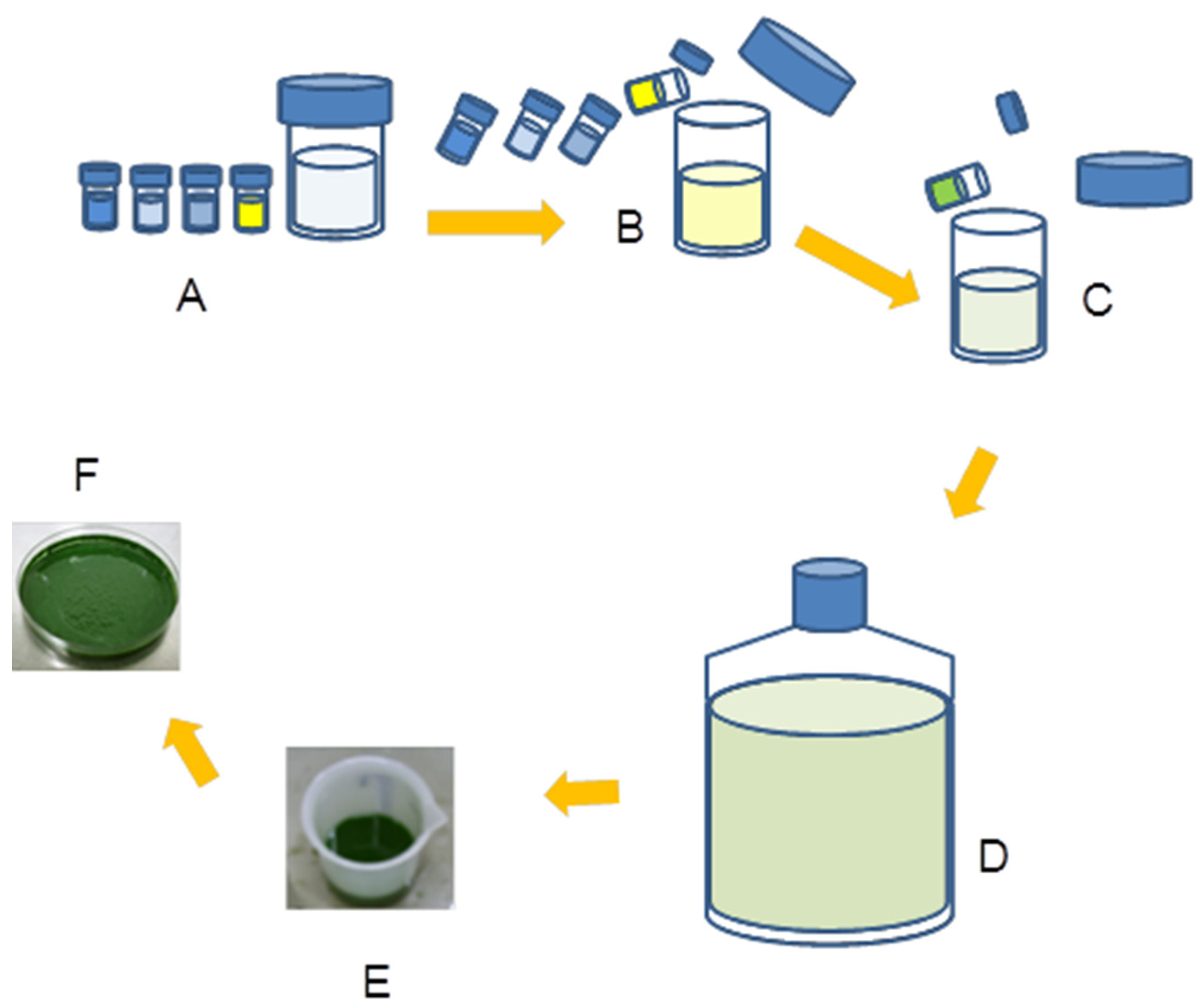
2.4. Cells Harvesting

The cells were harvested manually by passing the culture through a big funnel containing two layers: one of synthetic sponge and the other of multilayers of cotton tissue. After that, the wet biomass of A. platensis was placed in a Petri dish and left to dry using a sterile air flow of microbial cabinet under sterile conditions for 2 days. The steps of cultivation up to the obtaining of air-dried biomass are illustrated in Figure 1.

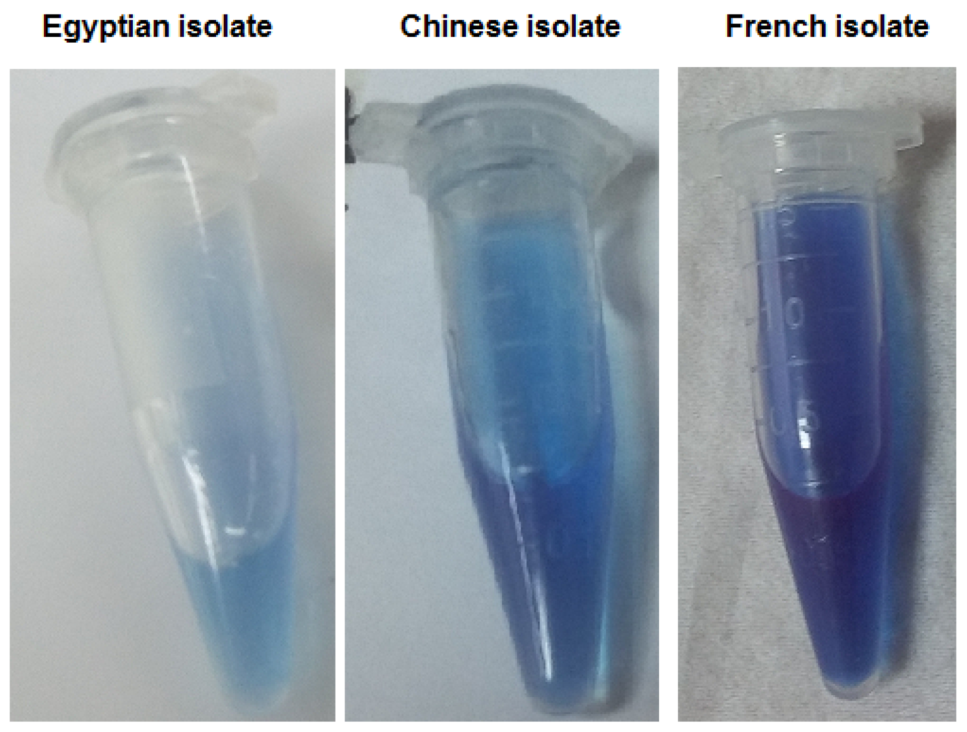
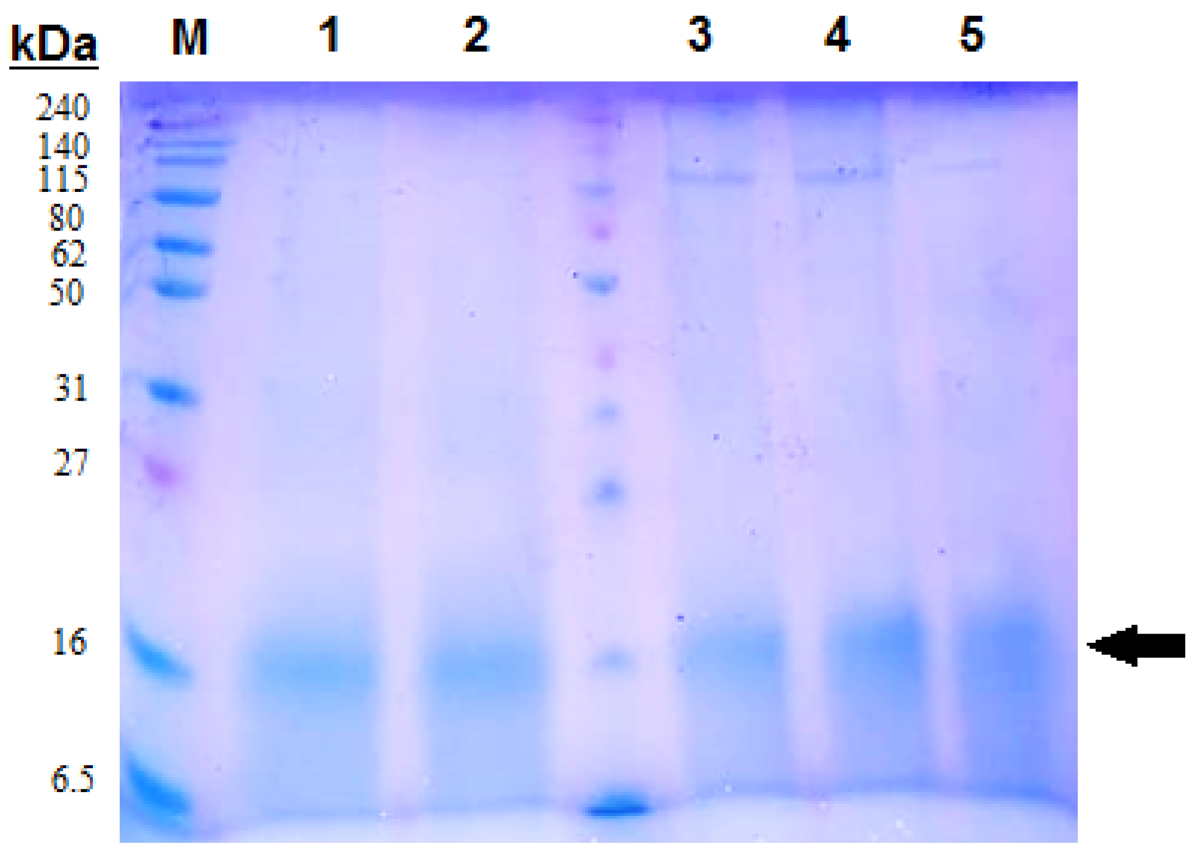
2.5. Extraction of C-phycocyanin from Dry Biomass of Three Microalgae Strains

The dried biomass (1 g) of Egyptian, French, and Chinese A. platensis was presoaked in a 20 mL sodium phosphate buffer of 0.1 M and pH 7 for 2 h to prepare for cell disruption. Following the presoaking step, maceration using a mortar and pestle and repeated freezing (at –40 °C for 2 h) and thawing (at 4 °C for 5 min) were carried out in the dark. Ultrasonication was performed in combination with conventional methods for primary extraction of C-phycocyanin, as described previously by Tavanandi et al. [22]. The sonicator (Bandelin Sonopuls Sonicator, Berlin, Germany) was programmed to provide 30 s pulses with 15 s pauses for a total period of 5 min.

2.6. Protein Content and Concentration of the Extracted C-phycocyanin

Following ultrasonication, the mixture was centrifuged at 13,000× g and 4 °C for 10 min to separate clear supernatants that contained extracted C-phycocyanin. The total protein concentration of the extracted C-phycocyanin from dry biomass of the three microalgae strains was measured by Bradford assay [23]. The supernatant samples were run on 12% SDS-PAGE. Absorbance of the supernatant samples from the three microalgae strains was evaluated at wavelengths of 600, 610, 615, 620, 630, 640, 650, and 652 nm (OPTIZEN Scan UV/VIS spectrophotometer, KLab Co., Daejeon, Korea) for C-phycocyanin.
The concentration of C-phycocyanin extracted from the three microalgae strains was estimated as previously reported by Siegelman and Kycia [24] and Sharmaa et al. [25] as follows:
C-phycocyanin = (A615 − 0.474 × A652)/5.34
where A615 is the absorbance measured at wavelength of 615 nm, and A652 is the absorbance measured at wavelength of 652 nm.

2.7. The Template Nucleotide Sequence of A. platensis Used in This Study

NCBI database (www.ncbi.nlm.nih.gov, last accessed on 20 January 2023) was used to obtain phycocyanin alpha subunit (cpcA) gene of Arthrospira platensis, and the obtained nucleotide sequence was saved in FASTA format and used as template for collecting sequences of nine Arthrospira sp. and Arthrospira platensis strains and variants from the order Oscillatoriales. The following sequence was selected as a template.
>ABD64608.1:1-162 phycocyanin alpha chain (Arthrospira platensis)
ATGAAAACCCCGCTGACCGAAGCGGTGAGCATTGCGGATAGCCAGGGCCGCTTTCTGAGCAGCACCGAAATTCAGGTGGCGTTTGGCCGCTTTCGCCAGGCGAAAGCGGGCCTGGAAGCGGCGAAAGCGCTGACCAGCAAAGCGGATAGCCTGATTACCGGCGCGGCGCAGGCGGTGTATAACAAATTTCCGTATACCACCCAGATGCAGGGCCCGAACTATGCGGCGAACCAGCGCGGCAAAGATAAATGCGCGCGCGATATTAGCTATTATCTGCGCATGGTGACCTATTGCCTGATTGCGGGCGGCACCGGCCCGATGGATGAATATCTGATTGCGGGCATTGATGAAATTAACCGCACCTTTGATCTGAGCCCGAGCTGGTATATTGAAGCGCTGAAATATATTAAAGCGAACCATGGCCTGAGCGGCGATGCGGCGGTGGAAGCGAACAGCTATCTGGATTATGCGATTAACGCGCTGAGC
In the database BLAST (https://blast.ncbi.nlm.nih.gov/Blast.cgi, last accessed on 14 January 2023), the template sequence was uploaded in the “Standard Nucleotide BLAST” search (https://blast.ncbi.nlm.nih.gov/Blast.cgi?PROGRAM=blastn&PAGE_TYPE=BlastSearch&LINK_LOC=blasthome, last accessed on 18 January 2023) [26]. Nine sequences were selected based on equality of length and similarity to the template nucleotide sequence and saved in FASTA format for further analysis.

2.8. The Template Amino Acid Sequence of A. platensis Used in This Study

The following sequence was obtained from the NCBI protein database (www.ncbi.nlm.nih.gov, last accessed on 20 January 2023) after searching for the phycocyanin alpha subunit (cpcA) amino acid sequence of A. platensis and collected from the result of the accession number ABD64608.1:1-162.
>ABD64608.1:1-162 phycocyanin alpha chain (Arthrospira platensis)
MKTPLTEAVSIADSQGRFLSSTEIQVAFGRFRQAKAGLEAAKALTSKADSLITGAAQAVYNKFPYTTQMQGPNYAANQRGKDKCARDISYYLRMVTYCLIAGGTGPMDEYLIAGIDEINRTFDLSPSWYIEALKYIKANHGLSGDAAVEANSYLDYAINALS.
The amino acid sequence was saved in FASTA format to be used as a template. In the database BLAST (https://blast.ncbi.nlm.nih.gov/Blast.cgi, last accessed on 18 January 2023), the template sequence was uploaded in the Standard Protein BLAST (https://blast.ncbi.nlm.nih.gov/Blast.cgi?PROGRAM=blastp&PAGE_TYPE=BlastSearch&LINK_LOC=blasthome, last accessed on 20 January 2023) [26]. Additionally, Translated BLAST: blastx (https://blast.ncbi.nlm.nih.gov/Blast.cgi?PROGRAM=blastx&PAGE_TYPE=BlastSearch&LINK_LOC=blasthome, last accessed on 24 January 2023) was used. A total of 25 protein sequences of Arthrospira sp. and Arthrospira platensis strains and variants from the order Oscillatoriales were selected based on the equality of length, presence of correct start codon, and similarity to the template and then saved in FASTA format for further analysis.

2.9. Analysis of the Nucleotide and Protein Sequences

The selected 9 nucleotide sequences and 25 amino acid sequences were analyzed using BioEdit version 7 for calculating the GC and AT content of the analyzed nucleotide sequences, performing and editing multiple sequence alignment and creating consensus sequence [27], using Molecular Evolutionary Genetics Analysis Version 11 (MEGA11) for alignment, building phylogenetic trees of species, and analyzing the amino acid and nucleotide composition of the studied sequences [28]. Phylogenetic tree visualization was performed using the Tree Figure Drawing Tool Version 1.4.2 (FigTree 1.4.2), a graphical viewer software, for each of the obtained phylogenetic trees (http://tree.bio.ed.ac.uk/software/figtree/, last accessed on 27 January 2023).

3. Results

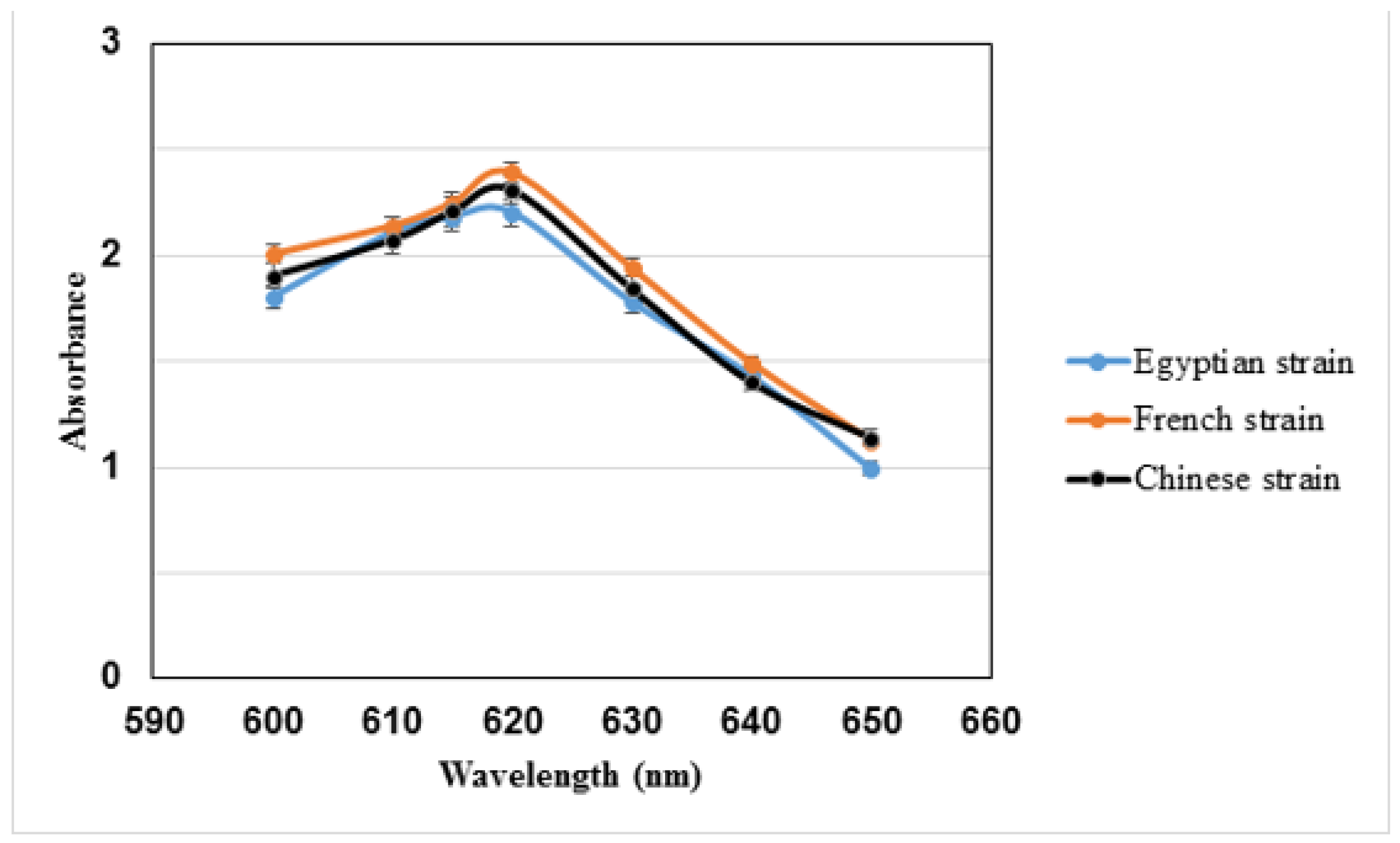
3.1. Extraction of C-phycocyanin

The total protein content of the extracted C-phycocyanin from dried biomass of Egyptian, French, and Chinese A. platensis as estimated by Bradford assay is presented in Table 1. As shown in Table 1, the extracted C-phycocyanin of the Egyptian strain had the highest total protein content, followed by the French strain and then the Chinese one. Extracted C-phycocyanin from dry biomass of the three microalgae strains is shown in Figure 2. As can be seen, there was a clear difference in color intensity between the extracted C-phycocyanin of the Egyptian strain (very faint blue extract) and the commercial ones (deep blue extracts). SDS-PAGE analysis of the C-phycocyanin extract is presented in Figure 3. The subunits of the extracted protein from the three microalgae strains were observed at slightly different molecular weights on SDS-PAGE. Visible spectrum (at wavelengths of 600–650 nm) of the extracted C-phycocyanin from dry biomass of the three microalgae strains is illustrated in Figure 4. The concentration of C-phycocyanin (mg/mL), calculated by the equation of Siegelman and Kycia [24], is demonstrated in Table 2. Purity of the extracted C-phycocyanin was calculated by dividing A615 (=2.186, 2.251, and 2.209) by A280 (=2.61, 3.25, and 2.633) for the Egyptian, French, and Chinese strains, respectively, as shown in Table 2. Regarding the concentration of C-phycocyanin, the French strain was the highest in concentration but the lowest in purity.

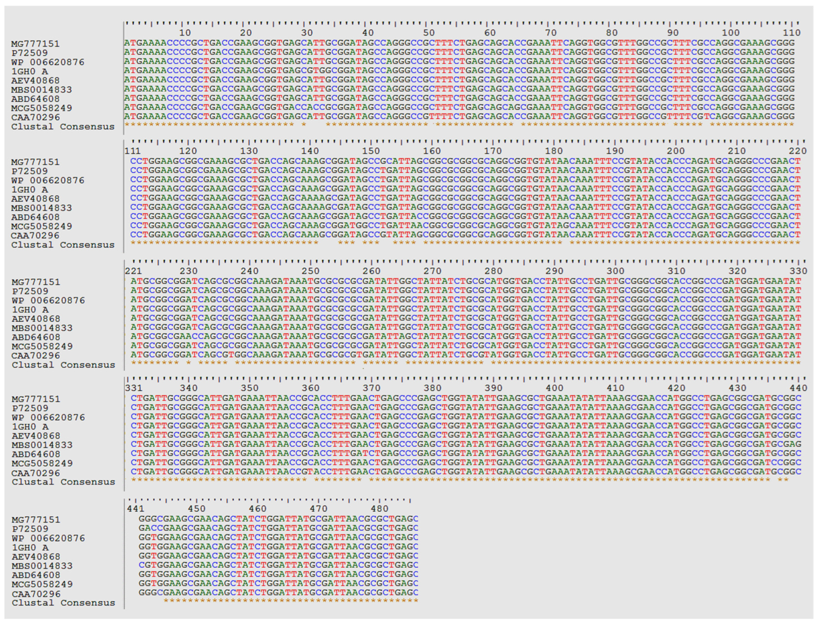
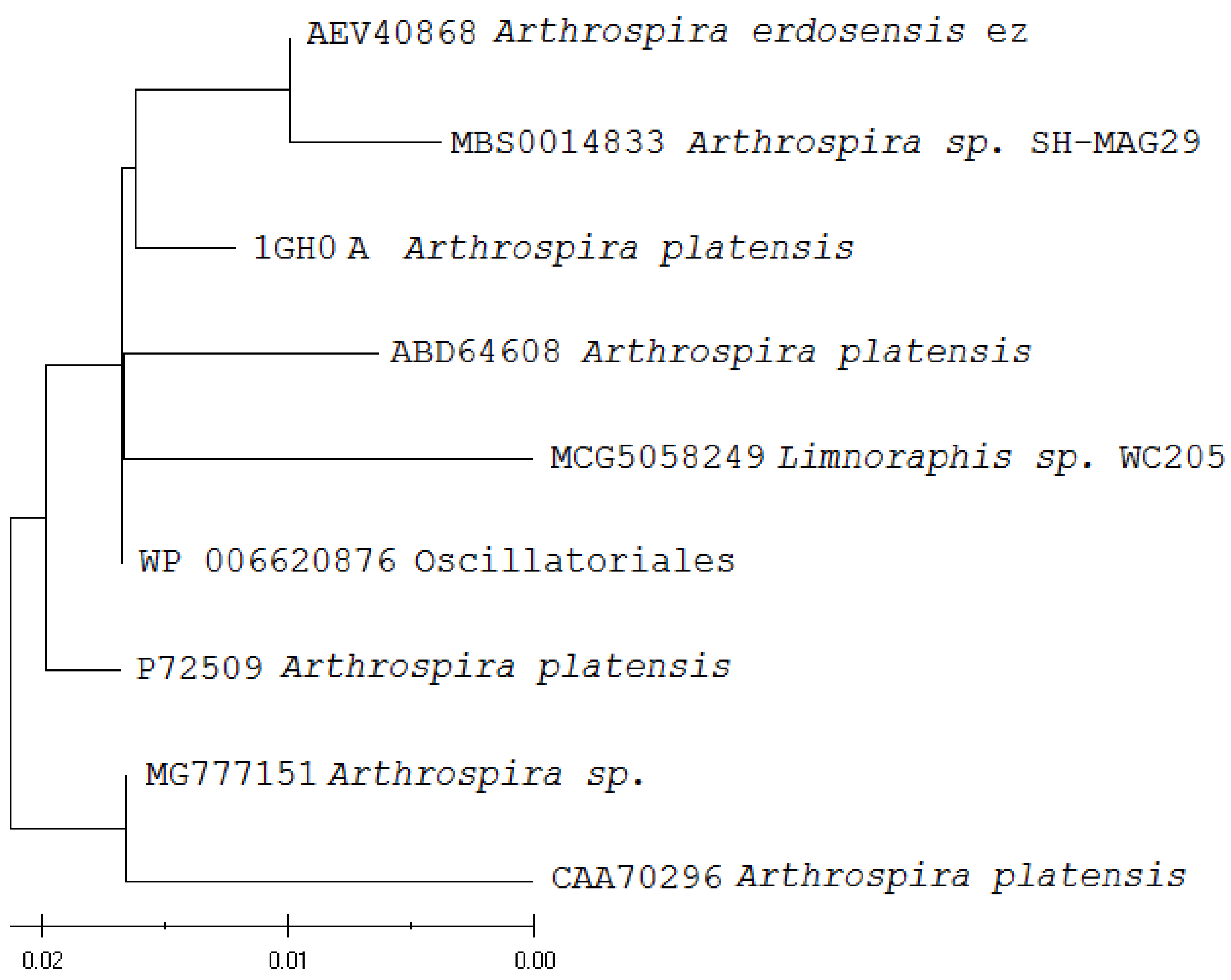
3.2. Bioinformatics Analysis of the Nucleotide Sequences of C-phycocyanin Subunit Alpha

After searching for many sequences in the NCBI database and carrying out BLAST analysis, only nine nucleotide sequences were selected to prove that phycocyanin subunit alpha sequences of Arthrospira species and Arthrospira platensis strains and variants are close to each other and different from other unrelated species of cyanobacteria. BioEdit and MEGA11 were used to run the alignment and build a phylogenetic tree. The multiple alignment of the nine selected nucleotide sequences revealed the presence of minor differences (only 31 from 486 nucleotides) among the aligned species and strains, as shown in Figure 5. This proved the conservation (455 from 486 nucleotides) of the sequence of phycocyanin subunit alpha among the nine aligned sequences. When the sequences of Arthrospira sp. CCALA 030 (Accession MG777151), Arthrospira erdosensis ez (Accession AEV40868), and Arthrospira sp. SH-MAG29 (Accession MBS0014833) were excluded from the alignment, the conservation of the phycocyanin subunit alpha sequence was even higher (461 from 486 nucleotides). This can be explained by the positions of the species Arthrospira sp. SH-MAG29 and the strain Arthrospira erdosensis ez in the upper end of the phylogenetic tree (Figure 6). The species Arthrospira sp. CCALA 030 (Accession MG777151) and the variant strain of Arthrospira platensis (Accession CAA70296) were found in the lower end of the tree. MEGA11 was used to analyze the nucleotide content of the investigated sequences. The nucleotide frequencies in the phycocyanin subunit alpha genes of the nine Arthrospira species and Arthrospira platensis strains and variants are presented in Table 3. As can be seen, the G content of 31.5% was found in Arthrospira sp. CCALA 030 (Accession MG777151) and three Arthrospira platensis variants (the species Limnoraphis sp. WC205 (Accession MCG5058249), Arthrospira platensis (Accession CAA70296), and Oscillatoriales (Accession WP 006620876)). The T content of 19.3% was found in only one strain of Arthrospira platensis (Accession ABD64608) and three other Arthrospira strains and species (Accessions WP 006620876, AEV40868, and MBS0014833). The C content of 25.1% was found in two strains of Arthrospira platensis (Accessions ABD64608 and P72509) in addition to Arthrospira sp. CCALA 030 (Accession MG777151), while that of 24.7% was more common in the analyzed species and strains (Accessions WP 006620876, 1GH0 A, AEV40868, and MBS0014833). The A content of 24.7% was found in two strains of Arthrospira platensis (Accessions ABD64608 and P72509) and Arthrospira erdosensis ez (Accession AEV40868), while that of 24.5% was observed in Arthrospira sp. CCALA 030 (Accession MG777151), Oscillatoriales (Accession WP 006620876), and Arthrospira platensis (Accession CAA70296). T-1 of 11.7%, C-1 of 20.4%, and A-3 of 10.5% were common in eight of the nine analyzed sequences, while A-2 of 31.5% was constant in all of them. Table 4 demonstrates the GC and AT content in phycocyanin subunit alpha nucleotide sequences of the analyzed A. platensis strains and variants. As can be seen, all the analyzed sequences had GC content of more than 54.5%.

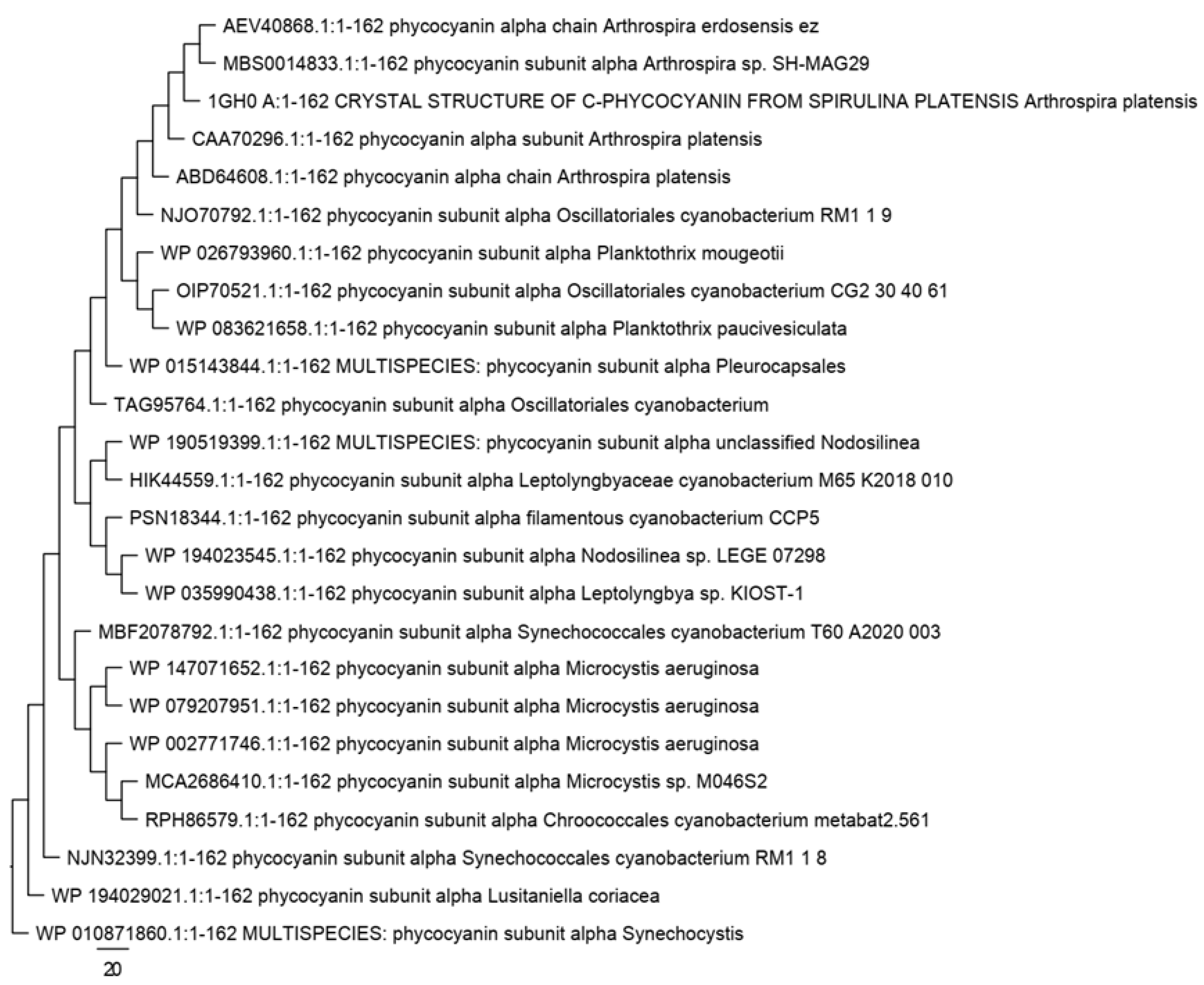
3.3. Bioinformatics Analysis of the Amino Acid Sequences of C-phycocyanin Subunit Alpha

From the NCBI database, 25 protein sequences of phycocyanin subunit alpha of Arthrospira sp. and Arthrospira platensis strains and variants from the order Oscillatoriales were selected to compare their amino acid composition. The alignment of these 25 sequences proved the presence of 97 conserved amino acids (about 60% sequence similarity) among the analyzed sequences, as shown in Figure 7. Phylogenetic tree of the amino acid sequences of phycocyanin subunit alpha of the 25 cyanobacterial species, including Arthrospira platensis strains and variants, is shown in Figure 8. The frequencies of amino acids (%) in phycocyanin subunit alpha protein sequences of the 25 cyanobacterial species are demonstrated in Table 5. As can be seen from Table 5, all essential amino acids (F, H, I, K, L, M, R, T, V, and W) were found in all the analyzed phycocyanin subunit alpha protein sequences. Alanine (A) was the most abundant amino acid (average 15.1%) among all the analyzed sequences, followed by glycine (G) and leucine (L) with average of 8.1% and then serine (S), tyrosine (Y), and threonine (T) with average of 7.7, 6.7, and 6.4%, respectively. The basic amino acid histidine (H) with content of 0.6% was nearly constant (24 of the 25 cyanobacterial species and strains), and the tryptophan (W) content of 0.6% was observed in all species and strains. Regarding acidic amino acids, aspartic acid (D) content of 5.6% and glutamic acid (E) content of 4.9% were found in 12 and 9 cyanobacterial species and strains, respectively, including Arthrospira species and strains of Arthrospira platensis. The frequencies of basic amino acids of 4.3% for arginine (R) and 5.6% for lysine (K) were present in 11 and 10 cyanobacterial species and strains, respectively, including Arthrospira sp., Arthrospira platensis, and their related strains. The contents of 3.1% phenylalanine (F) and 2.5% methionine (M) were found in 21 and 16 cyanobacterial species and strains, respectively. Table 6 demonstrates the unique and harsh environments from which the investigated cyanobacterial species and strains were isolated.

4. Discussion

Phycobiliproteins are large protein aggregates produced by cyanobacterial cells at a concentration of 40–60% of their total soluble protein. They are involved in harvesting of light for these cells during photosynthesis [36]. They can be categorized into phycoerythrin (maximum wavelength of 565 nm), phycocyanin (maximum wavelength of 620 nm), and allophycocyanin (maximum wavelength of 650 nm) based on their spectral properties [37]. Phycobiliproteins have antimicrobial, anti-inflammatory, antioxidant, and hepatoprotective effects [38,39]. They are commonly used as natural pigments and fluorescent proteins in several applications, such as food and cosmetic industries [40,41]. The phycocyanin gene is commonly used in the molecular identification and characterization of A. platensis strains [17].
This work investigated the variation between A. platensis strains and variants from the order Oscillatoriales based on the nucleotide and amino acid composition of the gene and protein sequences of C-phycocyanin alpha chain. The obtained data were linked to their habitat or source of isolation. C-phycocyanin was comparatively extracted from three different A. platensis strains: one isolated from the brackish Lake Mariout at southwest of Alexandria city in Egypt and two commercial French and Chinese strains. The Egyptian strain was cultivated in the laboratory using highly saline medium under static conditions at a temperature of 24 °C (day) and 18 °C (night). The extracted pigment was expected to vary based on the difference in isolation source of the three A. platensis strains and cultivation conditions used with the Egyptian isolate.
In 2013, salinity was verified to be an essential factor in addition to alkalinity for suppressing the growth of algae and cyanobacteria except for A. platensis, thus leading to this microalga dominating the environment [2]. In this study, an Egyptian isolate was cultivated in 1.5× (highly saline) Amara and Steinbuchel medium derived from a combination of George and Zarrouk media under the regular cycle of day and night and static cultivation condition, but the work was conducted in a photobioreactor [2]. The pigment extracted from the cultivated Egyptian strain was expected to be affected by these cultivation conditions, which was the case as the pigment was very faint in color compared to that extracted from the commercial strains. Interestingly, when the same strain was previously grown in Zarrouk medium at room temperature and exposed to fluorescent lamp/day–light, the extracted pigment using potassium phosphate buffer of 0.1 M and pH 7 was green in color, not even blue [17]. Therefore, further investigation should be carried out to control the quality of the pigment obtained from this microalga for biotechnological applications either by mimicking conditions of its natural environment or optimizing conditions in a bioreactor.
Genomes rich in GC are anticipated to be more adapted to high growth temperatures than those rich in AT as GC pairs are usually more stable than the AT ones [42]. In this study, Arthrospira platensis (Accession 1GH0 A) had GC content of 56.58% in the phycocyanin subunit alpha coding gene and was isolated from urban reservoir in Poland, which is characterized by high temperature and reduced air humidity [29,43]. A. platensis (Accession ABD64608) with GC content of 55.97% was isolated from India, while Arthrospira erdosensis ez (Accession AEV40868) with the same GC content was isolated from an alkaline lake in China. Arthrospira sp. SH-MAG29 (Accession MBS0014833) with GC content of 55.76% was isolated from shallow sediments of the arsenic-rich Salar de Huasco Lagoon in Chile, which is an extreme environment with high daily variations in temperature, high UV radiation, arsenic and salinity, and low pressure [30]. In addition, Arthrospira platensis (Accession CAA70296) with GC content of 54.94% was isolated from Thailand. These results prove that the GC content of the gene of phycocyanin subunit alpha of A. platensis strains and variants is linked with the extreme conditions in their habitat, such as high temperatures and alkalinity.
Amino acids play a vital role in metabolism and growth of microorganisms [44]. Amino acids are categorized as essential and nonessential; acidic, basic, and neutral; hydrophilic and hydrophobic; and polar and nonpolar amino acids based on their source and nature of their side chain [45]. Alanine is encoded exclusively or primarily by GC-biased codons, so it is closely related to the GC content [46]. In this work, all essential amino acids (F, H, I, K, L, M, R, T, V, and W) were found in all the analyzed protein sequences of phycocyanin subunit alpha of cyanobacterial species, including A. platensis strains and variants. Alanine was the most abundant amino acid (average of 15.1%) among all the analyzed sequences. Histidine was encoded equally by both GC-and AT-biased codons, but only one residue was found in nearly all the analyzed sequences. The increase in the acidic nature of proteins as an adaptation to hypersalinity has been previously reported [47]. Accordingly, the acidic amino acids aspartic acid and glutamic acid were more abundant than basic residues, especially histidine, in all the analyzed sequences. Overall, these findings confirm that the amino acid composition of the protein sequences of phycocyanin subunit alpha of A. platensis strains and variants are closely related to both GC content and the extreme conditions in their habitat, such as salinity.

5. Conclusions

Arthrospira platensis is an edible cyanobacterium that has great significance for different applications, including environmental, food, feed, biotechnological, and pharmaceutical. A. platensis shows dense growth under conditions of high salinity and alkalinity and dominates lakes in certain periods of the year. It can survive alone under many harsh environmental conditions, and its ability to adapt to environmental stresses exceeds that of other competitors. Phycobiliproteins (natural pigments), especially C-phycocyanin, are one of the most important bioactive products produced by Arthrospira platensis. C-phycocyanin is commonly used in the molecular identification and characterization of A. platensis due to the high conservation of its gene among cyanobacterial species. In this study, an Egyptian A. platensis strain was cultivated in our lab under static conditions in a highly saline medium, and then its C-phycocyanin was extracted and compared to two commercial strains. Additionally, the link between the amino acids and nucleotide composition of the alpha chain of this pigment and the adaptation of Arthrospira platensis to harsh environmental conditions was investigated. Overall, the extracted pigment from the Egyptian strain had a very faint color compared to the pigment of the commercial strains, which may be due to the difference in cultivation conditions and/or source of isolation. The analyzed species and strains had GC content of more than 54.5% in the C-phycocyanin alpha chain gene, which may be linked to adaptation of these cyanobacteria to high temperatures. Moreover, high frequencies of the acidic amino acids aspartic acid and glutamic acid in the C-phycocyanin alpha chain protein can be attributed to adaptation to hypersalinity. Understanding the differences between the strains and variants of Arthrospira platensis at the gene and protein levels of C-phycocyanin alpha chain and determining if these differences are environmentally based and affected by cultivation conditions on the pigment may help in optimizing biotechnological applications of this microalga and its pigment by mimicking the best-suited growth conditions for each habitat.

Author Contributions

N.A.E.-B. and A.A.A. conceived the research topic and designed the research; N.A.E.-B. and N.M.F.R. conducted the experimental work; N.A.E.-B. and A.A.A. collected and analyzed the data; N.A.E.-B. and A.A.A. wrote the manuscript; A.A.A. proofread and revised the manuscript; and N.A.E.-B. finalized the manuscript. All authors have read and agreed to the published version of the manuscript.

Funding

This work received no external funding.

Institutional Review Board Statement

Not applicable.

Informed Consent Statement

Not applicable.

Data Availability Statement

All data are contained within the article.

Conflicts of Interest

The authors declare no conflict of interest.

References

  1. Ciferri, O. Spirulina, the edible microorganism. Microbiol. Rev. 1983, 47, 551–578. [Google Scholar] [CrossRef] [PubMed]
  2. Amara, A.A.; Steinbüchel, A. New medium for pharmaceutical grade Arthrospira. Int. J. Bacteriol. 2013, 2013, 203432. [Google Scholar]
  3. Kebede, E. Response of Spirulina platensis (=Arthrospira fusiformis) from Lake Chitu, Ethiopia, to salinity stress from sodium salts. J. Appl. Phycol. 1997, 9, 551–558. [Google Scholar]
  4. Turpin, P.J.F. Spirulina oscillarioide. Dict. Sci. 1827, 50, 309–310. [Google Scholar]
  5. Rich, F. Notes on Arthrospira platetnsis. Rev. Algol. 1931, 6, 75–79. [Google Scholar]
  6. Dangeard, P. Sur une algue bleue alimentaire pour l’homme: Arthrospira platensis (Nordst.) Gomont. Act. Soc. Linn. Bordx. 1940, 91, 39–41. [Google Scholar]
  7. Shimamatsu, H. Mass production of Spirulina, an edible microalga. Hydrobiologia 2004, 512, 39–44. [Google Scholar] [CrossRef]
  8. Mühling, M.; Belay, A.; Whitton, B.A. Screening Arthrospira (Spirulina) strains for heterotrophy. J. Appl. Phycol. 2005, 17, 129–135. [Google Scholar] [CrossRef]
  9. Busnel, A.; Samhat, K.; Gérard, E.; Kazbar, A.; Marec, H.; Dechandol, E.; Le Gouic, B.; Hauser, J.-L.; Pruvost, J. Development and validation of a screening system for characterizing and modeling biomass production from cyanobacteria and microalgae: Application to Arthrospira platensis and Haematococcus pluvialis. Algal Res. 2021, 58, 102386. [Google Scholar] [CrossRef]
  10. AlFadhly, N.K.Z.; Alhelfi, N.; Altemimi, A.B.; Verma, D.K.; Cacciola, F. Tendencies affecting the growth and cultivation of genus Spirulina: An investigative review on current trends. Plants 2022, 11, 3063. [Google Scholar] [CrossRef]
  11. Capelli, B.; Cysewski, G.R. Potential health benefits of Spirulina microalgae: A review of the existing literature. Nutrafoods 2010, 9, 19–26. [Google Scholar] [CrossRef]
  12. Vo, T.S.; Ngo, D.H.; Kim, S.K. Chapter 19-Nutritional and pharmaceutical properties of microalgal Spirulina. In Handbook of Marine Microalgae; Kim, S.K., Ed.; Academic Press: Cambridge, MA, USA, 2015; pp. 299–308. [Google Scholar]
  13. Soni, R.A.; Sudhakar, K.; Rana, R.S. Spirulina—From growth to nutritional product: A review. Trends Food Sci. Technol. 2017, 69, 157–171. [Google Scholar] [CrossRef] [Green Version]
  14. Pez Jaeschke, D.; Rocha Teixeira, I.; Damasceno Ferreira Marczak, L.; Domeneghini Mercali, G. Phycocyanin from Spirulina: A review of extraction methods and stability. Food Res. Int. 2021, 143, 110314. [Google Scholar] [CrossRef] [PubMed]
  15. García, A.B.; Longo, E.; Bermejo, R. The application of a phycocyanin extract obtained from Arthrospira platensis as a blue natural colorant in beverages. J. Appl. Phycol. 2021, 33, 3059–3070. [Google Scholar] [CrossRef]
  16. Chentir, I.; Hamdi, M.; Li, S.; Doumandji, A.; Markou, G.; Nasri, M. Stability, bio-functionality and bio-activity of crude phycocyanin from a two-phase cultured Saharian Arthrospira sp. Strain. Algal Res. 2018, 35, 395–406. [Google Scholar] [CrossRef]
  17. Sharaf, M.; Amara, A.; Aboul-Enein, A.; Helmi, S.; Ballot, A.; Astani, A.; Schnitzler, P. Molecular authentication and characterization of the antiherpetic activity of the cyanobacterium Arthrospira fusiformis. Pharmazie 2010, 65, 132–136. [Google Scholar]
  18. Haverkamp, T.H.; Schouten, D.; Doeleman, M.; Wollenzien, U.; Huisman, J.; Stal, L.J. Colorful microdiversity of Synechococcus strains (picocyanobacteria) isolated from the Baltic Sea. ISME J. 2009, 3, 397–408. [Google Scholar] [CrossRef] [PubMed]
  19. Khan, Z.; Wan Maznah, W.O.; Faradina Merican, M.S.M.; Convey, P.; Najimudin, N.; Alias, S.A. A comparative study of phycobilliprotein production in two strains of Pseudanabaena isolated from Arctic and tropical regions in relation to different light wavelengths and photoperiods. Polar Sci. 2019, 20, 3–8. [Google Scholar] [CrossRef]
  20. Ruiz-Domínguez, M.C.; Jáuregui, M.; Medina, E.; Jaime, C.; Cerezal, P. Rapid Green Extractions of C-Phycocyanin from Arthrospira maxima for Functional Applications. Appl. Sci. 2019, 9, 1987. [Google Scholar] [CrossRef] [Green Version]
  21. Park, J.; Lee, H.; Dinh, T.B.; Choi, S.; De Saeger, J.; Depuydt, S.; Brown, M.T.; Han, T. Commercial Potential of the Cyanobacterium Arthrospira maxima: Physiological and Biochemical Traits and the Purification of Phycocyanin. Biology 2022, 11, 628. [Google Scholar] [CrossRef]
  22. Tavanandi, H.A.; Mittal, R.; Chandrasekhar, J.; Raghavarao, K.S.M.S. Simple and efficient method for extraction of C-Phycocyanin from dry biomass of Arthospira platensis. Algal Res. 2018, 31, 239–251. [Google Scholar] [CrossRef]
  23. Bradford, M.M. A rapid and sensitive method for the quantitation of microgram quantities of protein utilizing the principle of protein-dye binding. Anal. Biochem. 1976, 72, 248–254. [Google Scholar] [CrossRef]
  24. Siegelman, H.; Kycia, J.H. Alga biliproteins. In Handbook of Phycological Methods: Physiological and Biochemical Methods; Hellebust, J.A., Craigie, J.S., Eds.; Cambridge University Press: Cambridge, UK, 1978; pp. 72–78. [Google Scholar]
  25. Sharmaa, G.; Sarana, S.; Puri, N.; Jasuja, N.D.; Kumar, M. Optimization, purification and characterization of phycocyanin from Spirulina platensis. Int. J. Appl. Pure Sci. Agric. 2016, 2, 15–20. [Google Scholar]
  26. Madden, T. The BLAST Sequence analysis tool (Chapter 16). In The NCBI Handbook [Internet]; McEntyre, J., Ostell, J., Eds.; National Center for Biotechnology Information: Bethesda, MD, USA, 2002; p. 15. [Google Scholar]
  27. Hall, T.A. BioEdit: A user-friendly biological sequence alignment editor and analysis program for Windows 95/98/NT. Nucl. Acids Symp. Ser. 1999, 41, 95–98. [Google Scholar]
  28. Tamura, K.; Stecher, G.; Kumar, S. MEGA11: Molecular Evolutionary Genetics Analysis Version 11. Mol. Biol. Evol. 2021, 38, 3022–3027. [Google Scholar] [CrossRef]
  29. Wang, X.Q.; Li, L.N.; Chang, W.R.; Zhang, J.P.; Gui, L.L.; Guo, B.J.; Liang, D.C. Structure of C-phycocyanin from Spirulina platensis at 2.2 A resolution: A novel monoclinic crystal form for phycobiliproteins in phycobilisomes. Acta Crystallogr. D Biol. Crystallogr. 2001, 57, 784–792. [Google Scholar] [CrossRef]
  30. Castro-Severyn, J.; Pardo-Esté, C.; Mendez, K.N.; Fortt, J.; Marquez, S.; Molina, F.; Castro-Nallar, E.; Remonsellez, F.; Saavedra, C.P. Living to the High Extreme: Unraveling the Composition, Structure, and Functional Insights of Bacterial Communities Thriving in the Arsenic-Rich Salar de Huasco Altiplanic Ecosystem. Microbiol. Spectr. 2021, 9, e0044421. [Google Scholar] [CrossRef] [PubMed]
  31. Probst, A.J.; Castelle, C.J.; Singh, A.; Brown, C.T.; Anantharaman, K.; Sharon, I.; Hug, L.A.; Burstein, D.; Emerson, J.B.; Thomas, B.C.; et al. Genomic resolution of a cold subsurface aquifer community provides metabolic insights for novel microbes adapted to high CO2 concentrations. Environ. Microbiol. 2017, 19, 459–474. [Google Scholar] [CrossRef] [PubMed] [Green Version]
  32. Pérez-Carrascal, O.M.; Tromas, N.; Terrat, Y.; Moreno, E.; Giani, A.; Marques, L.C.B.; Fortin, N.; Shapiro, B.J. Single-colony sequencing reveals microbe-by-microbiome phylosymbiosis between the cyanobacterium Microcystis and its associated bacteria. Microbiome 2021, 9, 194. [Google Scholar] [CrossRef]
  33. Dalcin Martins, P.; Danczak, R.E.; Roux, S.; Frank, J.; Borton, M.A.; Wolfe, R.A.; Burris, M.N.; Wilkins, M.J. Viral and metabolic controls on high rates of microbial sulfur and carbon cycling in wetland ecosystems. Microbiome 2018, 6, 138. [Google Scholar] [CrossRef]
  34. Bouma-Gregson, K.; Olm, M.R.; Probst, A.J.; Anantharaman, K.; Power, M.E.; Banfield, J.F. Impacts of microbial assemblage and environmental conditions on the distribution of anatoxin-a producing cyanobacteria within a river network. ISME J. 2019, 13, 1618–1634. [Google Scholar] [CrossRef] [Green Version]
  35. Alcorta, J.; Alarcón-Schumacher, T.; Salgado, O.; Díez, B. Taxonomic Novelty and Distinctive Genomic Features of Hot Spring Cyanobacteria. Front. Genet. 2020, 11, 568223. [Google Scholar] [CrossRef]
  36. Kumar, D.; Dhar, D.W.; Pabbi, S.; Kumar, N.; Walia, S. Extraction and purification of C-phycocyanin from Spirulina platensis (CCC540). Indian J. Plant Physiol. 2014, 19, 184–188. [Google Scholar] [CrossRef] [PubMed] [Green Version]
  37. Glazer, A.N.; Bryant, D.A. Allophycocyanin B (λmax 671, 618 nm) Arch. Microbiol. 1975, 104, 15–22. [Google Scholar]
  38. Kulshreshtha, A.; Jacharia, A.J.; Jarouliya, U.; Bhadauriya, P.; Prasad, G.B.; Bisen, P.S. Spirulina in health care management. Curr. Pharmaceut. Biotechnol. 2008, 9, 400–405. [Google Scholar] [CrossRef] [Green Version]
  39. Hamad, G.M.; Abd El-Baky, N.; Sharaf, M.M.; Amara, A.A. Volatile compounds, fatty acids constituents, and antimicrobial activity of cultured Spirulina (Arthrospira fusiformis) isolated from Lake Mariout in Egypt. Sci. World J. 2023, 2023, 9919814. [Google Scholar] [CrossRef] [PubMed]
  40. Eriksen, N.T. Production of phycocyanin–A pigment with applications in biology, biotechnology, foods and medicine. Appl. Microbiol. Biotechnol. 2008, 80, 1–14. [Google Scholar] [CrossRef] [PubMed]
  41. Mishra, S.K.; Shrivastav, A.; Mishra, S. Effect of preservatives for food grade C-PC from Spirulina platensis. Process Biochem. 2008, 43, 339–345. [Google Scholar] [CrossRef]
  42. Hu, E.Z.; Lan, X.R.; Liu, Z.L.; Gao, J.; Niu, D.-K. A positive correlation between GC content and growth temperature in prokaryotes. BMC Genom. 2022, 23, 110. [Google Scholar] [CrossRef]
  43. Nowicka-Krawczyk, P.; Mühlsteinová, R.; Hauer, T. Detailed characterization of the Arthrospira type species separating commercially grown taxa into the new genus Limnospira (Cyanobacteria). Sci. Rep. 2019, 9, 694. [Google Scholar] [CrossRef] [Green Version]
  44. Wu, G. Amino acids: Metabolism, functions, and nutrition. Amino Acids 2009, 37, 1–17. [Google Scholar] [CrossRef] [PubMed]
  45. Kevin, A.; Indira, R.; Taralyn, T. Structure & Function—Amino Acids—Biology LibreTexts. Available online: https://bio.libretexts.org/Bookshelves/Biochemistry/Book:_Biochemistry_Free_For_All_(Ahern,_Rajagopal,_and_Tan)/2:_Structure_and_Function/2.2:_Structure_and_Function_-_Amino_Acids (accessed on 20 November 2020).
  46. Li, J.; Zhou, J.; Wu, Y.; Yang, S.; Tian, D. GC-content of synonymous codons profoundly influences amino acid usage. G3 (Bethesda) 2015, 5, 2027–2036. [Google Scholar] [CrossRef] [PubMed] [Green Version]
  47. Rhodes, M.E.; Fitz-Gibbon, S.T.; Oren, A.; House, C.H. Amino acid signatures of salinity on an environmental scale with a focus on the Dead Sea. Environ. Microbiol. 2010, 12, 2613–2623. [Google Scholar] [CrossRef] [PubMed]
Figure 1. Cultivation of Egyptian A. platensis strain in the laboratory under static conditions. (A) Preparation of membrane filter 1.5× medium components from parts A–C and the autoclaved part D; (B) mixing of the medium components under aseptic conditions; (C) adding the seed culture of A. platensis and the medium components to a sterile container; (D,E) static cultivation and collection of A. platensis; (F) air drying of the harvested cells.
Jmse 11 00663 g001
Figure 2. Extracted C-phycocyanin from dry biomass of the three microalgae strains.
Jmse 11 00663 g002
Figure 3. The 12% SDS-PAGE analysis of the extracted C-phycocyanin from dry biomass of the three microalgae strains. M: Protein molecular weight marker; 1, 2: C-phycocyanin extract from the Chinese strain; 3, 4: C-phycocyanin extract from the French strain; 5: C-phycocyanin extract from the Egyptian strain. The arrow points to C-phycocyanin subunits.
Jmse 11 00663 g003
Figure 4. Visible spectrum (at wavelengths 600–650 nm) of the extracted C-phycocyanin from dry biomass of the three microalgae strains.
Jmse 11 00663 g004
Figure 5. Multiple alignment of nucleotide sequences of phycocyanin subunit alpha of nine cyanobacterial species, including Arthrospira platensis strains and variants, using BioEdit. The letters A, T, C, and G symbolize the four nucleotides adenine, thymine, cytosine, and guanine, respectively; * represents conserved nucleotides.
Jmse 11 00663 g005
Figure 6. Phylogenetic tree of nucleotide sequences of phycocyanin subunit alpha of nine cyanobacterial species, including Arthrospira platensis strains and variants. The branching order and score were calculated by MEGA11 and visualized by FigTree 1.4.2.
Jmse 11 00663 g006
Figure 7. Multiple alignment of amino acid sequences of phycocyanin subunit alpha of 25 cyanobacterial species, including A. platensis strains and variants, using BioEdit. Amino acids are shown in one-letter coded form. A: alanine, C: cysteine, D: aspartic acid, E: glutamic acid, F: phenylalanine, G: glycine, H: histidine, I: isoleucine, K: lysine, L: leucine, M: methionine, N: asparagine, P: proline, Q: glutamine, R: arginine, S: serine, T: threonine, V: valine, W: tryptophan, Y: tyrosine. Consensus key: * (asterisk): positions that have a single, fully conserved residue; : (colon): conservation between groups that have strongly similar properties; . (period): conservation between groups that have weakly similar properties; blank spaces mean no consensus.
Jmse 11 00663 g007
Figure 8. Phylogenetic tree of amino acid sequences of phycocyanin subunit alpha of 25 cyanobacterial species, including A. platensis strains and variants. The branching order and score were calculated by MEGA11 and visualized by FigTree 1.4.2.
Jmse 11 00663 g008
Table 1. The total protein content of the extracted C-phycocyanin from dried biomass of the three microalgae strains as measured by Bradford assay.
StrainTotal Protein Concentration
(mg/g Dried Biomass)
Egyptian A. platensis1.233
French A. platensis1.188
Chinese A. platensis1.028
Table 2. The concentration and purity of the extracted C-phycocyanin from dried biomass of the three microalgae strains.
StrainC-phycocyanin Concentration
(mg/mL)
C-phycocyanin Purity
(Dividing A615 by A280)
Egyptian A. platensis0.3230.84
French A. platensis0.3330.69
Chinese A. platensis0.3270.84
Table 3. The frequencies of the nucleotides in phycocyanin subunit alpha nucleotide sequences of nine cyanobacterial species, including A. platensis strains and variants. All nucleotide frequencies were calculated by MEGA11 and are given as percentages.
Accession and SpeciesT(U)CAGTotalT-1C-1A-1G-1Pos #1T-2C-2A-2G-2Pos #2T-3C-3A-3G-3Pos #3
MG777151, Arthrospira sp. CCALA 03018.925.124.531.5486.011.720.431.536.4162.022.223.531.522.8162.022.831.510.535.2162.0
P72509, Arthrospira platensis19.125.124.731.1486.011.720.432.135.8162.022.824.131.521.6162.022.830.910.535.8162.0
WP 006620876, Oscillatoriales19.324.724.531.5486.011.720.431.536.4162.023.523.531.521.6162.022.830.210.536.4162.0
1GH0 A, Arthrospira platensis19.124.724.331.9486.011.720.430.937.0162.023.523.531.521.6162.022.230.210.537.0162.0
AEV40868, Arthrospira erdosensis ez19.324.724.731.3486.011.720.432.135.8162.023.522.831.522.2162.022.830.910.535.8162.0
MBS0014833, Arthrospira sp. SH-MAG2919.324.724.931.1486.011.720.432.735.2162.023.522.231.522.8162.022.831.510.535.2162.0
ABD64608, Arthrospira platensis19.325.124.730.9486.011.720.432.735.2162.023.524.131.521.0162.022.830.99.936.4162.0
MCG5058249, Limnoraphis sp. WC20518.925.324.331.5486.011.721.030.936.4162.022.824.131.521.6162.022.230.910.536.4162.0
CAA70296, Arthrospira platensis20.623.524.531.5486.01220.431.536.4162.02223.531.522.8162.02826.510.535.2162.0
Average %19.324.824.631.3486.01220.431.836.1162.02323.531.522.0162.02330.410.435.9162.0
Table 4. The GC and AT content of the analyzed nucleotide sequences of nine cyanobacterial species, including A. platensis strains and variants.
AccessionGC Content
(%)
AT Content
(%)
MG77715156.5843.42
P7250956.1743.83
WP 00662087656.1743.83
1GH0 A56.5843.42
AEV4086855.9744.03
MBS001483355.7644.24
ABD6460855.9744.03
MCG505824956.7943.21
CAA7029654.9445.06
Table 5. The frequencies of amino acids (%) in phycocyanin subunit alpha protein sequences of 25 cyanobacterial species, including A. platensis strains and variants, as calculated using MEGA11.
Phycocyanin Subunit AlphaACDEFGHIKLMNPQRSTVWYTotal
ABD64608.1:1-162 Arthrospira platensis14.81.25.64.33.17.40.66.85.68.02.54.33.14.34.37.46.23.10.66.8162
1GH0 A:1-162 Arthrospira platensis14.81.25.64.93.18.00.66.25.68.02.53.73.14.34.37.45.63.70.66.8162
AEV40868.1:1-162 Arthrospira erdosensis ez14.21.25.64.93.18.00.66.85.68.02.53.73.14.34.38.05.63.10.66.8162
MBS0014833.1:1-162 Arthrospira sp. SH-MAG2913.61.25.64.93.18.00.66.85.68.02.53.73.14.34.38.65.63.10.66.8162
CAA70296.1:1-162 Arthrospira platensis14.81.25.64.93.18.60.66.85.67.42.53.73.14.34.97.45.62.50.66.8162
NJO70792.1:1-162 Oscillatoriales cyanobacterium RM1 1 915.41.25.64.93.18.00.65.65.68.03.13.73.14.34.36.27.43.10.66.2162
OIP70521.1:1-162 Oscillatoriales cyanobacterium CG2 30 40 6114.81.24.35.63.18.00.65.65.67.41.93.73.14.34.36.88.04.30.66.8162
WP 026793960.1:1-162 Planktothrix mougeotii14.21.24.35.63.18.00.64.95.68.01.94.33.13.74.38.66.84.30.66.8162
WP 083621658.1:1-162 Planktothrix paucivesiculata14.81.24.35.63.18.60.64.95.68.61.23.73.14.34.36.87.44.30.66.8162
PSN18344.1:1-162 filamentous cyanobacterium CCP514.81.24.95.63.18.60.63.74.98.63.13.73.14.93.78.06.23.70.66.8162
WP 194023545.1:1-162 Nodosilinea sp. LEGE 0729814.81.24.95.63.18.60.63.14.98.03.13.73.14.33.78.06.84.90.66.8162
MCA2686410.1:1-162 Microcystis sp. M046S215.41.25.64.33.17.40.66.24.37.42.54.93.14.34.97.46.23.70.66.8162
WP 015143844.1:1-162 MULTISPECIES: Pleurocapsales15.41.94.94.92.58.00.66.24.98.03.15.63.14.93.76.85.62.50.66.8162
WP 147071652.1:1-162 Microcystis aeruginosa15.41.25.64.33.18.00.66.24.37.42.53.73.14.34.98.06.23.70.66.8162
RPH86579.1:1-162 Chroococcales cyanobacterium metabat2.56116.01.24.94.93.17.40.66.24.37.42.54.33.14.34.97.46.23.70.66.8162
WP 002771746.1:1-162 Microcystis aeruginosa16.01.25.64.33.16.80.66.24.97.42.53.73.14.34.38.66.23.70.66.8162
WP 079207951.1:1-162 Microcystis aeruginosa15.41.25.64.33.18.60.66.24.37.42.53.73.14.34.97.46.23.70.66.8162
WP 035990438.1:1-162 Leptolyngbya sp. KIOST-114.81.24.95.63.18.60.63.74.98.03.13.72.54.33.79.36.24.30.66.8162
MBF2078792.1:1-162 Synechococcales cyanobacterium T60 A2020 00314.81.25.64.93.17.40.66.24.97.42.53.73.16.24.37.45.63.70.66.8162
NJN32399.1:1-162 Synechococcales cyanobacterium RM1 1 814.81.25.64.33.18.00.64.35.68.62.53.73.74.33.18.66.83.70.66.8162
WP 190519399.1:1-162 MULTISPECIES: unclassified Nodosilinea16.71.94.95.62.58.60.63.14.98.62.54.32.53.73.77.46.24.90.66.8162
WP 010871860.1:1-162 MULTISPECIES: Synechocystis14.21.26.83.73.18.00.64.94.39.90.66.22.55.64.96.26.83.70.66.2162
WP 194029021.1:1-162 Lusitaniella coriacea14.81.24.96.22.59.31.24.34.98.62.53.72.53.13.79.95.64.30.66.2162
TAG95764.1:1-162 Oscillatoriales cyanobacterium16.01.24.34.93.18.00.64.34.38.62.53.73.14.94.36.28.63.70.66.8162
HIK44559.1:1-162 Leptolyngbyaceae cyanobacterium M65 K2018 01016.71.94.95.62.58.00.64.33.78.62.54.32.53.74.98.06.23.70.66.8162
Average %15.11.35.25.03.08.10.65.35.08.12.44.03.04.44.37.76.43.70.66.7162
Table 6. Characteristics of the investigated cyanobacterial organisms and source of isolation.
StrainCharacteristics of IsolateAccessionReference
Arthrospira platensis- Phycocyanin alpha chain
- Country: India
- Submitted by Plant Molecular Biology and Biotechnology, Tamil Nadu Agricultural University, India
ABD64608Unpublished
Arthrospira platensis (Gomont) Geitler 1925- Crystal structure of C-phycocyanin
- Isolation source: urban reservoir in Poland, Central Europe.
1GH0 A[29]
Arthrospira erdosensis ez- Phycocyanin alpha chain
- Submitted by College of Life Science and Technology, Inner Mongolia Normal University, P.R. China
- Isolation source: alkaline lake 1200–1600 m above sea level
AEV40868Unpublished
Arthrospira sp. SH-MAG29- Phycocyanin subunit alpha
- Isolation source: shallow sediments of the arsenic-rich Salar de Huasco
lagoon
- Submitted by Departamento de Ingenieria Quimica, Universidad Catolica del Norte, Chile
MBS0014833[30]
Arthrospira platensis- Phycocyanin alpha subunit
- Submitted by W. Jeamton, School of Bioresources and
Technology, King Mongkut’s Institute of Technology Thonburi (KMITT), Thailand
CAA70296Unpublished
Oscillatoriales cyanobacterium RM1_1_9- Phycocyanin subunit alpha
- Environmental sample
- Country: South Africa, Cape Recife
NJO70792Unpublished
Oscillatoriales cyanobacterium CG2_30_40_61- Phycocyanin subunit alpha
- Isolation source: Crystal Geyser (Utah, USA), a site where deeply sourced CO2 -saturated fluids are erupted at the surface
OIP70521[31]
Planktothrix mougeotiiPhycocyanin subunit alphaWP_026793960Unpublished
Planktothrix paucivesiculataPhycocyanin subunit alphaWP_083621658Unpublished
Filamentous cyanobacterium CCP5- Phycocyanin subunit alpha- Submitted by Earth, Atmospheric and Planetary Sciences, Massachusetts Institute of Technology, USA
- Isolation source: salt marsh
PSN18344Unpublished
Nodosilinea sp. LEGE 07298Phycocyanin subunit alphaWP_194023545Unpublished
Microcystis sp. M046S2- Phycocyanin subunit alpha
- Isolation source: fresh water surface (50 cm depth)
MCA2686410[32]
PleurocapsalesPhycocyanin subunit alphaWP_015143844Unpublished
Microcystis aeruginosaPhycocyanin subunit alphaWP_147071652Unpublished
Chroococcales cyanobacterium metabat2.561- Phycocyanin subunit alpha
- Isolation source: Prairie Pothole Region wetland sediments
RPH86579[33]
Microcystis aeruginosaPhycocyanin subunit alphaWP_002771746Unpublished
Microcystis aeruginosaPhycocyanin subunit alphaWP_079207951Unpublished
Leptolyngbya sp. KIOST-1Phycocyanin subunit alphaWP_035990438Unpublished
Synechococcales cyanobacterium T60_A2020_003- Phycocyanin subunit alpha
- Isolation source: nonacidic hot spring microbial mat
- Submitted by Departamento de Genetica Molecular y Microbiologia, Pontificia Universidad Catolica de Chile, Chile
MBF2078792Unpublished
Synechococcales cyanobacterium RM1_1_8-Phycocyanin subunit alpha
-Isolation source: stromatolite
- Submitted by Pharmaceutical Sciences, University of Wisconsin, USA
NJN32399Unpublished
Unclassified NodosilineaPhycocyanin subunit alphaWP_190519399Unpublished
SynechocystisPhycocyanin subunit alphaWP_010871860Unpublished
Lusitaniella coriaceaPhycocyanin subunit alphaWP_194029021Unpublished
Oscillatoriales cyanobacterium- Phycocyanin subunit alpha
- Isolation source: microbial mat material
- Environmental sample
- Country: USA, California, Eel River, Elder Creek
TAG95764[34]
Leptolyngbyaceae cyanobacterium M65_K2018_010-Phycocyanin subunit alpha
-Isolation source: Hot spring_65deg.
HIK44559[35]
Disclaimer/Publisher’s Note: The statements, opinions and data contained in all publications are solely those of the individual author(s) and contributor(s) and not of MDPI and/or the editor(s). MDPI and/or the editor(s) disclaim responsibility for any injury to people or property resulting from any ideas, methods, instructions or products referred to in the content.

