Article

Transcriptome Analysis of the Marine Nematode Litoditis marina in a Chemically Defined Food Environment with Stearic Acid Supplementation

1
CAS and Shandong Province Key Laboratory of Experimental Marine Biology, Institute of Oceanology, Chinese Academy of Sciences, Qingdao 266071, China
2
Laboratory of Marine Biology and Biotechnology, Qingdao National Laboratory for Marine Science and Technology, Qingdao 266237, China
3
Center for Ocean Mega-Science, Chinese Academy of Sciences, 7 Nanhai Road, Qingdao 266071, China
4
University of Chinese Academy of Sciences, Beijing 100049, China
*
Author to whom correspondence should be addressed.
J. Mar. Sci. Eng. 2022, 10(3), 428; https://doi.org/10.3390/jmse10030428
Submission received: 9 February 2022 / Revised: 8 March 2022 / Accepted: 8 March 2022 / Published: 16 March 2022

Abstract

:
Stearic acid represents one of the most abundant fatty acids in the Western diet and profoundly regulates health and diseases of animals and human beings. We previously showed that stearic acid supplementation promoted development of the terrestrial model nematode Caenorhabditis elegans in chemically defined CeMM food environment. However, whether stearic acid regulates development of other nematodes remains unknown. Here, we found that dietary supplementation with stearic acid could promote the development of the marine nematode Litoditis marina, belonging to the same family as C. elegans, indicating the conserved roles of stearic acid in developmental regulation. We further employed transcriptome analysis to analyze genome-wide transcriptional signatures of L. marina with dietary stearic acid supplementation. We found that stearic acid might promote development of L. marina via upregulation of the expression of genes involved in aminoacyl-tRNA biosynthesis, translation initiation and elongation, ribosome biogenesis, and transmembrane transport. In addition, we observed that the expression of neuronal signaling-related genes was decreased. This study provided important insights into how a single fatty acid stearic acid regulates development of marine nematode, and further studies with CRISPR genome editing will facilitate demonstrating the molecular mechanisms underlying how a single metabolite regulates animal development and health.

1. Introduction

Stearic acid (SA, C18:0) is a saturated long-chain fatty acid (FA) and one of the most abundant fatty acids in the Western diet [1,2]. Unlike palmitic acid (PA, C16:0), dietary stearic acid does not increase atherosclerosis risk, while stearic acid was reported to reduce LDL (low density lipoprotein) cholesterol in humans [3,4,5,6]. Studies have shown that elevated circulating stearic acid lipids levels are related to lower blood pressure [7], improved cardiac function [8], and reduced cancer risk [9,10,11,12,13], thus, stearic acid seems to benefit human health differently from other saturated fatty acids. In addition, recent reports showed that stearic acid ingestion promotes mitochondrial fusion and increases fatty acid β-oxidation [14,15].
Modern foods are composed of many metabolites and it is critical to know which are the most important ones to our health. Since human diets are very complex and it is hard to delineate how a single metabolite regulates the development, reproduction, and lifespan of the host, we have applied an axenic chemically defined diet CeMM (C. elegans maintenance medium) and the widely used biomedical model animal Caenorhabditis elegans to study how nutrition regulates animal development and health [16,17]. CeMM excludes variables associated with bacterial metabolism in the bacterial diet, making CeMM promising for use in drug screening [18,19,20]. In addition, the advantages of axenic cultivation and less need for astronaut intervention make CeMM widely used in the study of the effects of space flight on the development and physiology of tested animals such as C. elegans [21,22]. Given the composition and the amount of each component that can be manipulated in this chemically defined CeMM medium, it allows to study the precise mechanisms underlying how a single metabolite regulates animal health. We previously showed that dietary supplementation of the single fatty acid stearic acid significantly promotes development of C. elegans in a CeMM food environment [23]. However, whether stearic acid regulates the development of other nematodes and what the underlying mechanisms are remain unknown.
Litoditis marina is a free-living marine nematode, playing an essential role in marine ecosystems [24,25]. L. marina belongs to the same family as C. elegans, and has the advantages of short generation time, convenient laboratory culture, and its genome has been sequenced and annotated recently, making it a potential marine model animal [25,26].
In this study, we formulated sea-salt-CeMM that can support the growth of marine nematode L. marina and found that dietary supplementation with stearic acid could promote development of L. marina. We further employed RNA sequencing (RNA-seq) analysis to detect genome-wide transcriptional signatures of L. marina with dietary stearic acid supplementation. We found that stearic acid supplementation resulted in upregulation of the expression of genes involved in aminoacyl-tRNA biosynthesis, translation initiation and elongation, ribosome biogenesis, and transmembrane transporter in L. marina, whereas the expression of neuronal signaling-related genes was significantly downregulated with stearic acid supplementation. Our results provide clues for further research on the mechanisms underlying how a single metabolite regulates animal development and health.

2. Materials and Methods

2.1. Worms

The marine nematode L. marina wild strain HQ1 was isolated from intertidal sediments of Huiquan Bay, Qingdao. L. marina nematodes were cultured on seawater-NGM (SW-NGM) agar plates seeded with Escherichia coli OP50 as a food source [25,27]. Until this study, the L. marina nematodes had been maintained and propagated in the laboratory at 20 °C for about four years.

2.2. Sea-Salt-CeMM Preparation

The salinity of normal CeMM is too low to support the development of L. marina into adults. A CeMM medium supplemented with a final concentration of 15‰ sea-salt could support the growth and reproduction of L. marina. This sea-salt-CeMM medium was formulated as follows: 2× CeMM stock media was prepared as reported previously [16,23]. Then, the 15‰ sea-salt-CeMM plate was made by diluting the 2× CeMM stock to 1× via adding an equal volume of molten 3.4% agarose solution containing 30‰ sea-salt.

2.3. Dietary Stearic Acid Supplementation

Stearic acid (manufacturer: Aladdin) was prepared as 40 mM stocks in DMSO. For growth phenotype analysis, 100 μL of 2.5% solvent DMSO or 1 mM stearic acid suspension (prepared by diluting a 40 mM stock solution with sterile water) was added evenly on the surface of a 3.5 cm CeMM plate, and then dried in a dark incubator at 20 °C for one day before the experiments. For the dietary stearic acid supplementation RNA-seq experiment, 667 μL of 2.5% solvent DMSO or 1 mM stearic acid suspension was added evenly on the surface of 9 cm CeMM plate, then the plate was dried in a dark incubator at 20 °C for two days before use.

2.4. Synchronization of L. marina

The eggs were treated with a drop of worm bleaching solution (20% bleach, 0.5 M NaOH) on an empty SW-NGM plate. These eggs were then incubated overnight to obtain small scale synchronized L1.
For sample preparation of transcriptome, large-scale L1 synchronization was performed as described previously [27]. Briefly, a large number of eggs were treated for a short time with worm bleaching solution to kill the bacteria, which were then washed twice with sterile seawater and incubated overnight in sterile seawater to hatch. Finally, large numbers of synchronized L1 larvae were obtained through filtration via 500 grid nylon filter with 25 mm mesh size.

2.5. RNA-Seq Analysis

Synchronized L. marina L1s were transferred to corresponding 9 cm diameter 15‰ sea-salt-CeMM agarose plates supplemented with DMSO or stearic acid. The treated worms were cultured at 20 °C for 2.5 h and collected by washing with M9 buffer. Then, the obtained samples were frozen immediately in liquid nitrogen. Each treatment was performed with three biological replicates.
Total RNA was extracted using Trizol (Invitrogen, Carlsbad, CA, USA). Then, with three biological replicates for each treatment, a total of six RNA sequencing libraries were prepared with 1 μg RNA per library using NEBNext UltraTM RNA Library Prep Kit (New England Biolabs, Ipswich, MA, USA). Then, the sequencing libraries were sequenced on an Illumina platform.
Reads containing adaptors or poly-N and low-quality reads were removed to obtain clean data. The minimum of base score Q20 was over 99% and Q30 was over 93.6%. The clean data were mapped to reference genome [25] by Hisat2 (v2.0.5, with the default parameters) [28]. Gene expression levels were normalized with FPKM. The differentially expressed genes (DEGs) were analyzed by DESeq2 [29]. Fold change ≥ 1.3 and a false discovery rate (FDR) < 0.05 were applied. The adjusted p-values (padj) were calculated according to Benjamini and Hochberg [30] to control the FDR. Gene ontology (GO) enrichment analysis was carried out by the GOseq R packages [31]. The statistical enrichment of DEGs in KEGG pathways was performed by KOBAS [32].

2.6. Quantitative Real-Time PCR (qPCR) Analysis

Ten genes of our interest were randomly selected for qPCR validation, including upregulated genes EVM0009296/gars-1, EVM0009790/nars-1, EVM0003025/eif-6, EVM0016791/M01G5.3, EVM0007932/ZK795.3, EVM0017539/F14E5.1, and EVM0007205/mct-6 and downregulated genes EVM0014526/daf-37, EVM0003946/frpr-1, and EVM0009311/gnrr-5.
For each sample, 500 ng RNA was used for reverse transcription using a cDNA synthesis with gDNA Remover kit (Toyobo, FSQ-301), then the resulting cDNA was used for qPCR analysis performed on ABI QuantStudio 6 Flex system using SYBR Green (Toyobo, QPK-201). The internal reference gene used was EVM0013809/cdc-42. The information of all primers was shown in Supplementary File S1. Each experiment was performed in triplicate for each biological replication.

3. Results

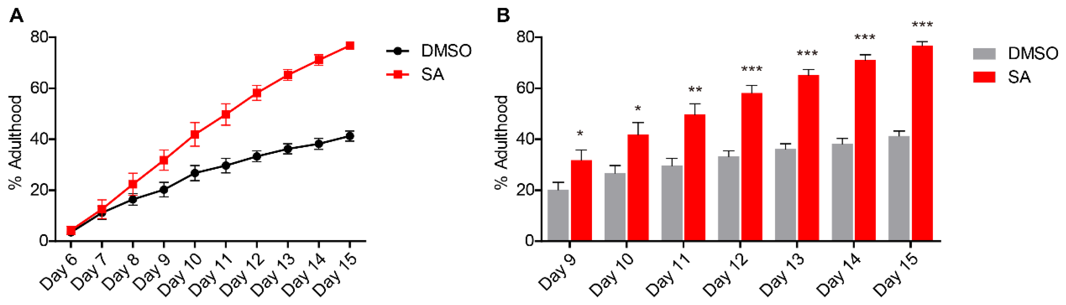
3.1. Atearic Acid Promotes Development of L. marina on CeMM

To determine whether stearic acid promotes development of L. marina on chemical defined food environment, we transferred newly hatched L1 larvae of L. marina to sea-salt-CeMM plates with stearic acid supplementation. We found that dietary stearic acid supplementation significantly promoted L. marina development on sea-salt-CeMM (Figure 1). Compared to the control (DMSO supplementation), the proportion of adults in the stearic acid supplementation group was significantly increased after 9 days post-hatching, and the adulthood rate at 15 days was significantly increased from about 40% (control) to nearly 80% (Figure 1). In line with the finding that stearic acid supplementation promotes development of C. elegans on CeMM [23], our data showed that stearic acid was able to promote the development of marine nematode L. marina on sea-salt CeMM medium, indicating a conserved developmental regulation role of stearic acid.

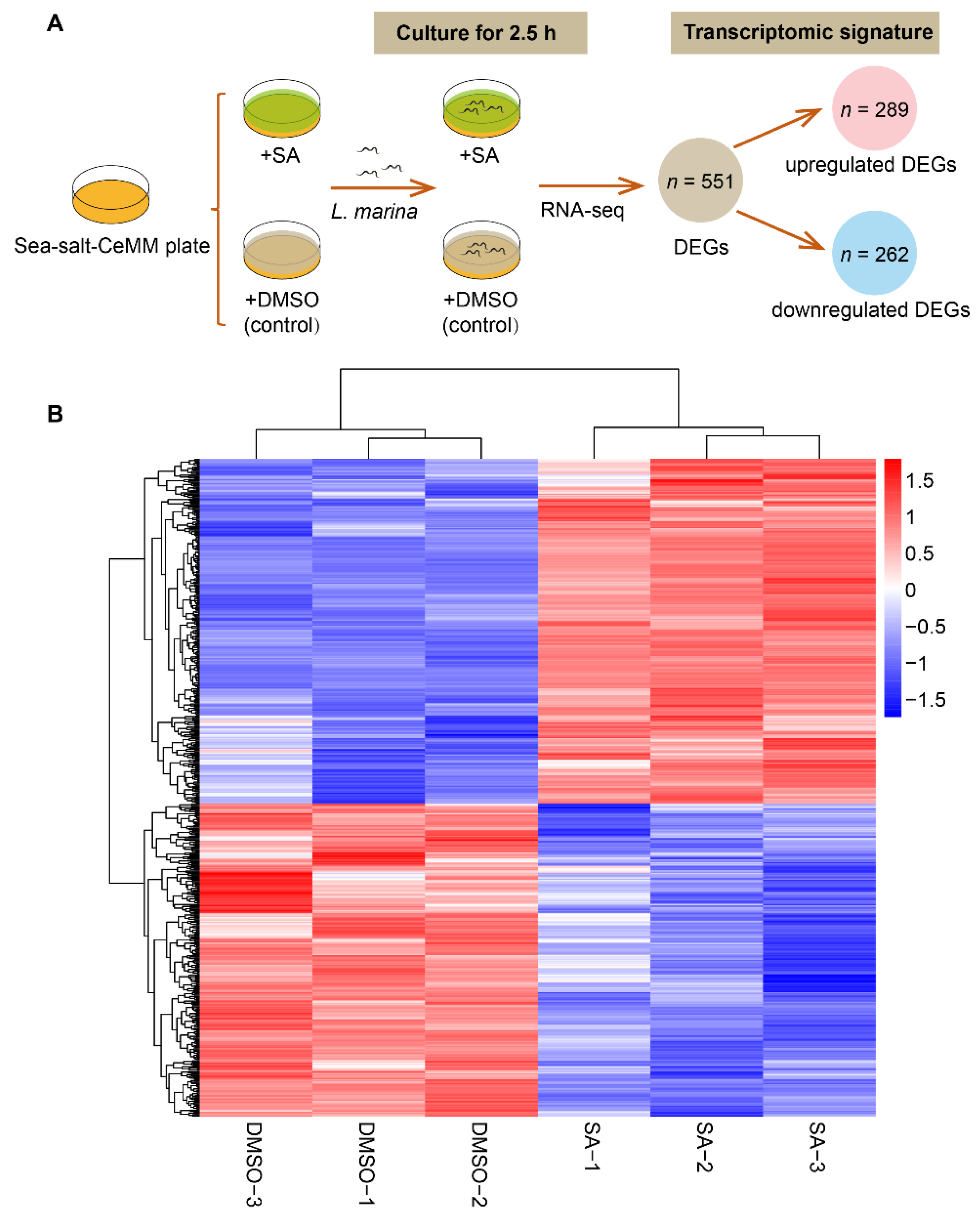
3.2. RNA-Seq Analysis in L. marina on Sea-Salt-CeMM with Stearic Acid Supplementation

To investigate transcriptomic responses of L. marina to dietary stearic acid supplementation, we performed high-throughput RNA-seq analysis. Newly hatched L. marina L1 larvae were cultured for 2.5 h on sea-salt-CeMM with stearic acid supplementation and DMSO (control) supplementation, respectively (Figure 2A). In total, 551 DEGs were identified, of which 289 were upregulated and 262 were repressed (Figure 2A,B). Details of significantly up- and downregulated DEGs are presented in Supplementary File S2.
KEGG and GO enrichment analysis results are shown, respectively in Figure 3 and Figure 4 and Supplementary File S3: Figures S1 and S2.

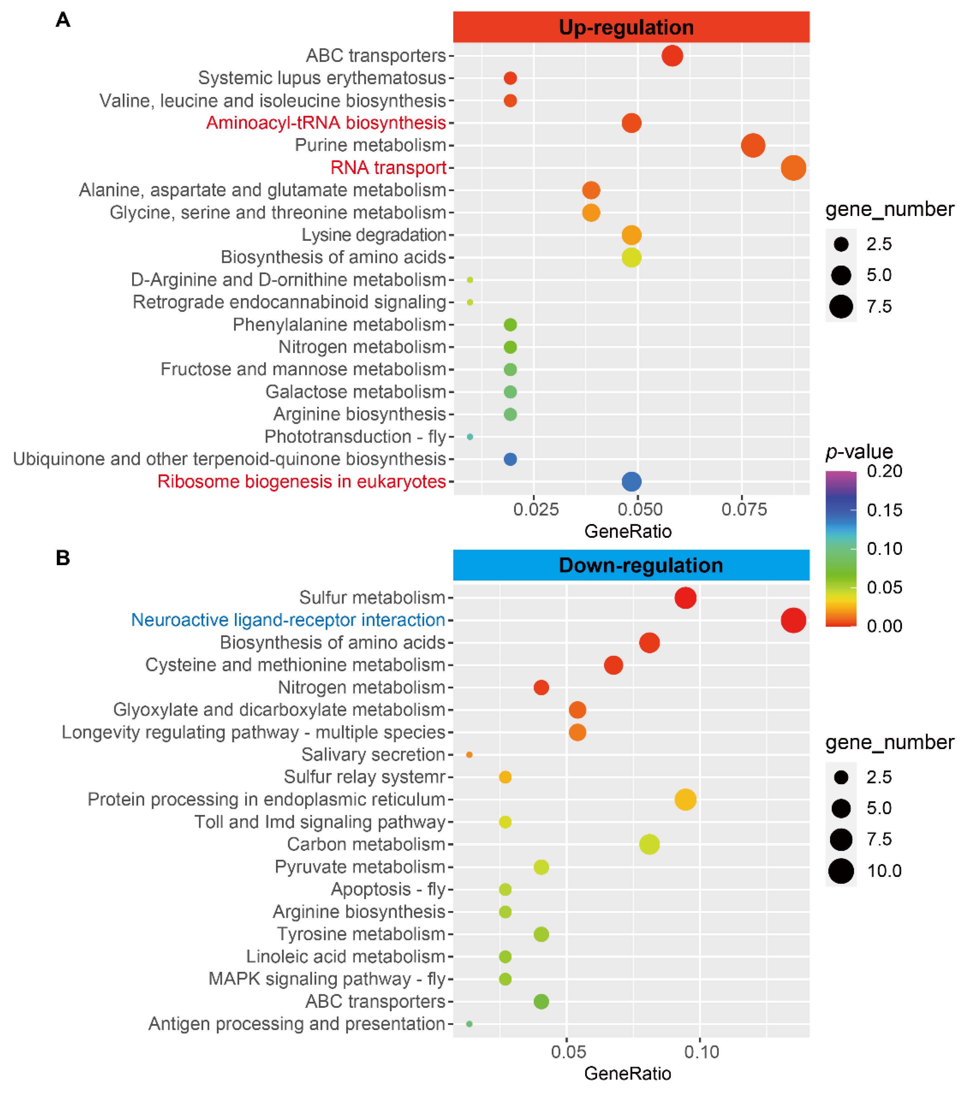
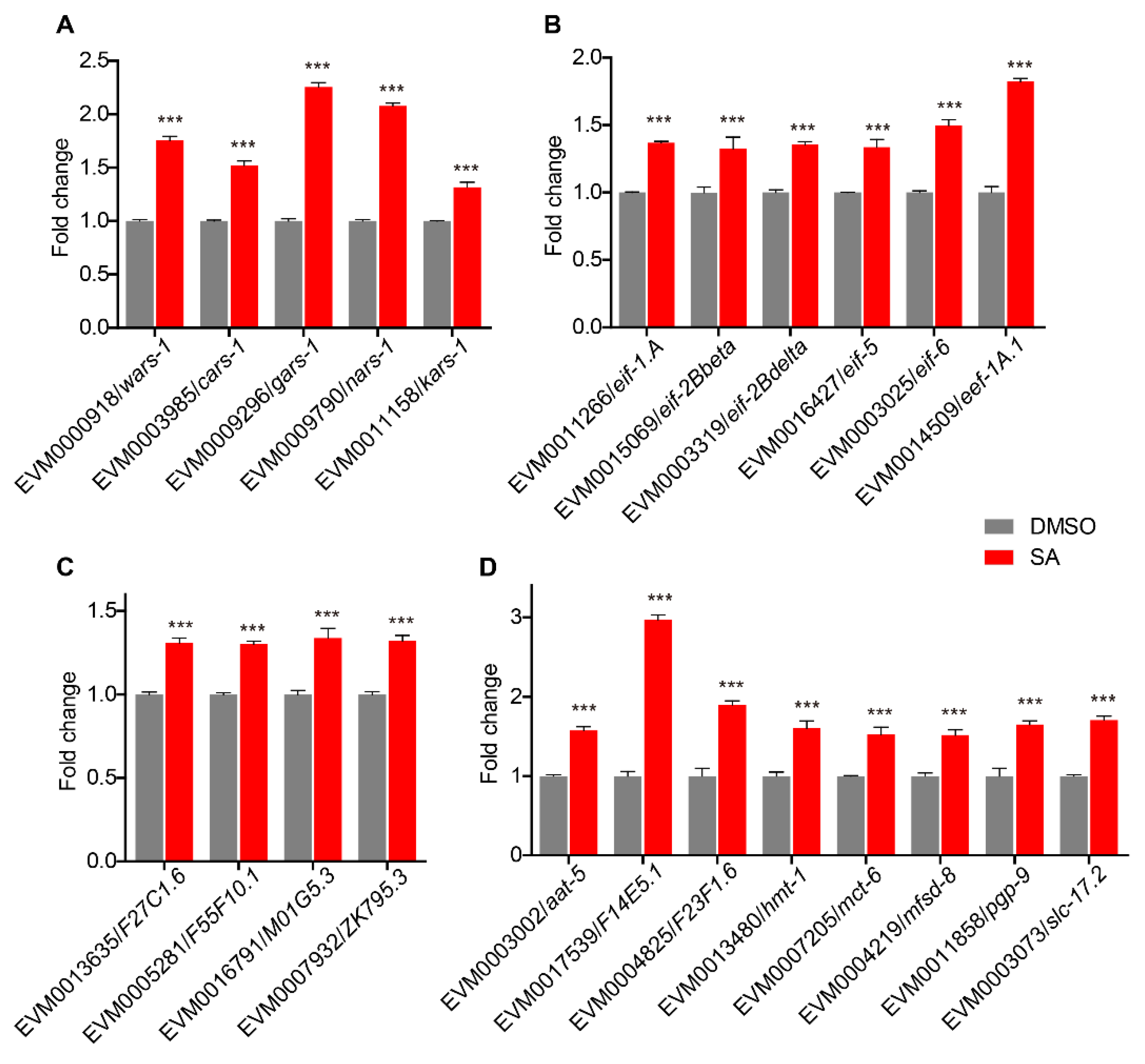
3.3. Upregulation of Aminoacyl-tRNA Biosynthesis Pathway Genes

Based on KEGG analysis, we observed that the “aminoacyl-tRNA biosynthesis” pathway-related genes were significantly enriched in upregulated DEGs (Figure 3A). The expression levels of five amino-acyl tRNA synthetase genes such as EVM0000918/wars-1 (tryptophanyl amino-acyl tRNA synthetase), EVM0003985/cars-1 (cysteinyl amino-acyl tRNA synthetase), EVM0009296/gars-1 (glycyl amino-acyl tRNA synthetase), EVM0009790/nars-1 (asparaginyl amino-acyl tRNA synthetase) and EVM0011158/kars-1 (lysyl amino-acyl tRNA synthetase) were significantly upregulated with dietary stearic acid supplementation (Figure 5A). Aminoacyl-tRNAs are essential substrates for protein translation [33]. Upregulation of aminoacyl-tRNA biosynthesis pathway genes might enable more proteins to be translated to support cell division and differentiation in development [33,34,35,36].

3.4. Upregulated Expression of Translation Initiation, Elongation, and Ribosome Biogenesis-Related Genes

We observed that “RNA transport” KEGG pathway genes were significantly upregulated in L. marina with dietary stearic acid supplementation (Figure 3A). RNA transport is a process in which RNA molecules are actively transported from one position within the cell to another, including the export of RNA from the nucleus to the cytoplasm through the nuclear pores, and the microtubule-assisted movement of specific RNAs along the axon [37,38,39]. Among eight upregulated DEGs enriched in the “RNA transport” pathway, five of which were translation initiation or elongation factors, including EVM0011266/eif-1.A (eukaryotic translation initiation factor 1A), EVM0015069/eif-2Bbeta, EVM0003319/eif-2Bdelta, EVM0016427/eif-5, and EVM0014509/eef-1A.1 (eukaryotic translation elongation factor 1α) (Figure 5B).
In addition, we found that the expression of several genes in the “ribosome biogenesis in eukaryotes” pathway was significantly increased in L. marina with dietary stearic acid supplementation (Figure 3A). One of them was EVM0003025/eif-6 (eukaryotic translation initiation factor 6), which is also a translation initiation factor. The other four were EVM0013635/F27C1.6, EVM0005281/F55F10.1, EVM0016791/M01G5.3, and EVM0007932/ZK795.3 (Figure 5C). Ribosome biogenesis is the process of producing ribosomes, the macromolecular machines that are responsible for mRNA translation into proteins [40,41].

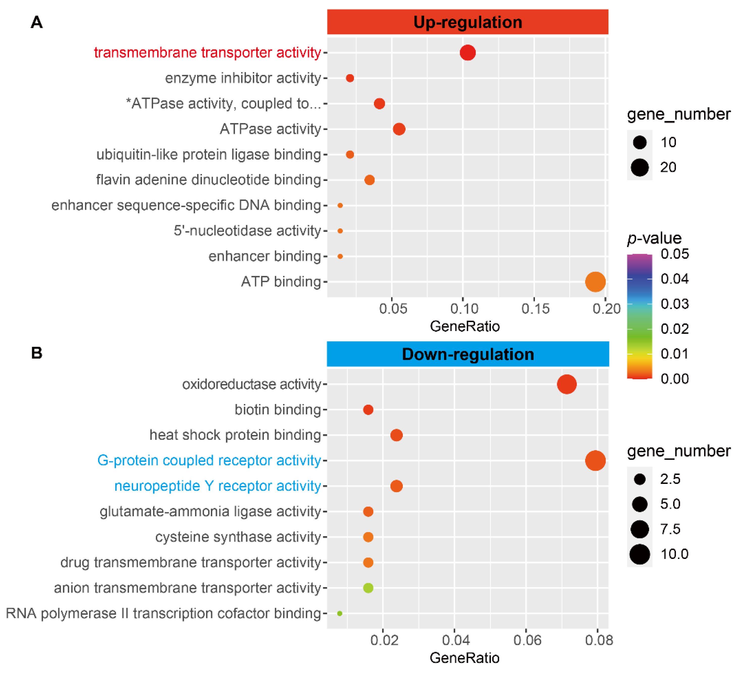
3.5. The Expression of Transmembrane Transporters Related Genes Was Upregulated

Based on GO functional enrichment analysis, we found that “transmembrane transporter activity” was the most significant enriched term for upregulated DEGs in MF (molecular function) category (Figure 4A). We observed that “transmembrane transporter activity”-related genes such as EVM0003002/aat-5, EVM0017539/F14E5.1, EVM0004825/F23F1.6, EVM0000321/F41C3.2, EVM0013480/hmt-1, EVM0007205/mct-6, EVM0004219/mfsd-8, EVM0011858/pgp-9, and EVM0003073/slc-17.2 were significantly increased in L. marina with dietary stearic acid supplementation (Figure 5D).

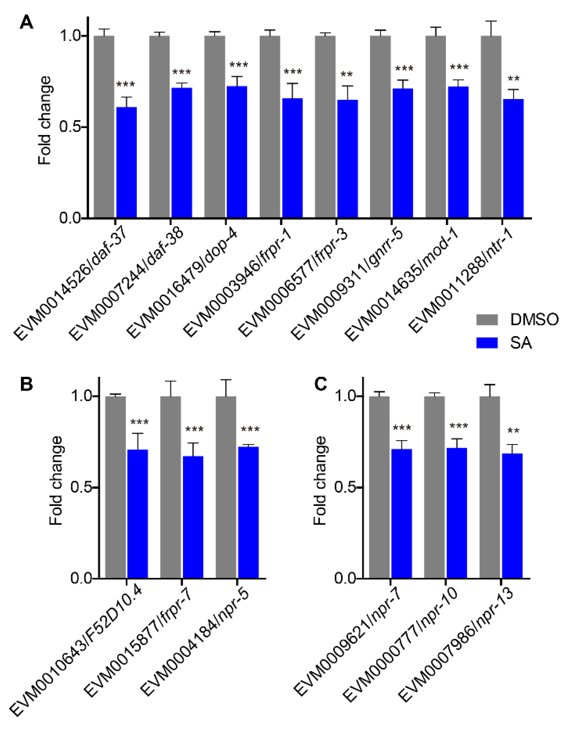
3.6. Downregulation of Neuronal Signaling Genes

We found that genes related to KEGG pathway term “neuroactive ligand-receptor interaction” and GO terms “G-protein coupled receptor activity” and “neuropeptide Y receptor activity” were significantly decreased in L. marina with dietary stearic acid supplementation (Figure 3B and Figure 4B). G protein-coupled receptors (GPCRs) are cell surface receptors that detect molecules outside the cell and could be activated by a variety of ligands such as hormones and neurotransmitters [42] and neuropeptide Y receptors belong to the class A GPCRs [43]. Significantly down-regulated “neuroactive ligand-receptor interaction” pathway genes were EVM0014526/daf-37, EVM0007244/daf-38, EVM0016479/dop-4, EVM0003946/frpr-1, EVM0006577/frpr-3, EVM0009311/gnrr-5, EVM0014635/mod-1, and EVM0011288/ntr-1 (Figure 6A). Downregulated “G-protein coupled receptor activity” genes include EVM0007244/daf-38, EVM0016479/dop-4, EVM0010643/F52D10.4, EVM0003946/frpr-1, EVM0006577/frpr-3, EVM0015877/frpr-7, EVM0009311/gnrr-5, EVM0014635/mod-1, EVM0004184/npr-5m and EVM0011288/ntr-1, among them EVM0007244/daf-38, EVM0016479/dop-4, EVM0003946/frpr-1, EVM0006577/frpr-3, EVM0009311/gnrr-5, EVM0014635/mod-1, and EVM0011288/ntr-1 were also enriched in “neuroactive ligand-receptor interaction” KEGG pathway (Figure 6B). Moreover, we observed that “neuropeptide Y receptor activity” term genes such as EVM0009621/npr-7, EVM0000777/npr-10, and EVM0007986/npr-13 were significantly repressed in L. marina with dietary stearic acid supplementation (Figure 6C).

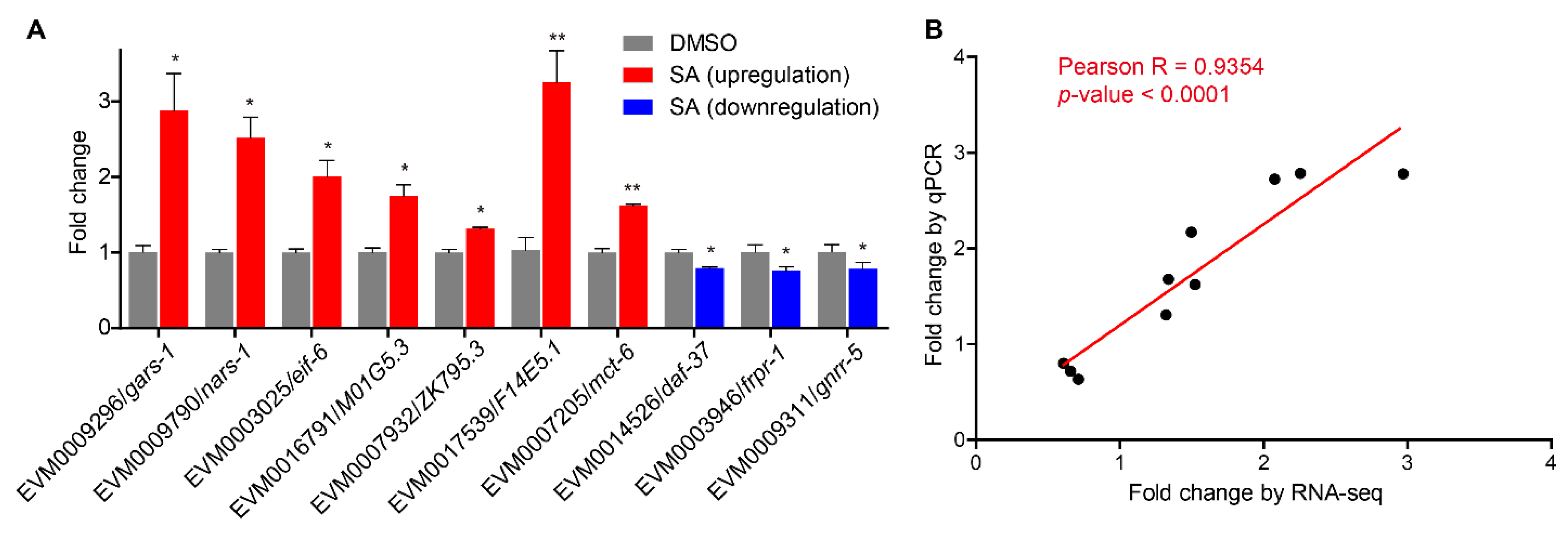
3.7. Quantitative Real-Time PCR Validation

We conducted qPCR to test the expression of interest genes identified from our RNA-seq results (Figure 7A). The qPCR results and RNA-seq results showed consistent trends (Figure 7B, Supplementary File S4). The expression of aminoacyl-tRNA biosynthesis genes EVM0009296/gars-1 and EVM0009790/nars-1, translation initiation factor gene EVM0003025/eif-6, ribosome biogenesis-related genes EVM0016791/M01G5.3 and EVM0007932/ZK795.3, and transmembrane transport-related genes EVM0017539/F14E5.1 and EVM0007205/mct-6 was significantly upregulated in L. marina with stearic acid supplementation, while the expression of neuronal signaling-related genes such as EVM0014526/daf-37, EVM0003946/frpr-1, and EVM0009311/gnrr-5 was significantly downregulated in L. marina with stearic acid supplementation.

4. Discussion

It was reported that stearic acid suppresses the development of the mammary gland in mice [44] and inhibits tumor development in rats [45]. A previous report showed that dietary stearic acid interferes with polyunsaturated fatty acid required for tumor development in mice models [46]. In addition, stearic acid inhibits the growth of human cervical cancer cells [47]. Another study showed that mice fed a diet containing stearic acid exhibit increased food intake and hedonic eating [48]. In Drosophila melanogaster, stearic acid supplementation improves animal physiology and mitochondrial function [49]. Certain concentrations of stearic acid can prolong the lifespan of C. elegans, while excessive stearic acid shortens the body length and longevity [50]. In this report, we found that dietary stearic acid supplementation promoted development of L. marina on sea-salt-CeMM (Figure 1), in line with our previous report that stearic acid supplementation promotes the growth of C. elegans on CeMM [23].

4.1. Stearic Acid Might Promote L. marina Development through Upregulation of Protein Synthesis Related Genes

Aminoacyl-tRNA synthetases (ARSs) ligate amino acids to the corresponding tRNAs in protein synthesis [36,51], and has been a growth marker for multiple zooplankton because of its positive correlation with individual growth rate [34,35]. Aminoacyl-tRNA synthetase defects lead to protein mistranslation and affect cellular physiology and development [52]. In cultured human HeLa cells, the disruption of MARS (multi-aminoacyl-tRNA synthetase complex) results in growth retardation due to a decreased rate of protein synthesis [53,54]. Drugs targeting methionyl-tRNA synthetases inhibit protein synthesis and consequently inhibit malaria parasite development [51]. In C. elegans, mutant of the glp-4 gene encoding valyl aminoacyl tRNA synthetase has reduced protein translation, displays slow growth, and shows defects in germline development [55]. A quantitative proteome analysis of C. elegans found that the level of valyl aminoacyl tRNA synthetase (encoded by glp-4) and aspartyl aminoacyl tRNA synthetase (encoded by dars-1) is significantly increased at 40 h after hatching compared to 20 h post hatching, suggesting that the increased aminoacyl-tRNA synthetases are critical for nematode development [56]. In the present study, we found that L. marina aminoacyl-tRNA synthetases genes, such as EVM0000918/wars-1, EVM0003985/cars-1, EVM0009296/gars-1, EVM0009790/nars-1, and EVM0011158/kars-1, were significantly upregulated with dietary stearic acid supplementation (Figure 5A). Our data indicate that stearic acid supplementation might promote development of L. marina via upregulating aminoacyl-tRNA synthetases to induce protein synthesis.
Eukaryotic initiation factors (eIFs) are essential in the initiation stage of eukaryotic translation, and contribute to the stabilization of ribosomal preinitiation complexes formation around the start codon [57,58]. Mutation of the eIF-5A homologue iff-2 or partial reduction of the expression of eIF-5B coding gene iffb-1 delays larval development in C. elegans [59,60]. Translation elongation factors help to elongate the polypeptide chain in translation [61]. A study in yeast has shown that elongation factor 1β mutant reduced total protein synthesis and growth defects [62]. C. elegans translation elongation factor 4 (EF4) localizes to mitochondria, and EF4 impairment delays development [63]. These reports suggest that translation initiation and elongation factors critically mediate the protein synthesis process and their deficiency might inhibit animal growth. In the present study, we found that several translation initiation and elongation factors such as EVM0011266/eif-1.A, EVM0015069/eif-2Bbeta, EVM0003319/eif-2Bdelta, EVM0016427/eif-5, and EVM0014509/eef-1A.1 were significantly upregulated in L. marina with dietary stearic acid supplementation (Figure 5B). Among them, previous studies have reported that RNAi inhibition of eif-1.A [64,65], eif-2Bbeta [66], eif-2Bdelta [64], or eif-6 [66] all slow down the growth of C. elegans. Therefore, our data indicate that stearic acid supplementation might promote development of L. marina on CeMM through inducing the expression of translation initiation and elongation factors genes.
Ribosomes are the fundamental macromolecular organelles that play a central role in the translation process, allowing the information encoded within mRNA to be converted into proteins [40]; a large number of ribosomes are required for cell growth [41]. A previous report showed that RBD-1 is essential for the early development of C. elegans through 18S ribosomal RNA processing [67]. The C. elegans nucleostemin promotes cell growth via increasing the ribosome biogenesis [68]. Here, we found that the expression of several ribosome biogenesis-related genes was significantly upregulated in L. marina with dietary stearic acid supplementation (Figure 5C), indicating that stearic acid supplementation might promote L. marina development via upregulation of ribosome biogenesis-related genes.

4.2. Stearic Acid Might Promote L. marina Development via Upregulation of Transmembrane Transporter Genes

Transmembrane transporters facilitate nutrient sensing and transport [69,70,71], and have been implicated in various diseases [72,73,74]. In this study, we observed that the expression of multiple transmembrane transporter genes such as aat-5, hmt-1, mct-6, mfsd-8, pgp-9, and slc-17.2 were significantly increased in marine nematode L. marina supplemented with stearic acid (Figure 5D). aat-5 encodes a L-amino acid transmembrane transporter, hmt-1 enables cadmium ion transmembrane transporter activity, mct-6 encodes a monocarboxylic acid transmembrane transporter, mfsd-8 is an ortholog of human MFSD8 and enables transmembrane transporter activity, pgp-9 is an ortholog of human ABCB1 and ABCB4, and slc-17.2 is predicted to be involved in anion transport (www.wormbase.org, accessed on 4 February 2022).
It was reported that RNAi knockdown of aat-5 could partially rescue the reduced brood size in C. elegans pept-1 (intestinal peptide transporter) mutant [75]. C. elegans hmt-1, a member of ABCB belonging to the ABC transporter superfamily, detoxifies cadmium, copper, and arsenic [76,77]. C. elegans ABCE is a transmembrane transporter, and RNAi knock-down the expression of abce-1 leads to slow growth [78]. MFSD (major facilitator superfamily domain) family members are known to be involved in energy consumption and homeostasis [79,80,81,82]. A previous report showed that Mfsd2a knockout mice are smaller and leaner [80], and mfsd-6 promotes PLM (posterior lateral microtubule) regrowth in C. elegans [83]. gem-1, encoding the SLC16 monocarboxylate transporter-related protein, promotes gonadal cell divisions in C. elegans [84]. T09F3.2 encodes a pyrimidine nucleotide transporter belonging to the SLC transporter family, and C. elegans T09F3.2 mutant exhibits slow growth and movement [85]. Together, our data suggest that stearic acid supplementation might promote development of L. marina via upregulation of certain transmembrane transporter genes.

4.3. Stearic Acid Might Promote Development of L. marina through Reduced Neuronal Signaling

Neuronal signaling is essential for nutrition sensation, feeding behavior, and developmental regulation in animals [16,86,87,88]. In particular, acetylcholine neuronal signaling plays an essential role in regulating diet consumption and energy homeostasis [89,90]. Impairment of cholinergic signaling increases food intake and leads to obesity [90]. Nicotine, as a known appetite suppressant, was reported to reduce food intake by activating nicotinic acetylcholine receptors (nAChRs) [91]. We previously reported that transmembrane channel-like gene tmc-1 attenuates the development of C. elegans via nicotinic acetylcholine receptor gene eat-2 [16], and this developmental regulating effect of tmc-1/eat-2 is achieved by altering fatty acid metabolism [23]. Our transcriptomic data showed that dietary stearic acid supplementation did not significantly change the expression of tmc-1 and eat-2 genes in L. marina, while we identified more than a dozen neuronal signaling receptor genes such as daf-37, daf-38, dop-4, frpr-1, frpr-3, frpr-7, gnrr-5, mod-1, npr-5, npr-7, npr-10, npr-13, and ntr-1 that were significantly downregulated in L. marina with dietary stearic acid supplementation (Figure 6). Both daf-37 and daf-38 encode G protein-coupled receptors, which cooperatively mediate pheromone perception and are involved in dauer formation in C. elegans [92]. DOP-4 protein is a dopamine neurotransmitter receptor [93], frpr-1, frpr-3, and frpr-7 all belong to the FMRFamide peptide receptor gene family, gnrr-5 is a homolog of human gonadotropin-releasing hormone receptor (GnRHR) gene, mod-1 is a serotonergic chloride channel, npr-5, npr-7, npr-10, and npr-13 belong to the neuropeptide Y receptor family, and ntr-1 is a homolog of human AVPR1B and AVPR2 (www.wormbase.org, accessed on 4 February 2022).
A previous report showed that the dop-4 mutants are shorter compared to the wild-type C. elegans [94]. External food supply promotes C. elegans avoidance responses to soluble repellents in a dop-4 dependent manner [95]. C. elegans FRPR-3 serves as a receptor for the FLP-20 (FMRF-like peptide 20) released by primary mechanosensory neurons, and regulates arousal and other behavioral states [96]. C. elegans AVK interneurons mediate food sensation by releasing the FLP-1 neuropeptides and the receptors are NPR-6 and FRPR-7 [97]. mod-1 is required for locomotor rate reduction when food-deprived C. elegans re-encounter food [98], and is involved in serotonin-induced fat loss [99]. NPR-1 is reported to regulate C. elegans foraging [100], and a previous report showed that npr-5 encodes a receptor gene for the FLP-18 neuropeptide, and flp-18 mutants exhibits defects in chemosensation and foraging [101]. It has been reported that FMRFamide neuropeptides inducing C. elegans sleep act via NPR-7 [102]. NPR-10 encodes a receptor for neuropeptides encoded by nlp-14, and NLP-14 activates NPR-10 to repress serotonin-induced aversive responses [103]. NTR-1 is a receptor for nematocin, which is a vasopressin/oxytocin-related neuropeptide. Previous reports showed that nematocin controls male mating behaviors, as well as salt chemotaxis of gustatory associative learning in C. elegans [104,105]. Given that impairing the function of acetylcholine neuronal signaling genes such as tmc-1 or eat-2 promote C. elegans development on CeMM [16,23], our data indicate that stearic acid supplementation might accelerate the growth of L. marina via repressing the expression of dopamine, serotonin, FMRFamide peptide, neuropeptide Y, and other neural receptor genes.

5. Conclusions

In conclusion, we found that dietary stearic acid supplementation promotes development of the marine nematode L. marina on an axenic chemical defined sea-salt-CeMM medium, indicating a conserved role of stearic acid in developmental regulation. Our results provide important insights into how a single fatty acid stearic acid regulates animal development, together with the advantage that the composition and amount of each chemical can be manipulated in the chemically defined CeMM medium. Further research with genome editing might illustrate the molecular mechanisms underlying how a single metabolite regulates animal development and physiology in response to global climate change with a dynamic nutritional environment.

Supplementary Materials

The following are available online at https://www.mdpi.com/article/10.3390/jmse10030428/s1, Supplementary File S1: Information of qPCR primers. Supplementary File S2: Details of differentially expressed genes. Supplementary File S3: Supplementary figures, Figure S1: GO-BP (Biological Process) enrichment analysis for DEGs; Figure S2: GO-CC (Cellular Component) enrichment analysis for DEGs. Supplementary File S4: Correlation analysis of the results of RNA-seq and qPCR for detected genes.

Author Contributions

Conceptualization, X.C. and L.Z.; formal analysis, X.C.; funding acquisition, L.Z.; investigation, X.C. and P.S.; supervision, L.Z.; visualization, X.C. and P.S.; writing—original draft, X.C.; writing—review and editing, L.Z. All authors have read and agreed to the published version of the manuscript.

Funding

This work was funded by the National Key R and D Program of China (No. 2018YFD0901301); “Talents from overseas Program, IOCAS” of the Chinese Academy of Sciences; “Qingdao Innovation Leadership Program” (Grant 16-8-3-19-zhc); and the Key deployment project of Centre for Ocean Mega-Research of Science, Chinese Academy of Sciences (COMS2019Q16).

Institutional Review Board Statement

Not applicable.

Informed Consent Statement

Not applicable.

Data Availability Statement

The datasets in this study were deposited in the NCBI Sequence Read Archive under the BioProject ID: PRJNA801406.

Acknowledgments

We are grateful to all members of the LZ laboratory for their helpful discussions.

Conflicts of Interest

The authors declare no conflict of interest.

References

  1. Sampath, H.; Ntambi, J.M. The fate and intermediary metabolism of stearic acid. Lipids 2005, 40, 1187–1191. [Google Scholar] [CrossRef] [PubMed]
  2. Spady, D.; Woollett, L.; Dietschy, J. Regulation of plasma LDL-cholesterol levels by dietary cholesterol and fatty acids. Annu. Rev. Nutr. 1993, 13, 355–381. [Google Scholar] [CrossRef] [PubMed]
  3. Siri-Tarino, P.W.; Sun, Q.; Hu, F.B.; Krauss, R.M. Saturated Fatty Acids and Risk of Coronary Heart Disease: Modulation by Replacement Nutrients. Curr. Atheroscler. Rep. 2010, 12, 384–390. [Google Scholar] [CrossRef] [PubMed] [Green Version]
  4. Hunter, J.E.; Zhang, J.; Kris-Etherton, P.M. Cardiovascular disease risk of dietary stearic acid compared with trans, other saturated, and unsaturated fatty acids: A systematic review. Am. J. Clin. Nutr. 2010, 91, 46–63. [Google Scholar] [CrossRef] [PubMed] [Green Version]
  5. Bonanome, A.; Grundy, S.M. Effect of Dietary Stearic Acid on Plasma Cholesterol and Lipoprotein Levels. N. Engl. J. Med. 1988, 318, 1244–1248. [Google Scholar] [CrossRef] [PubMed]
  6. Grande, F.; Anderson, J.T.; Keys, A. Comparison of Effects of Palmitic and Stearic Acids in the Diet on Serum Cholesterol in Man. Am. J. Clin. Nutr. 1970, 23, 1184–1193. [Google Scholar] [CrossRef] [PubMed] [Green Version]
  7. Simon, J.A.; Fong, J.; Bernert, J.T., Jr. Serum fatty acids and blood pressure. Hypertension 1996, 27, 303–307. [Google Scholar] [CrossRef]
  8. Kris-Etherton, P.M.; Griel, A.E.; Psota, T.L.; Gebauer, S.K.; Zhang, J.; Etherton, T.D. Dietary stearic acid and risk of cardiovascular disease: Intake, sources, digestion, and absorption. Lipids 2005, 40, 1193–1200. [Google Scholar] [CrossRef]
  9. Kühn, T.; Floegel, A.; Sookthai, D.; Johnson, T.; Rolle-Kampczyk, U.; Otto, W.; Von Bergen, M.; Boeing, H.; Kaaks, R. Higher plasma levels of lysophosphatidylcholine 18:0 are related to a lower risk of common cancers in a prospective metabolomics study. BMC Med. 2016, 14, 13. [Google Scholar] [CrossRef] [PubMed] [Green Version]
  10. Mondul, A.M.; Moore, S.C.; Weinstein, S.J.; Karoly, E.D.; Sampson, J.N.; Albanes, D. Metabolomic analysis of prostate cancer risk in a prospective cohort: The alpha-tocopherol, beta-carotene cancer prevention (ATBC) study. Int. J. Cancer 2015, 137, 2124–2132. [Google Scholar] [CrossRef]
  11. Cross, A.J.; Moore, S.; Boca, S.; Huang, W.-Y.; Xiong, X.; Stolzenberg-Solomon, R.; Sinha, R.; Sampson, J.N. A prospective study of serum metabolites and colorectal cancer risk. Cancer 2014, 120, 3049–3057. [Google Scholar] [CrossRef] [PubMed]
  12. Van Kappel, A.-L.; Winkvist, A.; Kaaks, R.; Lenner, P.; Riboli, E. Fatty-acid composition in serum phospholipids and risk of breast cancer: An incident case-control study in Sweden. Int. J. Cancer 1999, 83, 585–590. [Google Scholar] [CrossRef]
  13. Nůsková, H.; Serebryakova, M.V.; Ferrer-Caelles, A.; Sachsenheimer, T.; Lüchtenborg, C.; Miller, A.K.; Brügger, B.; Kordyukova, L.V.; Teleman, A.A. Stearic acid blunts growth-factor signaling via oleoylation of GNAI proteins. Nat. Commun. 2021, 12, 1–15. [Google Scholar] [CrossRef]
  14. Senyilmaz, D.; Virtue, S.; Xu, X.; Tan, C.Y.; Griffin, J.L.; Miller, A.; Vidal-Puig, A.; Teleman, A.A. Regulation of mitochondrial morphology and function by stearoylation of TFR1. Nature 2015, 525, 124–128. [Google Scholar] [CrossRef] [PubMed] [Green Version]
  15. Senyilmaz-Tiebe, D.; Pfaff, D.H.; Virtue, S.; Schwarz, K.V.; Fleming, T.; Altamura, S.; Muckenthaler, M.U.; Okun, J.G.; Vidal-Puig, A.; Nawroth, P. Dietary stearic acid regulates mitochondria in vivo in humans. Nat. Commun. 2018, 9, 1–10. [Google Scholar] [CrossRef] [PubMed]
  16. Zhang, L.; Gualberto, D.G.; Guo, X.; Correa, P.; Jee, C.; Garcia, L.R. TMC-1 attenuates C. elegans development and sexual behaviour in a chemically defined food environment. Nat. Commun. 2015, 6, 6345. [Google Scholar] [CrossRef] [Green Version]
  17. Cong, Y.; Yang, H.; Zhang, P.; Xie, Y.; Cao, X.; Zhang, L. Transcriptome Analysis of the Nematode Caenorhabditis elegans in Acidic Stress Environments. Front. Physiol. 2020, 11, 1107. [Google Scholar] [CrossRef]
  18. Goetsch, K.; Lu, N. Carbohydrate Requirement of Caenorhabditis Elegans and the Final Development of a Chemically Defined Medium. Nematologica 1993, 39, 303–311. [Google Scholar] [CrossRef] [Green Version]
  19. Szewczyk, N.J.; Kozak, E.; Conley, A.C. Chemically defined medium and Caenorhabditis elegans. BMC Biotechnol. 2003, 3, 19. [Google Scholar] [CrossRef] [Green Version]
  20. Szewczyk, N.J.; Udranszky, I.A.; Kozak, E.; Sunga, J.; Kim, S.K.; Jacobson, L.A.; Conley, C.A. Delayed development and lifespan extension as features of metabolic lifestyle alteration in C. elegans under dietary restriction. J. Exp. Biol. 2006, 209, 4129–4139. [Google Scholar] [CrossRef] [Green Version]
  21. Szewczyk, N.J.; Mancinelli, R.L.; McLamb, W.; Reed, D.; Blumberg, B.S.; Conley, C.A. Caenorhabditis elegans Survives Atmospheric Breakup of STS-107, Space Shuttle Columbia. Astrobiology 2005, 5, 690–705. [Google Scholar] [CrossRef] [PubMed] [Green Version]
  22. Szewczyk, N.; Tillman, J.; Conley, C.; Granger, L.; Segalat, L.; Higashitani, A.; Honda, S.; Honda, Y.; Kagawa, H.; Adachi, R.; et al. Description of International Caenorhabditis elegans Experiment first flight (ICE-FIRST). Adv. Space Res. 2008, 42, 1072–1079. [Google Scholar] [CrossRef] [PubMed] [Green Version]
  23. Cao, X.; Xie, Y.; Xue, B.; Yang, H.; Garcia, L.R.; Zhang, L. Fatty acid metabolic reprogramming promotes C. elegans Development. Biorxiv 2021. [Google Scholar] [CrossRef]
  24. Derycke, S.; De Meester, N.; Rigaux, A.; Creer, S.; Bik, H.; Thomas, W.; Moens, T. Coexisting cryptic species of the Litoditis marina complex (Nematoda) show differential resource use and have distinct microbiomes with high intraspecific variability. Mol. Ecol. 2016, 25, 2093–2110. [Google Scholar] [CrossRef] [Green Version]
  25. Xie, Y.; Zhang, P.; Xue, B.; Cao, X.; Ren, X.; Wang, L.; Sun, Y.; Yang, H.; Zhang, L. Establishment of a marine nematode model for animal functional genomics, environmental adaptation and developmental evolution. bioRxiv 2020. [Google Scholar] [CrossRef]
  26. Zhao, L.; Gao, F.; Gao, S.; Liang, Y.; Long, H.; Lv, Z.; Su, Y.; Ye, N.; Zhang, L.; Zhao, C.; et al. Biodiversity-based development and evolution: The emerging research systems in model and non-model organisms. Sci. China Life Sci. 2021, 64, 1236–1280. [Google Scholar] [CrossRef]
  27. Xie, Y.; Zhang, P.; Zhang, L. Genome-Wide Transcriptional Responses of Marine Nematode Litoditis marina to Hyposaline and Hypersaline Stresses. Front. Physiol. 2021, 12, 672099. [Google Scholar] [CrossRef]
  28. Kim, D.; Langmead, B.; Salzberg, S.L. HISAT: A fast spliced aligner with low memory requirements. Nat. Methods 2015, 12, 357–360. [Google Scholar] [CrossRef] [Green Version]
  29. Love, M.I.; Huber, W.; Anders, S. Moderated estimation of fold change and dispersion for RNA-seq data with DESeq2. Genome Biol. 2014, 15, 550. [Google Scholar] [CrossRef] [Green Version]
  30. Benjamini, Y.; Hochberg, Y. Controlling the False Discovery Rate: A Practical and Powerful Approach to Multiple Testing. J. R. Stat. Soc. Ser. B Methodol. 1995, 57, 289–300. [Google Scholar] [CrossRef]
  31. Young, M.D.; Wakefield, M.J.; Smyth, G.K.; Oshlack, A. Gene ontology analysis for RNA-seq: Accounting for selection bias. Genome Biol. 2010, 11, 1–12. [Google Scholar] [CrossRef] [PubMed] [Green Version]
  32. Mao, X.; Cai, T.; Olyarchuk, J.G.; Wei, L. Automated genome annotation and pathway identification using the KEGG Orthology (KO) as a controlled vocabulary. Bioinformatics 2005, 21, 3787–3793. [Google Scholar] [CrossRef] [PubMed]
  33. Ibba, M.; Söll, D. Aminoacyl-tRNA synthesis. Annu. Rev. Biochem. 2000, 69, 617–650. [Google Scholar] [CrossRef] [PubMed]
  34. McLaskey, A.K.; Keister, J.E.; Yebra, L. Individual growth rate (IGR) and aminoacyl-tRNA synthetases (AARS) activity as individual-based indicators of growth rate of North Pacific krill, Euphausia pacifica. J. Exp. Mar. Biol. Ecol. 2020, 527, 151360. [Google Scholar] [CrossRef]
  35. Yebra, L.; Hernández-León, S. Aminoacyl-tRNA synthetases activity as a growth index in zooplankton. J. Plankton Res. 2004, 26, 351–356. [Google Scholar] [CrossRef] [Green Version]
  36. Kwon, N.H.; Fox, P.L.; Kim, S. Aminoacyl-tRNA synthetases as therapeutic targets. Nat. Rev. Drug Discov. 2019, 18, 629–650. [Google Scholar] [CrossRef]
  37. Nakielny, S.; Fischer, U.; Michael, W.M.; Dreyfuss, G. RNA transport. Annu. Rev. Neurosci. 1997, 20, 269–301. [Google Scholar] [CrossRef] [Green Version]
  38. Basyuk, E.; Rage, F.; Bertrand, E. RNA transport from transcription to localized translation: A single molecule perspective. RNA Biol. 2021, 18, 1–17. [Google Scholar] [CrossRef]
  39. Fernandopulle, M.S.; Lippincott-Schwartz, J.; Ward, M.E. RNA transport and local translation in neurodevelopmental and neurodegenerative disease. Nat. Neurosci. 2021, 24, 622–632. [Google Scholar] [CrossRef]
  40. Thomson, E.; Ferreira-Cerca, S.; Hurt, E. Eukaryotic ribosome biogenesis at a glance. J. Cell Sci. 2013, 126, 4815–4821. [Google Scholar] [CrossRef] [Green Version]
  41. Lempiäinen, H.; Shore, D. Growth control and ribosome biogenesis. Curr. Opin. Cell Biol. 2009, 21, 855–863. [Google Scholar] [CrossRef] [PubMed]
  42. Ritter, S.L.; Hall, R. Fine-tuning of GPCR activity by receptor-interacting proteins. Nat. Rev. Mol. Cell Biol. 2009, 10, 819–830. [Google Scholar] [CrossRef] [PubMed] [Green Version]
  43. Michel, M.C.; Beck-Sickinger, A.; Cox, H.; Doods, H.N.; Herzog, H.; Larhammar, D.; Quirion, R.; Schwartz, T.; Westfall, T. XVI. International Union of Pharmacology recommendations for the nomenclature of neuropeptide Y, peptide YY, and pancreatic polypeptide receptors. Pharmacol. Rev. 1998, 50, 143–150. [Google Scholar] [PubMed]
  44. Meng, Y.; Yuan, C.; Zhang, J.; Zhang, F.; Fu, Q.; Zhu, X.; Shu, G.; Wang, L.; Gao, P.; Xi, Q.; et al. Stearic acid suppresses mammary gland development by inhibiting PI3K/Akt signaling pathway through GPR120 in pubertal mice. Biochem. Biophys. Res. Commun. 2017, 491, 192–197. [Google Scholar] [CrossRef] [PubMed]
  45. Habib, N.A.; Wood, C.B.; Apostolov, K.; Barker, W.; Hershman, M.J.; Aslam, M.; Heinemann, D.; Fermor, B.; Williamson, R.C.; Jenkins, W.E. Stearic acid and carcinogenesis. Br. J. Cancer 1987, 56, 455–458. [Google Scholar] [CrossRef] [PubMed]
  46. Bennett, A.S. Effect of dietary stearic acid on the genesis of spontaneous mammary adenocarcinomas in strain A/ST mice. Int. J. Cancer 1984, 34, 529–533. [Google Scholar] [CrossRef] [PubMed]
  47. Beesley, J.S.; Soutter, W.P.; White, J.O. Effect of Stearic Acid on Human Cervical Cancer Cell Growth. In Eicosanoids and Other Bioactive Lipids in Cancer, Inflammation and Radiation Injury; Springer: Boston, MA, USA, 1993; pp. 573–575. [Google Scholar]
  48. Li, Y.; Wu, H.; Zhang, R.; Shu, G.; Wang, S.; Gao, P.; Zhu, X.; Jiang, Q.; Wang, L. Diet containing stearic acid increases food reward-related behaviors in mice compared with oleic acid. Brain Res. Bull. 2020, 164, 45–54. [Google Scholar] [CrossRef] [PubMed]
  49. Bajracharya, R.; Bustamante, S.; Ballard, O.J.W. Stearic Acid Supplementation in High Protein to Carbohydrate (P:C) Ratio Diet Improves Physiological and Mitochondrial Functions of Drosophila melanogaster parkin Null Mutants. J. Gerontol. Ser. A 2019, 74, 1564–1572. [Google Scholar] [CrossRef]
  50. Wang, X.; Zhang, L.; Zhang, L.; Wang, W.; Wei, S.; Wang, J.; Che, H.; Zhang, Y. Effects of excess sugars and lipids on the growth and development of Caenorhabditis elegans. Genes Nutr. 2020, 15, 1–18. [Google Scholar] [CrossRef]
  51. Hussain, T.; Yogavel, M.; Sharma, A. Inhibition of protein synthesis and malaria parasite development by drug targeting of methionyl-tRNA synthetases. Antimicrob. Agents Chemother. 2015, 59, 1856–1867. [Google Scholar] [CrossRef] [Green Version]
  52. Lu, J.; Bergert, M.; Walther, A.; Suter, B. Double-sieving-defective aminoacyl-tRNA synthetase causes protein mistranslation and affects cellular physiology and development. Nat. Commun. 2014, 5, 1–13. [Google Scholar] [CrossRef] [PubMed] [Green Version]
  53. Kaminska M, Havrylenko S, Decottignies P, Gillet S, Le Maréchal P, Negrutskii B, Mirande M: Dissection of the structural organization of the aminoacyl-tRNA synthetase complex. J. Biol. Chem. 2009, 284, 6053–6060. [CrossRef] [Green Version]
  54. Havrylenko, S.; Legouis, R.; Negrutskii, B.; Mirande, M. Caenorhabditis elegans evolves a new architecture for the multi-aminoacyl-tRNA synthetase complex. J. Biol. Chem. 2011, 286, 28476–28487. [Google Scholar] [CrossRef] [Green Version]
  55. Rastogi, S.; Borgo, B.; Pazdernik, N.; Fox, P.; Mardis, E.R.; Kohara, Y.; Havranek, J.; Schedl, T. Caenorhabditis elegans glp-4 Encodes a Valyl Aminoacyl tRNA Synthetase. G3 Genes Genomes Genet. 2015, 5, 2719–2728. [Google Scholar] [CrossRef] [Green Version]
  56. Geillinger, K.E.; Kuhlmann, K.; Eisenacher, M.; Meyer, H.E.; Daniel, H.; Spanier, B. Dynamic Changes of the Caenorhabditis elegans Proteome during Ontogenesis Assessed by Quantitative Analysis with 15N Metabolic Labeling. J. Proteome Res. 2012, 11, 4594–4604. [Google Scholar] [CrossRef]
  57. Jackson, R.J.; Hellen, C.U.T.; Pestova, T.V. The mechanism of eukaryotic translation initiation and principles of its regulation. Nat. Rev. Mol. Cell Biol. 2010, 11, 113–127. [Google Scholar] [CrossRef] [Green Version]
  58. Aitken, C.E.; Lorsch, J.R. A mechanistic overview of translation initiation in eukaryotes. Nat. Struct. Mol. Biol. 2012, 19, 568–576. [Google Scholar] [CrossRef]
  59. Hanazawa, M.; Kawasaki, I.; Kunitomo, H.; Gengyo-Ando, K.; Bennett, K.L.; Mitani, S.; Iino, Y. The Caenorhabditis elegans eukaryotic initiation factor 5A homologue, IFF-1, is required for germ cell proliferation, gametogenesis and localization of the P-granule component PGL-1. Mech. Dev. 2004, 121, 213–224. [Google Scholar] [CrossRef]
  60. Yu, X.; Vought, V.E.; Conradt, B.; Maine, E.M. Eukaryotic translation initiation factor 5B activity regulates larval growth rate and germline development in Caenorhabditis elegans. Genesis 2006, 44, 412–418. [Google Scholar] [CrossRef]
  61. Andersen, G.R.; Nissen, P.; Nyborg, J. Elongation factors in protein biosynthesis. Trends Biochem. Sci. 2003, 28, 434–441. [Google Scholar] [CrossRef]
  62. Carr-Schmid, A.; Valente, L.; Loik, V.I.; Williams, T.; Starita, L.M.; Kinzy, T.G. Mutations in elongation factor 1β, a guanine nucleotide exchange factor, enhance translational fidelity. Mol. Cell. Biol. 1999, 19, 5257–5266. [Google Scholar] [CrossRef] [PubMed] [Green Version]
  63. Yang, F.; Gao, Y.; Li, Z.; Chen, L.; Xia, Z.; Xu, T.; Qin, Y. Mitochondrial EF4 links respiratory dysfunction and cytoplasmic translation in Caenorhabditis elegans. Biochim. Biophys. Acta 2014, 1837, 1674–1683. [Google Scholar] [CrossRef] [PubMed] [Green Version]
  64. Kamath, R.S.; Fraser, A.G.; Dong, Y.; Poulin, G.; Durbin, R.; Gotta, M.; Kanapin, A.; Le Bot, N.; Moreno, S.; Sohrmann, M.; et al. Systematic functional analysis of the Caenorhabditis elegans genome using RNAi. Nature 2003, 421, 231–237. [Google Scholar] [CrossRef] [PubMed]
  65. Melo, J.A.; Ruvkun, G. Inactivation of Conserved C. elegans Genes Engages Pathogen- and Xenobiotic-Associated Defenses. Cell 2012, 149, 452–466. [Google Scholar] [CrossRef] [Green Version]
  66. Fraser, A.G.; Kamath, R.S.; Zipperlen, P.; Martinez-Campos, M.; Sohrmann, M.; Ahringer, J. Functional genomic analysis of C. elegans chromosome I by systematic RNA interference. Nature 2000, 408, 325–330. [Google Scholar] [CrossRef]
  67. Saijou, E.; Fujiwara, T.; Suzaki, T.; Inoue, K.; Sakamoto, H. RBD-1, a nucleolar RNA-binding protein, is essential for Caenorhabditis elegans early development through 18S ribosomal RNA processing. Nucleic Acids Res. 2004, 32, 1028–1036. [Google Scholar] [CrossRef] [Green Version]
  68. Kudron, M.M.; Reinke, V. C. elegans Nucleostemin Is Required for Larval Growth and Germline Stem Cell Division. PLoS Genet. 2008, 4, e1000181. [Google Scholar] [CrossRef]
  69. Miguel-Aliaga, I. Nerveless and gutsy: Intestinal nutrient sensing from invertebrates to humans. Semin. Cell Dev. Biol. 2012, 23, 614–620. [Google Scholar] [CrossRef] [Green Version]
  70. Palm, W.; Thompson, C.B. Nutrient acquisition strategies of mammalian cells. Nature 2017, 546, 234–242. [Google Scholar] [CrossRef]
  71. Thwaites, D.T.; Anderson, C. H+-coupled nutrient, micronutrient and drug transporters in the mammalian small intestine. Exp. Physiol. 2007, 92, 603–619. [Google Scholar] [CrossRef] [Green Version]
  72. Prudente, S.; Flex, E.; Morini, E.; Turchi, F.; Capponi, D.; De Cosmo, S.; Tassi, V.; Guida, V.; Avogaro, A.; Folli, F.; et al. A Functional Variant of the Adipocyte Glycerol Channel Aquaporin 7 Gene Is Associated with Obesity and Related Metabolic Abnormalities. Diabetes 2007, 56, 1468–1474. [Google Scholar] [CrossRef] [PubMed] [Green Version]
  73. Cooper, R.; Sarioglu, S.; Sökmen, S.; Füzün, M.; Küpelioğlu, A.; Valentine, H.; Görken, I.B.; Airley, R.; West, C. Glucose transporter-1 (GLUT-1): A potential marker of prognosis in rectal carcinoma? Br. J. Cancer 2003, 89, 870–876. [Google Scholar] [CrossRef] [PubMed] [Green Version]
  74. Macheda, M.L.; Rogers, S.; Best, J.D. Molecular and cellular regulation of glucose transporter (GLUT) proteins in cancer. J. Cell. Physiol. 2005, 202, 654–662. [Google Scholar] [CrossRef] [PubMed]
  75. Benner, J. Amino acid homeostasis in Caenorhabditis elegans lacking the intestinal peptide transporter PEPT-1 and identification of PEPT-1 modulator proteins. Ph.D. Thesis, Technical University of Munich, Freising, Germany, 2011. [Google Scholar]
  76. Schwartz, M.S.; Benci, J.L.; Selote, D.S.; Sharma, A.K.; Chen, A.G.Y.; Dang, H.; Fares, H.; Vatamaniuk, O.K. Detoxification of Multiple Heavy Metals by a Half-Molecule ABC Transporter, HMT-1, and Coelomocytes of Caenorhabditis elegans. PLoS ONE 2010, 5, e9564. [Google Scholar] [CrossRef] [Green Version]
  77. Kim, S.; Sharma, A.K.; Vatamaniuk, O.K. N-terminal extension and C-terminal domains are required for ABCB6/HMT-1 protein interactions, function in cadmium detoxification, and localization to the endosomal-recycling system in Caenorhabditis elegans. Front. Physiol. 2018, 9, 885. [Google Scholar] [CrossRef] [Green Version]
  78. Zhao, Z.; Fang, L.L.; Johnsen, R.; Baillie, D.L. ATP-binding cassette protein E is involved in gene transcription and translation in Caenorhabditis elegans. Biochem. Biophys. Res. Commun. 2004, 323, 104–111. [Google Scholar] [CrossRef]
  79. Bagchi, S.; Perland, E.; Hosseini, K.; Lundgren, J.; Al-Walai, N.; Kheder, S.; Fredriksson, R. Probable role for major facilitator superfamily domain containing 6 (MFSD6) in the brain during variable energy consumption. Int. J. Neurosci. 2020, 130, 476–489. [Google Scholar] [CrossRef]
  80. Berger, J.H.; Charron, M.J.; Silver, D.L. Major Facilitator Superfamily Domain-Containing Protein 2a (MFSD2A) Has Roles in Body Growth, Motor Function, and Lipid Metabolism. PLoS ONE 2012, 7, e50629. [Google Scholar] [CrossRef] [Green Version]
  81. Lekholm, E.; Perland, E.; Eriksson, M.M.; Hellsten, S.V.; Lindberg, F.; Rostami, J.; Fredriksson, R. Putative Membrane-Bound Transporters MFSD14A and MFSD14B Are Neuronal and Affected by Nutrient Availability. Front. Mol. Neurosci. 2017, 10, 11. [Google Scholar] [CrossRef] [Green Version]
  82. Perland, E.; Lekholm, E.; Eriksson, M.M.; Bagchi, S.; Arapi, V.; Fredriksson, R. The Putative SLC Transporters Mfsd5 and Mfsd11 Are Abundantly Expressed in the Mouse Brain and Have a Potential Role in Energy Homeostasis. PLoS ONE 2016, 11, e0156912. [Google Scholar] [CrossRef] [Green Version]
  83. Kim, K.W.; Tang, N.H.; Piggott, C.A.; Andrusiak, M.G.; Park, S.; Zhu, M.; Kurup, N.; Cherra, S.J., III; Wu, Z.; Chisholm, A.D. Expanded genetic screening in Caenorhabditis elegans identifies new regulators and an inhibitory role for NAD+ in axon regeneration. Elife 2018, 7, e39756. [Google Scholar] [CrossRef] [PubMed]
  84. Kemp, B.J.; Church, D.L.; Hatzold, J.; Conradt, B.; Lambie, E.J. Gem-1 encodes an SLC16 monocarboxylate transporter-related protein that functions in parallel to the gon-2 TRPM channel during gonad development in Caenorhabditis elegans. Genetics 2009, 181, 581–591. [Google Scholar] [CrossRef] [PubMed] [Green Version]
  85. Ogurusu, T.; Sakata, K. The Caenorhabditis elegans homolog of human mitochondrial pyrimidine nucleotide transporter regulates glucose transport. Biochem. Biophys. Res. Commun. 2021, 557, 117–121. [Google Scholar] [CrossRef]
  86. Avery, L.; You, Y.-J. C. elegans Feeding (21 May 2012), WormBook, Ed. The C. elegans Research Community, WormBook. Available online: http://www.wormbook.org/chapters/www_feeding/feeding.html (accessed on 4 February 2022).
  87. Gallagher, T.; Kim, J.; Oldenbroek, M.; Kerr, R.; You, Y.-J. ASI Regulates Satiety Quiescence in C. elegans. J. Neurosci. 2013, 33, 9716–9724. [Google Scholar] [CrossRef] [Green Version]
  88. Ben Arous, J.; Laffont, S.; Chatenay, D. Molecular and sensory basis of a food related two-state behavior in C. elegans. PLoS ONE 2009, 4, e7584. [Google Scholar] [CrossRef] [Green Version]
  89. Yapici, N.; Cohn, R.; Schusterreiter, C.; Ruta, V.; Vosshall, L.B. A Taste Circuit that Regulates Ingestion by Integrating Food and Hunger Signals. Cell 2016, 165, 715–729. [Google Scholar] [CrossRef] [Green Version]
  90. Herman, A.M.; Ortiz-Guzman, J.; Kochukov, M.; Herman, I.; Quast, K.B.; Patel, J.; Tepe, B.; Carlson, J.C.; Ung, K.; Selever, J.; et al. A cholinergic basal forebrain feeding circuit modulates appetite suppression. Nature 2016, 538, 253–256. [Google Scholar] [CrossRef] [Green Version]
  91. Mineur, Y.S.; Abizaid, A.; Rao, Y.; Salas, R.; DiLeone, R.J.; Gündisch, D.; Diano, S.; De Biasi, M.; Horvath, T.L.; Gao, X.-B.; et al. Nicotine Decreases Food Intake Through Activation of POMC Neurons. Science 2011, 332, 1330–1332. [Google Scholar] [CrossRef] [Green Version]
  92. Park, D.; O’Doherty, I.; Somvanshi, R.K.; Bethke, A.; Schroeder, F.C.; Kumar, U.; Riddle, D.L. Interaction of structure-specific and promiscuous G-protein–coupled receptors mediates small-molecule signaling in Caenorhabditis elegans. Proc. Natl. Acad. Sci. USA 2012, 109, 9917–9922. [Google Scholar] [CrossRef] [Green Version]
  93. Sugiura, M.; Fuke, S.; Suo, S.; Sasagawa, N.; Van Tol, H.H.M.; Ishiura, S. Characterization of a novel D2-like dopamine receptor with a truncated splice variant and a D1-like dopamine receptor unique to invertebrates from Caenorhabditis elegans. J. Neurochem. 2005, 94, 1146–1157. [Google Scholar] [CrossRef]
  94. Nagashima, T.; Oami, E.; Kutsuna, N.; Ishiura, S.; Suo, S. Dopamine regulates body size in Caenorhabditis elegans. Dev. Biol. 2016, 412, 128–138. [Google Scholar] [CrossRef]
  95. Ezcurra, M.; Tanizawa, Y.; Swoboda, P. Schafer WR: Food sensitizes C. elegans avoidance behaviours through acute dopamine signalling. EMBO J. 2011, 30, 1110–1122. [Google Scholar] [CrossRef] [Green Version]
  96. Chew, Y.L.; Tanizawa, Y.; Cho, Y.; Zhao, B.; Alex, J.Y.; Ardiel, E.L.; Rabinowitch, I.; Bai, J.; Rankin, C.H.; Lu, H. An Afferent Neuropeptide System Transmits Mechanosensory Signals Triggering Sensitization and Arousal in C. elegans. Neuron 2018, 99, 1233–1246. [Google Scholar] [CrossRef] [Green Version]
  97. Oranth, A.; Schultheis, C.; Tolstenkov, O.; Erbguth, K.; Nagpal, J.; Hain, D.; Brauner, M.; Wabnig, S.; Costa, W.S.; McWhirter, R.D.; et al. Food Sensation Modulates Locomotion by Dopamine and Neuropeptide Signaling in a Distributed Neuronal Network. Neuron 2018, 100, 1414–1428.e10. [Google Scholar] [CrossRef] [Green Version]
  98. Ranganathan, R.; Cannon, S.; Horvitz, H.R. MOD-1 is a serotonin-gated chloride channel that modulates locomotory behaviour in C. elegans. Nature 2000, 408, 470–475. [Google Scholar] [CrossRef]
  99. Srinivasan, S.; Sadegh, L.; Elle, I.C.; Christensen, A.G.; Faergeman, N.J.; Ashrafi, K. Serotonin Regulates C. elegans Fat and Feeding through Independent Molecular Mechanisms. Cell Metab. 2008, 7, 533–544. [Google Scholar] [CrossRef] [Green Version]
  100. De Bono, M.; Bargmann, C.I. Natural variation in a neuropeptide Y receptor homolog modifies social behavior and food response in C. elegans. Cell 1998, 94, 679–689. [Google Scholar] [CrossRef] [Green Version]
  101. Cohen, M.; Reale, V.; Olofsson, B.; Knights, A.; Evans, P.; de Bono, M. Coordinated Regulation of Foraging and Metabolism in C. elegans by RFamide Neuropeptide Signaling. Cell Metab. 2009, 9, 375–385. [Google Scholar] [CrossRef] [Green Version]
  102. Chow, E.S.H. The Caenorhabditis elegans ALA Neuron: Its Transcriptome and Role in Inducing Sleep. Ph.D. Thesis, California Institute of Technology, Pasadena, CA, USA, 2014. [Google Scholar]
  103. Hapiak, V.; Summers, P.; Ortega, A.; Law, W.J.; Stein, A.; Komuniecki, R. Neuropeptides Amplify and Focus the Monoaminergic Inhibition of Nociception in Caenorhabditis elegans. J. Neurosci. 2013, 33, 14107–14116. [Google Scholar] [CrossRef]
  104. Garrison, J.L.; Macosko, E.Z.; Bernstein, S.; Pokala, N.; Albrecht, D.R.; Bargmann, C.I. Oxytocin/Vasopressin-Related Peptides Have an Ancient Role in Reproductive Behavior. Science 2012, 338, 540–543. [Google Scholar] [CrossRef] [Green Version]
  105. Beets, I.; Janssen, T.; Meelkop, E.; Temmerman, L.; Suetens, N.; Rademakers, S.; Jansen, G.; Schoofs, L. Vasopressin/Oxytocin-Related Signaling Regulates Gustatory Associative Learning in C. elegans. Science 2012, 338, 543–545. [Google Scholar] [CrossRef] [Green Version]
Figure 1. Stearic acid promoted L. marina growth on sea-salt-CeMM diet. (A,B) Daily adult rates of L. marina with dietary supplementation of SA (stearic acid) or DMSO (solvent control). (A) and (B) are different representations of the same set of data. DMSO, 6 trials, n = 52 worms on average in each trial; SA, 6 trials, n = 52 worms on average in each trial. All data shown are mean ± SEM (standard error of mean). * p < 0.05, ** p < 0.01, *** p < 0.001; two-sided t-test.
Jmse 10 00428 g001
Figure 2. Transcriptome characterization of L. marina supplemented with dietary stearic acid (SA). (A) Experimental design and the transcriptomic characterization of dietary stearic acid supplementation. (B) Hierarchical clustering of DEGs (differentially expressed genes) in L. marina supplementation with stearic acid and DMSO using the pheatmap package in R. Relative expression levels (z-score, gene expression level based on base-2 log-transformed FPKM value) were indicated for each gene (row) in each sample (column). Red indicates upregulation; blue indicates downregulation. The scale bar shows the z-score for DEGs.
Jmse 10 00428 g002
Figure 3. KEGG enrichment analysis for DEGs. (A) The top 20 enriched KEGG pathways for upregulated DEGs. (B) The top 20 enriched KEGG pathways for downregulated DEGs. Significance of enrichment is indicated by color from red to purple. Gene ratio is the ratio of the number of DEGs annotated to the KEGG pathway to the total number of DEGs.
Jmse 10 00428 g003
Figure 4. GO-MF (molecular function) enrichment analysis for DEGs. (A) The top 10 enriched GO-MF terms for upregulated DEGs. * The full name of this term is “ATPase activity, coupled to transmembrane movement of substances”. (B) The top 10 enriched GO-MF terms for downregulated DEGs. Significance of enrichment is indicated by color from red to purple. Gene ratio is the ratio of the number of DEGs annotated to the GO term to the total number of DEGs.
Jmse 10 00428 g004
Figure 5. Expression of related upregulated pathway genes. (A) Expression level of aminoacyl-tRNA biosynthesis genes. (B) Expression level of translation initiation and elongation factors genes. (C) Expression level of ribosome biogenesis-related genes. The expression level of upregulated gene EVM0003025/eif-6 enriched in this term is presented in (B). (D) Expression level of some transmembrane transporters-related genes. SA (stearic acid). The error bars represent SEM of the mean. *** padj < 0.001.
Jmse 10 00428 g005
Figure 6. Downregulated expression of neuronal signaling related genes. (A) Expression level of “neuroactive ligand-receptor interaction” downregulated genes. (B) Expression level of “G-protein coupled receptor activity” genes. The expression level of downregulated genes EVM0007244/daf-38, EVM0016479/dop-4, EVM0003946/frpr-1, EVM0006577/frpr-3, EVM0009311/gnrr-5, EVM0014635/mod-1, and EVM0011288/ntr-1 enriched in this term is presented in (A). (C) Expression level of “neuropeptide Y receptor activity” genes. SA (stearic acid). The error bars represent SEM of the mean. ** padj < 0.01, *** padj < 0.001.
Jmse 10 00428 g006
Figure 7. Validation of the RNA-seq results using qPCR. (A) qPCR analysis of interest genes identified from the RNA-seq results. Fold changes indicate the ratio of the SA (stearic acid) supplementation group to the control DMSO group. The error bars represent SEM of the mean, * p < 0.05, ** p < 0.01, two-sided t-test. (B) Correlation analysis of the results of RNA-seq and qPCR for detected genes. Each dot represents a gene, with detailed information shown in Supplementary File S4. Correlation analysis was performed using GraphPad Prism 5 software, Pearson R = 0.9354, with a p-value < 0.0001.
Jmse 10 00428 g007
Publisher’s Note: MDPI stays neutral with regard to jurisdictional claims in published maps and institutional affiliations.

