Article

Marine Archaeal Extracellular Polymeric Substances from Halococcus AMS12, Their Characterization, and Biological Properties

by
Kamala Kannan
1,
Sivaperumal Pitchiah
2,3,*,
Jeevankumar Guduri Joseph
2,
Dhanraj Ganapathy
3,
Subramanian Sundarrajan
4,* and
Seeram Ramakrishna
4
1
Centre for Marine Actinobacterial Research, Department of Physiology, Saveetha Dental College and Hospitals, Saveetha Institute of Medical & Technical Sciences, Saveetha University, Chennai 600077, Tamil Nadu, India
2
Marine Biomedical Research Lab and Environmental Toxicology Unit, Cellular and Molecular Research Centre, Saveetha Dental College and Hospitals, Saveetha Institute of Medical & Technical Sciences, Saveetha University, Chennai 600077, Tamil Nadu, India
3
Department of Prosthodontics & Implantology, Saveetha Dental College and Hospitals, Saveetha Institute of Medical & Technical Sciences, Saveetha University, Chennai 600077, Tamil Nadu, India
4
Center for Nanotechnology and Sustainability, Department of Mechanical Engineering, National University of Singapore, Singapore 117576, Singapore
*
Authors to whom correspondence should be addressed.
J. Mar. Sci. Eng. 2022, 10(11), 1788; https://doi.org/10.3390/jmse10111788
Submission received: 28 October 2022 / Revised: 18 November 2022 / Accepted: 19 November 2022 / Published: 21 November 2022
(This article belongs to the Special Issue Feature Papers in Marine Biology)

Abstract

:
In the present study, halophilic archaea were isolated from a marine sediment sample. Totally, 15 isolates (AMS 1–15) were identified by molecular identification as belonging to the ten genera. Further, their extracellular polymeric substances (EPS) were extracted (3.172 g/L), and their bioactivity was determined in terms of biosurfactant, emulsification, enzymatic and non-enzymatic antioxidants, and anticancer activity. The highest amount of EPS has been produced by Halococcus sp., AMS12. It is made up of 54.28% carbohydrates, 32.91% proteins, 2.41% lipids, and other compounds. Further, EPS has 43.69 ± 1.89 U/mg of gelatinase enzyme by degrading the substrate. The potential total antioxidant activity of 103.80 ± 0.02 (ascorbic acid equivalence (AAE)), total reducing power of 86.1 ± 0.25 AAE, 1,1-diphenyl-2-picrylhydrazyl (DPPH) activity of 97.23 ± 0.21%, the hydrogen peroxide scavenging of 60.8 ± 0.21%, and nitric oxide scavenging activity of 89.37 ± 0.24% were observed at 100 μg/mL of EPS. Hence, we conclude that the archeal EPS is multifunctional and useful for developing natural polymers for industrial, food, and pharmaceutical applications.

1. Introduction

Archaea have the ability to live in extreme environments with various adaptations. They have adapted to live in extreme environments, such as in soaring salt, pH, and pressure, to produce novel metabolites and are economically important [1]. These extremophilic archaea are identified from a moderate environment, which have the ability to fix nitrogen, reduce nitrate and nitrite, denitrify, and do all other activities [2]. The overview, general structure, and biological activities of EPS from archaea are given in Figure 1. Although, archaea have numerous prospective applications in different fields of biotechnology and it produces pigments, ammonia-oxidizing, hydrolytic enzymes such as lipase, DNAse, amylase, gelatinase, chitinase, protease, cellulose, and polymers [3], only a few of them are involved in a major part in the environment through halophilic and halo-tolerant enzymes [4]. However, the archaeal role in the environment is not yet clear, where they have competed with bacteria at a high-salt concentration by their enzymes and EPS. Generally, these EPS are produced with high molecular weight during their growth periods and thereby create a protective layer on the cell surfaces to form a biofilm [5]. The supple matrix of EPS from archaea has glycoproteins, enzymes, lipids, extracellular DNA, and other chelating substances. The heterogenic polymers have non-carbohydrates and monosaccharide units that differ from each species and each environment [6]. The chains of polysaccharides were built up by the repeating unit of oligosaccharides, and their sizes may vary according to the degree of polymerization [7]. These EPS are well known to be negatively charged, which has potential applications in pharmaceutical, food, and other industries. Additionally, the EPS from microbes remove organic contaminants and heavy metals from the environment [8]. In addition, they provide a defensive fence against UV radiation, desiccation, temperature fluctuation, pH changes, and other chemical substances. The EPS plays a key part in the biofilm formation on liquid and solid interfaces, and air–liquid interface [9] EPS producing thermophilic and halophilic archaea have been isolated from the hydrothermal vent and deep sea [2,10]. Extremophilic archaeal genera Archaeoglobus, Sulfolobus and Thermococcus have produced large amounts of EPS with antimicrobial activity [11]. The EPS from halophilic archaea have an odd range of sulfate and uronic acid content [12]. Although a large number of archaeal EPS were reported from various extreme environments, biosynthesis, composition, structure, and active properties [12], studies on archaea from marine sediment sample and their potential EPS production are very limited. Therefore, we focused on the archaeal domains of marine sediments and the bioactive potential of their EPS. In this study, we focused on the archaeal domains from marine sediments and the bioactive potential of EPS and their characterization.

2. Materials and Methods

2.1. Sample Collection

The sediment sample was collected from a depth of 25 m in the offshore region (13.003837° N, 80.329445° E) of Chennai using Van Veen grab of 0.04 m2. The sediment sample was collected in a sterile plastic container (Himedia-HPW016T), and it was safely transferred to the laboratory.

2.2. Isolation and Identification of Marine Archaea

One gram of sediment sample was diluted with 10 mL of sterile distilled water (10−1), and the same diluents were continued up to 10−6. From those diluents, 1 mL of the sample was pipetted into Petri plates containing halo archaeal agar (HAA) (per litre: 250 g of NaCl, 2 g of KCl, 20 g of MgSO4, 3 g of Na3C6H5O7, 10 g of yeast extract, 20 g of agar, and 7.2 pH). The plates were nurtured at a temperature of 35 ± 2 °C for 20 to 25 days. The archaeal population density was enumerated, and it has been stated as a CFU/mL (colony-forming unit per mL). Isolates were selected according to their distinct colony morphology and color of the colonies. Further, pure archaeal isolates were picked by recurrent modifications of different isolates on HAA plates [13].
The archaeal cell pellet was suspended in 1 mL of cell suspension solution (500 mM Tris HCl; 200 mM EDTA; 460 mM NaOH; pH 8) and 50 μL of RNase 100 μL of cell lysis solution (15% sodium dodecyl sulfate (SDS)) was incubated for 15 min at 55 °C and then added to the 25 μL of proteinase K incubated at 55 °C for 60 min, and 500 μL of 5 M NaCl was then added to incubate at 4 °C for 10 min. After incubation, 2 mL of TE buffer and 8 mL of ethanol were added to the aqueous layer of centrifuged samples and incubated for 2 min at room temperature and centrifuged for 15 min at 5000 rpm; subsequently, the dried condition pellet was dispersed in DNase free water [14]. The extracted DNA was visualized in agarose gel (0.7%) electrophoresis at 100 V. The archaeal 16SrDNA gene was amplified according to the Delong method [15] with 4 F and 1492 R archaeal primers. Amplified DNA contents were refined and sequenced by Bio-innovation Company (Mumbai). Further, the obtained sequence was analyzed using NCBI-BLAST search. Cell morphology and gram staining were measured by phase-contrast microscopy.

2.3. Optimal Growth Parameters of Archaea

Salt requirement (10–30%), temperature (35 °C–60 °C), and optimal growth pH (6 to 9) were determined in HAA medium, and the cultures were placed on a rotary shaker at 150 rpm at 30 °C without light sources for 120 h. Cell growth was noticed in halo-archaeal broth (per liter: 250 g of NaCl, 2 g of KCl, 20 g of MgSO4, 3 g of Na3C6H5O7, 10 g of yeast extract) for 168 h on a rotary shaker at 200 rpm. Every 24 h, 2 mL of broth were centrifuged, and the cells were suspended with halo-archaeal broth to measure the absorbance at 600 nm. Further, 50 µL of cell suspension was observed in the microscopic observation on the Neubauer counting chamber [16].

2.4. Production and Purification of EPS

The EPS were isolated from Halococcus sp., AMS12 in halo-archaeal broth at 45 ˚C for 120 h. The EPS was extracted using chemical and physical methods followed by methods used by Forster and Clarke [17] and Li and Yang [18] with slight modification. The EPS from archaea were separated by centrifugation at 10,000 rpm for 30 min, the entire process was maintained at 4 °C, and the pellets were further suspended using ice-cold ethanol (double volume) and incubated at 4 °C for overnight.
The archaeal EPS mixture in ethanol was obtained by the centrifugation method (13,000 rpm condition for 30 min at 4 °C). The pellet was washed with ethanol in absolute condition to further concentrate and purify. The excess salts and impurities were removed from the crude EPS using dialyzed membrane (HiMedia-Mumbai, LA401) for 48 h at 4 °C. The dialyzed EPS were freeze dried to make the powder [19], and total EPS was expressed in mg/L.

2.5. Characterization of Marine Archaeal EPS

The carbohydrate from the EPS was estimated by the phenol-sulphuric acid digestion method, glucose was maintained as standard [20], the protein level was determined by bicinchoninic assay (BCA), and BSA was used as standard. The nucleic acid was estimated by the method of [21] with a slight modification. In detail, the EPS solution was also diluted to the predefined volume after transferring through a 0.45 µm cellulose acetate membrane. A UV-Vis spectrophotometer was utilized to record the evolution of the dissolved solutions. Total uronic acid concentration levels were determined using a sulfamate/m-hydroxydiphenyl assay in a concentrated sulfuric acid [22], and total lipid contents were estimated [23] using ferric acetate-uranium acetate in a mixture of solvents, such as acetic acid, sulfuric acid, and ferrous sulfate. The lyophilized EPS surface morphological characteristics and shape were examined by FE-SEM [24].

2.6. Biosurfactant and Cell Membrane Protection Assay

Emulsification activity of EPS was measured by mixing 1 mL of EPS with an equal volume of aromatic hydrocarbons, such as benzene, and was vigorously vortexed for 3 min. Then, the mixture was incubated at 4 °C for 24 h. After incubation, the height of the emulsification was measured. Triton X-100 was used as a positive control, and cell-free PBS was used as a blank.
The emulsification index = height of emulsion (mm)/total height of mixture (mm) × 100
The cellular membrane defense activity of EPS from archaea was analyzed by the method of Sajjad et al. [25] with a slight modification. The red blood cells (RBC) were centrifuged at 5000 rpm for 5 min and then diluted with saline phosphate buffer (1:10). A 5 mL of EPS (0.1 to 0.5 mg/mL concentrations) was inoculated to diluted cells and kept for 10 min. Further, SDS was added to the reaction mixture made up to 10 mL and incubated for 30 min. The supernatant of the reaction mixture was collected by centrifugation at 5000 rpm for 5 min. The absorbance of the supernatant was measured at 600 nm. The percentage of cell–lysis was calculated by the given formula: % of cell lyses = (control-test/control) × 100. The diluted cell with SDS was used as the negative control and the cell with glycerol was a positive control.

2.7. Enzyme Activity of Marine Archaeal EPS

To determine the enzyme activity, the archaeal isolates were centrifuged at 10,000 rpm for 10 min at 4 °C, and the cell-free supernatant was used as a source of gelatinase enzyme. The assay was then carried out with a 1mg/mL concentration of gelatin as a substratum at 35 °C [25].

2.8. Antioxidant Activity of EPS

The antioxidant activity of the EPS from marine archaea was determined by the methods of Kamala et al. [26] at various concentrations in terms of DPPH scavenging activity, total antioxidant, hydrogen peroxide scavenging activity, total reducing power, and nitrous oxide inhibition activity. The enzymatic in-vitro antioxidant activity was performed by Sivaperumal et al. [27] at different concentrations in terms of superoxide dismutase (SOD), catalase (CAT), glutathione activity (GSH), lipid peroxidation, and malondialdehyde (MDA).

2.9. Cell Viability (MTT) and DCFH-DA Assay

Mouse fibroblast cell line (3T3) was purchased from National Centre for Cell Science (NCCS), Pune, and the obtained cells were incubated in T25 culture flasks of DMEM (Dulbecco’s Modified Eagle Medium) supplemented with 1% antibiotics and 10% FBS. Further, cells were incubated with 5% of CO2 (humidified atmosphere) and maintained at 37 °C. After accomplishment of confluence, the cells were used for further experiment. Cell viability was determined through MTT assay by the method of Bhavna et al. [28]. The viable cells in the control were 100%, and the viability of treated cells was calculated by the formula: % cell viability = [A570 nm of treated cells/A570 nm of control cells] × 100.

2.10. Dichloro-Dihydro-Fluorescein Diacetate DCFH-DA) Assay

The DCFH-DA assay was conducted to evaluate the reactive oxygen species (ROS) generation. The production of intracellular ROS in untreated and EPS-treated 3T3 cells was measured. Hydrogen peroxide treated cells were used as a positive control for ROS generation. A total volume of 2 mL was obtained by diluting an aliquot of 8 × 10⁶ cells/mL in PBS with 7.4 pH condition. The mixture of one ml cell suspension and 100 µL of DCFH-DA (5 uM/mL) were incubated for 30 min at 37 °C. An inverted fluorescence microscope was used to observe and photograph the stained cells from each group.

3. Results

3.1. Isolation and Identification of Marine Archaea

A total of 34 archaeal isolates were obtained from marine sediment samples using HAA medium (Figure 2a). Out of them, 15 isolate had distinct colony morphology which had been taken for further studies. The potential study strain of Halococcus sp. AMS12 showed the cocci-shaped morphology (Figure 2b) in the microscopic view. Genomic DNA was isolated and amplified with archaeal primers (4 F with 1492 R) at 1488 bp. Archaeal 16S rDNA analysis revealed the presence of members from Euryarchaeota phyla, which were identified as Halococcus sp. AMS1, Halorubrum sp. AMS2, Natonorubrum sp. AMS3, Halobacterium sp. AMS4, Halomicrobium sp. AMS5, Halorhabdus sp. AMS6, Natrinema sp. AMS7, Haloferax sp. AMS8, Haloplanus sp. AMS9, Natrinema sp. AMS10, Haloferax sp. AMS11, Halococcus sp. AMS12, Halomicrobium sp. AMS13, Natrinema sp. AMS14, and Haloarcula sp. AMS15. Phylogenetic analysis of the 16S rDNA of these isolates had II clusters; cluster I had eight genera (Halococcus, Halorubrum, Halobacterium, Halomicrobium, Haloferax, Haloplanus, Halorhabdus, and Haloarcula). Cluster II had two genera (Natrinema and Natronorubrum), and Escherichia coli (AB269763) was used for our group. All the isolates were submitted to the gene bank to obtain an accession number (KU995301-KU995315) with 90–99.9% similarity with the query sequences.
Totally, 26 nucleotide sequences were used for the phylogenic study which has 1400 base pairs in the ending dataset [29]. The optimal branch length of the phylogenic tree was 1.0678 [30]. The percentage of repeat trees in which the associated taxa clustered together in the bootstrap test (1000 replicates) was shown next to the branches. The phylogenetic tree was constructed by ten clusters; each genus separated into a single cluster; cluster I was constructed with seven isolated from five genera with five reference sequences (Halomicrobium (HM063952), Haloarcula (EF645680), Halorhabdus (NR113464), Halobacterium (AB663362), and Halococcus (AB935407)) at 100% bootstrap level with respective isolates. Cluster II has four isolates belonging to three genera with reference sequences (Haloferax (D14128), Halorubrum (AY510708), and Haloplanus (NR113462); cluster III has four isolates belonging to Natrinema with the reference sequence (AB663465) and Escherichia coli (AB269763) was used as the out group (Figure 3).

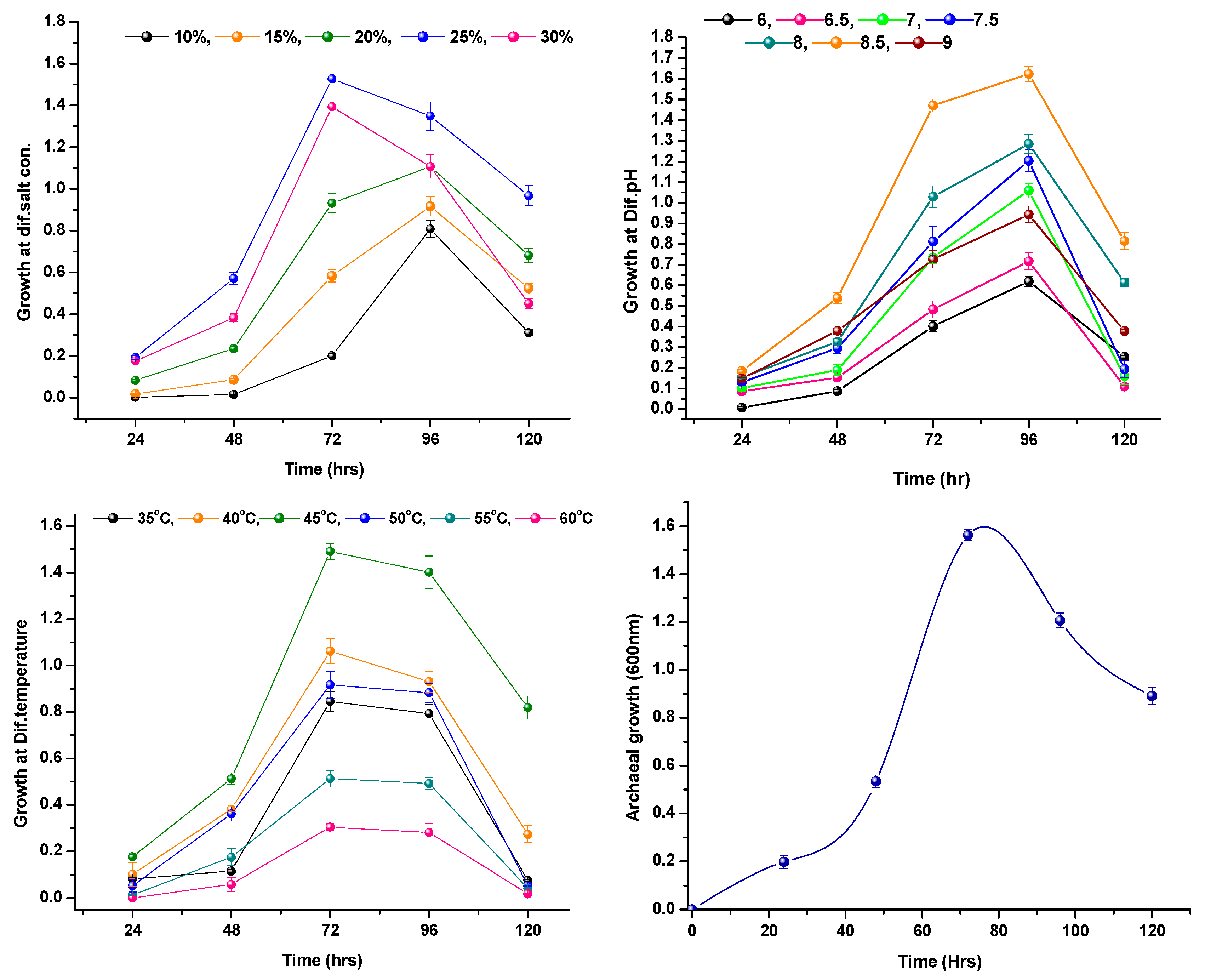
3.2. Optimal Growth Condition of Halococcus sp. AMS12

A total of 15 archaeal isolates (AMS1 to AMS 15) were used to screen the maximum EPS production. Compared to other isolates, Halococcus sp. AMS12 produced a maximum quantity of EPS (3.172 g/L). For further study, we used the potential isolates of Halococcus sp. AMS12 (Figure 4). The Halococcus sp. AMS12 optimal growth condition was noted in the presence of various salt concentrations ranging from 10% to 30% of NaCl, but the optimal growth was observed at 25% NaCl concentration. The result confirmed the halophilic nature of the isolate Halococcus sp AMS12. The growth level of AMS12 at various pH has been given in Figure 5 which showed the optimal growth at pH 7.5. Similarly, the growth in different temperatures was also investigated, and the optimal growth was observed at 45 °C. The optimal growth was observed after 48 to 76 h at 45 °C with a 25% salt concentration at pH 7.5.

3.3. Production and Characterization of Marine Archaeal EPS

During the log phase, the isolate of AMS12 produced 3.172 g/L of EPS (Figure 5) which contained 54.28% of carbohydrate, 32.91% of proteins, 2.41% of lipids, 1.3% nucleic acid, 0.76% of uronic acid, and 8.34% of unidentified compounds (Figure 6). In the archaeal EPS, contributions of carbohydrates are a higher percentage followed by proteins and other substances. The unidentified compound may be phosphorous and other minor contents [31]. Further, the surface morphology of EPS was analyzed through FE-SEM and mentioned in Figure 7. The FESEM image indicates that the surface morphology is rough, and hence, it will have strong binding capacity. Therefore, the EPS showed good biological activity.

3.4. Biosurfactant Activity and Cell Membrane Protection

The bio-surfactant activity was determined in terms of emulsification at various concentrations of archaeal EPS. The significant activity was 52% from archaeal EPS, and the titronX-100 shows 67% of emulsifying activity. The lowest emulsifying activities of 18% and 24% were observed at 25 µg/mL concentration of EPS and standard of TitronX-100, respectively (Figure 8). Cell membrane stability of archaeal EPS was estimated with human RBC, which showed 12% cell lysis in the presence of detergent at 100 µg/mL of EPS concentration, followed by 28% cell lysis in the presence of glycerol and 94% cell lysis with the addition of detergent (SDS). The minimal concentration (25 µg/mL) of detergent lysed 52% of cells, whereas 42% of cells were lysed with the addition of archaeal EPS.

3.5. Enzymatic Activity of Marine Halococcus sp. AMS12

The extracellular enzyme from halophilic archaea received special attention due to the high stability in pH and temperature resistance, low water activity, and solvent resistance, which also facilitate survival in unfavorable conditions [32]. In the present study, the hydrolyzing activity of EPS from Halococcus sp. AMS12 exhibited 43.69 ± 1.89 U/mg gelatinase enzyme by degrading the substrates.

3.6. Antioxidant Activity of EPS Obtained from Halococcus sp. AMS12

In the present study, in vitro antioxidant activity was observed by the various antioxidant assays. The potential total antioxidant activity of 103.80±0.02 (AAE), total reducing power of 86.1 ± 0.25 (AAE), DPPH of 97.23 ± 0.21%, hydrogen peroxide scavenging of 60.8 ± 0.21%, and nitric oxide scavenging activity of 89.37 ± 0.24% were observed at 100 μg/mL of archaeal EPS (Figure 9). The stable free-radical DPPH started to react with archaeal EPS and donated a hydrogen atom which led to reduction form (diphenyl picryhydrazine), which had a deep yellow color. Through the inhalation of vapor, hydrogen peroxide will enter the human body, which is the source of lipid peroxidation, and it may lead to DNA damage in the body. The enzymatic antioxidant activity of EPS exhibited 17 U/mg of catalase, 32 U/mg of superoxide dismutase, 58 U/mg of lipid peroxidation, 117 U/mg of malondialdehyde (MDA), and 129 U/mg of glutathione activity (GSH) (Figure 9).

3.7. Cell Viability (MTT) and DCFH-DA Assay

The 3T3 cell viability of EPS from the halo archaeal isolate was determined after 24 h incubation though the MTT assay (Figure 10). The results revealed that the EPS inhibited 13.19% of cell death against 3T3 cell lines with 86.11 ± 4.16% of cell viability at 100 µg/mL of EPS concentration (Figure 11). The result suggested the archaeal EPS did not affect the noncancer cell lines.

4. Discussion

4.1. Isolation and Identification of Marine Archaea

In the marine environment, archaeal community has a key role in sulfur metabolism, methane oxidation, and nitrification [33]. Thus, the account of the archaeal community in coastal water, estuarine, basins mangrove, and the open sea is investigated to determine the role in marine habitats. However, the archaeal diversity in marine coastal sediment was little known [34]. Hence, the present study pays attention to the admittance of archaeal diversity from coastal sediment samples. Red-pigmented archaeal isolates, such as Haloarcula vallismortis and Haloarcula qudrata, were isolated from salter soil, which have optimal growth at 25% of salt concentration with a wide range of antibiotic resistance [35]. Similarly, the maximum growth of Halococcus sp. AMS112 was also observed in the medium with 25% of salt content. Archaeal genera, such as Pyrococcus, Pyrodictium, Igneococcus, Thermococcus, Methanococcus, Archaeoglobus, and Methanopyrus, were isolated from deep-sea hydrothermal systems, and aquifex were isolated from hydrothermal vents around 3500 m below the seabed of the North Sea of Alaska [36]. Similarly, archaeal communities were isolated from poly-aromatic hydrocarbon and poly-chlorinated sand samples from the Johnston atoll [37] and geothermal stream vent aerosols [38]. Methanogenic archaea were isolated from marine sediment samples collected at 800 m depth at the hydrated ridge in Oregon [39]. Unculturable Euryarchaeota and Crenarchaeaota were isolated from the Skan Bay of Alaska. In this study, both culture-dependent and culture-independent analyses were used to explore the methanogens from the sediment sample [40]. The phylogenetic diversity of archaea from a sediment sample collected from the tidal creek of the coastal salt marsh was studied by Munson et al. [30] with archeal-specific primers. Likewise, the archaeal community from polychlorinated biphenyl and polycyclic aromatic hydrocarbon contaminated sand samples were collected from the Johnston atoll by a molecular technique (SSU rDNA) [41]. The Euryarchaeota phylum was reported from an oil-contaminated shoreline sediment sample, which has taken a major part (94%) of total microbial diversity [42].

4.2. Optimal Growth Condition of Halococcus sp. AMS12

Halophilic archaeal isolates, such as Haloarcula, Natrinema, Halorubrum, Natronomonas, and Halovivax, were isolated from the Salt Lake which has optimal growth at 20% of salt concentration, and they are incapable of growing in a salt concentration lower than 15% [43]. Similar results were observed from the halophilic archaea isolated from Lonar Lake, which have optimal growth at pH range between 8 to 10 and with 25% salt; no growth was observed when the concentration was less than 15% [16]. The maximum growth of Halobacterium sp. from fish sauce was noted at the range of 15–20% salt concentration, and the growth pattern greatly depends on the concentration of NaCl [44]. The optimal growth for Halorhabdus sp., AMS6 was recorded at pH 8.2 for 168 h. Halo archaeal isolates from Thai fish sauce grew well in 20–25% of salt concentration, which exhibited an enzyme with food and biotechnological applications [45].

4.3. Production and Characterization of Marine Archaeal EPS

The carbohydrate content in the EPS may perform the inhibition process of pathogen and host [46]. Other protein and lipid components might be involved in cell adhesion, receptor function, and cell recognition [47]. Halophilic and thermophilic archaeal EPS have been considered as stable sources for gelling and emulsifying agents in food industries [11,48]. Popescu and Dumitru [49] reported that halophilic archaeal EPS may reduce the contamination in an extreme halophilic environment. Halo archaeal EPS have been considered as base material for petrochemical plastic [50].

4.4. Biosurfactant Activity and Cell Membrane Protection

The amphiphilic surface-active agent (bio-surfactant from archaea) has a high stability with exclusive composition and thereby plays a vital role in biofilm formation [51]. Earlier studies reported the bio-surfactant producing halophiles from a hypersaline environment. The halotolerant bacteria Bacillus spp. produced biosurfactants that have improved the oil revival [52]. Similarly, bio-surfactants from extreme thermophilic microbes, such as Methanobacterium thermoautotrophicum, are stable and active in a wide range of salt (up to 20%) and pH range (5–10) [53]. The bio-surfactant-producing microbes are harmless and biodegradable [54]. Additionally, the extracellular production of the bio-surfactant may be limited to a small area on the surface coupled with the cell membrane [55]. Desai et al. [16] reported the stable emulsifying agent from Halobacterium sp. has simultaneous anti-freezing and anti-dedication properties. These surface-active substances improve the stability of microbes and biofilm formation [56]. In addition, surface-active molecules on the cell surfaces improve the hydrophobicity of the cell [57]. In 1998, Watanabe et al. [58] reported that the intra and extra cellular biopolymers protected the membrane of RBCs. These biopolymers improved the survival of organisms from the unfavorable environment and provide resistance to tolerate pollutants [59]. In addition, the EPS from Bacillus circulans have potential antimicrobial activity and cell-membrane-protecting activity [60]. In addition, Shivani et al. [61] reported that the bacterial exopolysaccharides can protect the cells and make them resistant to antimicrobial agents. Similar results were found in the present study which provided evidence of archaeal EPS having significant membrane protecting activity towards human RBCs.

4.5. Enzymatic Activity of Marine Halococcus sp. AMS12

Halophilic protease enzyme-producing archaea have been reported by Zorgani et al. [44]. Similarly, hydrolytic enzyme-producing archaea Natrinema sp. was isolated from Lonar Lake at 20% of salt concentration at pH 9.5 [62] and isolated from solar slattern. The halo archaeal protease is applied to accelerate the fish sauce fermentation, which minimizes the time of fish sauce maturation [63]. Likewise, Singh and Singh [64] reported extracellular protease, amylase-producing archaea, and poly (hydroxy butyrate) (PHB). In addition, hydrophobic protease and thermostable enzymes were reported from haloarchaea [65]. Promising enzyme activity of EPS was very important, and it can be precise to predict active bioactive fragments [66].

4.6. Antioxidant Activity of EPS Obtained from Halococcus sp. AMS12

Generally, antioxidant activity should not conclude with a single assay, and various in vitro and in vivo models have been observed to have antioxidant potential from particular samples [67]. Therefore, in the present study, in vitro and in vivo analyses of EPS from archaea were conducted. Total antioxidant activities are the method to determine the presence of total antioxidants in the sample, which are evaluated by the discoloration. The total reducing power of archaeal EPS was maintained by increasing the concentration, which shows significant reducing capacity and prevention of free-radical chain reaction [68]. Antioxidants are used mostly to prevent oxidative tissue damage from processed food. Most synthetic antioxidants are accountable for carcinogenesis and liver damage [69]. In the present study, antioxidant activity was evaluated at various concentrations. Sodium nitroprusside will oxidize at alkaline pH (7.2) to synthesize nitric oxide (NO) free radicals. These NO radicals are converted into nitrate and nitrite by the oxygen supply from the archaeal EPS [70].
Metabolites from microbes have various bioactive potentials, such as antioxidant, immunosuppressive, enzyme inhibitory, anti-inflammatory, and other therapeutic uses [71]. In the present observation, archaeal EPS has the capacity to form polyoxometalates from molybdate and convert the trivalent ferric iron into divalent ferrous iron by the oxidation process [72]. Similarly, ROS formation of the cell membrane was generated by the reaction of hydrogen peroxide with divalent ferrous, and copper ions were inhibited by the archaeal EPS [73]. Superoxide and hydroxyl radical scavenging capacities of the three marine EPS were reported by Mao et al. [74]. Reductase activity was found in the cytoplasmic membrane of Halobacterium salinarum, and further research revealed that oxidoreductase was associated with the membrane as a loose bounded enzyme [75]. Similarly, Wakagi et al. [76] reported the oxidoreductase catalyzes from Sulfolobus sp. Likewise, Wang et al. [77] reported oxidase encoding genes from Metallosphaera species and 19 other genera of archaea [78]. Chelating activity of oxidoreductase on ROS was very significant, which was proven by Wheaton et al. [79] and Mesle et al. [80]. The reaction of sodium nitroprusside with oxygen forms nitrite and further reaction of oxygen will form nitric oxide and peroxy nitrate, which damage the nucleic acids, other proteins, and lipids in the cell (73).

4.7. Cell Viability (MTT) and DCFH-DA Assay

Haloarchaeo biusiranensis has no effect on noncancer cell lines and the significant reduction of cancerous cell viability against the prostate cancer cell line DU145, the breast cancer cell line, MDA-MB468, and the lung cancer cell line, A549 [81]. The halophilic archaeal strain, Halogeo metricumlimi, produced carotenoid, which has antioxidant, antihemolytic, and anticancer activity [82]. Previous studies demonstrated the archaeal inhibition of cancer cell proliferation by their bioactive metabolites. Similarly, Halobacterium halobium inhibited the proliferation of cancer cells [83]. In addition, DCFH-DA assay has been conducted to reveal the potential intracellular ROS inhibition of EPS against the 3T3 cell line. The microscopic results indicated that the inhibition of intracellular ROS by the reduction of fluorescence (Figure 11). ROS-induced positive control has bright fluorescence which was reduced by the EPS-treated samples, and the activity was improved at 100 µg/mL of Halococcus sp. AMS12 EPS.

5. Conclusions

EPS from marine archaea Halococcus sp. AMS12 have multifunctional bioactivity in terms of hydrolytic activity of protein substrate, bio-surfactant activity, cell membrane protection, and enzymatic and nonenzymatic antioxidant activities. Furthermore, archaeal EPS are more stable and less toxic, and the current study proved low toxicity effects in fibroblast cells. The present study showed that archaeal EPS have bioactive potential, which points to their use in the biomedical, cosmetic, and food preservation industries.

Author Contributions

K.K.: investigation, data collection, funding, writing original draft, and editing; S.P.: conceptualization, statistical analysis, and manuscript review and editing J.G.J.: manuscript editing; D.G.: language correction; S.S.: manuscript review and final draft editing; S.R.: language editing. All authors have read and agreed to the published version of the manuscript.

Funding

Project No. TAR/2019/000143. Science and Engineering Research Board (SERB), Govt. of India.

Institutional Review Board Statement

Not applicable.

Informed Consent Statement

Not applicable.

Data Availability Statement

Not applicable.

Acknowledgments

The authors are thankful to Saveetha Dental College and Hospital, Saveetha Institute of Medical and Technical Sciences (SIMATS), Saveetha University, Chennai. The corresponding authors are grateful and acknowledge the Science and Engineering Research Board-DST for funding (File no: TAR/2019/000143).

Conflicts of Interest

The authors declare no conflict of interest.

References

  1. Bullock, C. The Archaea-a biochemical perspective. Biochem. Mol. Biol. Educ. 2000, 28, 186–191. [Google Scholar]
  2. Francis, C.A.; Roberts, K.J.; Beman, J.M.; Santoro, A.E.; Oakley, B.B. Ubiquity and diversity of ammonia-oxidizing archaea in water columns and sediments of the ocean. Proc. Natl. Acad. Sci. USA 2005, 102, 14683–14688. [Google Scholar] [CrossRef] [PubMed] [Green Version]
  3. Adair, K.; Blazewicz, S.J.; Hungate, B.A.; Hart, S.C.; Dijkstra, P.; Schwartz, E. A positive relationship between the abundance of ammonia oxidizing archaea and natural abundance δ15N of ecosystems. Soil Biol. Biochem. 2013, 65, 313–315. [Google Scholar] [CrossRef]
  4. Kumar, S.; Karan, R.; Kapoor, S.; Singh, S.P.; Khare, S.K. Screening and isolation of halophilic bacteria producing industrially important enzymes. Braz. J. Microbiol. 2012, 43, 1595–1603. [Google Scholar] [CrossRef] [PubMed] [Green Version]
  5. Tsuneda, S.; Aikawa, H.; Hayashi, H.; Yuasa, A.; Hirata, A. Extracellular polymeric substances responsible for bacterial adhesion onto solid surface. FEMS Microbiol. Lett. 2003, 223, 287–292. [Google Scholar] [CrossRef] [Green Version]
  6. Kazak, H.; Oner, E.T.; Dekker, R.F.H. Extremophiles as sources of exopolysaccharides. In Hand book of Carbohydrate Polymers: Development, Properties and Applications; Ito, R., Matsuo, Y., Eds.; Nova Science Publishers: New York, NY, USA, 2010; pp. 605–619. [Google Scholar]
  7. Ruffing, A.; Chen, R.R. Metabolic engineering of microbes for oligosaccharide and polysaccharide synthesis. Microb. Cell Factories 2006, 5, 25. [Google Scholar] [CrossRef] [Green Version]
  8. Zhang, D.Y.; Wang, J.L.; Pan, X.L. Cadmium sorption by EPSs produced by anaerobic sludge under sulfate-reducing conditions. J. Hazard. Mater. 2006, 138, 589–593. [Google Scholar] [CrossRef]
  9. Flemming, H.C.; Wingender, J. The biofilm matrix. Nat. Rev. Microbiol. 2010, 8, 623–633. [Google Scholar] [CrossRef]
  10. Rusch, A.; Gaidos, E. Nitrogen cycling bacteria and archaea in the carbon sediment of a coral reef. Geobiology 2013, 11, 472–484. [Google Scholar] [CrossRef]
  11. Nicolaus, B.; Kambourova, M.; Oner, E.T. Exopolysaccharides from extremophiles: From fundamentals to biotechnology. Environ. Technol. 2010, 31, 1145–1158. [Google Scholar] [CrossRef]
  12. Poli, A.; Kazak, H.; Gurleyendag, B. High level synthesis of levan by a novel Halomonas species growing on defined media. Carbohydr. Polym. 2009, 78, 651–657. [Google Scholar] [CrossRef]
  13. Akolkar, A.V.; Deepa, D.; Desai, A.J. Acceleration of fish sauce fermentation by Halobacterium sp. SP1 (1). J. Appl. Microbiol. 2009, 109, 44–53. [Google Scholar] [CrossRef] [PubMed]
  14. Hamid, M.; Abedi, D.; Mohmoudpour, H.R.; Akbari, V. Comparison of five methods for extraction of genomic DNA from a marine archaea Pyrococcus furiosus. Pak. J. Med. Sci. 2013, 29, 390–394. [Google Scholar]
  15. Delong, E.F. Archaea in coastal marine environments. Proc. Natl. Acad. Sci. USA 1992, 89, 5685–5689. [Google Scholar] [CrossRef] [PubMed] [Green Version]
  16. Desai, C.; Patel, P.; Markande, A.R.; Kamala, K.; Sivaperumal, P. Exploration of haloarchaea for their potential application in food industry. Int. J. Environ. Sci. Technol. 2020, 17, 4455–4464. [Google Scholar] [CrossRef]
  17. Forster, C.F.; Clarke, A.R. The production of polymer from activated sludge by ethanolic extraction and its relation to treatment plant operation. Water Pollut. Control (Maidstone) 1983, 82, 430–433. [Google Scholar]
  18. Li, X.Y.; Yang, S.F. Influence of loosely bound extracellular polymeric substances (EPS) on the flocculation, sedimentation and dewaterability of activated sludge. Water Res. 2007, 41, 1022–1030. [Google Scholar] [CrossRef]
  19. Sivaperumal, P.; Kamala, K.; Rajaram, R. Adsorption of Cesium ion by marine actinobacterium Nocardiopsis sp. 13H and their Extracellular Polymeric Substances (EPS) role in Bioremediation. Environ. Sci. Pollut. Res. 2018, 25, 4254–4267. [Google Scholar] [CrossRef]
  20. Dubois, M.; Gilles, K.A.; Hamilton, J.K.; Rebers, P.A.; Smith, F. Colorimetric method for determination of sugars and related substances. Anal. Chem. 1956, 28, 350. [Google Scholar] [CrossRef]
  21. Burton, K. A study of the conditions and mechanism of the diphenylamine reaction for the colorimetric estimation of deoxyribonucleic acid. Biochem. J. 1956, 62, 315–323. [Google Scholar] [CrossRef] [Green Version]
  22. Hung, C.C.; Santschi, P.H. Spectrophotometric determination of total uronic acids in seawater using cation exchange separation and pre-concentration by lyophilization. Anal. Chim. Acta 2001, 427, 111–117. [Google Scholar] [CrossRef]
  23. Parekh, A.L.; Jung, D.H. Cholesterol determination with ferric acetate—Uranium. Anal. Chem. 1970, 42, 1423. [Google Scholar] [CrossRef]
  24. Kamala, K.; Sivaperumal, P.; Thilagaraj, R.; Natarajan, E. Bioremediation of Sr2+ ion radionuclide by using marine Streptomyces sp. CuOff24 Extracellular Polymeric Substances (EPS). J. Chem. Technol. Biotechnol. 2019, 95, 893–903. [Google Scholar]
  25. Sajjad, W.; Qadir, S.; Ahmad, M.; Rafiq, M.; Hasan, F.; Tehan, R. Ectoine: A compatible solute in radio−halophilic Stenotrophomonas sp. WMA−LM 19 strain to prevent ultraviolet−induced protein damage. J. Appl. Microbiol. 2018, 125, 457–467. [Google Scholar] [CrossRef] [PubMed]
  26. Kamala, K.; Sivaperumal, P.; Gobalakrishnan, R.; Swarnakumar, N.S.; Rajaram, R. Isolation and characterization of biologically active alkaloids from marine actinobacteria Nocardiopsis sp. NCS1. Biocatal. Agric. Biotechnol. 2015, 4, 63–69. [Google Scholar] [CrossRef]
  27. Sivaperumal, P.; Kamala, K.; Rajaram, R.; Mishra, S.S. Melanin from marine Streptomyces (MVCS13) with potential effect against ornamental fish pathogens of Carassius auratus (Linnaeus, 1758). Biocatal. Agric. Biotechnol. 2014, 3, 134–141. [Google Scholar] [CrossRef]
  28. Bhavna, V.S.C.; Chopra, B.S.; Raut, S.; Sagar, A.; Asish, M.D.B.; Kharti, N. Antioxidant and wound healing property of gelsolin in 3T3-L1 Cells. Oxidative Med. Cell. Longev. 2020, 2020, 4045365. [Google Scholar]
  29. Tamura, K.; Stecher, G.; Kumar, S. MEGA6: Molecular evolutionary genetics analysis version 6.0. Mol. Biol. Evol. 2013, 12, 2725–2729. [Google Scholar] [CrossRef] [Green Version]
  30. Munson, M.A.; Nedwell, D.B.; Embley, T.M. Phylogentic diversity of archaea in sediment samples from coastal salt marsh. Appl. Environ. Microbiol. 1997, 63, 4729–4733. [Google Scholar] [CrossRef] [Green Version]
  31. Felsenstein, J. Confidence limits on phylogenies: An approach using the Bootstrap. Evolution 1985, 39, 783–791. [Google Scholar] [CrossRef]
  32. Paola, L. Marine polysaccharide in pharmaceutical applications: An overview. Mar. Drugs 2010, 8, 2435–2465. [Google Scholar]
  33. Cao, H.; Auguet, J.C.; Gu, J.D. Global ecological pattern of ammonia oxidizing archaea. PLoS ONE 2013, 8, e52853. [Google Scholar] [CrossRef] [Green Version]
  34. Uma, G.; Mariavincent, M.B.; Vincent, S.G.P.; Selvaraj, J.N.; Thavasimuthu, C. Nature and bioprospecting of haloalkaliphilics: A review. World J. Microbiol. Biotechnol. 2020, 36, 66. [Google Scholar] [CrossRef] [PubMed]
  35. Asha, K.R.T.; Vinitha, D.A.J.; Kiran, S.G.; Manjusha, W.A.; Sukumaran, N.; Selvin, J. Isolation and cultivation of Halophilic archaea rom solar salterns located in peninsular coast of India. Int. J. Microbiol. 2005, 1, 1–4. [Google Scholar]
  36. Bertoldo, C.; Antranikian, G. Biotechnology of archaea: Biotechnology Marine biotechnology. In Encyclopedia of Life Support System; Doelle, H.W., MarinBerovic, S.R., Eds.; Technical University Hamburg: Hamburg, Germany, 2000; Volume IX, pp. 186–219. [Google Scholar]
  37. Harada, M.R.; Yoza, B.A.; Masutani, S.M.; Li, Q.X. Diversity of archaea communities within contaminated sane samples from Johnston atoll. Bioremed. J. 2013, 17, 182–189. [Google Scholar] [CrossRef]
  38. Ellis, D.G.; Bizzoco, R.W.; Kelley, S.T. Halophilicarchaea determined from geothermal vent aerosols. Environ. Microbiol. 2008, 10, 1582–1590. [Google Scholar] [CrossRef]
  39. Kendall, M.M.; Boone, D.R. Cultivation of methanogens from shallow marine sediments at Hydrate ridge, Oregon. Archaea 2006, 2, 31–38. [Google Scholar] [CrossRef]
  40. Kendall, M.M.; Wardlaw, G.D.; Tang, C.F.; Bonin, A.S.; Liu, Y.; Valentine, D.L. Diversity of archaea in marine sediments from Skan bay, Alaska, including cultivated methanogens and description of Methanogenium boonei sp. nov. Appl. Environ. Microbiol. 2007, 73, 407–414. [Google Scholar] [CrossRef] [Green Version]
  41. Roling, W.F.M.; Couto de Brito, I.R.; Swannell, R.P.J.; Head, I.M. Response of archaeal communities in beach sediments to spilled oil and bioremediation. Appl. Environ. Microbiol. 2004, 70, 2614–2620. [Google Scholar] [CrossRef] [Green Version]
  42. Kakhki, A.M.; Amoozegar, M.A.; Khaledi, E.M. Diversity of haloarchaeal strains isolated from salt alke. Int. J. Environ. Sci. Technol. 2011, 8, 705–714. [Google Scholar] [CrossRef] [Green Version]
  43. Patil, J.; Bajekal, S. Diversity of hydrolytic enzymes in haloalkaliphilic archaea isolated from Lonarlake. Int. J. Sci. Res. 2013, 2, 414–415. [Google Scholar]
  44. Namwong, S.; Tanaupawat, S. Characterization of lipase producing Haloarchula strain from thai fish sauce. Chiang Mai J. Sci. 2017, 44, 39–49. [Google Scholar]
  45. Bergey, E.; Stinson, M. Heparin-inhibitable basement membrane-binding protein of Streptococcus pyogenes. Infect. Immun. 1998, 56, 1715–1721. [Google Scholar] [CrossRef] [PubMed] [Green Version]
  46. Wight, T.N.; Kinsella, M.G.; Qwarnstrom, E. The role of proteglycans in cell adhesion, migration and proliferation. Curr. Opin. Cell Biol. 1992, 4, 793–801. [Google Scholar] [CrossRef] [Green Version]
  47. Charlesworth, J.C.; Burns, B.P. Extremophilic adaptation and biotechnological applications in diverse environments. AIMS Microbiol. 2015, 2, 251–261. [Google Scholar] [CrossRef]
  48. Popescu, G.; Dumitru, L. Biosorption of some heavy metals from media with high salt concentrations by halophilic Archaea. Biotechnol. Biotechnol. Equip. 2009, 23, 791–795. [Google Scholar] [CrossRef]
  49. Poli, A.; Di Donato, P.; Nicolaus, G.R. Synthesis, production, and biotechnological applications of exopolysaccharides and polyhydroxyalkanoates by Archaea. Archaea 2011, 2011, 693253. [Google Scholar] [CrossRef] [Green Version]
  50. Gana, K.S.; Gana, M.L.; Khemii, S.; Naimi, F.F.; Bouanane, N.A.; Penninckx, M.; Hacene, H. Isolation and characterization of archaea able to produce biosurfactant. J. Ind. Microbiol. Biotechnol. 2009, 36, 727–738. [Google Scholar] [CrossRef]
  51. Yakimov, M.M.; Timmis, K.N.; Wray, V.; Fredrickso, H.L. Characterization of new lipopeptide surfactant produced by thermotolerant and halotolerant Subsurface Bacillus licheniformis BAS 50. Appl. Environ. Microbiol. 1995, 61, 1706–1713. [Google Scholar] [CrossRef]
  52. Trebbau de Acevedo, G.; McInerney, M.J. Emulsifying activity in thermophilic and extremely thermophlic microorganisms. J. Ind. Microbiol. Biotechnol. 1996, 16, 1–7. [Google Scholar]
  53. Poremba, K.; Gunkel, W.; Lang, S.; Wagner, F. Toxicity testing of synthetic and biogenic surfactants on marine microorganisms. Environ. Toxicol. Water Qual. 1991, 6, 157–163. [Google Scholar] [CrossRef]
  54. Surekha, K.S.; Banat, I.M.; Dhakephalkar, P.K.; Banpurkar, A.G.; Chopade, B.A. Biosurfactants, Bioemulsifiers and exopolysaccharides from marine microorganisms. Biotechnol. Adv. 2010, 28, 436–450. [Google Scholar]
  55. Djeridi, I.; Militon, C.; Grossi, V.; Cuny, P. Evidence of surfactant production by the haloarchaeaon Haloferax sp. MSNC14 hydrocarbon containing media. Extremophiles 2013, 17, 669–675. [Google Scholar] [CrossRef] [PubMed]
  56. Perfumo, A.; Smyth, T.J.P.; Marchant, R.; Banat, I.M. Production and roles of biosurfactants and bioemulsifiers in accessing hydrophobic substrates. In Handbook of Hydrocarbon and Lipid Microbiology; Kenneth, N.T., Ed.; Springer: London, UK; Berlin/Heidelberg, Germany, 2010; Chapter 47; Volume 2 part 7, pp. 1501–1512. [Google Scholar]
  57. Watanabe, K.; Tabuchi, M.; Morinaga, Y.; Yoshinaga, F. Structural features and properties of bacterial cellulose produced in agitated culture. Cellulose 1998, 5, 187–200. [Google Scholar] [CrossRef]
  58. Maunders, E.; Welch, M. Matrix exopolysaccharides; the sticky side of biofilm formation. FEMS Microbiol. Lett. 2017, 364, fnx120. [Google Scholar] [CrossRef] [Green Version]
  59. Das, P.; Mukherjee, S.; Sen, R. Antimicrobial potential of a lipopeptide biosurfactant derived from a marine Bacillus circulans. J. Appl. Microbiol. 2008, 4, 675–684. [Google Scholar] [CrossRef]
  60. Shivani, S.; Saptashwa, D.; Narayanan, K.B.; Rajnish, K.N. Bacterial exopolysaccharides from biofilms: Role in antimicrobial resistance and treatments. J. Genet. Eng. Biotechnol. 2021, 19, 140. [Google Scholar]
  61. Zorgani, A.; Patron, K.; Desvaux, M. New insight in the structural features of haloadaptation in α-amylases from halophilicarchaea following homology modeling: Folded and stable confirmation maintained through low hydrophobicity and high negative charged surface. J. Comput.-Aided Mol. Des. 2014, 28, 721–734. [Google Scholar] [CrossRef]
  62. Kamala, K.; Sivaperumal, P.; Bilal, A.P.; Mohammad, K.S. Identification of haloarchaea during fermentation of Sardinella longiceps for being the starter culture to accelerate fish sauce production. Int. J. Food Sci. Technol. 2021, 56, 5717–5725. [Google Scholar] [CrossRef]
  63. Singh, A.; Singh, A.K. Isolation, characterization and exploring biotechnological potential of halophilicarchaea from salterns of western India. 3 Biotech 2018, 8, 45. [Google Scholar] [CrossRef]
  64. Beblovranesevic, K.; Galinski, E.; Rachel, R.; Huber, H.; Rettberg, P. Influence of osmotic stress on desiccation and irradiation tolerance of hyper thermophilic microorganisms. Arch. Microbiol. 2017, 199, 17–28. [Google Scholar] [CrossRef] [PubMed]
  65. Trincone, A. Molecular fishing: Marine oligosaccharides. Front. Mar. Sci. 2014, 1, 26. [Google Scholar] [CrossRef]
  66. Badarinath, A.V.; Mallikarjuna, K.R.; Chetty, C.M.S.; Ramkanth, S.; Rajan, T.V.S.; Gnanaprakash, K. A review on In-vitro antioxidant methods: Comparisions, correlations and considerations. Int. J. PharmTech Res. 2010, 2, 1276–1285. [Google Scholar]
  67. Meir, S.; Kanner, J.; Akiri, B.; Hadas, S.P. Determination and involvement of aqueous reducing compounds in oxidative defense system of various senescing leaves. J. Agric. Food Chem. 1995, 43, 1813–1819. [Google Scholar] [CrossRef]
  68. Liu, F.; Ng, T.B. Antioxidative and free radical scavenging activities of selected medicinal herbs. Life Sci. 2000, 66, 725–735. [Google Scholar] [CrossRef]
  69. Qi, H.M.; Zhang, Q.B.; Zhao, T.T.; Chen, R.; Zhang, H.; Niu, X.; Li, Z. Antioxidant activity ofdifferent sulfate content derivatives of polysaccharide extracted from Ulvapertusa (Chlorophyta) in vitro. Int. J. Biol. Macromol. 2005, 37, 195–199. [Google Scholar] [CrossRef]
  70. Virginia, H.; Sarah, L.E.; Rachel, J.S.; Nathaniel, T.; Josseph, S.; Adam, E.; Cecilia, G. Mitochondrial nitric oxide synthase: Role in pathophysiology. IUBMB Life 2003, 55, 599–603. [Google Scholar]
  71. Williams, D.E.; Davies, J.; Patrick, B.O.; Bottriell, H.; Tarling, T.; Roberge, M.; Andersen, R.J. Caldoniamides A-G, Tryptophan- derived alkaloids produced in culture by Streptomyces uncialis. Org. Lett. 2008, 10, 3501–3504. [Google Scholar] [CrossRef]
  72. Kamala, K.; Karuppiah, V.; Sivakumar, K. Comparative evolution of in vitro antioxidant potent of the marine actinobacteria from Gulf of mannar biosphere reserve. Int. J. Pharm. Biol. Sci. 2013, 4, 207–216. [Google Scholar]
  73. Sai Ut, S.; Benjakul, S.; Sumpavapon, P. Screening gelatinolytic enzyme producing bacteria for production of hydrolysate with antioxidant activity. Int. Proc. Chem. Biol. Environ. Eng. 2013, 53, 55–59. [Google Scholar]
  74. Mao, G.D.; Thomas, P.D.; Lopaschuk, G.D.; Poznansky, M.J. Superoxide Dismutase (SOD)-Catalase Conjugates—Role of Hydrogen Peroxide and the Fenton Reaction in SOD Toxicity. J. Biol. Chem. 1993, 268, 416–420. [Google Scholar] [CrossRef]
  75. Schafer, G.; Stefan, A.; Ralf, M. Archaeal complex II: Classical and nonclassical succinate quinone reductase within unusual features. Biochim. Biophys. Acta 2002, 1553, 57–73. [Google Scholar] [CrossRef] [Green Version]
  76. Wakagi, T.; Hiroshi, N.; Masayuki, M.; Shinya, F. Archaeal Mo-containing glyceraldehyde oxidoreductse isozymes exhibit diverse substrate specificities through unique subunit assemblies. PLoS ONE 2016, 11, e0147333. [Google Scholar] [CrossRef] [PubMed]
  77. Wang, P.; Li, L.Z.; Qin, Y.L.; Liang, Z.L.; Li, X.T.; Yin, H.Q.; Liu, L.J.; Liu, S.J.; Jiang, C.Y. Comparative genomic analysis reveals the metabolism and evolution of the thermophilic archaeal genus Metallosphaera. Front. Microbiol. 2020, 11, 1192. [Google Scholar] [CrossRef] [PubMed]
  78. Dai, X.; Wang, H.; Zhang, Z.; Li, K.; Zhang, X.; Mora-Lopez, M.; Jiang, C.; Liu, C.; Wang, L.; Zhu, Y.; et al. Genome sequencing of Sulfolobus so A20 from costa Rica and comparative analyses of the putative pathways of carbon, nitrogen and sulfur metabolism in various Sulfolobus strains. Front. Microbiol. 2016, 7, 1902. [Google Scholar] [CrossRef] [PubMed] [Green Version]
  79. Wheaton, G.H.; Mukherjee, A.; Kelly, R.M. Transcriptomes of the extremely thermoacidophilic archaeon Metallosphaera sedula exposed to metal shock reveal generic and specific metal responses. Appl. Environ. Microbiol. 2016, 82, 4613–4627. [Google Scholar] [CrossRef] [PubMed]
  80. Mesle, M.; Beam, J.P.; Jay, Z.J.; Bodle, B.; Bogenschutz, E.L.; Inskeep, W.P. Hydrogen peroxide cycling in high-temperature acidic geothermal springs and potential implications for oxidative stress response. Front. Mar. Sci. 2017, 4, 130. [Google Scholar] [CrossRef] [Green Version]
  81. Safarpour, A.; Ebrahimi, M.; Fazeli, S.A.S.; Amoozegar, M.A. Supernatant metabolites from halophilicarchaea to reduce tumorgenesis in prostate cancer In-vitro and In-vivo. Iran. J. Pharm. Res. 2019, 18, 241–253. [Google Scholar]
  82. Hou, J.; Cui, H.L. In vitro antioxidant, antihemolytic and anticancer activity of the carotenoids from halophilicarchaea. Curr. Microbiol. 2018, 75, 266–271. [Google Scholar] [CrossRef]
  83. Leone, A.; Zefferino, R.; Longo, C.; Leo, L.; Zacheo, G. Supercritical CO2-extracted tomato oleoresins enhance gap junction intercellular communications and recover from mercury chloride inhibition in keratinocytes. J. Agric. Food Chem. 2010, 58, 4769–4778. [Google Scholar] [CrossRef]
Figure 1. Archaeal EPS structure and their different biological activities.
Jmse 10 01788 g001
Figure 2. Archaeal colonies on halo-archaeal agar (HAA) and micro morphology of Halococcus sp. AMS12: (a) isolated colonies; (b) microscopic view.
Jmse 10 01788 g002
Figure 3. Phylogenetic analysis of EPS-producing marine archaeal from sediment sample.
Jmse 10 01788 g003
Figure 4. Total EPS production of archaeal isolates AMS12 on HAA.
Jmse 10 01788 g004
Figure 5. Optimal growth condition of marine archaea Halococcus AMS12.
Jmse 10 01788 g005
Figure 6. Components of EPS from marine archaea Halococcus sp. AMS12.
Jmse 10 01788 g006
Figure 7. The surface morphology of purified EPS obtained from marine archaea Halococcus sp. AMS12 by SEM analysis. (a) EPS observation at 10-micron magnification and (b) at 20-micron magnification.
Jmse 10 01788 g007
Figure 8. Cell-membrane protection and bio-surfactant activity of Halococcus sp. AMS12.
Jmse 10 01788 g008
Figure 9. Antioxidant activity of marine archaeal EPS from Halococcus sp. AMS12.
Jmse 10 01788 g009
Figure 10. Cell viability assay of marine archaeal EPS obtained from Halococcus sp. AMS12.
Jmse 10 01788 g010
Figure 11. ROS inhibition assay using EPS obtained from Halococcus sp. AMS12.
Jmse 10 01788 g011
Publisher’s Note: MDPI stays neutral with regard to jurisdictional claims in published maps and institutional affiliations.

