
A Perspective Review on Green Nanotechnology in Agro-Ecosystems: Opportunities for Sustainable Agricultural Practices & Environmental Remediation
 , , , ,
  ,
,  , , and
Abstract
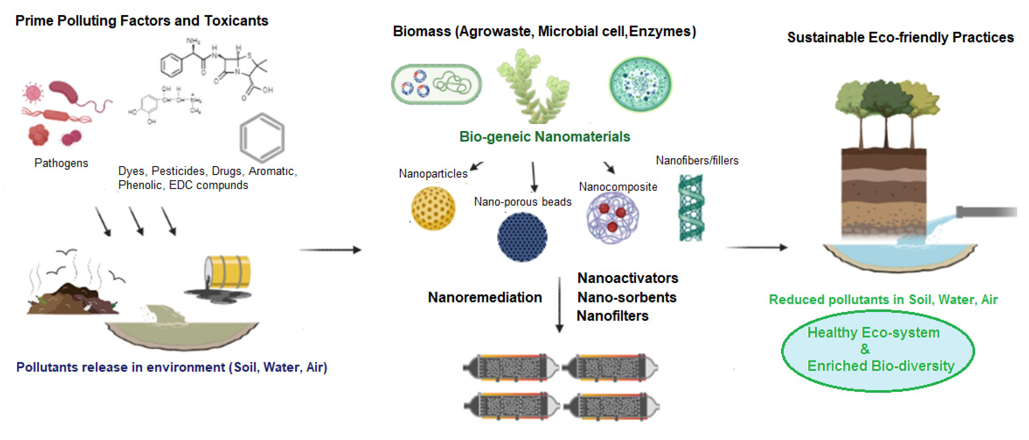
:1. Introduction
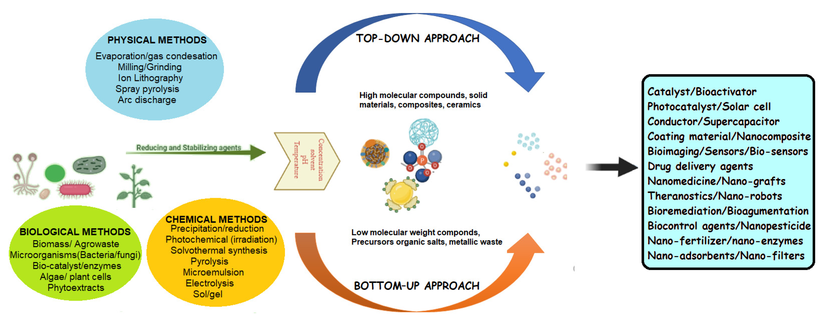
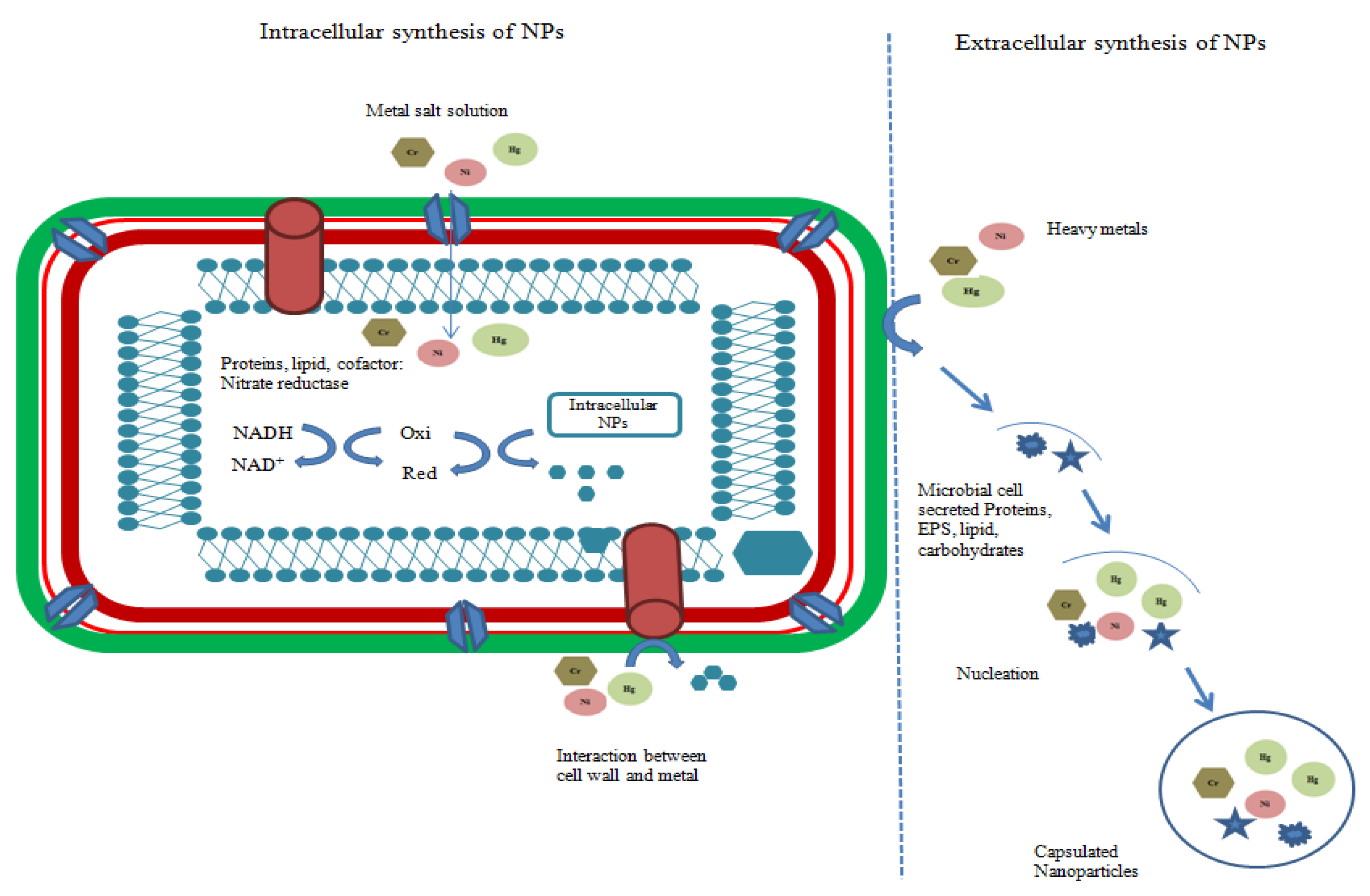
2. Biogenic Nanoparticles and Their Synthesis
2.1. Conventional Synthesis Method
2.2. Green Synthesis Method
2.3. Nanoparticle Synthesis by Bacteria

| Bacterial Strain | Biogenic Nanoparticles | Size and Shape | Localization | Reference |
|---|---|---|---|---|
| Jeotgalicoccus coquina ZC15 | Platinum | 5.74 nm, Spherical | Extracellular | [24] |
| Kocuria rosea MN23 | Platinum | 5.85 nm, Spherical | Extracellular | [24] |
| Pseudomonas kunmingensis ADR19 | Platinum | 3.95 nm, Spherical | Extracellular | [24] |
| Pseudomonas putida KT2440 | Platinum | 8.06 nm, Spherical | Extracellular | [24] |
| Psychrobacter faecalis FZC6 | Platinum | 2.49 nm, Spherical | Extracellular | [24] |
| Sporosarcina psychrophila KC19 | Platinum | 4.24 nm, Spherical | Extracellular | [24] |
| Vibrio fischeri B11177 | Platinum | 3.84 nm, Spherical | Extracellular | [24] |
| Bacillus pumilus, Bacillus paralicheniformis and Sphingomonas paucimobilis. | Silver | 4–20 nm, Spherical to oval | Extracellular | [20] |
| Bacillus cereus | Silver | 5–7.06 nm, Spherical | Extracellular | [24] |
| Bacillus pumilus, Bacillus persicus, and Bacillus licheniformis | Silver | 77–92 nm, Triangular, hexagonal, and spherica | Extracellular | [25] |
| Bacillus subtilis KMS2–2 | Silver | 18–100 nm, Spherical | Extracellular | [26] |
| Sporosarcina koreensis DC4 | Silver and Gold | 30–50 nm, Spherical | Extracellular | [27] |
| Paracoccus haeundaensis BC74171T | Gold | 20.93 ± 3.46 nm, Spherical | Extracellular | [6] |
| Bacillus subtilis | Zinc | 10–15 nm, Spherical | Extracellular | [28] |
| Bacillus Subtilis | Zinc | 16–20 nm, Spherical | Extracellular | [29] |
| Bacillus sp. | Selenium | 2–50 nm, Spherical | Extracellular | [30] |
| Azospirillum brasilense | Selenium | 2–50 nm, Spherical | Extracellular | [31] |
| Streptomyces zaomyceticus Oc−5 and Streptomyces pseudogriseolus Acv−11 | Copper | 78–80.0 nm, spherical | Extracellular | [32] |
| Escherichia sp | Copper | 22.33 to 39 nm, Spherical | Extracellular | [33] |
2.4. Nanoparticle Synthesis by Fungi
2.5. Nanoparticle Synthesis by Algae
2.6. Nanoparticle Synthesis by Plants
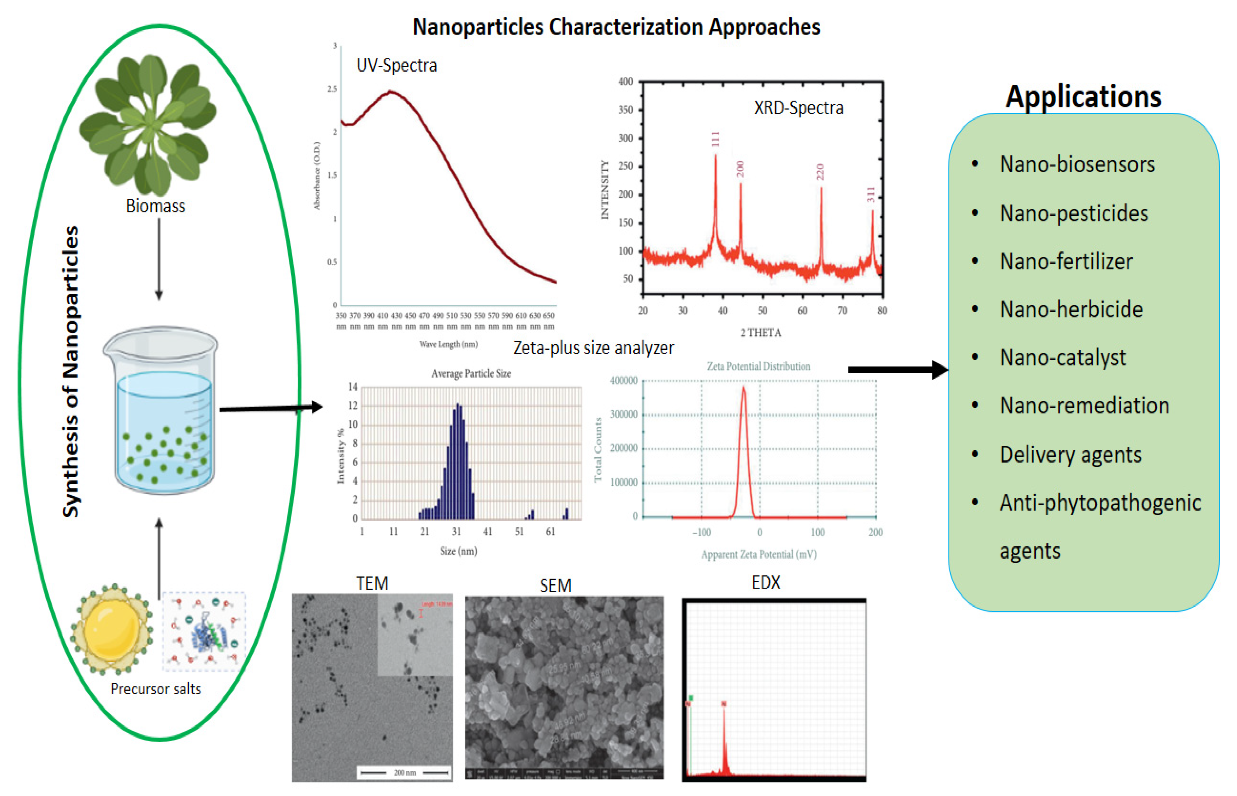
3. Characterization of Biogenic Nanoparticles

4. Application of Biogenic Nanoparticles in Sustainable Agricultural Practices
4.1. Application of Biogenic NPs as Nanofertilizers
4.2. Application of Biogenic NPs as Nanopesticides
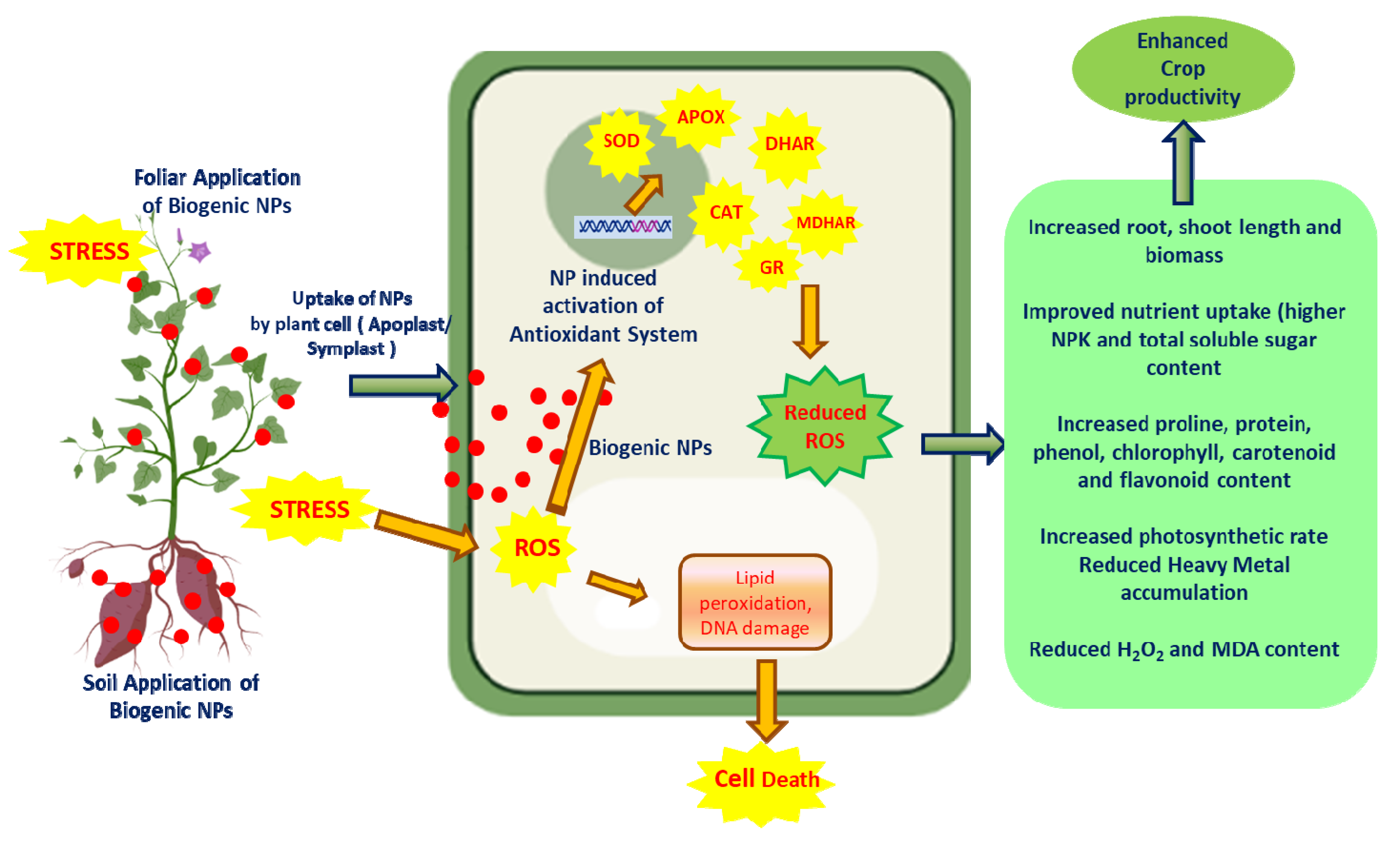
4.3. Applications of NPs in Abiotic Stress Mangament
4.4. Applications of NPs as Nanobiosensors
4.5. Application of Biogenic Nanoparticles in Bioremediation
5. Conclusions and Future Prospects
Author Contributions
Funding
Institutional Review Board Statement
Data Availability Statement
Acknowledgments
Conflicts of Interest
References
- Ahmed, T.; Noman, M.; Manzoor, N.; Shahid, M.; Abdullah, M.; Ali, L.; Wang, G.; Hashem, A.; Al-Arjani, A.F.; Alqarawi, A.A.; et al. Nanoparticle-based amelioration of drought stress and cadmium toxicity in rice via triggering the stress responsive genetic mechanisms and nutrient acquisition. Ecotoxicol. Environ. Saf. 2021, 209, 111829. [Google Scholar] [CrossRef] [PubMed]
- Khan, M.I.R.; Palakolanu, S.R.; Chopra, P.; Rajurkarm, A.B.; Gupta, R.; Iqbal, N.; Maheshwari, C. Improving drought tolerance in rice: Ensuring food security through multi- dimensional approaches. Physiol. Plant. 2021, 172, 645–668. [Google Scholar] [CrossRef] [PubMed]
- Chaudhary, P.; Chaudhary, A.; Bhatt, P.; Kumar, G.; Khatoon, H.; Rani, A.; Kumar, S.; Sharma, A. Assessment of Soil Health Indicators Under the Influence of Nanocompounds and Bacillus spp. in Field Condition. Front. Environ. Sci. 2022, 9, 769871. [Google Scholar] [CrossRef]
- Nejatzadeh, F. Effect of silver nanoparticles on salt tolerance of Satureja hortensis L. during in vitro and in vivo germination tests. Heliyon 2021, 7, e05981. [Google Scholar] [CrossRef] [PubMed]
- Abdelkhalek, A.; El-Gendi, H.; Alotibi, F.O.; Al-Askar, A.A.; Elbeaino, T.; Behiry, S.I.; Abd-Elsalam, K.A.; Moawad, H. Ocimum basilicum-mediated synthesis of silver nanoparticles induces innate immune responses against cucumber mosaic virus in squash. Plants 2022, 11, 2707. [Google Scholar] [CrossRef]
- Patil, S.; Chandrasekaran, R. Biogenic nanoparticles: A comprehensive perspective in synthesis, characterization, application and its challenges. J. Genet. Eng. Biotechnol. 2020, 18, 67. [Google Scholar] [CrossRef]
- Mughal, B.; Zaidi, S.Z.J.; Zhang, X.; Hassan, S.U. Biogenic Nanoparticles Synthesis, Characterisation and Applications. Appl. Sci. 2021, 11, 2598. [Google Scholar] [CrossRef]
- Sengani, M.; Chakraborty, S.; Balaji, M.P.; Govindasamy, R.; Alahmadi, T.A.; Obaid, S.A.; Karuppusamy, I.; Chi, N.T.L.; Brindhadevi, K.; Devi, R.V. Anti-diabetic efficacy and selective inhibition of methyl glyoxal, intervention with biogenic Zinc oxide nanoparticle. Environ. Res. 2023, 216, 114475. [Google Scholar] [CrossRef]
- Jaiswal, K.K.; Banerjee, I.; Dutta, S.; Verma, R.; Gunti, L.; Awasthi, S.; Bhushan, M.; Kumar, V.; Alajmi, M.F.; Hussain, A. Microwave-assisted polycrystalline Ag/AgO/AgCl nanocomposites synthesis using banana corm (rhizome of Musa sp.) extract: Characterization and antimicrobial studies. J. Ind. Eng. Chem. 2022, 107, 145–154. [Google Scholar] [CrossRef]
- Chakraborty, S.; Singh, A.; Roychoudhury, A. Biogenic nanoparticles and generation of abiotic stress-resilient plants: A new approach for sustainable agriculture. Plant Stress 2022, 6, 100117. [Google Scholar] [CrossRef]
- Kavitha, G.; Kumar, J.V.; Pavithra, S.; Komal, M.; Nivetha, M.S.; Kayalvizhi, R.; Abirami, N. Biogenic synthesis of argentum nanocomposites for visible light photocatalyst of dye degradation. Chem. Phys. Lett. 2022, 809, 140159. [Google Scholar] [CrossRef]
- Heinemann, M.G.; Rosa, C.H.; Rosa, G.R.; Dias, D. Biogenic synthesis of gold and silver nanoparticles used in environmental applications: A review. Trends Environ. Anal. Chem. 2021, 30, e00129. [Google Scholar] [CrossRef]
- Saha, P.; Kim, B.S. Plant extract and agricultural waste-mediated synthesis of silver nanoparticles and their biochemical activities. Green Synth. Silver Nanomat. 2022, 285–315. [Google Scholar] [CrossRef]
- Mittal, D.; Kumar, A.; Balasubramaniam, B.; Thakur, R.; Siwal, S.S.; Saini, R.V.; Gupta, R.K.; Saini, A.K. Synthesis of Biogenic silver nanoparticles using plant growth promoting bacteria: Potential use as biocontrol agent against phytopathogens. Biomat. Polym. Horiz. 2022, 1, 22–31. [Google Scholar] [CrossRef]
- Rai, M.; Ingle, A.P.; Trzcinska-Wencel, J.; Wypij, M.; Bonde, S.; Yadav, A.; Kratošová, G.; Golinska, P. Biogenic Silver Nanoparticles: What We Know and What Do We Need to Know? Nanomaterials 2021, 11, 2901. [Google Scholar] [CrossRef]
- Revati, K.; Pandey, B.D. Microbial synthesis of iron-based nanomaterials—A review. Bull. Mater. Sci. 2011, 34, 191–198. [Google Scholar]
- Durán, N.; Marcato, P.D.; Durán, M.; Yadav, A.; Gade, A.; Rai, M. Mechanistic aspects in the biogenic synthesis of extracellular metal nanoparticles by peptides, bacteria, fungi, and plants. Appl. Microbiol. Biotechnol. 2011, 90, 1609–1624. [Google Scholar] [CrossRef]
- Wadhwani, S.A.; Shedbalkar, U.U.; Singh, R.; Chopade, B.A. Biogenic selenium nanoparticles: Current status and future prospects. Appl. Microbiol. Biotechnol. 2016, 100, 2555–2566. [Google Scholar] [CrossRef]
- Nangia, Y.; Wangoo, N.; Goyal, N.; Sharma, S.; Wu, J.S.; Dravid, V.; Shekhawat, G.S.; Suri, C.R. Facile biosynthesis of phosphate capped gold nanoparticles by a bacterial isolate Stenotrophomonas maltophilia. Appl. Phys. Lett. 2009, 94, 233901. [Google Scholar] [CrossRef]
- Allam, N.G.; Ismail, G.A.; El-Gemizy, W.M.; Salem, M.A. Biosynthesis of silver nanoparticles by cell-free extracts from some bacteria species for dye removal from wastewater. Biotechnol. Lett. 2019, 41, 379–389. [Google Scholar] [CrossRef]
- Gahlawat, G.; Choudhary, A.R. A review on the biosynthesis of metal and metal salt nanoparticles by microbes. RSC Adv. 2019, 9, 12944–12967. [Google Scholar] [CrossRef] [PubMed]
- Graves, J.L.; Tajkarimi, M.; Cunningham, Q.; Campbell, A.; Nonga, H.; Harrison, S.H. Rapid evolution of silver nanoparticles resistance in Escherichia colli. Front. Genet. 2015, 6, 42. [Google Scholar] [CrossRef] [PubMed]
- Shi, L.; Rosso, K.M.; Clarke, T.A.; Richardson, D.J.; Zachara, J.M.; Fredrickson, J.K. Molecular underpinnings of Fe (III) oxide reduction by Shewanella Oneidensis MR-1. Front. Microbiol. 2012, 3, 50. [Google Scholar] [CrossRef] [PubMed]
- Eramabadi, P.; Masoudi, M.; Makhdoumi, A.; Mashreghi, M. Microbial cell lysate supernatant (CLS) alteration impact on platinum nanoparticles fabrication, characterization, antioxidant and antibacterial activity. Mat. Sci. Eng. C 2020, 117, 111292. [Google Scholar] [CrossRef]
- Elbeshehy, E.K.F.; Elazzazy, A.M.; Aggelis, G. Silver nanoparticles synthesis mediated by new isolates of Bacillus spp., nanoparticle characterization and their activity against Bean Yellow Mosaic Virus and human pathogens. Front. Microbiol. 2015, 6, 453. [Google Scholar] [CrossRef]
- Mathivanan, K.; Selva, R.; Chandirika, J.U.; Govindarajan, R.K.; Srinivasan, R.; Annadurai, G.; Duc, P.A. Biologically synthesized silver nanoparticles against pathogenic bacteria: Synthesis, calcination and characterization. Biocatal. Agric. Biotechnol. 2019, 22, 101373. [Google Scholar] [CrossRef]
- Singh, J.; Kumar, S.; Alok, A.; Upadhyay, S.K.; Rawat, M.; Tsang, D.C.W. The Potential of Green Synthesized Zinc Oxide Nanoparticles as Nutrient Source for Plant Growth. J. Clean. Prod. 2019, 214, 1061–1070. [Google Scholar] [CrossRef]
- Dhandapani, P.; Prakash, A.A.; AlSalhi, M.S.; Maruthamuthu, S.; Devanesan, S.; Rajasekar, A. Ureolytic bacteria mediated synthesis of hairy ZnO nanostructure as photocatalyst for decolorization of dyes. Mater. Chem. Phys. 2020, 243, 122619. [Google Scholar] [CrossRef]
- Sabir, S.; Zahoor, M.A.; Waseem, M.; Siddique, M.H.; Shafique, M.; Imran, M.; Hayat, S.; Malik, I.R.; Muzammil, S. Biosynthesis of ZnO Nanoparticles using Bacillus subtilis: Characterization and nutritive significance for promoting plant growth in Zea mays L. Dose Res. 2020, 18, 1559325820958911. [Google Scholar] [CrossRef]
- Bharathi, S.; Kumaran, S.; Suresh, G.; Ramesh, M.; Thangamani, V.; Pughazhventhan, S.R. Extracellular synthesis of nanoselenium from fresh water bacteria Bacillus sp., and its validation of antibacterial and cytotoxic potential. Biocatal. Agric. Biotechnol. 2020, 27, 101655. [Google Scholar] [CrossRef]
- Tugarova, A.V.; Mamchenkova, P.V.; Khanadeev, V.A.; Kamnev, A.K. Selenite reduction by the rhizobacterium Azospirillum brasilense, synthesis of extracellular selenium nanoparticles and their characterisation. New Biotechnol. 2020, 58, 17–24. [Google Scholar] [CrossRef]
- Hassan, S.E.; Fouda, A.; Radwan, A.A.; Salem, S.S.; Barghoth, M.G.; Awad, M.A.; Abdo, A.M.; El-Gamal, M.S. Endophytic actinomycetes Streptomyces spp. mediated biosynthesis of copper oxide nanoparticles as a promising tool for biotechnological applications. J. Biol. Inorg. Chem. 2019, 24, 377–393. [Google Scholar] [CrossRef]
- Noman, M.; Shahid, M.; Ahmed, T.; Niazi, M.B.K.; Hussain, S.; Song, F. Use of biogenic copper nanoparticles synthesized from a native Escherichia sp. as photocatalysts for azo dye degradation and treatment of textile effluents. Environ. Pollut. 2020, 257, 113514. [Google Scholar] [CrossRef]
- Mukherjee, P.; Roy, M.; Mandal, B.P.; Dey, G.K.; Mukherjee, P.K.; Ghatak, J.; Tyagi, A.K.; Kale, S.P. Green synthesis of highly stabilized nanocrystalline silver particles by a nonpathogenic and agriculturally important fungus T. asperellum. Nanotechnology 2008, 19, 103–110. [Google Scholar]
- Jalal, M.; Ansari, M.A.; Alzohairy, M.A.; Ali, S.G.; Khan, H.M.; Almatroudi, A.; Raees, K. Biosynthesis of silver nanoparticles from oropharyngeal Candida glabrata Isolates and their antimicrobial activity against clinical strains of bacteria and fungi. Nanomaterials 2018, 8, 586. [Google Scholar] [CrossRef]
- Kalpana, V.N.; Kataru, B.A.S.; Sravani, N.; Vigneshwari, T.; Panneerselvam, A.; Rajeswari, V.D. Biosynthesis of zinc oxide nanoparticles using ulture filtrates of Aspergillus niger: Antimicrobial textiles and dye degradation studies. Open Nanomat. 2018, 3, 48–55. [Google Scholar]
- Krishna, G.; Srileka, V.; Charya, M.A.S.; Serea, E.S.A.; Shalan, A.E. Biogenic synthesis and cytotoxic effects of silver nanoparticles mediated by white rot fungi. Heliyon 2021, 7, e06470. [Google Scholar] [CrossRef]
- Vijayanandan, A.S.; Balakrishnan, R.M. Biosynthesis of cobalt oxide nanoparticles using endophytic fungus Aspergillus nidulans. J. Environ. Manag. 2018, 218, 442–450. [Google Scholar] [CrossRef]
- Mukherjee, P.; Ahmad, A.; Mandal, D.; Senapati, S.; Sainkar, S.R.; Khan, M.I.; Ramani, R.; Parischa, R.; Ajayakumar, P.V.; Alam, M. Bioreduction of AuCl4− ions by the fungus, Verticillium sp. and surface trapping of the gold nanoparticles formed. Angew. Chem. Int. Ed. 2001, 40, 3585–3588. [Google Scholar] [CrossRef]
- Spagnoletti, F.N.; Spedalieri, C.; Kronberg, F.; Giacometti, R. Extracellular biosynthesis of bactericidal Ag/AgCl nanoparticles for crop protection using the fungus Macrophomina phaseolina. J. Environ. Manag. 2019, 231, 457–466. [Google Scholar] [CrossRef]
- Win, T.T.; Khan, S.; Fu, P. Fungus-(Alternaria sp.) mediated silver nanoparticles synthesis, characterization, and screening of antifungal activity against some phytopathogens. J. Nanotechnol. 2020, 29, 8828878. [Google Scholar] [CrossRef]
- Tesser, M.E.; Guilger, M.; Bilesky-José, N.; Risso, W.E.; de Lima, R.; dos Reis Martinez, C.B. Biogenic metallic nanoparticles (Ag, TiO2, Fe) as potential fungicides for agriculture: Are they safe for the freshwater mussel Anodontites trapesialis? Chemosphere 2022, 309, 136664. [Google Scholar] [CrossRef] [PubMed]
- Muñoz, A.J.; Espínola, F.; Ruiz, E.; Cuartero, M.; Castro, E. Biotechnological use of the ubiquitous fungus Penicillium sp. 8L2, Biosorption of Ag (I) and synthesis of silver nanoparticles. J. Environ. Manag. 2022, 316, 115281. [Google Scholar] [CrossRef]
- Das, P.; Barua, S.; Sarkar, S.; Chatterjee, S.K.; Mukherjee, S.; Goswami, L.; Das, S.; Bhattacharya, S.; Karak, N.; Bhattacharya, S.S. Mechanism of toxicity and transformation of silver nanoparticles: Inclusive assessment in earthworm-microbe-soil-plant system. Geoderma 2018, 314, 73–84. [Google Scholar] [CrossRef]
- Mahanty, S.; Bakshi, M.; Ghosh, S.; Chatterjee, S.; Bhattacharyya, S.; Das, P.; Das, S.; Chaudhuri, P. Green synthesis of iron oxide nanoparticles mediated by filamentous fungi isolated from Sundarban mangrove ecosystem, India. BioNanoScience 2019, 9, 637–651. [Google Scholar] [CrossRef]
- Grandmontagne, D.; Navarro, D.; Neugnot-Roux, V.; Ladevèze, S.; Berrin, J.G. The Secretomes of Aspergillus japonicus and Aspergillus terreus Supplement the Rovabio® Enzyme Cocktail for the Degradation of Soybean Meal for Animal. Feed. J. Fungi 2021, 7, 278. [Google Scholar] [CrossRef]
- Sharma, B.; Purkayastha, D.D.; Hazra, S.; Gogoi, L.; Bhattacharjee, C.R.; Ghosh, N.N.; Rout, J. Biosynthesis of gold nanoparticles using a freshwater green alga, Prasiolacrispa. Mat. Lett. 2014, 116, 94–97. [Google Scholar] [CrossRef]
- Senapati, S.; Syed, A.; Moeez, S.; Kumar, A.; Ahmad, A. Intracellular synthesis of gold nanoparticles using alga Tetraselmis kochinensis. Mat. Lett. 2012, 79, 116–118. [Google Scholar] [CrossRef]
- Jena, J.; Pradhan, N.; Aishvarya, V.; Nayak, R.R.; Dash, B.P.; Sukla, L.B.; Panda, P.K.; Mishra, B.K. Biological sequestration and retention of cadmium as CdS nanoparticles by the microalga Scenedesmus-24. J. Appl. Phycol. 2012, 27, 2251–2260. [Google Scholar] [CrossRef]
- Subramaniyam, V.; Ramraj, S.; Bose, S.; Thavamani, P.; Megharaj, M.; Chen, Z.; Naidu, R. Chlorococcum sp. MM11- a novel Phyco-nanofactoryfor the synthesis of iron NP. J. Appl. Phycol. 2015, 27, 1861–1869. [Google Scholar] [CrossRef]
- Rao, M.D.; Pennathur, G. Green synthesis and characterization of cadmium sulphide nanoparticles from Chlamydomonas reinhardtii and their application as photocatalysts. Mat. Res. Bull. 2017, 85, 64–73. [Google Scholar] [CrossRef]
- Sayadi, M.H.; Salmani, N.; Heidari, A.; Rezaei, M.R. Bio-synthesis of palladium nanoparticle using Spirulina platensis alga extract and its application as adsorbent. Surf. Inter. 2018, 10, 136–143. [Google Scholar] [CrossRef]
- Massironi, A.; Morelli, A.; Grassi, L.; Puppi, D.; Braccini, S.; Maisetta, G.; Chiellini, F. Ulvan as novel reducing and stabilizing agent from renewable algal biomass: Application to green synthesis of silver nanoparticles. Carbohydr. Polym. 2019, 203, 310–321. [Google Scholar] [CrossRef]
- Itroutwar, P.D.; Kasivelu, G.; Raguraman, V.; Malaichamy, K.; Sevathapandian, S.K. Effects of biogenic zinc oxide nanoparticles on seed germination and seedling vigor of maize (Zea mays). Biocatal. Agricul. Biotechnol. 2020, 29, 101778. [Google Scholar] [CrossRef]
- Öztürk, B.Y.; Gürsu, B.Y.; Dag, I. Antibiofilm and antimicrobial activities of green synthesized silver nanoparticles using marine red algae Gelidium corneum. Process Biochem. 2020, 89, 208–219. [Google Scholar] [CrossRef]
- Abdel-Raouf, N.; Al-Enazi, N.M.; Ibraheem, I.B.M.; Alharbi, R.M.; Alkhulaifi, M.M. Biosynthesis of silver nanoparticles by using of the marine brown alga Padina pavonia and their characterization. Saudi J. Biol. Sci. 2019, 26, 1207–1215. [Google Scholar] [CrossRef]
- Annamalai, J.; Nallamuthu, T. Green synthesis of silver nanoparticles: Characterization and determination of antibacterial potency. Appl. Nanosci. 2016, 6, 259–265. [Google Scholar] [CrossRef]
- Choudhary, M.K.; Kataria, J.; Sharma, S. Evaluation of the kinetic and catalytic properties of biogenically synthesized silver nanoparticles. J. Clean. Prod. 2018, 198, 882–890. [Google Scholar] [CrossRef]
- Ahmad, N.; Sharma, S.; Alam, M.K.; Singh, V.N.; Shamsi, S.F.; Mehta, B.R. Rapid synthesis of silver nanoparticles using dried medicinal plant of basil. Colloids Surf. B 2010, 81, 81–86. [Google Scholar] [CrossRef]
- Kumar, P.V.; Kala, S.M.J.; Prakash, K.S. Green synthesis of gold nanoparticles using croton Caudatus geisel leaf extract and their biological studies. Mater. Lett. 2019, 236, 19–22. [Google Scholar] [CrossRef]
- Kumar, B.; Smita, K.; Cumbal, L.; Debut, A. Synthesis of silver nanoparticles using Sacha inchi (Plukenetia volubilis L.) leaf extracts. Saudi J. Biol. Sci. 2014, 21, 605–609. [Google Scholar] [CrossRef] [PubMed]
- Gavade, N.L.; Kadam, A.N.; Suwarnkar, M.B.; Ghodake, V.P.; Garadkar, K.M. Biogenic synthesis of multi-applicative silver nanoparticles by using Ziziphus Jujuba leaf extract. Spectrochim. Acta A Mol. Biomol. Spectrosc. 2015, 136, 953–960. [Google Scholar] [CrossRef] [PubMed]
- Joseph, S.; Mathew, B. Microwave-assisted green synthesis of silver nanoparticles and the study on catalytic activity in the degradation of dyes. J. Mol. Liq. 2015, 204, 184–191. [Google Scholar] [CrossRef]
- Periyasami, G.; Palaniappan, S.; Karuppiah, P.; Rahaman, M.; Karthikeyan, P.; Aldalbahi, A.; Al-Dhabi, N.A. Biogenic Silver Nanoparticles Fabricated by Euphorbia granulate Forssk’s Extract: Investigating the Antimicrobial, Radical Scavenging, and Catalytic Activities. J. Nanomat. 2022, 10, 3864758. [Google Scholar] [CrossRef]
- Sharma, T.S.; Selvakumar, K.; Hwa, K.Y.; Sami, P.; Kumaresan, M. Biogenic fabrication of gold nanoparticles using Camellia japonica L. leaf extract and its biological evaluation. J. Mat. Res. Technol. 2019, 8, 1412–1418. [Google Scholar] [CrossRef]
- Guo, M.; Li, W.; Yang, F.; Liu, H. Controllable biosynthesis of gold nanoparticles from a Eucommia ulmoides bark aqueous extract. Spectrochim. Acta A Mol. Biomol. Spectrosc. 2015, 142, 73–79. [Google Scholar] [CrossRef]
- Nasrollahzadeh, M.; Sajadi, S.M.; Vartooni, A.R.; Bagherzadeh, M. Green synthesis of Pd/CuO nanoparticles by Theobroma cacao L. seeds extract and their catalytic performance for the reduction of 4- nitrophenol and phosphine-free Heck coupling reaction under aerobic conditions. J. Colloid Interface Sci. 2015, 448, 106–113. [Google Scholar] [CrossRef]
- Darroudi, M.; Sarani, M.; Oskuee, R.K.; Zak, A.K.; Hosseini, H.A.; Gholami, L. Green synthesis and evaluation of metabolic activity of starch mediated nanoceria. Ceram. Int. 2014, 40, 2041–2045. [Google Scholar] [CrossRef]
- Kumar, B.; Smita, K.; Debut, A.; Cumbal, L. Andean Sacha Inchi (Plukenetia volubilis L.) leaf-mediated synthesis of Cu2O nanoparticles: A low-cost approach. Bioengineering 2020, 7, 54. [Google Scholar] [CrossRef]
- Prasad, K.S.; Gandhi, P.; Selvaraj, K. Synthesis of green nano iron particles (GnIP) and their application in adsorptive removal of As (III) and As (V) from aqueous solution. Appl. Surface. Sci. 2014, 317, 1052–1059. [Google Scholar] [CrossRef]
- Wang, T.; Jin, X.; Chen, Z.; Megharaj, M.; Naidu, R. Green synthesis of Fe nanoparticles using eucalyptus leaf extracts for treatment of eutrophic wastewater. Sci. Total Environ. 2014, 466–467, 210–213. [Google Scholar] [CrossRef]
- Venkateswarlu, S.; Kumar, B.N.; Prasad, C.H.; Venkateswarlu, P.; Jyothi, N.V.V. Bio-inspired green synthesis of Fe3O4 spherical magnetic nanoparticles using Syzygiumcumini seed extract. Phys. B 2014, 449, 67–71. [Google Scholar] [CrossRef]
- Qu, J.; Yuan, X.; Wang, X.; Shao, P. Zinc accumulation and synthesis of ZnO nanoparticles using Physalis alkekengi. Environ. Pollut. 2011, 159, 1783–1788. [Google Scholar] [CrossRef]
- Suresh, A.K. Extracellular bio-production and characterization of small monodispersed CdSe quantum dot nanocrystallites. Spectrochim. Acta Part A Mol. Biomol. Spectrosc. 2014, 130, 344–349. [Google Scholar] [CrossRef]
- Abdelkhalek, A.; Al-Askar, A.A. Green synthesized ZnO nanoparticles mediated by Mentha spicata extract induce plant systemic resistance against Tobacco mosaic virus. Appl. Sci. 2020, 10, 5054. [Google Scholar] [CrossRef]
- Srivastava, V.; Sharma, Y.C.; Sillanpää, M. Green synthesis of magnesium oxide nanoflower and its application for the removal of divalent metallic species from synthetic waste water. Ceram. Int. 2015, 41, 6702–6709. [Google Scholar] [CrossRef]
- Abdelkhalek, A.; Qari, S.H.; Abu-Saied, M.A.; Khalil, A.M.; Younes, H.A.; Nehela, Y.; Behiry, S.I. Chitosan nanoparticles inactivate alfalfa mosaic virus replication and boost innate immunity in Nicotiana glutinosa plants. Plants 2021, 10, 2701. [Google Scholar] [CrossRef]
- Joudeh, N.; Linke, D. Nanoparticle classification, physicochemical properties, characterization, and applications: A comprehensive review for biologists. J. Nanobiotechnol. 2022, 20, 262. [Google Scholar] [CrossRef]
- Tombuloglu, H.; Slimani, Y.; Tombuloglu, G.; Alshammari, T.; Almessiere, M.; Korkmaz, A.D.; Baykal, A.; Samia, A.C.S. Engineered magnetic nanoparticles enhance chlorophyll content and growth of barley through the induction of photosystem genes. Environ. Sci. Pollut. Res. 2020, 27, 3431–3432. [Google Scholar] [CrossRef]
- Raj, S.; Trivedi, R.; Soni, V. Biogenic Synthesis of Silver Nanoparticles, Characterization and Their Applications—A Review. Surfaces 2022, 5, 67–90. [Google Scholar] [CrossRef]
- Tombuloglu, H.; Slimani, Y.; Alshammari, T.; Tombuloglu, G.; Almessiere, M.; Baykal, A.; Ercan, I.; Ozcelik, S.; Demirci, T. Magnetic Behavior and Nutrient Content Analyses of Barley (Hordeum vulgare L.) Tissues upon CoNd0.2Fe1.8O4 Magnetic Nanoparticle Treatment. J. Soil Sci. Plant Nutr. 2020, 20, 357–366. [Google Scholar] [CrossRef]
- Balasubramanian, S.; Kala, S.M.J.; Pushparaj, T.L. Biogenic synthesis of gold nanoparticles using Jasminum auriculatum leaf extract and their catalytic, antimicrobial and anticancer activities. J. Drug Deliv. Sci. Technol. 2020, 57, 101620. [Google Scholar] [CrossRef]
- Khan, I.; Saeed, K.; Khan, I. Nanoparticles: Properties, applications and toxicities. Arab. J. Chem. 2019, 12, 908–931. [Google Scholar] [CrossRef]
- Nwoko, K.C.; Liang, X.; Perez, M.A.; Krupp, E.; Gadd, G.M.; Feldmann, J. Characterisation of selenium and tellurium nanoparticles produced by Aureobasidium pullulans using a multi-method approach. J. Chromatogr. A 2021, 1642, 462022. [Google Scholar] [CrossRef] [PubMed]
- Tombuloglu, H.; Anıl, I.; Akhtar, S.; Turumtay, H.; Sabit, H.; Slimani, Y.; Almessiere, M.; Baykal, A. Iron oxide nanoparticles translocate in pumpkin and alter the phloem sap metabolites related to oil metabolism. Sci. Hortic. 2020, 265, 109223. [Google Scholar] [CrossRef]
- Nguyen, T.M.T.; Huynh, T.T.T.; Dang, C.H.; Mai, D.T.; Nguyen, T.T.N.; Nguyen, D.T. Novel biogenic silver nanoparticles used for antibacterial effect and catalytic degradation of contaminants. Res. Chem. Intermed. 2020, 46, 1975–2090. [Google Scholar] [CrossRef]
- Purohit, A.; Sharma, R.; Ramakrishnan, R.S.; Sharma, S.; Kumar, A.; Jain, D. Biogenic synthesis of Silver Nanoparticles (AgNPs) Using Aqueous Leaf Extract of Buchanania lanzan Spreng and Evaluation of Their Antifungal Activity against Phytopathogenic Fungi. Bioinorg. Chem. Appl. 2022, 2022, 6825150. [Google Scholar] [CrossRef]
- Liu, R.; Lal, R. Potentials of engineered nanoparticles as fertilizers for increasing agronomic productions. Sci. Total Environ. 2015, 514, 131–139. [Google Scholar] [CrossRef]
- Agri, U.; Chaudhary, P.; Sharma, A.; Kukreti, B. Physiological response of maize plants and its rhizospheric microbiome under the influence of potential bioinoculants and nanochitosan. Plant Soil. 2022, 474, 451–468. [Google Scholar] [CrossRef]
- Avila-Quezada, G.D.; Ingle, A.P.; Golińska, P.; Rai, M. Strategic applications of nano-fertilizers for sustainable agriculture: Benefits and bottlenecks. Nanotechnol. Rev. 2022, 11, 2123–2140. [Google Scholar] [CrossRef]
- Shinde, S.; Paralikar, P.; Ingle, A.P.; Rai, M. Promotion of seed germination and seedling growth of Zea mays by magnesium hydroxide nanoparticles synthesized by the filtrate from Aspergillus niger. Arab. J. Chem. 2020, 13, 3172–3182. [Google Scholar] [CrossRef]
- Salih, A.M.; Qahtan, A.A.; Al-Qurainy, F.; Al-Munqedhi, B.M. Impact of Biogenic Ag-Containing Nanoparticles on Germination Rate, Growth, Physiological, Biochemical Parameters, and Antioxidants System of Tomato (Solanum tuberosum L.) In Vitro. Processes 2022, 10, 825. [Google Scholar] [CrossRef]
- Fouda, M.M.; Abdelsalam, N.R.; El-Naggar, M.E.; Zaitoun, A.F.; Salim, B.M.; Bin-Jumah, M.; Allam, A.; Abo-Marzoka, S.A.; Kandil, E.E. Impact of high throughput green synthesized silver nanoparticles on agronomic traits of onion. Int. J. Biol. Macromol. 2020, 149, 1304–1317. [Google Scholar] [CrossRef]
- Krishnaraj, C.; Jagan, E.G.; Ramachandran, R.; Abirami, S.M.; Mohan, N.; Kalaichelvan, P.T. Effect of biologically synthesized silver nanoparticles on Bacopa monnieri (Linn.) Wettst. plant growth metabolism. Proc. Biochem. 2012, 47, 51–658. [Google Scholar] [CrossRef]
- Shaik, A.M.; David, R.M.; Reddy, D.R.S. Green synthesis of zinc oxide nanoparticles using aqueous root extract of Sphagneticola trilobata Lin and investigate its role in toxic metal removal, sowing germination and fostering of plant growth. Inorg. Nano-Metal Chem. 2020, 50, 569–579. [Google Scholar] [CrossRef]
- Del Buono, D.; Di Michele, A.; Costantino, F.; Trevisan, M.; Lucini, L. Biogenic ZnO nanoparticles synthesized using a novel plant extract: Application to enhance physiological and biochemical traits in maize. Nanomaterials 2021, 11, 1270. [Google Scholar] [CrossRef]
- Abou-Zeid, H.; Ismail, G. The role of priming with biosynthesized silver nanoparticles in the response of Triticum aestivum L. to salt stress. Egypt. J. Bot. 2018, 58, 73–85. [Google Scholar] [CrossRef]
- Kasivelu, G.; Selvaraj, T.; Malaichamy, K.; Kathickeyan, D.; Shkolnik, D.; Chaturvedi, S. Nano-Micronutrients [γ-Fe2O3(Iron) and ZnO (Zinc)]: Green Preparation, Characterization, Agro-Morphological Characteristics and Crop Productivity Studies in Two Crops (Rice and Maize). New J. Chem. 2020, 44, 11373–11383. [Google Scholar] [CrossRef]
- Acharya, P.; Jayaprakasha, G.K.; Crosby, K.M.; Jifon, J.L.; Patil, B.S. Nanoparticle-mediated seed priming improves germination, growth, yield, and quality of watermelons (Citrullus lanatus) at multi-locations in Texas. Sci. Rep. 2020, 10, 5037. [Google Scholar] [CrossRef]
- Batool, S.U.; Javed, B.; Zehra, S.S.; Mashwani, Z.U.R.; Raja, N.I.; Khan, T.; Alamri, S. Exogenous applications of bio-fabricated silver nanoparticles to improve biochemical, antioxidant, fatty acid and secondary metabolite contents of sunflower. Nanomaterials 2021, 11, 1750. [Google Scholar] [CrossRef]
- Ahmed, S.; Ahmad, M.; Swami, B.L.; Ikram, S. A review on plants extract mediated synthesis of silver nanoparticles for antimicrobial applications: A green expertise. J. Adv. Res. 2016, 7, 17–28. [Google Scholar] [CrossRef] [PubMed]
- Castillo-Henríquez, L.; Alfaro-Aguilar, K.; Ugalde-Álvarez, J.; Vega-Fernández, L.; Montes de Oca-Vásquez, G.; Vega-Baudrit, J.R. Green synthesis of gold and silver nanoparticles from plant extracts and their possible applications as antimicrobial agents in the agricultural area. Nanomaterials 2020, 10, 1763. [Google Scholar] [CrossRef] [PubMed]
- Devatha, C.P.; Jagadeesh, K.; Patil, M. Effect of Green synthesized iron nanoparticles by Azardirachta indica in different proportions on antibacterial activity. Environ. Nanotech. Monit. Manag. 2018, 9, 85–94. [Google Scholar] [CrossRef]
- González-Merino, A.M.; Hernández-Juárez, A.; Betancourt-Galindo, R.; Ochoa-Fuentes, Y.M.; Valdez-Aguilar, L.A.; Limón-Corona, M.L. Antifungal activity of zinc oxide nanoparticles in Fusarium oxysporum–Solanum lycopersicum pathosystem under controlled conditions. J. Phytopat. 2021, 169, 533–544. [Google Scholar] [CrossRef]
- Mishra, S.; Singh, B.R.; Singh, A.; Keswani, C.; Naqvi, A.H.; Singh, H.B. Biofabricated silver nanoparticles act as a strong fungicide against Bipolaris sorokiniana causing spot blotch disease in wheat. PLoS ONE 2014, 9, e97881. [Google Scholar] [CrossRef]
- Saharan, V.; Sharma, G.; Yadav, M.; Choudhary, M.K.; Sharma, S.S.; Pal, A.; Raliya, R.; Biswas, P. Synthesis and in vitro antifungal efficacy of Cu-chitosan nanoparticles against pathogenic fungi of tomato. Int. J. Biol. Macromol. 2015, 75, 346–353. [Google Scholar] [CrossRef]
- Abdellatif, K.F.; Abdelfattah, R.H.; El-Ansary, M.S.M. Green nanoparticles engineering on root-knot nematode infecting eggplants and their effect on plant DNA modification. Iran. J. Biotechnol. 2016, 14, 250. [Google Scholar] [CrossRef]
- Pascoli, M.; Jacques, M.T.; Agarrayua, D.A.; Avila, D.S.; Lima, R.; Fraceto, L.F. Neem oil based nanopesticide as an environmentally-friendly formulation for applications in sustainable agriculture: An ecotoxicological perspective. Sci. Total Environ. 2019, 677, 57–67. [Google Scholar] [CrossRef]
- Syed, B.; Prasad, M.N.; Satish, S. Synthesis and characterization of silver nanobactericides produced by Aneurinibacillus migulanus 141, a novel endophyte inhabiting Mimosa pudica L. Arab. J. Chem. 2019, 12, 3743–3752. [Google Scholar] [CrossRef]
- Vanti, G.L.; Masaphy, S.; Kurjogi, M.; Chakrasali, S.; Nargund, V.B. Synthesis and application of chitosan-copper nanoparticles on damping off causing plant pathogenic fungi. Inter. J. Biol. Macromol. 2020, 156, 1387–1395. [Google Scholar] [CrossRef]
- Ghazy, N.A.; El-Hafez, A.; Omnia, A.; El-Bakery, A.M.; El-Geddawy, D.I. Impact of silver nanoparticles and two biological treatments to control soft rot disease in sugar beet (Beta vulgaris L). Egypt. J. Biol. Pest. Control. 2021, 31, 3. [Google Scholar] [CrossRef]
- Mishra, R.C.; Kalra, R.; Dilawari, R.; Goel, M.; Barrow, C.J. Bio-Synthesis of Aspergillus terreus Mediated Gold Nanoparticle: Antimicrobial, Antioxidant, Antifungal and In Vitro Cytotoxicity Studies. Materials 2022, 15, 3877. [Google Scholar] [CrossRef]
- Sreelakshmi, B.; Induja, S.; Adarsh, P.; Rahul, H.; Arya, S.; Aswana, S.; Haripriya, R.; Aswathy, B.; Manoj, P.; Vishnudasan, D. Drought stress amelioration in plants using green synthesised iron oxide nanoparticles. Mater. Today Proc. 2020, 41, 723–727. [Google Scholar] [CrossRef]
- Zafar, S.; Hasnain, Z.; Aslam, N.; Mumtaz, S.; Jaafar, H.Z.; Wahab, P.E.M.; Qayum, M.; Ormenisan, A.N. Impact of Zn nanoparticles synthesized via green and chemical approach on okra (Abelmoschus esculentus L.) growth under salt stress. Sustainability 2021, 13, 3694. [Google Scholar] [CrossRef]
- Iqbal, M.; Raja, N.I.; Mashwani, Z.U.R.; Hussain, M.; Ejaz, M.; Yasmeen, F. Effect of Silver Nanoparticles on Growth of Wheat Under Heat Stress. Iran J. Sci. Technol. Trans A Sci. 2019, 43, 387–395. [Google Scholar] [CrossRef]
- Kalyani, N.; Goel, S.; Jaiswal, S. On-site sensing of pesticides using point-of-care biosensors: A review. Environ. Chem. Lett. 2021, 19, 345–354. [Google Scholar] [CrossRef]
- Jebril, S.; Fdhila, A.; Dridi, C. Nanoengineering of eco-friendly silver nanoparticles using five different plant extracts and development of cost-effective phenol nanosensor. Sci. Rep. 2021, 11, 22060. [Google Scholar] [CrossRef]
- Thakur, M.; Wang, B.; Madan, L.; Verma, L. Development and applications of nanobiosensors for sustainable agricultural and food industries: Recent developments, challenges and perspectives. Environ. Technol. Inn. 2022, 26, 102371. [Google Scholar] [CrossRef]
- James, A.; Franco, F.; Merca, E.M.; Rodriguez, S.; Johny, F.; Balidon-Veronica, P.; Divina, M.; Evangelyn, A.; Alocilja, C.; Fernando, L.M. DNA based electrochemical nanobiosensor for the detection of Phytophthora palmivora causing black pod rot in cacao (Theobroma cacao L.) pods. Physiol. Mol. Plant Pathol. 2019, 107, 14–20. [Google Scholar] [CrossRef]
- Duhan, J.S.; Kumar, R.; Kumar, N.; Kaur, P.; Nehra, K.; Duhan, S. Nanotechnology: The new perspective in precision agriculture. Biotechnol. Rep. 2017, 15, 11–23. [Google Scholar] [CrossRef]
- Giraldo, J.P.; Wu, H.; Newkirk, G.M.; Kruss, S. Nanobiotechnology approaches for engineering smart plant sensors. Nat. Nanotechnol. 2019, 14, 541–553. [Google Scholar] [CrossRef] [PubMed]
- Lew, T.T.S.; Koman, V.B.; Silmore, K.S.; Seo, J.S.; Gordiichuk, P.; Kwak, S.Y. Real-time detection of wound-induced H2O2 signalling waves in plants with optical nanosensors. Nat. Plants 2020, 6, 404–415. [Google Scholar] [CrossRef] [PubMed]
- Kaushal, M.; Wani, S.P. Nanobiosensors: Frontiers in Precision Agriculture; Prasad, R., Ed.; Springer Nature Ltd.: Singapore, 2017; pp. 279–291. [Google Scholar]
- Ramnani, P.N.; Saucedo, M.; Mulchandani, A. Carbon nanomaterial-based electrochemical biosensors for label-free sensing of environmental pollutants. Chemosphere 2016, 143, 85–98. [Google Scholar] [CrossRef] [PubMed]
- Malik, S.; Dhasmana, A.; Preetam, S.; Mishra, Y.K.; Chaudhary, V.; Bera, S.P.; Ranjan, A.; Bora, J.; Kaushik, A.; Minkina, T.; et al. Exploring Microbial-Based Green Nanobiotechnology for Wastewater Remediation: A Sustainable Strategy. Nanomaterials 2022, 12, 4187. [Google Scholar] [CrossRef] [PubMed]
- Singh, P.; Singh, H.; Kim, Y.J.; Mathiyalagan, R.; Wang, C.; Yang, D.C. Extracellular synthesis of silver and gold nanoparticles by Sporosarcina koreensis DC4 and their biological applications. Enzym. Microb. Technol. 2016, 86, 75–83. [Google Scholar] [CrossRef]
- Nazari, N.; Kashi, F.J. A novel microbial synthesis of silver nanoparticles: Its bioactivity, Ag/Ca-Alg beads as an effective catalyst for decolorization Disperse Blue 183 from textile industry effluent. Sep. Purificat. Technol. 2021, 259, 118117. [Google Scholar] [CrossRef]
- Qiu, X.; Wang, S.; Miao, S.; Suo, H.; Xu, H.; Hu, Y. Co-immobilization of laccase and ABTS onto amino-functionalized ionic liquid-modified magnetic chitosan nanoparticles for pollutants removal. J. Hazar. Mat. 2021, 401, 123353. [Google Scholar] [CrossRef]
- Soto-Robles, C.A.; Nava, O.; Cornejo, L.; Lugo-Medina, E.; Vilchis-Nestor, A.R.; Castro-Beltrán, A.; Luque, P.A. Biosynthesis, Characterization and Photocatalytic Activity of ZnO Nanoparticles Using Extracts of Justicia spicigera for the Degradation of Methylene Blue. J. Mol. Struct. 2021, 1225, 129101. [Google Scholar] [CrossRef]
- Saini, M.; Yadav, S.; Rani, N.; Mushtaq, A.; Rawat, S.; Saini, K.; Maity, D. Biosynthesized zinc oxide nanoparticles using seed and bark extract of Azadirachta indica for antibacterial, photocatalytic and supercapacitor applications. Mat. Sci. Eng. B 2022, 282, 115789. [Google Scholar] [CrossRef]
- Ramakrishna, M.; Babu, D.R.; Gengan, R.M.; Chandra, S.; Rao, G.N. Green synthesis of gold nanoparticles using marine algae and evaluation of their catalytic activity. J. Nanost. Chem. 2016, 6, 1–13. [Google Scholar] [CrossRef]
- Balaraman, P.; Balasubramanian, B.; Kaliannan, D.; Durai, M.; Kamyab, H.; Park, S.; Chelliapan, S.; Lee, C.T.; Maluventhen, V.; Maruthupandian, A. Phyco-synthesis of silver nanoparticles mediated from marine algae Sargassum myriocystum and its potential biological and environmental applications. Waste Biomass Valorizat. 2020, 11, 5255–5271. [Google Scholar] [CrossRef]
- Aboelfetoh, E.F.; El-Shenody, R.A.; Ghobara, M.M. Eco-friendly synthesis of silver nanoparticles using green algae (Caulerpa serrulata): Reaction optimization, catalytic and antibacterial activities. Environ. Monit. Assess. 2017, 189, 349. [Google Scholar] [CrossRef]
- Agarwal, P.; Gupta, R.; Agarwal, N. Advances in synthesis and applications of microalgal nanoparticles for wastewater treatment. J. Nanotechnol. 2019, 7392713. [Google Scholar] [CrossRef]
- Ahmed, T.; Noman, M.; Shahid, M.; Niazi, M.B.K.; Hussain, S.; Manzoor, N.; Wang, X.; Li, B. Green synthesis of silver nanoparticles transformed synthetic textile dye into less toxic intermediate molecules through LC-MS analysis and treated the actual wastewater. Environ. Res. 2020, 191, 110142. [Google Scholar] [CrossRef]
- Liang, Y.; Demir, H.; Wu, Y.; Aygun, A.; Tiri, R.N.E.; Gur, T.; Yuan, Y.; Xia, C.; Demir, C.; Sen, F.; et al. Facile synthesis of biogenic palladium nanoparticles using biomass strategy and application as photocatalyst degradation for textile dye pollutants and their in-vitro antimicrobial activity. Chemosphere 2022, 306, 135518. [Google Scholar] [CrossRef]


| Organism | Biogenic Nanoparticles | Size and Shape | Applications | References |
|---|---|---|---|---|
| Fungi | ||||
| Verticillium | Gold | 20 nm, Spherical Both extracellular and Intracellular | Catalysis and precursors for synthesis of coatings for electronic applications | [39] |
| Trichoderma asperellum | Silver | 13–18 nm, Spherical | -- | [34] |
| Macrophomina phaseolina. | Silver | 5–30 nm, Spherical | Bactericidal for phytopathogens and seed protection agent | [40] |
| Alternaria sp. | Silver | 3–10 nm, Spherical | Biocontrol agent | [41] |
| Trichoderma harzianum | Silver, Iron and Titanium | 15–20 nm, Spherical | Biocontrol agent of Sclerotinia sclerotiorum, heavy metal absorption | [42] |
| Penicillium sp. 8L2 | Silver | 2–9 nm, Spherical | Biosorption of heavy metal, Biocidal agents | [43] |
| Candida glabrata | Silver | 2–15 nm, Spherical | Antimicrobial agent | [35] |
| Aspergillus niger | Zinc | 41 nm, Spherical | Dye Decolorizing and antimicrobial agent | [36] |
| Aspergillus niger BSC−1 | Iron | 20–40 nm, Spherical | Removal of Chromium from aqueous solution | [44] |
| Trichoderma asperellum, Phialemoniopsis ocularis and Fusarium incarnatum | Iron | 13–30 nm, Spherical | Chemosorption of Chromium | [45] |
| Aspergillus terreus S1 | Magnesium | ≤38 nm | Precipitation and adsorption | [46] |
| Ganoderma enigmaticum and Trametes ljubarskyi | Silver | 5–40 nm, Spherical | Cytotoxic agents | [37] |
| Aspergillus nidulans | Cobalt | 20.29 nm, Spherical | Energy storgae | [38] |
| Algae | ||||
| Prasiola crispa | Gold | 5–25 nm, Spherical | Nanosensor, Nano-catalyst | [47] |
| Tetraselmis kochinensis | Gold | 5–35 nm, Spherical, Intracellular synthesis | Nano-catalyst | [48] |
| Scenedesmus−24 | Cadmium sulphide | 120–175 nm oval, Intracellular synthesis | Heavy metal sequesteration | [49] |
| Chlorococcum sp. MM11 | Iron | 20–50 nm, spherical | Chromium remedaition | [50] |
| Chlamydomonas reinhardtii | Cadmium sulphide | 5 nm, Spherical | Photocatalytic degradation agent | [51] |
| Spirulina plantesis | Palladium | 10–20 nm, Spherical | Adsorption of heavy metal | [52] |
| Ulvan | Silver | 33 nm | Bactericidal | [53] |
| Turbinaria ornata | Zinc oxide | - | Alleviation zinc deficiency and improved the agronomical parameters of maize seedling | [54] |
| Gelidium corneum | Silver | 20–50 nm | Antibiofilm and antimicrobial agent | [55] |
| Padina pavonia | Silver | 49.58–86.37 nm, Variable shape | -- | [56] |
| Biogenic Nanoparticles | Plant Source | Size and Shape | Applications | References |
|---|---|---|---|---|
| Silver | Plukenetia volubilis L. | 4–25 nm, Spherical | Photocatalytic degradation and Antioxidant agent | [61] |
| Ziziphus jujuba | 20–30 nm | Nano-catalyst | [62] | |
| Biophytum sensitivum | 19.06 nm, Spherical | Dye degradation | [63] | |
| Ocimum basilicum | 26.3 to 83 nm, Spherical | Biocontrol agents for Viral infections (Cucumber mosaic virus) | [5] | |
| Euphorbia granulata | 5–20 nm, Spherical | Antioxidant agent, Nano-catalyst | [64] | |
| Taraxacum officinale | ~15 nm, Spherical | Biocontrol agent | [15] | |
| Gold | Camellia japonica | 40 nm, Spherical and triangular | Antimicrobial agents, Nano-catalyst | [65] |
| Aerva lanata | ~18 nm, Spherical | Nano-catalyst | [63] | |
| Eucommia ulmoides | 16.4 nm, Spherical | Dye degradation | [66] | |
| Copper | Theobroma cacao L. | Spherical, ≤32 nm | Nano-catalyst | [67] |
| Plant based Starch | 50 nm, Spherical clusters | Biosensor | [68] | |
| Plukenetia volubilis L. | 6–10 nm, Spherical | Photocatalytic degradation agent | [69] | |
| Iron | Mentha spicata L. | 20–45 nm, Spherical | Removal of Arsenic (III&V) from contaminated water bodies | [70] |
| Eucalyptus sp. | 20–80 nm, Spherical | Treatment of swine wastewater | [71] | |
| Syzygium cumini | 9–20 nm, Spherical | Biomedical | [72] | |
| Zinc oxide | Physalis alkekengi L. | 72.5 nm, Spherical | Phytoremediation | [73] |
| Artocarpus gomenzianus | 5–15nm, Spherical | Photocatalytic degradation agent | [74] | |
| Mentha spicata | 11–88 nm, Spherical | Biocontrol agent against Tobacco mosaic virus | [75] | |
| Magnesium oxide | Acacia gum | ≤100 nm, Spherical | Heavy metal removal | [76] |
| Lead | Theobroma cacao | 40 nm, Spherical | Nano-catalyst | [67] |
| Chitosan/Dextran | Nicotiana glutinosa | 91.68 nm, Spherical | Biocontrol agent against Alfalfa Mosaic Virus | [77] |
| Biogenic Nanoparticles | Host Plant | Pathogen | Response | References |
|---|---|---|---|---|
| Silver | Wheat | Bipolaris sorokiniana | Inhibit pathogens and prevent disease | [105] |
| Copper | Tomato | Fusarium | Increased plant growth and reduce disease severity | [106] |
| Silver | Brinjal | Meloidogyne javanica | Inhibit the growth of nematode | [107] |
| Zinc oxide | Cowpea | Callosobruchus | Inhibit the growth of pathogens | [82] |
| Neem oil-loaded zein | Allium cepa | Pesticidal | Inhibit pathogens | [108] |
| Copper | Potato | Pythium ultimum | Inhibit fungal pathogens | [32] |
| Silver | Mimosa putida | Pseudomonas aeruginosa, Staphylococcus | Inhibit the growth of pathogens | [109] |
| Chitosan coupled copper | Chili | Rhizoctonia solani, Pythium aphanidermautum | Inhibit the growth of pathogen and improved plant growth | [110] |
| Silver | Sugar beet | Pectobacteriumcarotovorum | Inhibit pathogens and improved sugar quality | [111] |
| Gold | - | Fusarium oxysporum, Rhizoctoiniasolani | Inhibit the growth of fungal pathogens | [112] |
Disclaimer/Publisher’s Note: The statements, opinions and data contained in all publications are solely those of the individual author(s) and contributor(s) and not of MDPI and/or the editor(s). MDPI and/or the editor(s) disclaim responsibility for any injury to people or property resulting from any ideas, methods, instructions or products referred to in the content. |
© 2023 by the authors. Licensee MDPI, Basel, Switzerland. This article is an open access article distributed under the terms and conditions of the Creative Commons Attribution (CC BY) license (https://creativecommons.org/licenses/by/4.0/).
Share and Cite
Bhandari, G.; Dhasmana, A.; Chaudhary, P.; Gupta, S.; Gangola, S.; Gupta, A.; Rustagi, S.; Shende, S.S.; Rajput, V.D.; Minkina, T.; et al. A Perspective Review on Green Nanotechnology in Agro-Ecosystems: Opportunities for Sustainable Agricultural Practices & Environmental Remediation. Agriculture 2023, 13, 668. https://doi.org/10.3390/agriculture13030668
Bhandari G, Dhasmana A, Chaudhary P, Gupta S, Gangola S, Gupta A, Rustagi S, Shende SS, Rajput VD, Minkina T, et al. A Perspective Review on Green Nanotechnology in Agro-Ecosystems: Opportunities for Sustainable Agricultural Practices & Environmental Remediation. Agriculture. 2023; 13(3):668. https://doi.org/10.3390/agriculture13030668
Chicago/Turabian StyleBhandari, Geeta, Archna Dhasmana, Parul Chaudhary, Sanjay Gupta, Saurabh Gangola, Ashulekha Gupta, Sarvesh Rustagi, Sudhir S. Shende, Vishnu D. Rajput, Tatiana Minkina, and et al. 2023. "A Perspective Review on Green Nanotechnology in Agro-Ecosystems: Opportunities for Sustainable Agricultural Practices & Environmental Remediation" Agriculture 13, no. 3: 668. https://doi.org/10.3390/agriculture13030668