Article

Lycium barbarum Polysaccharide Inhibits E. coli-Induced Inflammation and Oxidative Stress in Mammary Epithelial Cells of Dairy Cows via SOCS3 Activation and MAPK Suppression

1
College of Animal Science and Technology, Yangzhou University, Yangzhou 225009, China
2
Joint International Research Laboratory of Agriculture and Agri-Product Safety of Ministry of Education of China, Yangzhou University, Yangzhou 225009, China
*
Author to whom correspondence should be addressed.
Agriculture 2022, 12(5), 598; https://doi.org/10.3390/agriculture12050598
Submission received: 11 March 2022 / Revised: 14 April 2022 / Accepted: 20 April 2022 / Published: 24 April 2022
(This article belongs to the Special Issue Breeding, Genetics and Safety Production of Dairy Cattle)

Abstract

:
Escherichia coli (E. coli) is one of the main causative agents of mastitis in dairy cows. Lycium barbarum polysaccharide (LBP) has a variety of physiological effects as it has antioxidants, it is hypoglycemic, it has anti-aging properties, it is neuroprotective, immune boosting, and it has anti-inflammatory effects in vivo and in vitro. In this study, we examined whether LBP affects the expression of pro-inflammatory factors, and the mitogen-activated protein kinase (MAPK) signaling pathway via activation of the suppressor of cytokine signaling-3 (SOCS3) in E. coli-induced primary bovine mammary epithelial cell (pbMEC) inflammatory responses. The experiment was designed with the control group (NC), cells were treated with E. coli for 6 h as the E. coli group (E. coli), and cells were pretreated with 100 μg/mL or 300 μg/mL of LBP for 24 h, followed by the addition of E. coli for 6 h as the E. coli + low level (E + LL) or E. coli + high level (E + HL) groups. The addition of LBP did not alter the cell viability of pbMEC in a dose-dependent assay. Pretreatment with LBP significantly decreased the expression of pro-inflammatory genes (IL1B, MAPK14, COX-2, iNOS) and proteins (COX-2, IL-1β, TNF-α) in the cells challenged by E. coli as compared with the control group (p < 0.05). E. coli stimulation significantly increased the production of reactive oxygen species (ROS) and malondialdehyde (MDA) in pbMEC, and decreased the antioxidants’ capacity with regard to decreased superoxide dismutase (SOD) and total antioxidant capacity (T-AOC); however, pretreatment with LBP reversed the oxidative stress and inhibition of antioxidants in cells challenged by E. coli. Moreover, LBP reversed the upregulated expression of the components of the MAPK pathway (increased phosphorylation level of p38, JNK, and ERK), followed by E. coli stimulation. Consistently, cells exposed to E. coli strengthened the staining of p38, whereas pretreatment of LBP weakened the staining of p38 in cells challenged by E. coli. Notably, the expression of SOCS3 was increased by LBP added to the cells in a dose-dependent manner. Additionally, the level of decreased expression of proinflammatory factors (IL-1β, TNF-α, and COX-2) was higher in the E + LL group than in the E + HL group. These results indicate that LBP pretreatment is effective in the alleviation of E. coli-induced inflammatory and oxidative responses in pbMEC through activation of SOCS3 and depression of MAPK signaling. As such, this might help us to develop molecular strategies for mitigating the detrimental effects of clinical bovine mastitis.

1. Introduction

Bovine mastitis is one of the serious diseases that causes huge economic losses for dairy farming. This is mainly because the invasion of pathogenic microorganisms such as E. coli into mammary tissue leads to a storm of inflammation and tissue damage [1,2,3,4]. Studies have shown that E. coli-induced inflammation of the mammary gland has a large effect on cows’ milk production and milk quality [5]. At present, antibiotics are still one of the most effective in the treatment for bovine mastitis; however, the drug resistance of pathogens, as well as drug residues in dairy products, have been an issue for the dairy industry and public health. Hence, looking for alternatives such as natural substances to treat bovine mastitis is of great importance [6,7].
The primary bovine mammary epithelial cells (pbMEC) constitute an important line of defense against the invasion of pathogens [8,9]. Moreover, pbMEC tries to express a variety of pathogen patterns, identify receptors, and activate downstream signaling pathways to trigger the production of inflammatory cytokines [10,11]. Cytokines such as COX-2, IL6, and TNF-α have been reported to be induced by activating the nuclear factor kappa-B (NF-κB) and MAPK signaling pathways [12]. Thus, the suppression of inflammation is usually mediated by inhibiting the NF-κB and MAPK signaling pathways [13]. Nevertheless, SOCS family cytokine signal transduction inhibition is a negative regulatory protein that may reduce the overactivation of MAPK and NF-κB [14,15]. Furthermore, it has been revealed that SOCS1 and SOCS3 reduce the inflammatory responses in BV-2 microglia by inhibiting the activation of the TLR/NF-κB [16,17,18].
LBP is a major active ingredient and water-soluble polysaccharide extracted from Goji berries [19,20]. A study has shown the anti-inflammatory, antioxidant, and antiapoptotic properties of LBP [21]. Studies have also reported that it appears to protect human lens epithelial cells and mouse tissues from oxidative damage [22,23]. Recent studies with bovine hepatocytes have shown that LBP inhibits the activation of NF-κB to decrease chemokines [24]. Preliminary research in our laboratory has shown the anti-inflammatory potential of LBP. On the basis of available data, we hypothesized that LBP might be effective at protecting cells against inflammatory responses and oxidative stress induced by E. coli in pbMEC. Hence, the aim of this study was to investigate the protective effect of LBP against E. coli-induced inflammation responses and the oxidative stress of pbMEC through SOCS3 activation and MAPK suppression. The study may provide new insights into therapeutic strategies for combating clinical mastitis using LBP as diet supplement or medical agent and highlighted the link between SOCS3 and regulation of mammary inflammation.

2. Materials and Methods

2.1. Experimental Design

We optimized the concentration and time aspects of the treatment of E. coli or LBP, cells were cultured with E. coli (1 × 10⁷/mL) for 6 h as positive control (E. coli); cells were pre-treated with (100 μg/mL) LBP for 24 h, then washed, followed by the addition of E. coli exposure for 6 h (E + LL group, E + LL). Similarly, for cells pretreated with (300 μg/mL) LBP for 24 h, followed by the addition of E. coli and exposure for 6 h (E + HL group, E + HL), an equal amount of PBS was added to the culture as the negative control (NC). The concentration screening of LBP and E. coli has been previously studied in this laboratory. Before adding LBP and E. coli (1 × 10⁷/mL), cells were incubated using serum-free medium for 24 h.

2.2. Chemicals

LBP was purchased from Solarbio (P7850, Solarbio Life Sciences, Beijing, China) with a purity of > 50%, and E. coli was isolated and identified as a B1 type from the milk of clinical bovine mastitis.

2.3. Cell Culture Conditions

pbMEC were isolated and obtained by our laboratory from bovine mammary tissue of three lactating cows without signs of clinical disease. The pbMEC in good growth condition were inoculated in 6 well plates (2.5 × 10⁵/well) using complete medium (90% RPMI 1640, (Gibco, Temecula, CA, USA), 10% fetal bovine serum (Thermo Fisher Scientific, Waltham, MA, USA), and antibiotics (penicillin 100 IU/mL; streptomycin 100 μg/mL). The medium was replaced with a fresh medium, free of antibiotics, during E. coli stimulation. Cells were incubated at conditions of 5% CO2 and 37 °C.

2.4. Cell Viability

Cell viability was measured using a cell counting kit-8 (CCK-8) (Vazyme, Nanjing, China). When the cell density was appropriate (1 × 10⁶), different concentrations of LBP were added at 0, 25, 100, 300, 500, and 1000 μg/mL for 24 h, then 10 μL of CCK-8 solution was added for 2 h, and the absorbance was measured at 450 nm wavelength.

2.5. RNA Extraction and Quantitative RT-PCR

Total RNA from cells was extracted using RNA-easy Isolation Reagent (Vazyme, Nanjing, China). The extracted RNA was performed using a HiScript III 1st Strand cDNA Synthesis Kit (Vazyme, Nanjing, China) to reverse transcribe cDNA. qRT-PCR was performed using HiScript II One Step qRT-PCR SYBR Green Kit (Vazyme, Nanjing, China) on an Applied Biosystems 7300 Real-Time PCR System with Primer 5.0 to design primers for IL1B, MAPK14, COX2, iNOS, IL6, TNFA UXT, RPS9, and GAPDH, whose sequences are listed in Supplementary Table S1. The CT values were recorded by RT-PCR using AceQ® Probe Master Mix, and the results were analyzed using the 2ΔΔCt method.

2.6. ROS Determination

The intracellular ROS levels were measured using a Reactive Oxygen Species Assay Kit (Beyotime Biotechnology, Nantong, China). The 2,7-Dichlorodihydrofluorescein diacetate (DCFH-DA) probe was diluted to 10 µM/L according to a ratio of 1:1000, cells were collected and suspended in the DCFH-DA solution, and incubated in the incubator at 37 °C for 20 min. The mixture was then centrifuged at 1000 rpm for 5 min. Subsequently, pellets were resuspended in PBS and measured at 488 nm excitation and 525 nm emission.

2.7. Antioxidant Enzyme Activity

The cells were collected to fully disrupt and release the antioxidants therein, centrifuged at 4 °C and 12,000× g for 5 min, and the supernatant was taken for subsequent determination. According to the manufacturer’s protocol (Shanghai Beyotime Biotechnology, Shanghai, China), MDA levels, T-AOC, and SOD were detected with biochemical kits (Shanghai Beyotime Biotechnology, Shanghai, China). For all kits, after adding the working solution, it was mixed well and centrifuged accordingly, as indicated in the instructions of the kits. Absorbance values were measured and used to calculate output by the formulas according to the protocols.

2.8. Immunofluorescence

Cell crawls were placed in 12 well plates and inoculated with (2 × 10⁴) cells per well. The medium was discarded, washed in PBS, fixed in 4% paraformaldehyde for 15 min, washed 3 times in PBS, incubated in a 0.5% Triton X-100 permeation solution for 15 min at RT, closed in a 5% bovine serum albumin (BSA) blocking buffer for 1 h at room temperature, washed three times in PBS, and incubated with a primary antibody at 4 °C overnight. It was then washed three times with PBS, and incubated for 1 h in the dark at 37 °C using a goat anti-rabbit Cy3-labeled secondary antibody. Nuclei were stained with diamidino-phenyl-indole (DAPI) (1 μg/mL) (D8417, Sigma-Aldrich, St. Louis, MO, USA) for 5 min, washed three times in PBS, transferred to slides, and sealed with a fluorescent quencher. Images were then captured using Leica DMi8 fluorescent microscopy.

2.9. Western Blot

Cells in 6-well plates were placed on ice with radio immunoprecipitation assay (RIPA) lysate (Beyotime, Shanghai, China) for total protein extraction. Then, 10% SDS-PAGE gel was used to separate each target proteins. The blots were incubated for 2 h at RT with horseradish peroxidase coupled (HRP) secondary antibodies, followed by the incubation of primary antibodies overnight at 4 °C, which were then washed 3 times with 1 × TBS + Triton mixed solution (TBST). Differences in protein transfer efficiency between blots were normalized with a Histone-H3 quantification. The gray values of the bands of each target protein were quantified using ImageJ system analysis software (National Institutes of Health, Bethesda, MD, USA). Primary antibodies (p65, p-p65, MAPK, p-MAPK, ERK, p-ERK, JNK, p-JNK, Socs3, IL-1β, TNF-α, Histone-H3) raised from rabbit were purchased from Cell Signaling Technology (Danvers, MA, USA; #8242, #3033, #8690, #4631, #4695, #4370, #9252, #4671, #52,113, #12,703, #6945, #4499), and were diluted 1:1000 for incubation.

2.10. Statistics

Data are expressed as the mean ± standard error of the means (mean ± SEM). All data were analyzed using one-way ANOVA with Dunnett’s post-test by SPSS Statistics for Win (IBM Inc., New York, NY, USA). Differences with p values < 0.05 were considered statistically significant. Experiments were performed in triplicate.

3. Results

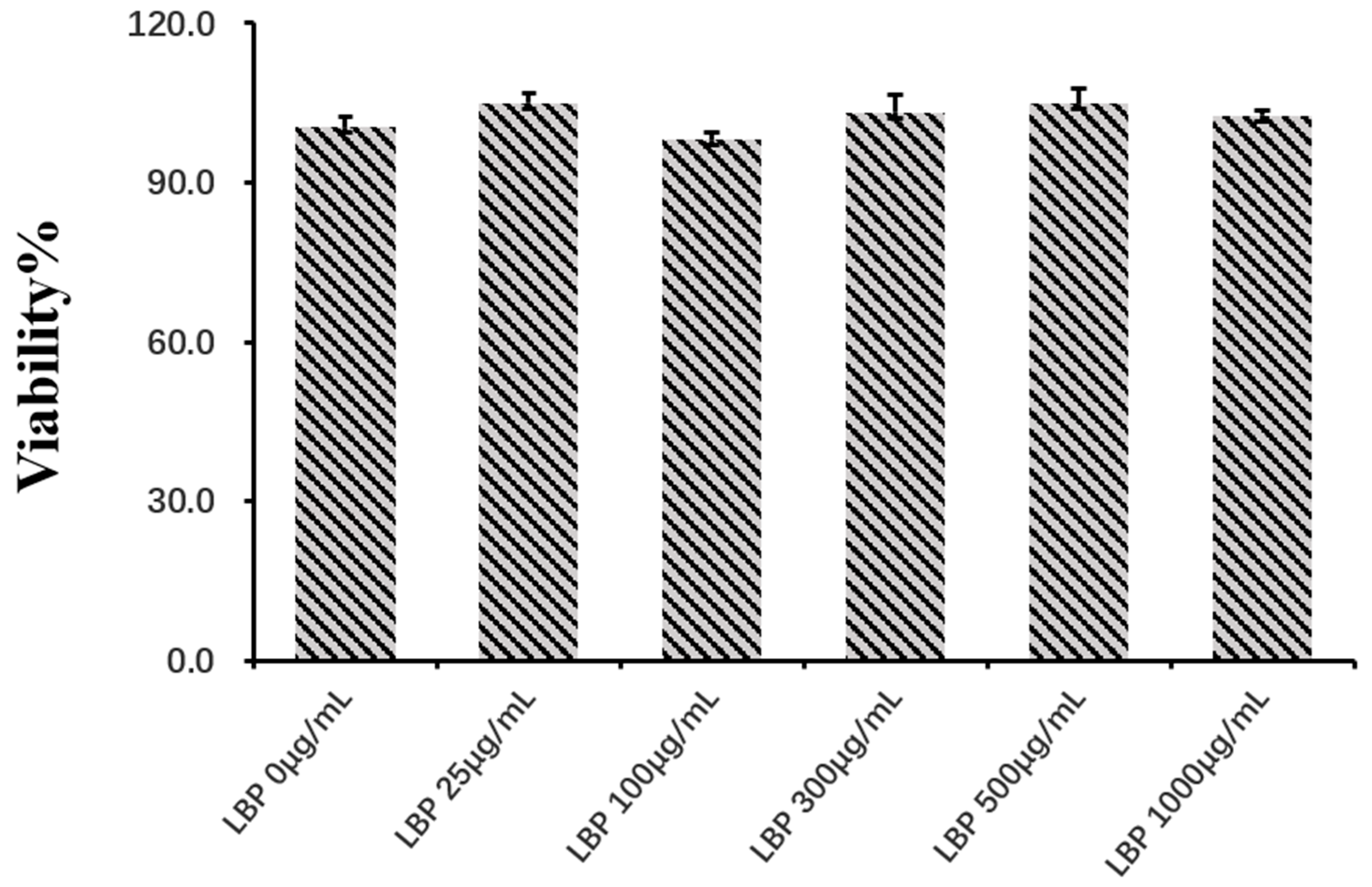
3.1. The Effect of Cell Viability of LBP on pbMEC

pbMEC were treated with different concentrations of LBP at 0, 25, 100, 300, 500, and 1000 μg/mL for 24 h. The effect of different doses of LBP on pbMEC viability was measured by CCK-8. Compared with the control group (0 μg/mL), cell viability was not affected by pretreatment with LBP from 25 to 1000 μg/mL. As the concentration of LBP increased, it did not cause cell apoptosis (p > 0.05, Figure 1).

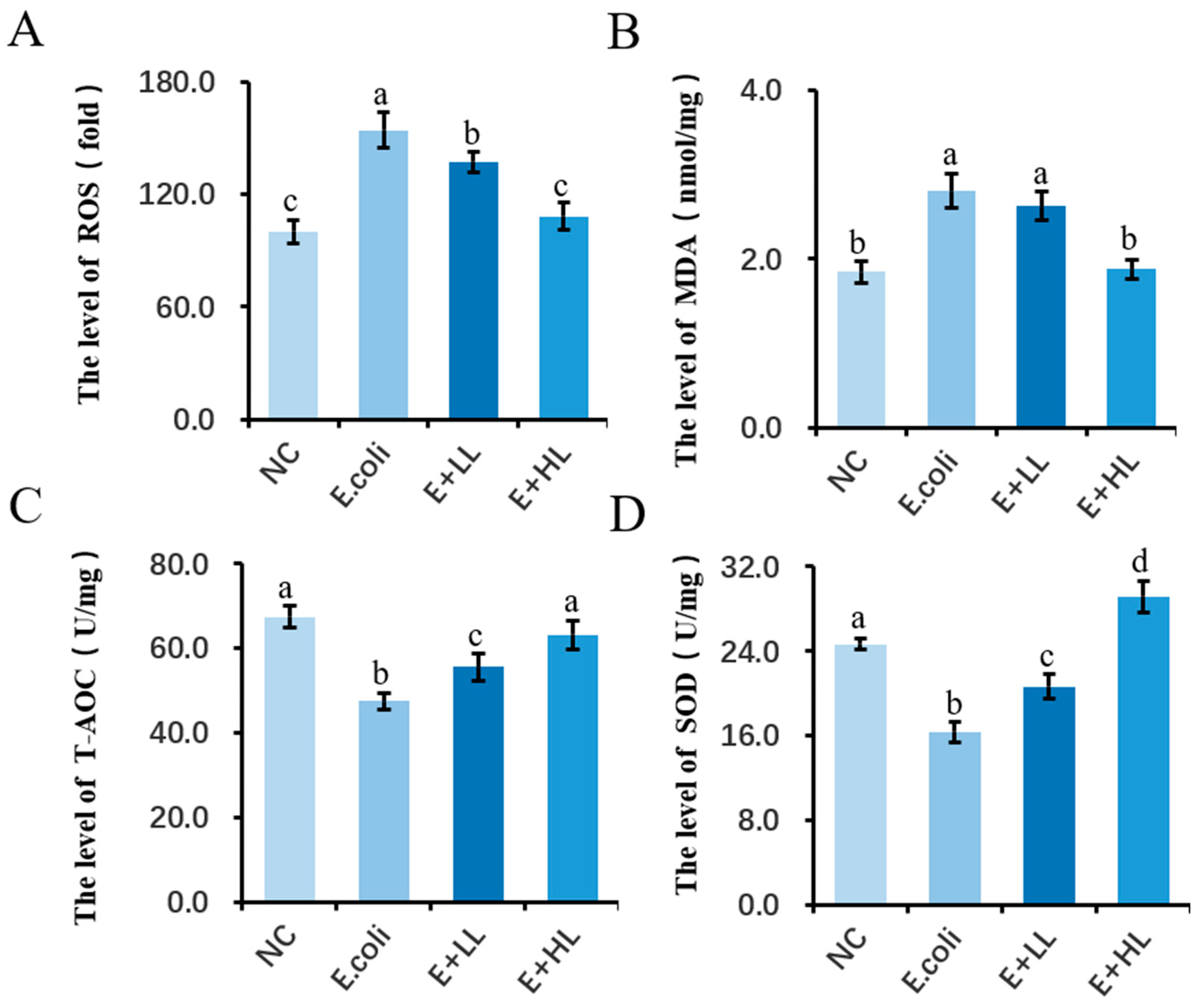
3.2. The Effect of LBP on the Antioxidant Capacity of pbMEC

The results showed that E. coli treatment upregulated the content of ROS and MDA, whereas LBP reversed the upregulation of ROS and MDA in a dose-dependent manner. Moreover, LBP pretreatment at 100 μg/mL did not reduce the level of MDA as compared with the E. coli group. We found E. coli treatment decreased the activity of SOD and T-AOC. As a result, the decreased T-AOC level was reversed in a similar manner to that in cells pretreated with LBP, and the effect of LBP treatment is dose dependent (p = 0.0002, p = 0.005, p = 0.128; Figure 2C). Compared with the E. coli group, 100 μg/mL of LBP altered the SOD level, whereas 300 μg/mL of LBP significantly increased the SOD activity (p = 0.007, p = 0.0001; Figure 2D).

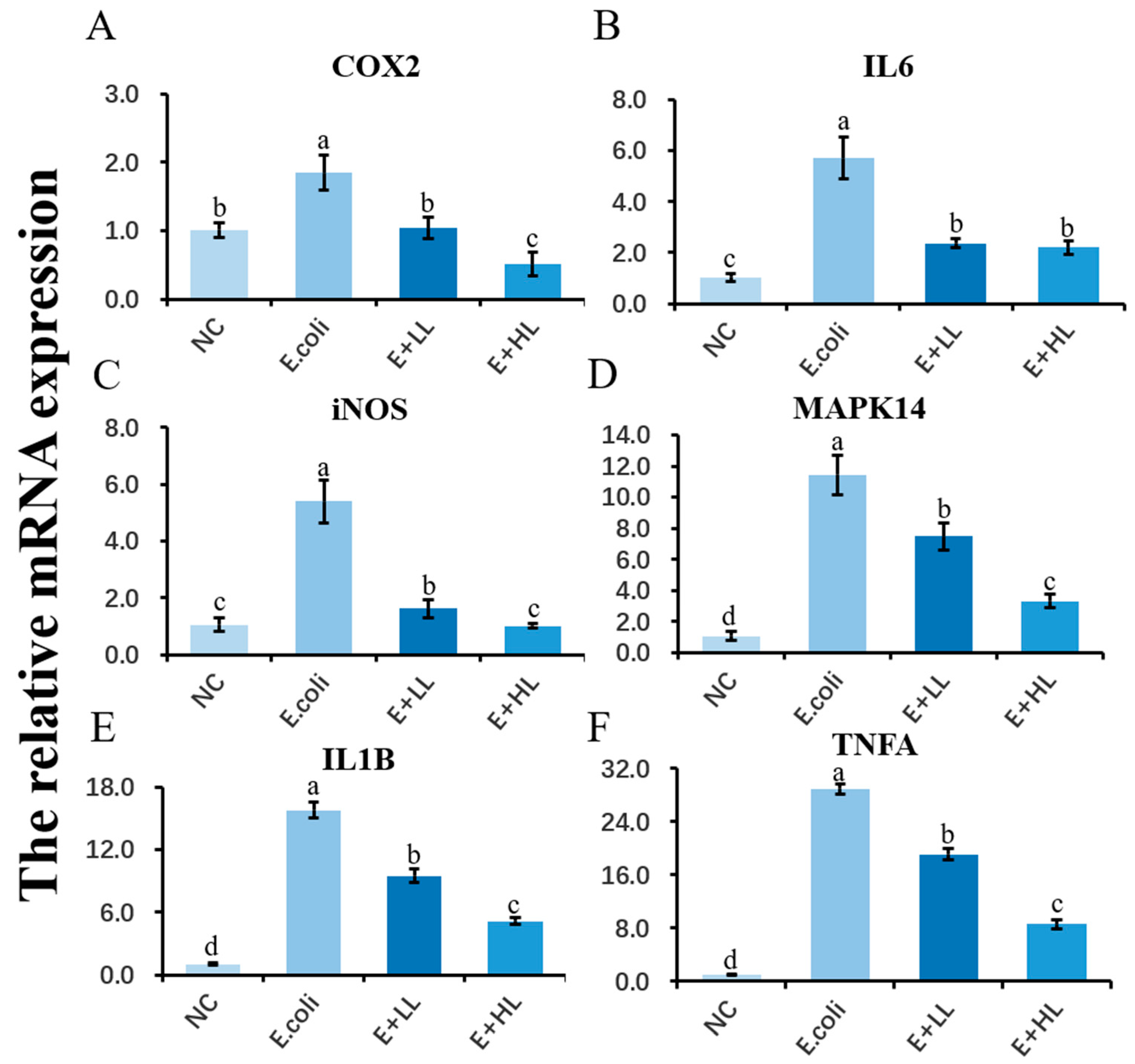
3.3. Effect of LBP on the Expression of Genes Related to Inflammatory Responses in E. coli Stimulated pbMEC

In this study, LBP pretreatment attenuated E. coli-induced proinflammatory cytokine gene expression in pbMEC. The LBP-induced alteration of COX2, IL1B, IL6, TNFA, and iNOS gene expression in pbMEC stimulated by E. coli was examined by RT-PCR. Compared with the control group, E. coli treatment upregulated the mRNA abundance of COX2, IL1B, IL6, TNFA, and iNOS. pbMEC were treated with different doses of LBP at 100 μg/mL or 300 μg/mL, and markedly downregulated the proinflammatory genes that were induced by E. coli challenge. Moreover, the inhibitory effect of LBP on inflammatory gene expression was more effective for cells pretreated with the increased doses of LBP, as a result of a lower expression of proinflammatory genes in E + HH than those in the E + LL group. Similarly, E. coli treatment upregulated the mRNA abundance of MAPK14, whereas pretreatment with different doses of LBP at 100 μg/mL or 300 μg/mL, markedly attenuated the elevation of MAPK14 induced by E. coli (p = 0.007, p = 0.002, p = 0.018; Figure 3D).

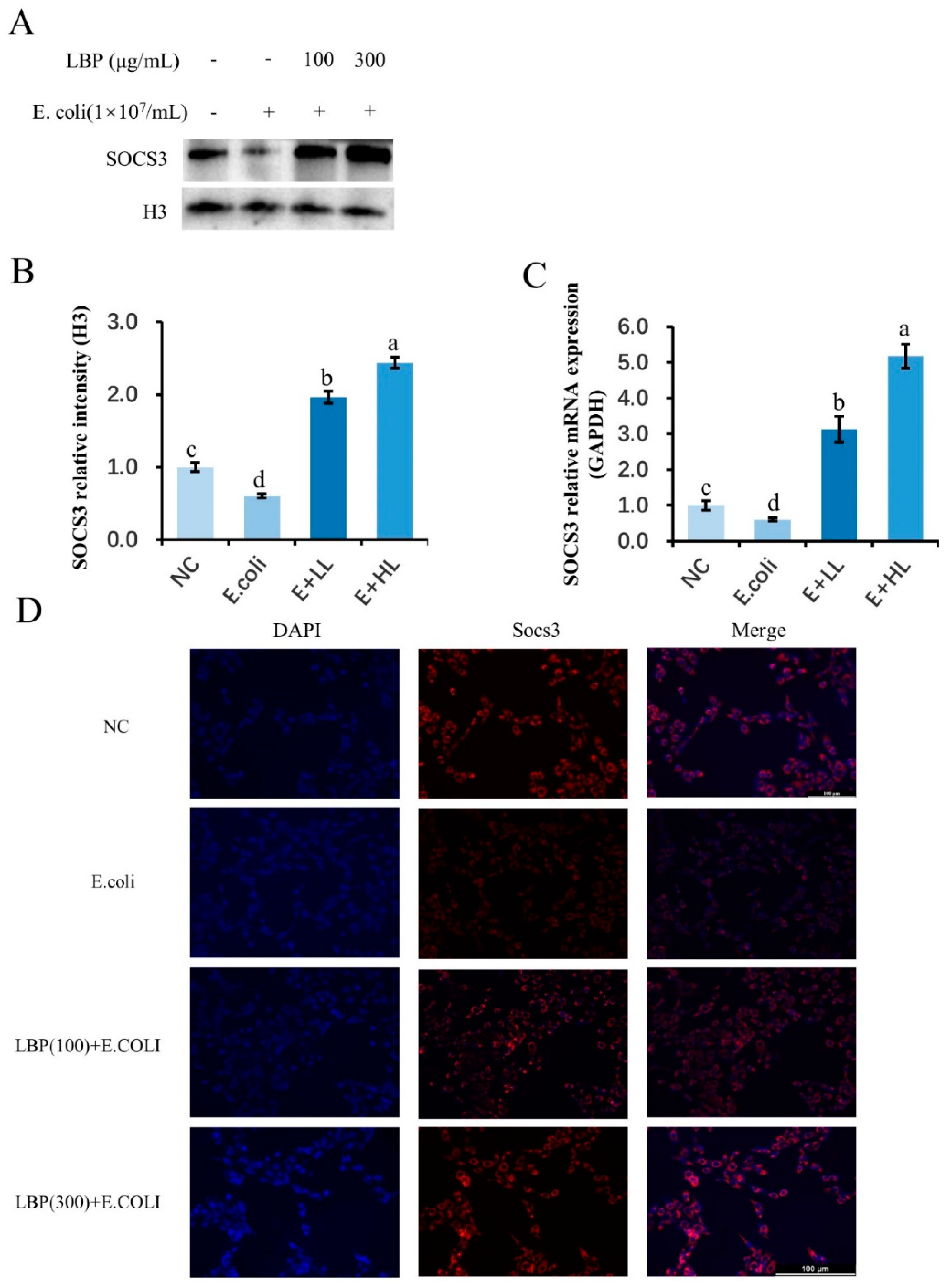
3.4. Pretreatment with LBP Activated SOCS3 in pbMEC Challenged with E. coli

E. coli significantly decreased the protein expression of SOCS3, whereas pretreatment with LBP in the E + LL and E + HH groups restored the inhibited expression of SOCS3 (p < 0.05; Figure 4A,B). Moreover, LBP pretreatment at 100 and 300 μg/mL raised the abundance of SOCS3 to approximately twofold that of the control group. Consistently, gene expression of SOCS3 was also affected by pretreatment of LBP, as a result of the reversed expression of SOCS3 that was downregulated by E. coli stimulation (p = 0.011; Figure 4C). Additionally, results in the immunofluorescence assay agreed with the protein expression generated by Western blot. The weakened staining of SOCS3 in the E. coli group was restored by the pretreatment of LBP in the E + LL and E + HH groups (Figure 4D).

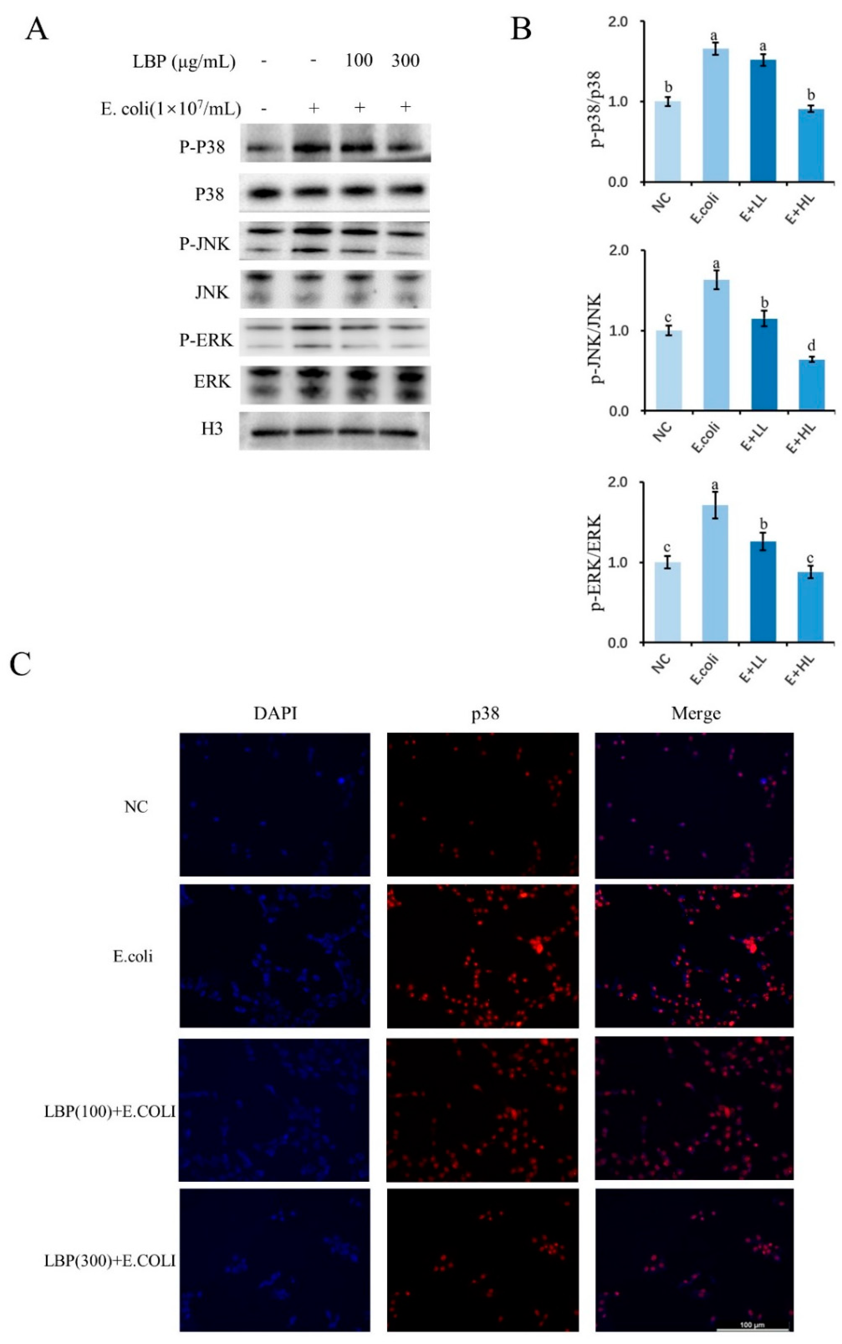
3.5. Effect of LBP on the MAPK Pathway-Related Protein Expression of pbMEC under Different Treatments

MAPK is a classic signaling pathway in charge of inflammatory responses. We investigated the components of the MAPK signaling by Western blot and immunofluorescence. When compared with the control group, E. coli treatment elevated the proportion of p-p38, p-JNK, and p-ERK to total p38, JNK, and ERK protein (p = 0.005, p = 0.002, p = 0.012, respectively; Figure 5B). Compared with the E. coli-treated group, 100 μg/mL LBP treatment did not reduce the ratio of p-p38 to p38 (p = 0.062). Instead, 100 μg/mL LBP treatment significantly reduced the ratio of p-JNK to JNK and p-ERK to ERK (p = 0.032, p = 0.046, respectively; Figure 5B). In contrast, as the pretreatment of LBP with a concentration of 300 μg/mL, the ratio of p-p38 to p38, p-JNK to JNK, and p-ERK to ERK decreased significantly in cells treated with E. coli (p < 0.05; Figure 5A,B). Consistently, immunofluorescence staining results revealed that cells exposed to E. coli strengthened the staining of p38, whereas LBP pretreatment attenuated the staining of p38 in nuclei. Notably, the expression of phosphorylated p38 was decreased in a dose-dependent manner of LBP added to the cells, indicating that the effect of LBP at a concentration of 300 μg/mL is better than that of 100 μg/mL (Figure 5C).

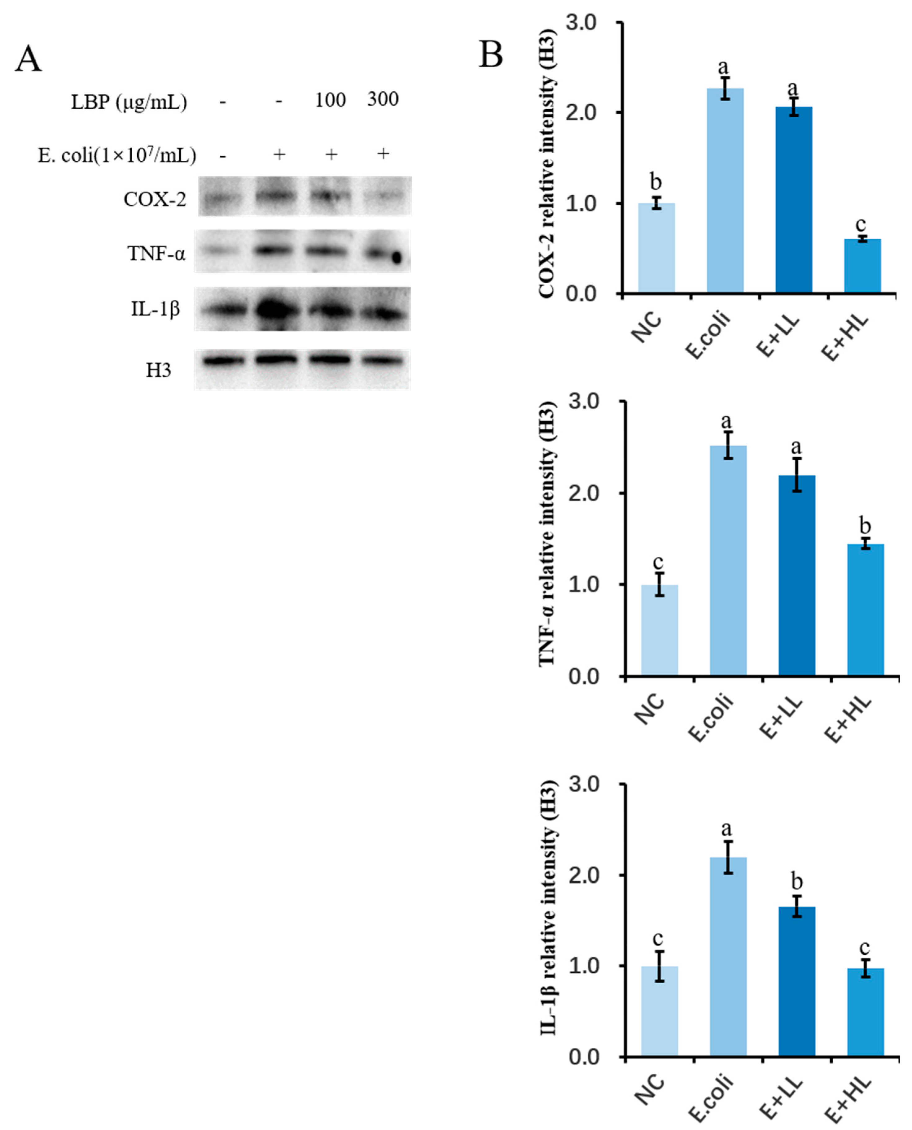
3.6. LBP Pretreatment Inhibits E. coli-Induced Activation of Inflammatory Genes

We investigated the effect of pretreated LBP on pro-inflammatory factors by using Western blot. Compared with the control group, the E. coli challenge increased the protein abundance of COX-2, IL-1β, and TNF-α, and 100 μg/mL, and LBP pretreatment did not reduce the protein expression of COX-2 or IL-1β (p = 0.287, p = 0.094, respectively; Figure 6A,B); however, 300 μg/mL of LBP supplemented to cells significantly inhibited the expression of COX-2 and IL-1β. Furthermore, the abundance of TNF-α was partially inhibited by LBP pretreatment in a dose-dependent manner followed by E. coli stimulation (p < 0.05; Figure 6A,B).

4. Discussion

Bovine mastitis is a common infectious disease caused mainly by microbial infections. E. coli is one of the most common pathogens, mainly causing subclinical mastitis [25]. Several studies agree with the upregulation of TNFA, IL6, and IL1B mRNA abundance in the mammary gland during coliform mastitis, when cows are in the transition period [26,27]. It is well known that antibiotics are currently the main treatment for mastitis, as traditional antibiotics develop resistance during treatment [6]. Hence, research on developing novel and effective antibacterial drugs might be an alternative strategy for the treatment of mastitis. LBP has been widely used for defending oxidative stress and alleviating inflammation [23,28]. In this study, we investigated the anti-inflammatory effects and mechanism of LBP on E. coli-stimulated pbMEC inflammatory responses via activation of SOCS3 and inactivation of MAPK pathway.

4.1. LBP Attenuates the E. coli-Induced Inflammatory and Oxidative Responses in pbMEC

Since LBP did not reveal a cytotoxic effect on pbMEC at doses ranging from 0 to 1000 μg/mL, we selected the concentration of LBP at either 100 or 300 μg/mL in the formal experiment according to reported studies [29]. LBP has been shown to reduce the expression of TNFA, IL6, and IL1B in E. coli-induced mouse macrophages [30]. In a mouse model of steatohepatitis, LBP treatment affects the secretion of NF-κB-mediated inflammatory cytokines and inflammasome, such as NLRP3/6 [31]. LPS comprises endotoxins of Gram-negative bacteria that envelop E. coli; thus, it is widely used to establish a cellular inflammation model for microbial infections in vitro [32]. In this study, treatment with LPS increases the gene expression of TNFA, IL6, and IL1B in pbMEC, whereas LBP pretreatment reversed the elevated pro-inflammatory gene expression. Additionally, LPS is a typical potent stimulus of the immune response, which leads to an increase in the expression of iNOS and COX2 in macrophages [33,34]. Notably, in the current study, LBP pretreatment lowered the upregulation of iNOS and COX2 gene expression in pbMEC induced by E. coli. This result indicates that LBP suppresses the E. coli-induced expression of proinflammatory gene expression and supports the protection of inflammation cascades.
ROS is a byproduct of the physiological metabolism of mitochondria which can participate in the transduction of signaling pathways, tissue damage, and related physiological processes [35]. When ROS are produced in large quantities, it may cause oxidative stress in cells and lead to autophagy or apoptosis [36]. LBP is a well-known powerful antioxidant; the study has found that increasing doses of LBP can improve the SOD activity of mouse blood serum, liver, and brain tissues, while reducing the content of MDA [37]. It has also recently been demonstrated that LBP can reduce oxidative stress in HepG2 cells [38]; therefore, we determined whether the antioxidant capacity of LBP is related to the elimination of ROS in the cellular environment by detecting the activity of antioxidant enzymes. SOD is a specific enzyme that can scavenge free radicals in the biological antioxidant system [39]. The current study showed that cells treated with E. coli produced a large amount of ROS, whereas LBP pretreatment significantly attenuated MDA content and restored oxidative capacity by increasing SOD activity and T-AOC in pbMEC. The results are indicative of the role of LBP in reducing the oxidative responses induced by E. coli and stabilizing the homeostasis of cells with regard to the defense of oxidative damage. Although LBP affects the immune and oxidative response in vitro, the limitations of the current study regarding the reflection of the real situation in the infected udder using LBP, could be a potential therapy until more robust tests are made.

4.2. LBP Inhibits Inflammatory and Oxidative Responses in pbMEC through SOCS3 and MAPK Molecules

SOCS3 is a protein that negatively regulates the cytokine signal transduction pathway [40]. Recent studies have shown that SOCS3 can be modulated by a variety of inflammatory stimuli, and that it blocks the signal interactions of immune molecules. For example, the MAPK signaling pathway plays a key role in the bacteria-induced gene expression of SOCS3 in primary human macrophages [41]. Interestingly, the study found that SOCS3 interference blocked the upregulation of TNFA and IL1B in the LPS-induced 3T3-L1 adipocyte inflammation model [42]; however, the role of SOCS3 in the regulation of inflammatory responses in bovine mastitis is still unclear. In this study, we found that pretreatment with LBP reversed the decrease in SOCS3 mRNA and protein expression induced by E. coli challenge; hence, this result indicates the involvement of SOCS3 in E. coli-stimulated pbMEC and its potential role in the modulation of inflammation by LBP.
It is well known that the NF-κB and MAPK signaling pathways play a crucial role in the innate immune response [43]. We have reported in a previous study that LBP inhibits LPS-induced NF-κB activation in pbMEC; however, the MAPK signaling pathway may act as an indispensable regulator to further study the potential mechanism of LBP that inhibits E. coli-induced inflammation in pbMEC. In mammalian cells, three MAPK families have been clearly characterized: extracellular signal-regulated kinase (ERK), C-Jun N-terminal kinase (JNK), and p38 kinase [44,45]. The current study shows that LBP reverses the activation of inflammatory factors through its suppression of MAPK signaling pathways. The activation of p38, ERK, and JNK was also restrained in a dose-dependent fashion by LBP. The immunofluorescence results emphasized that the activation of the p38 protein by E. coli stimulation is inhibited by pretreatment with LBP. Several studies have confirmed that the activation of the MAPK signaling pathway results in the phosphorylation of transcription regulators, which indirectly or directly modulates the secretion of cytokines such as TNF-α and IL-1β [46,47]. The fact that pretreatment of LBP suppressed the abundance of COX-2, IL-1β, and TNF-α protein in pbMEC stimulated by E. coli, is in line with the inhibitory effects of LBP on MAPK signaling.
Overall, our results indicated that LBP suppressed the expression of proinflammatory cytokines by preventing MAPK activation in E. coli-stimulated pbMEC, showing that the anti-inflammatory mechanism of LBP might be associated with SOCS3-mediated suppressing in MAPK signaling pathways. The results highlight the potential role of LBP as a therapeutic extract in combating clinical bovine mastitis.

Supplementary Materials

The following supporting information can be downloaded at: https://www.mdpi.com/article/10.3390/agriculture12050598/s1, Table S1: Primers used for real-time qPCR [11,48].

Author Contributions

R.L., T.X. and H.Z. analyzed and processed the data. R.L., X.L., J.Z. and X.W. performed the experiment, and T.X. and Z.Y. were major contributors to the drafting of the manuscript. Z.Y. and T.X. provided financing throughout the experiment and generated the outlines of the research. All authors have read and agreed to the published version of the manuscript.

Funding

This study was funded by the National Natural Science Foundation of China (32,102,731; 31,872,324).

Institutional Review Board Statement

The animal study protocol was approved by the Ethics Committee of Yangzhou University (YZU-2021003-168) on 10 July 2021.

Informed Consent Statement

Not applicable.

Data Availability Statement

The data presented in this study are available on request from the corresponding author.

Acknowledgments

The authors would like to thank all those who helped us during this project for their valuable advice and support.

Conflicts of Interest

The authors declare no conflict of interest.

References

  1. Merriman, K.E.; Powell, J.L.; Santos, J.E.P.; Nelson, C.D. Intramammary 25-hydroxyvitamin D 3 treatment modulates innate immune responses to endotoxin-induced mastitis. J. Dairy Sci. 2018, 101, 7593–7607. [Google Scholar] [CrossRef] [PubMed]
  2. Tomazi, T.; Coura, F.M.; Goncalves, J.L.; Heinemann, M.B.; Santos, M.V. Antimicrobial susceptibility patterns of Escherichia coli phylogenetic groups isolated from bovine clinical mastitis. J. Dairy Sci. 2018, 101, 9406–9418. [Google Scholar] [CrossRef] [PubMed]
  3. Guo, M.; Gao, Y.; Xue, Y.; Liu, Y.; Zeng, X.; Cheng, Y.; Ma, J.; Wang, H.; Sun, J.; Wang, Z.; et al. Bacteriophage Cocktails Protect Dairy Cows Against Mastitis Caused by Drug Resistant Escherichia coli Infection. Front. Cell. Infect. Microbiol. 2021, 11, 690377. [Google Scholar] [CrossRef] [PubMed]
  4. Guerra, S.T.; Paula, C.; Bolaos, C.; Hernandes, R.T.; Ribeiro, M.G. Virulence factors of Escherichia coli: An overview of animal and human infections with emphasis in bovine mastitis. Semin. Cienc. Agrar. 2019, 40, 2087. [Google Scholar] [CrossRef]
  5. Blum, S.E.; Dan, E.H.; Jacoby, S.; Krifuks, O.; Leitner, G. Physiological response of mammary glands to Escherichia coli infection: A conflict between glucose need for milk production and immune response. Sci. Rep. 2020, 10, 9602. [Google Scholar] [CrossRef]
  6. Seal, B.S.; Lillehoj, H.S.; Donovan, D.M.; Gay, C.G. Alternatives to antibiotics: A symposium on the challenges and solutions for animal production. Anim. Health Res. Rev. 2013, 14, 78–87. [Google Scholar] [CrossRef] [Green Version]
  7. Mingmongkolchai, S.; Panbangred, W. Bacillus probiotics: An alternative to antibiotics for livestock production. J. Appl. Microbiol. 2018, 124, 1334–1346. [Google Scholar] [CrossRef]
  8. Jedrzejczak, M.; Szatkowska, I. Bovine mammary epithelial cell cultures for the study of mammary gland functions. In Vitro Cell. Dev. Biol.-Anim. 2014, 50, 389–398. [Google Scholar] [CrossRef] [Green Version]
  9. Kehrli, M.E.; Shuster, D.E. Factors Affecting Milk Somatic Cells and Their Role in Health of the Bovine Mammary Gland. J. Dairy Sci. 1994, 77, 619–627. [Google Scholar] [CrossRef]
  10. Gomes, F.; Henriques, M. Control of Bovine Mastitis: Old and Recent Therapeutic Approaches. Curr. Microbiol. 2016, 72, 377–382. [Google Scholar] [CrossRef] [Green Version]
  11. Wang, J.; Guo, C.; Wei, Z.; He, X.; Kou, J.; Zhou, E.; Yang, Z.; Fu, Y. Morin suppresses inflammatory cytokine expression by downregulation of nuclear factor-κB and mitogen-activated protein kinase (MAPK) signaling pathways in lipopolysaccharide-stimulated primary bovine mammary epithelial cells. J. Dairy Sci. 2016, 99, 3016–3022. [Google Scholar] [CrossRef] [PubMed] [Green Version]
  12. Ren, X.; Han, L.; Li, Y.; Zhao, H.; Zhang, Z.; Zhuang, Y.; Zhong, M.; Wang, Q.; Ma, W.; Wang, Y. Isorhamnetin attenuates TNF-α-induced inflammation, proliferation, and migration in human bronchial epithelial cells via MAPK and NF-κB pathways. Anat Rec (Hoboken). 2021, 304, 901–913. [Google Scholar] [CrossRef]
  13. Yang, Z.; Yin, R.; Cong, Y.; Yang, Z.; Zhou, E.; Wei, Z.; Liu, Z.; Cao, Y.; Zhang, N. Oxymatrine Lightened the Inflammatory Response of LPS-Induced Mastitis in Mice Through Affecting NF-κB and MAPKs Signaling Pathways. Inflammation 2014, 37, 2047–2055. [Google Scholar] [CrossRef] [PubMed]
  14. Shu, F.; Junfu, W.; Xiaoqun, X.; Xuemei, C.; Junwen, L.; Qinghong, S.; Meng, L.; Huali, W.; Changsheng, Z. The Expression of SOCS and NF-ϰB p65 in Hypopharyngeal Carcinoma. Iran. J. Public Health 2018, 47, 1874–1882. [Google Scholar]
  15. Elliott, J.; Johnston, J.A. SOCS: Role in inflammation, allergy and homeostasis. Trends Immunol. 2004, 25, 434–440. [Google Scholar] [CrossRef]
  16. Souza, J.A.C.d.; Nogueira, A.V.B.; Souza, P.P.C.d.; Kim, Y.J.; Lobo, C.S.; Oliveira, G.J.P.L.d.; Cirelli, J.A.; Garlet, G.P.; Rossa, C.; López-Collazo, E. SOCS3 Expression Correlates with Severity of Inflammation, Expression of Proinflammatory Cytokines, and Activation of STAT3 and p38 MAPK in LPS-Induced Inflammation In Vivo. Mediat. Inflamm. 2013, 2013, 650812. [Google Scholar]
  17. Delgado-Ortega, M.; Melo, S.; Meurens, F. Expression of SOCS1-7 and CIS mRNA in porcine tissues. Vet. Immunol. Immunopathol. 2011, 144, 493–498. [Google Scholar] [CrossRef]
  18. Hu, J.; Wang, W.; Hao, Q.; Zhang, T.; Yin, H.; Wang, M.; Zhang, C.; Zhang, C.; Zhang, L.; Zhang, X.; et al. Suppressors of cytokine signalling (SOCS)-1 inhibits neuroinflammation by regulating ROS and TLR4 in BV2 cells. Inflamm. Res. 2020, 69, 27–39. [Google Scholar] [CrossRef]
  19. Mingzhao, D.; Xinyu, H.; Ling, K.; Baohai, Z.; Chaopu, Z. Lycium barbarum Polysaccharide Mediated the Antidiabetic and Antinephritic Effects in Diet-Streptozotocin-Induced Diabetic Sprague Dawley Rats via Regulation of NF-κB. BioMed Res. Int. 2016, 2016, 3140290. [Google Scholar]
  20. Xiao-rui, Z.; Chun-hui, Q.; Jun-ping, C.; Gang, L.; Lin-juan, H.; Zhong-fu, W.; Wen-xia, Z.; Yong-xiang, Z. Lycium barbarum polysaccharide LBPF4-OL may be a new Toll-like receptor 4/MD2-MAPK signaling pathway activator and inducer. Int. Immunopharmacol. 2014, 19, 132–141. [Google Scholar]
  21. Péter, F.; Ildikó, S.; Hajnalka, L.; Mariann, H.; Sándor, S.; György, P. Association of chemerin with oxidative stress, inflammation and classical adipokines in non-diabetic obese patients. J. Cell. Mol. Med. 2014, 18, 1313–1320. [Google Scholar]
  22. Qi, B.; Ji, Q.; Wen, Y.; Lian, L.; Guo, X.; Hou, G.; Wang, G.; Zhong, J.; Ferenc, G. Lycium barbarum Polysaccharides Protect Human Lens Epithelial Cells against Oxidative Stress–Induced Apoptosis and Senescence. PLoS ONE 2014, 9, e110275. [Google Scholar] [CrossRef] [PubMed]
  23. Li, X.M.; Ma, Y.L.; Liu, X.J. Effect of the Lycium barbarum polysaccharides on age-related oxidative stress in aged mice. J. Ethnopharmacol. 2007, 111, 504–511. [Google Scholar] [CrossRef] [PubMed]
  24. Sun, Y. Biological activities and potential health benefits of polysaccharides from Poria cocos and their derivatives. Int. J. Biol. Macromol. 2014, 68, 131–134. [Google Scholar] [CrossRef]
  25. Sargeant, J.M.; Gillespie, J.R.; Oberst, R.D.; Phebus, R.K.; Hyatt, D.R.; Bohra, L.K.; Galland, J.C. Results of a longitudinal study of the prevalence of Escherichia coli O157:H7 on cow-calf farms. Am. J. Vet. Res. 2000, 61, 1375–1379. [Google Scholar] [CrossRef] [Green Version]
  26. Shuster, D.E.; Kehrli, M.E.; Stevens, M.G. Cytokine production during endotoxin-induced mastitis in lactating dairy cows. Am. J. Vet. Res. 1993, 54, 80–85. [Google Scholar]
  27. Larson, M.A.; Weber, A.; Mcdonald, T.L. Bovine serum amyloid A3 gene structure and promoter analysis: Induced transcriptional expression by bacterial components and the hormone prolactin. Gene 2006, 380, 104–110. [Google Scholar] [CrossRef]
  28. Xiao, J.; Xing, F.; Huo, J.; Fung, M.L.; Liong, E.C.; Ching, Y.P.; Xu, A.; Chang, R.; So, K.F.; Tipoe, G.L. Lycium barbarum polysaccharides therapeutically improve hepatic functions in non-alcoholic steatohepatitis rats and cellular steatosis model. Sci. Rep. 2014, 4, 5587. [Google Scholar] [CrossRef] [Green Version]
  29. Cuadrado, A.; Nebreda, A. Mechanisms and functions of p38 MAPK signalling. Biochem. J. 2010, 429, 403–417. [Google Scholar] [CrossRef] [Green Version]
  30. Peng, Q.; Liu, H.; Shi, S.; Li, M. Lycium ruthenicum polysaccharide attenuates inflammation through inhibiting TLR4/NF-κB signaling pathway. Int. J. Biol. Macromol. 2014, 67, 330–335. [Google Scholar] [CrossRef]
  31. Xiao, J.; Wang, F.; Liong, E.C.; So, K.F.; Tipoe, G.L. Lycium barbarum polysaccharides improve hepatic injury through NFkappa-B and NLRP3/6 pathways in a methionine choline deficient diet steatohepatitis mouse model. Int. J. Biol. Macromol. 2018, 120, 1480–1489. [Google Scholar] [CrossRef] [PubMed]
  32. Granucci, F. The Family of LPS Signal Transducers Increases: The Arrival of Chanzymes. Immunity 2018, 48, 4–6. [Google Scholar] [CrossRef] [PubMed] [Green Version]
  33. Dang, Y.; Xu, Y.; Wu, W.; Li, W.; Sun, Y.; Yang, J.; Zhu, Y.; Zhang, C. Tetrandrine Suppresses Lipopolysaccharide-Induced Microglial Activation by Inhibiting NF-κB and ERK Signaling Pathways in BV2 Cells. PLoS ONE 2014, 9, e102522. [Google Scholar] [CrossRef] [PubMed] [Green Version]
  34. Qian, G.; Li, Y.; He, M.; Guo, W.; Kan, X.; Xu, D.; Liu, J.; Fu, S. Peiminine Protects against Lipopolysaccharide-Induced Mastitis by Inhibiting the AKT/NF-κB, ERK1/2 and p38 Signaling Pathways. Int. J. Mol. Sci. 2018, 19, 2637. [Google Scholar]
  35. Wang, Y.; Branicky, R.; Noë, A.; Hekimi, S. Superoxide dismutases: Dual roles in controlling ROS damage and regulating ROS signaling. J. Cell Biol. 2018, 217, 1915–1928. [Google Scholar] [CrossRef]
  36. Willson, J.A.; Arienti, S.; Sadiku, P.; Reyes, L.; Coelho, P.; Morrison, T.; Rinaldi, G.; Dockrell, D.H.; Whyte, M.K.B.; Walmsley, S.R. Neutrophil HIF-1α stabilization is augmented by mitochondrial ROS produced via the glycerol 3-phosphate shuttle. Blood 2022, 139, 281–286. [Google Scholar] [CrossRef]
  37. Tian, X.; Liang, T.; Liu, Y.; Ding, G.; Zhang, F.; Ma, Z. Extraction, Structural Characterization, and Biological Functions of Lycium Barbarum Polysaccharides: A Review. Biomolecules 2019, 9, 389. [Google Scholar] [CrossRef] [Green Version]
  38. Tuo, X.U.; Ling, H.Y.; Long, J.; Jian-Qin, H.E.; Yang, S.S.; Zhu, Z.M.; Yan, C.Q.; Feng, S.D.; Physiology, D.O. Ameliorative effects and the mechanism of lycium barbarum polysaccharide on insulin resistance of HepG2 cell. Chin. J. Appl. Physiol. 2017, 33, 568–571. [Google Scholar]
  39. Kwon, K.; Jung, J.; Sahu, A.; Tae, G. Nanoreactor for cascade reaction between SOD and CAT and its tissue regeneration effect. J. Control Release 2022, 344, 160–172. [Google Scholar] [CrossRef]
  40. Tamiya, T.; Kashiwagi, I.; Takahashi, R.; Yasukawa, H.; Yoshimura, A. Suppressors of cytokine signaling (SOCS) proteins and JAK/STAT pathways: Regulation of T-cell inflammation by SOCS1 and SOCS3. Arter. Thromb Vasc Biol 2011, 31, 980–985. [Google Scholar] [CrossRef] [Green Version]
  41. Latvala, S.; Miettinen, M.; Kekkonen, R.A.; Korpela, R.; Julkunen, I. Lactobacillus rhamnosus GG and Streptococcus thermophilus induce suppressor of cytokine signalling 3 (SOCS3) gene expression directly and indirectly via interleukin-10 in human primary macrophages. Clin. Exp. Immunol. 2011, 165, 94–103. [Google Scholar] [CrossRef]
  42. Liu, Z.; Gan, L.; Zhou, Z.; Jin, W.; Sun, C. SOCS3 promotes inflammation and apoptosis via inhibiting JAK2/STAT3 signaling pathway in 3T3-L1 adipocyte. Immunobiology 2015, 220, 947–953. [Google Scholar] [CrossRef] [PubMed]
  43. Liu, L.; Ye, L.; Liu, L.; Bian, Y.; Li, Z.; Gao, X.; Li, Q. 14-3-3γ Regulates Lipopolysaccharide-Induced Inflammatory Responses and Lactation in Dairy Cow Mammary Epithelial Cells by Inhibiting NF-κB and MAPKs and Up-Regulating mTOR Signaling. Int. J. Mol. Sci. 2015, 16, 16622–16641. [Google Scholar] [CrossRef] [PubMed] [Green Version]
  44. Cargnello, M.; Roux, P.P. Activation and Function of the MAPKs and Their Substrates, the MAPK-Activated Protein Kinases. Microbiol. Mol. Biol. Rev. 2011, 75, 50–83. [Google Scholar] [CrossRef] [PubMed] [Green Version]
  45. Junttila, M.R.; Li, S.P.; Westermarck, J. Phosphatase-mediated crosstalk between MAPK signaling pathways in the regulation of cell survival. FASEB J. 2008, 22, 954–965. [Google Scholar] [CrossRef] [PubMed] [Green Version]
  46. Zahra, A.; Vaiva, K.; Volodymyr, S. Dual control of MAPK activities by AP2C1 and MKP1 MAPK phosphatases regulates defence responses in Arabidopsis. J. Exp. Bot. 2022, 73, 2369–2384. [Google Scholar]
  47. Liu, K.; Liu, E.; Lin, L.; Hu, Y.; Yuan, Y.; Xiao, W. L-Theanine mediates the p38MAPK signaling pathway to alleviate heat-induced oxidative stress and inflammation in mice. Food Funct. 2022, 13, 2120–2130. [Google Scholar] [CrossRef]
  48. Liu, M.; Song, S.; Li, H.; Jiang, X.; Yin, P.; Wan, C.; Liu, X.; Liu, F.; Xu, J. The protective effect of caffeic acid against inflammation injury of primary bovine mammary epithelial cells induced by lipopolysaccharide. J. Dairy Sci. 2014, 97, 2856–2865. [Google Scholar] [CrossRef] [Green Version]
Figure 1. Determination of cell viability in different doses of LBP via CCK-8 assay. Effects of LBP on pbMEC viability. pbMEC were treated with different concentrations of LBP at 0, 25, 100, 300, 500, and 1000 μg/mL for 24 h. Determination of cell viability in different doses of LBP via Cell Counting Kit-8 assay. Results are presented as means ± SEM. Superscripts represent that the difference between groups was significant according to statistical analysis (p < 0.05).
Agriculture 12 00598 g001
Figure 2. The effect of LBP on the antioxidant capacity of pbMEC. Different concentrations of LBP at 100 μg/mL or 300 μg/mL were added to pbMEC, followed by a challenge with 1 × 10⁷/mL E. coli for an additional 6 h. (A) Determination of ROS content in pbMEC. (B) Detection of MDA content in pbMEC. (C) Detection of T-AOC content in pbMEC. (D) Detection of SOD content in pbMEC. Results are presented as means ± SEM. Letters in superscripts represent that the difference between groups was significant according to statistical analysis (p < 0.05).
Agriculture 12 00598 g002
Figure 3. The effect of LBP on mRNA expression related to inflammatory responses. The inflammation injury of pbMEC challenged by E. coli. pbMEC were treated with different doses of LBP at 100 μg/mL or 300 μg/mL), followed by a challenge with 1 × 10⁷/mL E. coli for an additional 6 h. (A) mRNA quantitative expression of COX2. (B) mRNA quantitative expression of IL6. (C) mRNA quantitative expression of iNOS. (D) mRNA quantitative expression of MAPK14. (E) mRNA quantitative expression of IL1B. (F) mRNA quantitative expression of TNFA. The biological replicates for PCR running were triplicate, along with three unique experiments. Results are presented as means ± SEM. Superscripts represent that the difference between groups was significant according to statistical analysis (p < 0.05).
Agriculture 12 00598 g003
Figure 4. Effects of LBP on SOCS3 protein expression. pbMEC were treated with different doses of LBP at 100 μg/mL or 300 μg/mL followed by a challenge with 1 × 10⁷/mL E. coli for an additional 6 h. (A) Western blot analyses of SOCS3. (B) Relative protein abundance of SOCS3 to Histone H3. (C) Relative mRNA expression level of SOCS3. (D) Fluorescence for detecting SOCS3 abundance. Antibodies against SOCS3 (Cy3 labeled secondary antibody, red) and counterstained with DAPI (blue) were conducted for target protein and nuclear staining respectively. The biological replicates for PCR running were triplicate, along with triple unique experiments. Results are presented as means ± SEM. Superscripts represent that the difference between groups was significant according to statistical analysis (p < 0.05).
Agriculture 12 00598 g004
Figure 5. Expression of proteins related to MAPK signaling. Different doses of LBP at 100 μg/mL or 300 μg/mLwere added to pbMEC, followed by a challenge with 1 × 10⁷/mL E. coli for an additional 6 h. (A) Western blots of the components of MAPK signaling. (B) The ratio of p-p38, p-JNK and p-ERK to total p38, JNK and ERK abundance. (C) Fluorescence detection for SOCS3 (Cy3 labeled, red) was performed, and the nucleus was stained with dye DAPI (blue). Results are expressed as means ± SEM. Results are presented as means ± SEM. Superscripts represent that the difference between groups was significant according to statistical analysis (p < 0.05).
Agriculture 12 00598 g005
Figure 6. Effects of LBP on inflammatory factor protein expression. pbMEC were treated with different doses of LBP at 100 μg/mL or 300 μg/mL, followed by a challenge with 1 × 10⁷/mL E. coli for an additional 6 h. (A) Western blots of COX-2, IL-1β, and TNF-α. (B) Relative protein abundance of COX-2, IL-1β, and TNF-α to Histone H3. Results are expressed as means ± SEM. Results are presented as means ± SEM. Superscripts represent that the difference between groups was significant according to statistical analysis (p < 0.05).
Agriculture 12 00598 g006
Publisher’s Note: MDPI stays neutral with regard to jurisdictional claims in published maps and institutional affiliations.

