Article

Evaluation of the Antagonistic Effect of Pseudomonas Rhizobacteria on Fusarium Wilt of Chickpea

by
Meriem Wafaa Khalifa
1,
Noureddine Rouag
2,* and
Mariem Bouhadida
3
1
Laboratory of Characterisation and Valorisation of Natural Resources, University Mohamed El Bachir El Ibrahimi, Bordj Bou-Arreridj 34000, Algeria
2
Laboratory of Applied Microbiology, Department of Agronomy, Faculty of Nature and Life Sciences, University of Ferhat Abbas, Setif 19000, Algeria
3
Field Crops Laboratory, National Institute of Agricultural Research of Tunisia (INRAT), University of Carthage, Tunis 1054, Tunisia
*
Author to whom correspondence should be addressed.
Agriculture 2022, 12(3), 429; https://doi.org/10.3390/agriculture12030429
Submission received: 2 February 2022 / Revised: 6 March 2022 / Accepted: 16 March 2022 / Published: 19 March 2022

Abstract

:
This study aimed to isolate rhizobacteria belonging to the genus Pseudomonas with plant-growth-promoting properties that can be used in the control of chickpea wilt disease caused by Fusarium oxysporum f. sp. ciceris (Foc). The production of antifungal compounds by the isolated rhizobacteria was assessed against two Foc isolates, coded Foc-S1 and Foc-S2. Strains E1FP13, E1FP4, and E1PP7 were the most effective against Foc-S1, with percentages of 52.77%, 48.37%, and 47.97%, respectively, while E1PP6, E1FP13, and E1PP15 were the most effective against Foc-S2 with percentages of 52.20%, 52.09%, and 45.38%, respectively. All five isolates were identified as Pseudomonas species using 16S rRNA sequencing. The microscopic examination of the impact of the Pseudomonas strains on Foc revealed that all five strains caused morphological changes in Foc, such as granulation and condensation of the cytoplasm, fragmentation, and deformation of the hyphae. The strains produced several plant-growth-promoting compounds, such as cellulase, hydrogen cyanide, indole acetic acid, ammonia, siderophores, lipase, protease, and solubilized phosphate. They were also able to significantly increase chickpea growth and reduce wilt disease, with E1FP13 resulting in the highest disease reductions of 55.77% (Foc-S1) and 53.33% (Foc-S2). The results revealed that our isolates can make promising biocontrol agents for controlling chickpea wilt disease.

1. Introduction

Chickpea is considered to be one of the most valuable pulse crops around the world [1]. It provides energy and nutrients, such as proteins, lipids, carbohydrates, and minerals, and it also helps to maintain good health through non-nutritive components [2]. In a diet without animal proteins, it can present a solid substitute to vegetarians [3]. It is used in the livestock industry [1] and also contributes to soil fertility through azote fixation when included in crop rotations [4].
Fusarium oxysporum f. sp. ciceris (Foc) (Padwick) Matuo and K. Sato is a fungus that causes Fusarium wilt [5], a soil- and seed-borne disease [6]. It affects almost all chickpea-producing regions in the world [7]. It is a challenging pathogen, as it may survive in the soil and on crop remainders as chlamydospores for up to six years without its host [8]. The fungus can cause losses of yield up to 100% when conditions are adequate [9].
Several management strategies have been employed to control wilt disease. Crop rotation effectiveness has significantly decreased due to the persisting nature of the pathogen in the soil [8,10], and the use of resistant cultivars can have serious limitations due to the variability of the pathogen [11]. Chemical pesticides are no longer appealing due to their serious side effects on the applicator, the consumer, and the environment overall [12,13]. The search for a safer and more effective alternative has been the aim of many researchers in the past few years; thus, interest in biological control has been increasing [14,15]. Among the diverse bacteria that exist in soil, the genus Pseudomonas seems to grab the attention of researchers due to its outstanding capacity in the management of phytopathogens through a multitude of mechanisms, ranging from the production of antibiotics, siderophores, and lytic enzymes to the release of volatile antifungal compounds into the atmosphere [16]. Numerous studies assessing the effect of these bacteria on phytopathogens have been published. P. luteola and P. fluorescens were reported by Abed et al. [14] for their ability to inhibit the growth of Foc. P. fluorescens-5 was also mentioned for its ability to inhibit the growth of F. oxysporum f. sp. cumini [17]. P. aeruginosa isolates ISO1 and ISO2 showed significant levels of antagonism toward Fusarium solani [15]. P. frederiksbergensis CMAA 1323 was also shown to control the growth of Botrytis cinerea affecting strawberry pseudofruits [18].
The overall goal of the present work was to isolate rhizobacteria belonging to the genus Pseudomonas from the rhizosphere of chickpea that could be used in the control of chickpea wilt disease. Specifically, we assessed their antagonistic activities against two Foc isolates, as well as their plant-growth-promoting properties (PGPP). The effect of the antagonists on the morphology of Foc isolates was also microscopically examined. The impact of the Pseudomonas isolates on chickpea growth and the control of wilt disease were evaluated under greenhouse conditions.

2. Materials and Methods

2.1. Source of Foc Isolates

Foc-S1 and Foc-S2 were isolated from chickpea plants showing typical wilt disease symptoms. The plants were collected from an infected chickpea field at the agriculture department of Ferhat Abbas University of Setif, Algeria, during the spring season of 2017. The Foc isolates were identified microscopically following the key of Nelson et al. [19] and through PCR using Fusarium oxysporum [20] and Foc [21] specific primers. The PCRs were carried out in a 25 µL volume containing: 1 µL of each primer (0.2 µM), 2.5 µL DNA template (25 ng), 8 µL of ultra-pure H2O, and 12.5 µL of CSL-JADNA PCR reaction mixture. DNA amplifications were performed in a Applied Biosystems SimpliAmp thermal cycler (CA, USA) according to the specific cycle of each set of primers, as described by Mishra et al. [20] for F. oxysporum and Jimenez-Gasco and Jimenez-Diaz [21] for Foc. For electrophoresis, 2% (w/v) agarose gels in 10× TBE with 3 µL ethidium bromide stain were used. GeneOn 100bp Plus ladder was used as a molecular weight marker. Gels were examined under UV light in Vilbert Lourmat™ Ebox, and developed samples bands were compared with those of the molecular weight marker. The pathogenicity of the isolates was confirmed by conducting a pathogenicity test following the protocol of Hassan et al. [22]. Seeds from the chickpea line ILC 482 (susceptible to Foc) were used in the test and were provided by the National Institute of Agronomic Research of Algeria.

2.2. Isolation of Pseudomonas from the Rhizosphere of Chickpea

The isolation of Pseudomonas from the rhizosphere was conducted as follows: 1 g of the soil surrounding the roots was mixed with 10 mL of sterile distilled water; serial dilutions were then prepared by mixing 1 mL of the previous solution with 9 mL of sterile distilled water, and so on, until dilution 10−5. An amount of 1 mL of each dilution was spread on King B (KB) agar and incubated at 30 °C for 48 h [23]. After incubation, individual colonies were streaked onto new Petri dishes for purification. Isolates showing typical macroscopic, microscopic, and biochemical characteristics of Pseudomonas, as described by Cowan and Steel [24], were kept and conserved in glycerol stock at −20 °C for upcoming tests.

2.3. In Vitro Evaluation of Antagonistic Activities of Isolated Pseudomonas

Pseudomonas strains were evaluated for the production of antifungal compounds using co-culture assay following the protocol of Erdogan and Benlioglu [25] with some modifications: a 5 mm disc taken from seven-day-old Foc culture was placed onto one side of a 9 cm Petri dish containing Potato Dextrose Agar (PDA), and on the other side, 10 µL of bacterial suspension (1.5 × 10⁸ CFU mL−1) was streaked at a distance of 3 cm from the mycelium plug. The production of volatile antifungal compounds was tested using the divided plate method, following the protocol of Dilantha Fernando and Linderman [26] with some modifications: a 5 mm disc taken from the Foc culture was placed onto one half of the divided plate containing PDA; then, 10 µL of bacterial suspension (1.5 × 10⁸ CFU mL−1) was streaked onto the other half containing KB agar. The plates were incubated at 30 °C for seven days, including plates without bacteria, which served as controls. Fungal growth was measured after seven days of incubation. Three replicates were used for each strain in both assays. The inhibition percentage was calculated using the formula of Etebarian et al. [27]:
% inhibition = [(diameter of Foc in control plate diameter of Foc in sample plate)/diameter of Foc in control plate] × 100.

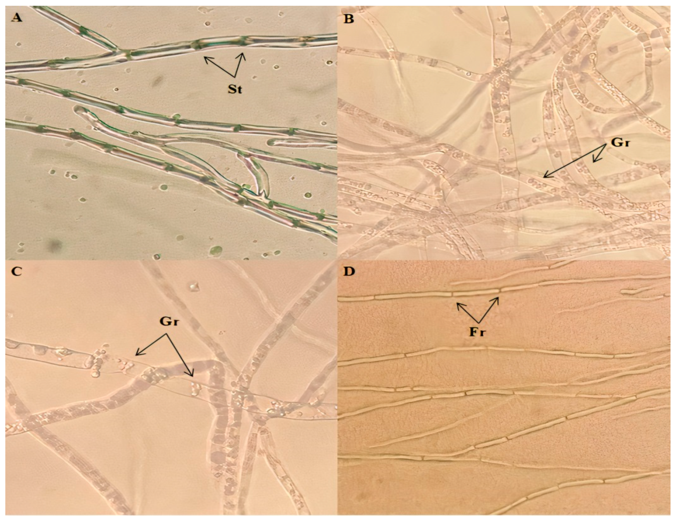
2.4. Effect of Pseudomonas Strains on Hyphae Morphology of Foc Isolates

To examine the impact of the bacterial antagonists on Foc isolates, a microscopic examination of the hyphae morphology of the Foc isolates treated with Pseudomonas strains (antagonism plates) and from the control plates was performed for the Pseudomonas strains with the highest inhibition percentages of each Foc.

2.5. Evaluation of PGPP

The Pseudomonas strains with the highest inhibition percentages of each Foc were selected and checked for their PGPP. Three replicates were used for each of the following tests.

2.5.1. Protease Production

Protease production was evaluated by inoculating Pseudomonas strains with 10% skim milk agar. The appearance of a clear zone around the colonies after incubation for three days at 30 °C was considered a sign of proteolytic activity [28].

2.5.2. α-. Amylase Production

α-amylase production was evaluated by streaking Pseudomonas isolates on starch agar. After incubation at 30 °C for 48 h, the plates were amended with Gram’s iodine solution, and isolates exhibiting clear zones around the colonies were marked as α-amylase producers [29].

2.5.3. Lipase Production

Lipase production was evaluated by inoculating Pseudomonas strains on a medium composed of (g L−1): peptone (10), NaCl (5), CaCl2 H2O (0.1), agar (18), and Tween 80 at a final concentration of 1%. The appearance of opaque halos around the colonies after incubation for six days at 30 °C was noted as an indication of lipase production [30].

2.5.4. Cellulase Production

Cellulase production was evaluated by streaking Pseudomonas isolates on Carboxymethylcellulose (CMC) agar. After incubation at 30 °C for seven days, Gram’s iodine solution was applied. Isolates showing clear halos around the colonies were marked as cellulase positive [31].

2.5.5. Hydrogen Cyanide (HCN) Production

HCN production was assessed as follows: sterile filter papers were soaked in HCN detection solution and placed on the lids of Petri plates containing KB agar supplemented with 4.4 g L−1 glycine and streaked with Pseudomonas isolates. The plates were then incubated at 30 °C for six days. The change in the paper color from yellow to light brown, brown, or reddish-brown was considered a sign of HCN production [32].

2.5.6. Indole Acetic Acid (IAA) Production

IAA production was estimated by streaking Pseudomonas isolates on Luria–Bertani agar supplemented with 5 mM L-tryptophan; the agar was then covered with sterile filter paper. After incubation at 30 °C for three days, the paper was recovered and treated with Salkowski reagent for 30 min. The change in the filter paper color from white to a reddish color was noted as a sign of IAA production [33].

2.5.7. Phosphate Solubilization

Phosphate solubilization was first screened by inoculating Pseudomonas strains on NBRIP agar. Strains showing clear halos around the colonies after incubation at 30 °C for 14 days were marked as phosphate solubilizers [34]. Quantification of the solubilized phosphate was estimated in NBRIP broth. Concentrations of the formed soluble phosphate were extrapolated by a standard curve drawn using known concentrations of KH2PO4 [35].

2.5.8. Ammonia Production

Pseudomonas strains were screened for their ability to produce ammonia by inoculation into tubes containing peptone water. After incubation at 30 °C for four days, Nessler’s reagent was added to the tubes. Ammonia production was indicated by the development of a yellow to a brown color [36].

2.5.9. Chitinase Production

Chitinase production was evaluated by streaking Pseudomonas isolates on chitinase medium. Isolates showing clear zones around the colonies after incubation at 30 °C for five days were reported as chitinase producers [37].

2.5.10. Siderophores Production

Siderophores production was evaluated as follows: Chrome Azurol S (CAS) blue dye solution was prepared as described by Louden et al. [38]. A modified protocol of Hu and Xu [39] was used for the preparation of plates as follows: blue CAS agar was prepared by adding sterile blue dye solution into sterile agar at 10% of the final volume. Pseudomonas strains were streaked onto nutrient agar and incubated at 30 °C for 18 h. After incubation, plates were overlaid with the previously prepared blue CAS agar. The change of color from blue to yellow-orange was considered as positive siderophores production.

2.6. Effect of Selected Pseudomonas Strains on the Shoot and Root Growth of Chickpea

This test was performed for the Pseudomonas strains with the highest inhibition percentages of each Foc. Seed bacterization was carried out by soaking disinfected chickpea seeds (ILC 482) overnight in a bacterial suspension (1.5 × 10⁸ CFU mL−1), to which CMC was added at a 1% concentration to assure the adherence of Pseudomonas strains onto the seeds [40]. Three replicates were used for each bacterial treatment, as well as for the control. Each replicate consisted of a pot (D: 15.5 cm and H: 13.5 cm) containing a sterilized mixture of soil, sand, and compost. Three seeds were used for each pot. Inoculated seeds were used for the treatments, and seeds soaked only in CMC water were used as control. Pots were irrigated every five days with 200 mL of water. After 30 days, shoot and root length, as well as shoot and root dry weight (after drying at 70 °C for a day), were measured.

2.7. Control of Wilt Disease of Chickpea by Selected Pseudomonas Strains

A pot experiment under greenhouse conditions (day/night cycle of 12/12 h, a temperature of 20 ± 2 °C, and 60 ± 2% relative humidity) was performed to evaluate the effect of selected Pseudomonas strains on chickpea wilt disease. The previously described sterilized soil mixture was infected with Foc isolates following the protocol of Hassan et al. [22]: five PDA discs of four-day-old Foc were transferred into flasks containing an autoclaved mixture of 100 g of barley and 100 mL of distilled water. After incubation at 30 °C for 14 days with shaking, the mixture was added to plastic pots containing the sterilized soil mixture at a 2% concentration. Seed bacterization of chickpea line ILC 482 was performed as previously described. Three replicates were used for each bacterial treatment, as well as for the control. Each replicate consisted of a pot containing the infected soil mixture, and three seeds were used for each pot. Inoculated seeds were used for the treatments, and non-inoculated seeds served as control.
A scale was assigned to each plant based on the level of symptoms, (0: no symptoms; 1: yellowing or wilting of 1/3 of the plant; 2: yellowing or wilting of 2/3 of the plant; 3: yellowing or wilting of the whole plant; 4: plant dead) [41]. Disease severity (DS) was calculated as follows:
DS (%) = [(Σ v × n)/(N × V)] × 100.
The letter n is the number of plants in each scale value; v is the scale value; V is the maximum scale value, and N is the total number of plants observed [42]. The disease reduction (DR) percentage was calculated as follows:
DR = [(A − B)/A] × 100.
The letter A is the percentage of disease in the control, and B is the percentage of disease with the application of bacteria [43].

2.8. Molecular Identification of Selected Pseudomonas

16S rRNA sequencing was performed to identify the selected Pseudomonas isolates as follows: DNA extraction was carried out using the commercial kit NucleoMag DNA Bacteria for DNA purification from bacteria and yeast (Macherey-Nagel Germany) by following the manufacturer’s instructions. PCR was performed using primers 27f (5′-AGAGTTTGATCMTGGCTCAG-3′) and 1492r (5′- TACGGYTACCTTGTTACGACTT -3′) in a reaction mixture containing: 2 μL genomic DNA (25 ng μL−1), 0.2 μL Taq polymerase Promega (1U), 1 μL of each primer (0.5 μM), 0.2 μL dNTP (0.2 mM), 1.5 μL MgCl2 (1.5 mM), 5 μL Taq Promega buffer, and ultra-pure water to a final volume of 25 μL. Conditions for PCR were as follows: initial denaturation at 95 °C/5 min followed by 35 cycles of denaturation at 95 °C/30 s; annealing at 53 °C/30 s and extension at 72 °C/45 s; and a final extension at 72 °C/7 min. The amplification products were separated by electrophoresis on a 1.5% agarose gel after staining with ethidium bromide (0.5 μg mL−1). Fragments were visualized and photographed under UV using the Gel doc system from Biorad. PCR products were purified using NucleoSpin® Gel and PCR Clean-up kit following the manufacturer’s protocol. Sequencing was conducted using Applied Biosystems’ BigDye v3.1 kit and by following the method of Sanger et al. [44].

2.9. Data Analysis

To evaluate the significance between treatments, the results were statistically processed through the Student’s t-test, analysis of variance (ANOVA), and Tukey’s B test at p < 0.05 significance. Principal component analysis (PCA) was also performed to assess the variability of traits.

3. Results

3.1. In Vitro Evaluation of Antagonistic Activities of Isolated Pseudomonas

Seventeen Pseudomonas strains were isolated from the rhizosphere of chickpeas and evaluated for their antagonistic activities against the Foc isolates in vitro.
The results revealed a difference between the percentages of inhibition registered with each antagonism assay. The growth of Foc-S1 was significantly more reduced in the divided plate assay (p < 0.0001), with an average inhibition rate equal to 44.31% against 20.40% in the co-culture assay. Concerning Foc-S2, there was no significant difference between the two assays (p = 0.085), with an average inhibition equal to 26.75% in the co-culture assay against 19.91% in the divided plate assay (Table 1).
Isolates E1FP13, E1FP4, and E1PP7 gave the highest inhibition percentages against Foc-S1, with values equal to 52.77%, 48.37%, and 47.97%, respectively. All three values were registered with the divided plate assay (action of volatile compounds). In the case of Foc-S2, isolates E1PP6, E1FP13, and E1PP15 registered the highest inhibition rates of 52.20%, 52.09%, and 45.38%, respectively, with co-culture assay (mainly through the action of diffusible compounds) for E1PP6 and E1FP13 and with divided plate assay (action of volatile compounds) for E1PP15 (Table 1) (Supplementary Materials, Figure S1).
The ANOVA analysis showed that the results in both assays (co-culture assay and divided plate assay) for both pathogens (Foc-S1 and Foc-S2) were highly significant (p < 0.0001). All traits were subjected to PCA to estimate the contribution of each trait and to assess the total level of variability. Two components (PC1-PC2) gave eigenvalues >1.0 and contributed to the explanation of 75.19% of the information. PC1 explained 50.19% of the total variability; the two pathogens were linked to it, as well as the two assays. PC2 explained 25.00% of the total contribution to variability; all strains of Pseudomonas were linked to it.

3.2. Effect of Pseudomonas Strains on Hyphae Morphology of Foc Isolates

The comparison between the microscopic observations of the Foc isolates in the antagonism plates and the control plates revealed a clear difference between the two. In the case of Foc-S1, the hyphae treated with E1FP13 appeared discontinued (fragmented), empty, and without septa (Figure 1D), while for E1PP7 and E1FP4, the hyphae appeared empty of their content with the presence of granulations (vesicles); the septa were also absent (Figure 1B,C). In the case of Foc-S2, the hyphae treated with E1PP15 were devoid of septa with condensed cytoplasm (Figure 2D). With regard to E1PP6 and E1FP13, the hyphae appeared empty, devoid of septa for the most part, with the presence of granulations; some areas were also deformed (swollen) (Figure 2B,C). Both controls (Figure 1A and Figure 2A) displayed the characteristic Foc appearance without any changes in the hyphae morphology.

3.3. Evaluation of PGPP

All selected Pseudomonas strains were positive for phosphate solubilization; the quantitative estimation showed highly significant variability between the strains (p < 0.0001) with values ranging from 57.66 to 240.69 μg P mL−1. The highest solubilization values were recorded with E1FP4 and E1PP7, which released 240.69 μg P mL−1 and 223.09 μg P mL−1, respectively. All isolates were found positive for the production of siderophores, IAA, and ammonia. Three isolates, E1PP7, E1PP15, and E1PP6, were found positive for the production of HCN. The screening for the different enzymes revealed that all isolates were negative for α-amylase and chitinase production but were positive for cellulase production. The results of the production of lipase and protease were very variable, with E1PP7 and E1PP15 exhibiting the highest activity (Table 2).

3.4. Effect of Selected Pseudomonas Strains on the Shoot and Root Growth of Chickpea

The inoculation of seeds with the selected Pseudomonas revealed that, aside from E1PP15, all isolates were able to significantly increase shoot length, as well as shoot dry weight in comparison to the control (p < 0.0001). Isolate E1PP7 gave the best results with a shoot length of 35.77 cm (increased by 42.68%) and a shoot dry weight of 0.32 g (increased by 88.24%). Aside from E1PP6, all isolates were able to significantly increase root length in comparison to the control (p < 0.0001), with E1PP7 giving the highest length of 20.53 cm (increased by 83.80%) followed by E1FP13 with a length of 17.97 cm (increased by 60.88%). All isolates were able to significantly increase root dry weight in comparison to the control (p = 0.003). There was no significant difference between root dry weights of seeds treated with isolates E1FP13, E1FP4, E1PP7, and E1PP15. However, seeds treated with isolate E1PP6 registered the lowest root dry weight. Nonetheless, it was still significantly higher than the root dry weight of the control (Table 3) (Supplementary Materials, Figure S4).

3.5. Control of Wilt Disease of Chickpea by Selected Pseudomonas Strains

The pot experiment revealed that the DS was significantly lower (p = 0.002) in plants inoculated with the selected Pseudomonas strains (E1FP13, E1FP4, and E1PP7) in comparison to the uninoculated control infected with Foc-S1. There was no significant difference (p = 0.489) in the DR between E1FP13, E1FP4, and E1PP7, with values equal to 55.77%, 48.08%, and 46.16%, respectively (Table 4) (Supplementary Materials, Figure S2).
The DS was also significantly lower (p = 0.0002) in plants inoculated with the selected Pseudomonas strains (E1FP13, E1PP6, and E1PP15) in comparison to the uninoculated control infected with Foc-S2. There was a significant difference (p = 0.021) among the DR results, with isolate E1FP13 giving the highest percentage of 53.33%, followed by E1PP6 (46.67%) and E1PP15 (30%) (Table 4) (Supplementary Materials, Figure S3).

3.6. Molecular Identification of Selected Pseudomonas

The sequences of selected Pseudomonas strains were compared with other sequences using BLAST. The search was limited to sequences from type material, and the identification was based on homology percentage with the reference sequences. Results are presented in Table 5, with accession numbers of each isolate received from NCBI after sequences submission.
Phylogenetic trees were also generated by the neighbor-joining method using the MEGA-X program with Bootstrap values based on 1000 replications. Sequences from the BLAST search with the highest similarity percentages were selected and used for the construction of the trees. Rhizobacter gummiphilus was used as an out-group bacterium. Results of the analysis revealed that the closest relative to isolates E1PP6, E1FP13, and E1FP4 was Pseudomonas frederiksbergensis with which they had the highest similarity percentages (>99.50%), while isolates E1PP7 and E1PP15 were closely related to Pseudomonas granadensis (100% and 99.77% identity, respectively) and Pseudomonas soyae (99.72% and 99.65% identity, respectively). Results can be found in Supplementary Materials (Figures S5–S9).

4. Discussion

In this study, Pseudomonas isolates were used as potential biocontrol agents against Foc. Several studies have demonstrated the efficacy of biocontrol agents in controlling this damaging phytopathogen [45,46,47]. The biocontrol activity of our Pseudomonas strains against Foc was confirmed through multiple assays.
In the in vitro antagonism test, the Pseudomonas strains reduced the mycelial growth of the Foc isolates with varying inhibition percentages. The results of the antagonistic effect of isolated Pseudomonas strains revealed that the growth of Foc-S1 was significantly more reduced in the divided plate assay (effect of volatile compounds) than in the co-culture assay, whereas for Foc-S2, there was no significant difference between the two assays. Typically, a higher growth inhibition percentage would be expected with the co-culture assay rather than with the divided plate assay, considering that in the co-culture assay, Foc is exposed to both diffusible and volatile antifungal metabolites, whereas in the divided plate assay, Foc is only exposed to the volatile antifungal metabolites. However, our results stated above revealed otherwise. This can be explained by the diffusion of antibacterial metabolites by Foc into the medium during the co-culture assay, which negatively affected the ability of Pseudomonas strains to inhibit fungal growth, thus resulting in a lower inhibition percentage. Meanwhile, in the divided plate assay, the partition in Petri dishes protects the Pseudomonas strains from potential antibacterial compounds released by Foc. The most known metabolite is fusaric acid, a mycotoxin produced by several species in the genus Fusarium [48]. The production of fusaric acid by Foc was reported by Türkkan and Dolar [49]. This mycotoxin is capable of decreasing the production of 2, 4-diacetylphloroglucinol (2,4-DAPG), an antifungal metabolite synthesized by the antagonistic strain P. protegens Pf-5 [50]. Similar results to ours have been reported by Kumari and Khanna [51].
E1FP13, E1FP4, and E1PP7 were the Pseudomonas strains with the highest inhibition percentages against Foc-S1. They registered the highest inhibition rates with volatile compounds, which is consistent with their ability to produce volatile antifungal compounds. The assessment of their PGPP revealed that E1FP13 and E1FP4 produced ammonia, while E1PP7 produced ammonia and HCN. A recent study conducted by Vlassi and Nesler [52] confirmed the involvement of ammonia released by Lysobacter capsici AZ78 in the inhibition of Rhizoctonia solani. HCN emitted by P. putida R32 and P. chlororaphis R47 was shown to be responsible for inhibiting the mycelial growth of the phytopathogen Phytophthora infestans [53]. HCN targets the electron transport chain of the pathogens, which leads to a deficiency in their ATP production, thus limiting their growth and development [51].
Microscopic examination of the effect of E1FP13 on Foc-S1 revealed fragmented and empty hyphae without septa. Similarly, Attia et al. [54] have reported hyphal fragmentation of Alternaria solani after treatment with plant-growth-promoting rhizobacteria. These results can be interpreted by the lysis of the hyphae that led to the loss of their content, which explains the empty appearance. P. monteilii PsF84 was shown to cause hyphal lysis of Fusarium oxysporum through the action of 2, 4-Di-tert-butylphenol, a volatile antifungal compound [55]. In the case of E1PP7 and E1FP4, the hyphae appeared empty of their content with the presence of granulations (vesicles). The same results were observed by Barka et al. [56] when Botrytis cinerea was treated with Pseudomonas sp. strain PsJN. In another study, a volatile antifungal compound released by P. brassicacearum and identified as dl-Limonene was also shown to cause the formation of granulations inside Sclerotinia sclerotiorum hyphae [57]. The alterations observed with Foc-S1’s hyphae were caused by the volatile antifungal compounds released by E1FP13, E1FP7, and E1FP4. These compounds can be ammonia and HCN, as they can be other volatile antifungal compounds produced by these strains [58,59,60].
Isolates E1PP6 and E1FP13 gave the highest inhibition rates against Foc-S2 with diffusible compounds, while E1PP15 acted through the action of volatile compounds. The results of PGPP revealed that E1PP15 was able to produce HCN and ammonia, which correlates positively with its volatile antagonistic activity. Isolates E1PP6 and E1FP13 were both able to produce siderophores, which are low molecular weight compounds that chelate ferric iron [61]. Di Francesco and Baraldi [62] reported that competition for iron through the release of siderophores by antagonists reduced the mycelial growth of the pathogenic fungus Monilinia laxa. In another study, it was shown that rhizobacteria that had a strong antagonistic effect against Pyricularia oryzae were the ones that registered the best siderophores production [63]. Besides the production of siderophores, isolate E1FP13 was also positive for the production of lipase, an enzyme responsible for the degradation of the fungal cell wall [64]. Isolate E1PP6 did not produce any of the tested diffusible lytic enzymes, which suggests that other enzymes, such as β-glucanase, involved in the degradation of the fungal cell wall [65] or other mechanisms, such as the production of cyclo (Pro-Val), a cyclic dipeptide with antifungal activity produced by P. frederiksbergensis CMAA 1323 [18], and the production of antibiotics [66], may have been employed.
The microscopic examination of the effect of E1PP15 on Foc-S2 revealed that this isolate caused a condensation of the hyphae’s cytoplasm; the same observation was reported by Giorgio et al. [57] when Sclerotinia sclerotiorum was treated with Pseudomonas spp. USB2104. E1PP6 and E1FP13 caused granulations, emptiness, and deformations (swelling) of the hyphae, which were also observed by Arora et al. [67] when Rhizoctonia solani was treated with Pseudomonas PGC2. Likewise, empty and swollen hyphae were reported by Chiranjeevi et al. [68] when Rhizoctonia bataticola was treated with the crude metabolite extract of Bacillus subtillis. The granulations could be cytoplasm remains formed due to cytoplasm leakage following hyphal damage caused by the hydrolytic enzyme, lipase, produced by E1FP13. In the case of E1PP6, which did not produce any of the tested lytic enzymes, other mechanisms, as mentioned above, may have been responsible for the observed morphological changes.
All five isolates were shown to synthesize IAA, a phytohormone that increases the plant’s nutrients intake by enhancing the root surface [69]. All isolates were positive for the production of cellulase, involved in the increase in organic matter in the soil [70]. All five isolates were also able to solubilize phosphate; however, the qualitative assay results did not match those of the quantitative assay, as the highest solubilization values were observed with the strains that gave the weakest solubilization halo in the plate assay. The same results were observed by Nautiyal [34]. Phosphate plays a vital role in plant development. Despite its abundance in the soil, most of it remains unavailable to plants. Phosphate-solubilizing microorganisms can hydrolyze the insoluble phosphate forms into a soluble form that can be easily assimilated by plants [71].
The evaluation of the effect of selected Pseudomonas strains on the shoot and root growth of chickpea revealed that isolates E1PP6, E1FP13, E1FP4, and E1PP7 were able to significantly increase shoot length and shoot dry weight, which correlates positively with their ability to produce several PGP compounds, such as ammonia, cellulase, siderophores, and solubilize phosphate. Although E1PP15 was also shown to produce these compounds, it did not improve shoot growth like the rest of the isolates. This result may be explained by the fact that, unlike in the in vitro tests, E1PP15 was not able to synthesize these metabolites in vivo. Concerning the growth of the roots, isolates E1PP15, E1FP13, E1FP4, and E1PP7 were able to significantly increase root length and root dry weight, which concurs well with their ability to produce IAA. IAA plays an important role in the development of plant roots; in a recent study, P. moraviensis wild type was reported for its ability to increase wheat root area in comparison to its IAA-deficient mutants [72]. Regarding E1PP6, this isolate did not improve the root length; however, it significantly increased the roots’ dry weight, and this can be interpreted by an increase in the growth of lateral roots through the production of IAA. The positive effect of IAA-producing bacteria on the number of plant’s lateral roots has been demonstrated by Herlina et al. [73]. Our results are in line with other studies that have demonstrated the capacity of Pseudomonas strains to improve chickpea growth [74,75,76]. Our study confirms the ability of the Pseudomonas isolates to promote chickpea growth by increasing shoot and root length, as well as their dry weight.
The pot experiment revealed that all selected Pseudomonas strains were able to significantly decrease the disease severity caused by both Foc isolates. The in vitro antagonistic effects (percentages of inhibition) of E1FP13, E1FP4, and E1PP7 against Foc-S1 were not significantly different from their in vivo effects (DR percentages). Similarly, there was no significant difference between the in vitro and in vivo antagonistic effects of E1PP6 and E1FP13 against Foc-S2. However, the antagonistic effect of isolate E1PP15 was significantly lower in the in vivo test, which can be attributed to lower production of antifungal compounds than that expressed in the in vitro assay. A recent study conducted by Besset-Manzoni et al. [77] showed that biocontrol agents may not always perform the same way in the in vivo antagonism assay as they do in the in vitro antagonism assay. In this study, the selected Pseudomonas strains were shown to control Fusarium wilt of chickpea caused by Foc; various studies have demonstrated the potential of Pseudomonas strains in the protection against phytopathogens [14,15,17,53]. Our results revealed that the Pseudomonas isolates produced various antifungal compounds and caused some damage to the Foc hyphae. Moreover, the isolates improved chickpea growth through the production of several metabolites that play an important role in the provision and acquisition of nutrients. The improvement of plant growth contributes to the biocontrol process, as it confers a better resistance to plants against pathogens, thus limiting their negative impact on the plants. The combination of all these mechanisms explains the significant reduction in disease severity by our isolates when applied as seed treatments.
The BLAST results and the phylogenetic analysis confirmed that the selected rhizobacteria belong to the genus Pseudomonas. Isolates E1PP6, E1FP13, and E1FP4 were closely related and best matched with P. frederiksbergensis, a bacterium that is usually associated with abiotic stress reduction, such as the degradation of pesticides [78] and the enhancement of cold stress and salt stress tolerance [79]. A few studies have been carried out on the antagonistic and PGPP of P. frederiksbergensis. A study performed by Ferchichi et al. [80] revealed that P. frederiksbergensis LB113 was able to produce HCN and inhibit mycelium growth of Macrophomina phaseolina and Alternaria alternate in PDA. P. frederiksbergensis PgBE39 and PgBE45 were reported for their antifungal activity against Cylindrocarpon destructans and Botrytis cinerea [81], while P. frederiksbergensis G62 was reported by Ben Zineb et al. [82] for its ability to produce siderophores, HCN, IAA, and solubilize phosphate. Isolates E1PP7 and E1PP15 were closely related to two Pseudomonas species, P. soyae and P. granadensis, with the latter having the highest similarity percentages with the two isolates. P. granadensis strain 100 was reported by Riera et al. [83] for its ability to produce siderophores, while P. granadensis PMK4 was shown to produce IAA, siderophores, and solubilize phosphate [84].

5. Conclusions

In brief, our results revealed that the Pseudomonas strains have the potential to be used as biocontrol agents in the management of wilt disease and as plant-growth-promoting bacteria in chickpea plants.

6. Future Perspective

In the future, it would be of interest to further study the ability of our Pseudomonas strains to control Foc under different stress conditions, such as cold, high salinity soils, and soils polluted with pesticides, and to test their effect alone and in consortium with other bacteria against Foc and other phytopathogens. Other experiments and field evaluations of the isolated Pseudomonas strains should be carried out to reveal in detail the underlying mechanisms of their biocontrol activity.

Supplementary Materials

The following supporting information can be downloaded at: https://www.mdpi.com/article/10.3390/agriculture12030429/s1, Figure S1: Inhibition of Foc growth by isolated Pseudomonas. (1) Antagonism plate, (2) Plate without bacteria (control); Figure S2: Biocontrol of Foc-S1 by isolated Pseudomonas. (1) Treatment with E1FP13, (2) Treatment with E1PP7, (3) Control, (4) Treatment with E1FP4; Figure S3: Biocontrol of Foc-S2 by isolated Pseudomonas. (1) Treatment with E1PP6, (2) Treatment with E1PP15, (3) Control, (4) Treatment with E1FP13; Figure S4: Promotion of chickpea growth by Pseudomonas isolates. (1) Control, (2) Treatment with E1FP13, (3) Treatment with E1PP7, (4) Treatment with E1PP6, (5) Treatment with E1FP4, (6) Treatment with E1PP15; Figure S5: Phylogenetic tree of strain E1PP6 constructed by neighbor-joining method with Bootstrap values supporting the branches shown at the nodes; the bar indicates the number of substitutions per site; Figure S6: Phylogenetic tree of strain E1FP13 constructed by neighbor-joining method with Bootstrap values supporting the branches shown at the nodes; the bar indicates the number of substitutions per site; Figure S7: Phylogenetic tree of strain E1PP7 constructed by neighbor-joining method with Bootstrap values supporting the branches shown at the nodes; the bar indicates the number of substitutions per site; Figure S8: Phylogenetic tree of strain E1PP15 constructed by neighbor-joining method with Bootstrap values supporting the branches shown at the nodes; the bar indicates the number of substitutions per site; Figure S9: Phylogenetic tree of strain E1FP4 constructed by neighbor-joining method with Bootstrap values supporting the branches shown at the nodes; the bar indicates the number of substitutions per site.

Author Contributions

Conceptualization, M.W.K. and N.R.; Formal analysis, M.W.K. and N.R.; Investigation, M.W.K.; Supervision, N.R. and M.B.; Writing—original draft, M.W.K.; Writing—review and editing, M.W.K., N.R. and M.B. All authors have read and agreed to the published version of the manuscript.

Funding

This research received no external funding.

Institutional Review Board Statement

Not applicable.

Informed Consent Statement

Not applicable.

Data Availability Statement

All the main data supporting the results of this study are reported in the present article.

Acknowledgments

The authors wish to express their gratitude to Mokhtari Zineb and Benhamida Aissa for their technical support. The authors are also very thankful to all the laboratory technicians of the Field crops laboratory (INRAT) for their assistance during the molecular identification of Foc.

Conflicts of Interest

The authors declare no conflict of interest.

References

  1. Rawal, V.; Navarro, D.K. The Global Economy of Pulses; FAO: Rome, Italy, 2019. [Google Scholar] [CrossRef]
  2. Wood, J.A.; Grusak, M.A. Nutritional value of chickpea. In Chickpea Breeding and Management; Yadav, S.S., Redden, R.J., Chen, W., Eds.; CAB International: Wallingford, UK, 2007; pp. 101–142. [Google Scholar] [CrossRef]
  3. Latham, M.C. Human Nutrition in the Developing World; FAO: Rome, Italy, 1997. [Google Scholar]
  4. Aslam, M.; Mahmood, I.A.; Peoples, M.B.; Schwenke, G.D.; Herridge, D.F. Contribution of chickpea nitrogen fixation to increased wheat production and soil organic fertility in rain-fed cropping. Biol. Fertil. Soils 2003, 38, 59–64. [Google Scholar] [CrossRef]
  5. Landa, B.B.; Navas-Cortés, J.A.; del Mar Jimenez-Gasco, M.; Katan, J.; Retig, B.; Jiménez-Díaz, R.M. Temperature response of chickpea cultivars to races of Fusarium oxysporum f. sp. ciceris, causal agent of Fusarium wilt. Plant Dis. 2006, 90, 365–374. [Google Scholar] [CrossRef] [PubMed] [Green Version]
  6. Knights, E.J. Chickpea overview. In Encyclopedia of Grain Science; Wrigley, C., Corke, H., Walker, C., Eds.; Elsevier Ltd.: Amsterdam, The Netherlands, 2004; Volume 1, pp. 280–287. [Google Scholar] [CrossRef]
  7. Haware, M.P.; Jiménez-Diaz, R.M.; Amin, K.S.; Phillips, J.C.; Halila, H. Integrated management of wilt and root rots of chickpea. In Proceedings of the Chickpea in the Nineties, ICRISAT (International Crops Research Institute for the Semi-Arid Tropics), Patancheru, Andhra Pradesh, India, 4–8 December 1990; pp. 129–134. [Google Scholar]
  8. Gopalakrishnan, S.; Srinivas, V. Management of soil-borne diseases of grain legumes through broad-spectrum actinomycetes having plant growth-promoting and biocontrol traits. In Plant Microbe Interface; Varma, A., Tripathi, S., Prasad, R., Eds.; Springer: Berlin/Heidelberg, Germany, 2019; pp. 129–144. [Google Scholar] [CrossRef]
  9. Landa, B.B.; Navas-Cortés, J.A.; Jiménez-Díaz, R.M. Integrated management of Fusarium wilt of chickpea with sowing date, host resistance, and biological control. Phytopathology 2004, 94, 946–960. [Google Scholar] [CrossRef] [PubMed] [Green Version]
  10. Arfaoui, A.; Sifi, B.; Boudabous, A.; El Hadrami, I.; Cherif, M. Identification of Rhizobium isolates possessing antagonistic activity against Fusarium oxysporum f. sp. ciceris, the causal agent of Fusarium wilt of chickpea. J. Plant Pathol. 2006, 88, 67–75. [Google Scholar]
  11. Jiménez-Gasco, M.; Navas-Cortés, J.A.; Jiménez-Díaz, R.M. The Fusarium oxysporum f. sp. ciceris/Cicer arietinum pathosystem: A case study of the evolution of plant-pathogenic fungi into races and pathotypes. Int. Microbiol. 2004, 7, 95–104. [Google Scholar] [PubMed]
  12. Ramezani, H. Efficacy of some fungal and bacterial bioagents against Fusarium oxysporum f. sp. ciceri on chickpea. Plant Prot. J. 2009, 1, 108–113. [Google Scholar]
  13. Hayes, W.J.; Laws, E.R. Handbook of Pesticide Toxicology; Academic Press Inc.: New Delhi, India, 1991; Volume 1. [Google Scholar]
  14. Abed, H.; Rouag, N.; Mouatassem, D.; Rouabhi, A. Screening for Pseudomonas and Bacillus antagonistic rhizobacteria strains for the biocontrol of Fusarium wilt of chickpea. Eurasian J. Soil Sci. 2016, 5, 182–191. [Google Scholar] [CrossRef] [Green Version]
  15. Al-Ghafri, H.M.; Velazhahan, R.; Shahid, M.S.; Al-Sadi, A.M. Antagonistic activity of Pseudomonas aeruginosa from compost against Pythium aphanidermatum and Fusarium solani. Biocontrol. Sci. Technol. 2020, 30, 642–658. [Google Scholar] [CrossRef]
  16. Kumari, S.; Khanna, V. Biocidal mechanisms in biological control of Fusarium wilt in chickpea (Cicer arietinum L.) by antagonistic rhizobacteria: A current perspective in soil borne fungal pest management. Int. J. Curr. Microbiol. App. Sci. 2019, 8, 1494–1510. [Google Scholar] [CrossRef]
  17. Rathore, R.; Vakharia, D.N.; Rathore, D.S. In vitro screening of different Pseudomonas fluorescens isolates to study lytic enzyme production and growth inhibition during antagonism of Fusarium oxysporum f. sp. cumini, wilt causing pathogen of cumin. Egypt. J. Biol. Pest Control 2020, 30, 1–8. [Google Scholar] [CrossRef]
  18. Melo, I.S.; Souza, W.R.; Silva, L.J.; Santos, S.N.; Assalin, M.R.; Zucchi, T.D.; Queiroz, S.D.N. Antifungal activity of Pseudomonas frederiksbergensis CMAA 1323 isolated from the Antarctic hair grass Deschampsia antarctica. Br. Microbiol. Res. J. 2016, 14, 1–11. [Google Scholar] [CrossRef] [Green Version]
  19. Nelson, P.E.; Toussoun, T.A.; Marasas, W.F.O. Fusarium oxysporum Schlecht. Emend. Snyd. & Hans. In Fusarium Species: An Illustrated Manual for Identification; The Pennsylvania State University Press: Pennsylvania, PA, USA, 1983; pp. 142–145. [Google Scholar]
  20. Mishra, P.K.; Fox, R.T.; Culham, A. Development of a PCR-based assay for rapid and reliable identification of pathogenic Fusaria. FEMS Microbiol. Lett. 2003, 218, 329–332. [Google Scholar] [CrossRef] [PubMed]
  21. Jiménez-Gasco, M.D.M.; Jiménez-Díaz, R.M. Development of a specific polymerase chain reaction-based assay for the identification of Fusarium oxysporum f. sp. ciceris and its pathogenic races 0, 1A, 5, and 6. Phytopathology 2003, 93, 200–209. [Google Scholar] [CrossRef] [PubMed] [Green Version]
  22. Hassan, N.; Shimizu, M.; Hyakumachi, M. Occurrence of root rot and vascular wilt diseases in Roselle (Hibiscus sabdariffa L.) in Upper Egypt. Mycobiology 2014, 42, 66–72. [Google Scholar] [CrossRef] [PubMed] [Green Version]
  23. Gowsalya, A.; Ponnusami, V.; Sugumaran, K.R. Isolation of bacteria from soil sample for exo-polysaccharide production. Int. J. Chemtech. Res. 2014, 6, 2925–2928. [Google Scholar]
  24. Cowan, S.T.; Steel, K.J. Diagnostic tables for the common medical bacteria. Epidemiol. Infect. 1961, 59, 357–373. [Google Scholar] [CrossRef] [PubMed] [Green Version]
  25. Erdogan, O.; Benlioglu, K. Biological control of Verticillium wilt on cotton by the use of fluorescent Pseudomonas spp. under field conditions. Biol. Control 2010, 53, 39–45. [Google Scholar] [CrossRef]
  26. Dilantha Fernando, W.G.; Linderman, R.G. Inhibition of Phytophthora vignae and stem and root rot of cowpea by soil bacteria. Biol. Agric. Hortic. 1995, 12, 1–14. [Google Scholar] [CrossRef]
  27. Etebarian, H.-R.; Sholberg, P.L.; Eastwell, K.C.; Sayler, R.J. Biological control of apple blue mold with Pseudomonas fluorescens. Can. J. Microbiol. 2005, 51, 591–598. [Google Scholar] [CrossRef] [PubMed]
  28. Sheeba, J.; Dhamotharan, R.; Baskar, K. Isolation, screening and characterization of plant growth promoting bacteria and their antifungal effect on Rhizoctonia solani (JG Kühn 1858). Adv. Plants Agric. Res. 2017, 7, 369–375. [Google Scholar] [CrossRef]
  29. Zimbro, M.J.; Power, D.A.; Miller, S.M.; Wilson, G.E.; Johnson, J.A. (Eds.) Difco & BBL Manual: Manual of Microbiological Culture Media, 2nd ed.; Becton, Dickinson and Company: Maryland, MD, USA, 2009. [Google Scholar]
  30. Sierra, G. A simple method for the detection of lipolytic activity of micro-organisms and some observations on the influence of the contact between cells and fatty substrates. Antonie Leeuwenhoek 1957, 23, 15–22. [Google Scholar] [CrossRef] [PubMed]
  31. Khatiwada, P.; Ahmed, J.; Sohag, M.H.; Islam, K.; Azad, A.K. Isolation, screening and characterization of cellulase producing bacterial isolates from municipal solid wastes and rice straw wastes. J. Bioprocess Biotech. 2016, 6, 280–284. [Google Scholar] [CrossRef] [Green Version]
  32. Bakker, A.W.; Schippers, B. Microbial cyanide production in the rhizosphere in relation to potato yield reduction and Pseudomonas spp.-mediated plant growth-stimulation. Soil Biol. Biochem. 1987, 19, 451–457. [Google Scholar] [CrossRef]
  33. Bric, J.M.; Bostock, R.M.; Silverstone, S.E. Rapid in situ assay for indoleacetic acid production by bacteria immobilized on a nitrocellulose membrane. Appl. Environ. Microbiol. 1991, 57, 535–538. [Google Scholar] [CrossRef] [PubMed] [Green Version]
  34. Nautiyal, C.S. An efficient microbiological growth medium for screening phosphate solubilizing microorganisms. FEMS Microbiol. Lett. 1999, 170, 265–270. [Google Scholar] [CrossRef] [PubMed]
  35. Saravanakumar, K.; Arasu, V.S.; Kathiresan, K. Effect of Trichoderma on soil phosphate solubilization and growth improvement of Avicennia marina. Aquat. Bot. 2013, 104, 101–105. [Google Scholar] [CrossRef]
  36. Cappuccino, J.C.; Sherman, N. Microbiology: A Laboratory Manual, 3rd ed.; Benjamin Cummings Pub. Co.: New York, NY, USA, 1992. [Google Scholar]
  37. Murthy, N.; Bleakley, B. Simplified method of preparing colloidal chitin used for screening of chitinase-producing microorganisms. Internet J. Microbiol. 2012, 10, 1–5. [Google Scholar] [CrossRef]
  38. Louden, B.C.; Haarmann, D.; Lynne, A.M. Use of blue agar CAS assay for siderophore detection. J. Microbiol. Biol. Educ. 2011, 12, 51–53. [Google Scholar] [CrossRef] [Green Version]
  39. Hu, Q.-P.; Xu, J.-G. A simple double-layered Chrome Azurol S Agar (SD-CASA) plate assay to optimize the production of siderophores by a potential biocontrol agent Bacillus. Afr. J. Microbiol. Res. 2011, 5, 4321–4327. [Google Scholar] [CrossRef]
  40. Kaur, R.; Singh, R.S.; Alabouvette, C. Antagonistic activity of selected isolates of fluorescent Pseudomonas against Fusarium oxysporum f. sp. ciceris. Asian J. Plant Sci. 2007, 6, 446–454. [Google Scholar] [CrossRef] [Green Version]
  41. Zemouli-Benfreha, F.; Djamel-eddine, H.; Merzoug, A. Fusarium wilt of chickpea (Cicer arietinum L.) in North-West Algeria. Afr. J. Agric. Res. 2014, 9, 168–175. [Google Scholar] [CrossRef]
  42. Townsend, G.R. Methods for estimating losses caused by diseases in fungicide experiments. Plant Dis. Rep. 1943, 27, 340–343. [Google Scholar]
  43. Bektas, I.; Kusek, M. Biological control of onion basal rot disease using phosphate solubilising rhizobacteria. Biocontrol Sci. Technol. 2021, 31, 190–205. [Google Scholar] [CrossRef]
  44. Sanger, F.; Nicklen, S.; Coulson, A.R. DNA sequencing with chain-terminating inhibitors. Proc. Natl. Acad. Sci. USA 1977, 74, 5463–5467. [Google Scholar] [CrossRef] [PubMed] [Green Version]
  45. Maitlo, S.A.; Rajput, N.A.; Syed, R.N.; Khanzada, M.A.; Rajput, A.Q.; Lodhi, A.M. Microbial control of Fusarium wilt of chickpea caused by Fusarium oxysporum f. sp. ciceris. Pak. J. Bot. 2019, 51, 2261–2268. [Google Scholar] [CrossRef]
  46. Trivedi, S.; Srivastava, M.; Ratan, V.; Mishra, A.; Dixit, S.; Pandey, S. Evaluation of microbial consortia on systemic resistance against chickpea wilt. Bangladesh J. Bot. 2020, 49, 653–661. [Google Scholar] [CrossRef]
  47. Ankati, S.; Srinivas, V.; Pratyusha, S.; Gopalakrishnan, S. Streptomyces consortia-mediated plant defense against Fusarium wilt and plant growth-promotion in chickpea. Microb. Pathog. 2021, 157, 104961. [Google Scholar] [CrossRef] [PubMed]
  48. Bacon, C.W.; Porter, J.K.; Norred, W.P.; Leslie, J.F. Production of fusaric acid by Fusarium species. Appl. Environ. Microbiol. 1996, 62, 4039–4043. [Google Scholar] [CrossRef] [PubMed] [Green Version]
  49. Türkkan, M.; Dolar, F.S. Determination of fusaric acid production by Fusarium oxysporum f. sp. ciceris with thin layer chromatography and spectrophotometric methods. Anadolu Tarım Bilimleri Derg. 2010, 25, 146–150. [Google Scholar]
  50. Quecine, M.C.; Kidarsa, T.A.; Goebel, N.C.; Shaffer, B.T.; Henkels, M.D.; Zabriskie, T.M.; Loper, J.E. An interspecies signaling system mediated by fusaric acid has parallel effects on antifungal metabolite production by Pseudomonas protegens Strain Pf-5 and antibiosis of Fusarium spp. Appl. Environ. Microbiol. 2016, 82, 1372–1382. [Google Scholar] [CrossRef] [PubMed] [Green Version]
  51. Kumari, S.; Khanna, V. Biological management of vascular wilt of chickpea (Cicer arietinum L.) incited by Fusarium oxysporum f. sp. ciceris by antagonistic rhizobacteria co-inoculated with native Mesorhizobium. Int. J. Curr. Microbiol. Appl. Sci. 2018, 7, 920–941. [Google Scholar] [CrossRef]
  52. Vlassi, A.; Nesler, A.; Parich, A.; Puopolo, G.; Schuhmacher, R. Volatile-mediated inhibitory activity of rhizobacteria as a result of multiple factors interaction: The case of Lysobacter capsici AZ78. Microorganisms 2020, 8, 1761. [Google Scholar] [CrossRef] [PubMed]
  53. Anand, A.; Chinchilla, D.; Tan, C.; Mène-Saffrané, L.; L’Haridon, F.; Weisskopf, L. Contribution of hydrogen cyanide to the antagonistic activity of Pseudomonas strains against Phytophthora infestans. Microorganisms 2020, 8, 1144. [Google Scholar] [CrossRef] [PubMed]
  54. Attia, M.S.; El-Sayyad, G.S.; Abd Elkodous, M.; El-Batal, A.I. The effective antagonistic potential of plant growth-promoting rhizobacteria against Alternaria solani-causing early blight disease in tomato plant. Sci. Hortic. 2020, 266, 109289. [Google Scholar] [CrossRef]
  55. Dharni, S.; Maurya, A.; Samad, A.; Srivastava, S.K.; Sharma, A.; Patra, D.D. Purification, characterization, and in vitro activity of 2, 4-di-tert-butylphenol from Pseudomonas monteilii PsF84: Conformational and molecular docking studies. J. Agric. Food Chem. 2014, 62, 6138–6146. [Google Scholar] [CrossRef] [PubMed]
  56. Barka, E.A.; Gognies, S.; Nowak, J.; Audran, J.-C.; Belarbi, A. Inhibitory effect of endophyte bacteria on Botrytis cinerea and its influence to promote the grapevine growth. Biol. Control 2002, 24, 135–142. [Google Scholar] [CrossRef]
  57. Giorgio, A.; De Stradis, A.; Lo Cantore, P.; Iacobellis, N.S. Biocide effects of volatile organic compounds produced by potential biocontrol rhizobacteria on Sclerotinia sclerotiorum. Front. Microbiol. 2015, 6, 1056. [Google Scholar] [CrossRef] [Green Version]
  58. Zhang, Y.; Li, T.; Xu, M.; Guo, J.; Zhang, C.; Feng, Z.; Peng, X.; Li, Z.; Xing, K.; Qin, S. Antifungal effect of volatile organic compounds produced by Pseudomonas chlororaphis subsp. aureofaciens SPS-41 on oxidative stress and mitochondrial dysfunction of Ceratocystis fimbriata. Pestic. Biochem. Physiol. 2021, 173, 104777. [Google Scholar] [CrossRef]
  59. Verma, S.K.; Kingsley, K.L.; Bergen, M.S.; Kowalski, K.P.; White, J.F. Fungal disease prevention in seedlings of rice (Oryza sativa) and other grasses by growth-promoting seed-associated endophytic bacteria from invasive Phragmites australis. Microorganisms 2018, 6, 21. [Google Scholar] [CrossRef] [PubMed] [Green Version]
  60. Guevara-Avendaño, E.; Bejarano-Bolívar, A.A.; Kiel-Martínez, A.-L.; Ramírez-Vázquez, M.; Méndez-Bravo, A.; von Wobeser, E.A.; Sánchez-Rangel, D.; Guerrero-Analco, J.A.; Eskalen, A.; Reverchon, F. Avocado rhizobacteria emit volatile organic compounds with antifungal activity against Fusarium solani, Fusarium Sp. associated with Kuroshio Shot Hole Borer, and Colletotrichum gloeosporioides. Microbiol. Res. 2019, 219, 74–83. [Google Scholar] [CrossRef] [PubMed]
  61. Neilands, J.B. Methodology of siderophores. In Proceedings of the Siderophores from Microorganisms and Plants; Structure and Bonding; Springer: Berlin/Heidelberg, Germany, 1984; Volume 58, pp. 1–24. [Google Scholar] [CrossRef]
  62. Di Francesco, A.; Baraldi, E. How siderophore production can influence the biocontrol activity of Aureobasidium pullulans against Monilinia laxa on peaches. Biol. Control 2021, 152, 104456. [Google Scholar] [CrossRef]
  63. Nabila; Kasiamdari, R.S. Antagonistic activity of siderophore-producing bacteria from black rice rhizosphere against rice blast fungus Pyricularia oryzae. Microbiol. Biotechnol. Lett. 2021, 49, 217–224. [Google Scholar] [CrossRef]
  64. Zouari, I.; Masmoudi, F.; Medhioub, K.; Tounsi, S.; Trigui, M. Biocontrol and plant growth-promoting potentiality of bacteria isolated from compost extract. Antonie Leeuwenhoek 2020, 113, 2107–2122. [Google Scholar] [CrossRef] [PubMed]
  65. Dewi, R.T.K.; Mubarik, N.R.; Suhartono, M.T. Medium optimization of β-glucanase production by Bacillus subtilis SAHA 32.6 used as biological control of oil palm pathogen. Emir. J. Food Agric. 2016, 28, 116–125. [Google Scholar] [CrossRef] [Green Version]
  66. Sharma, D.; Gupta, M.; Gupta, S.; Kashyap, P.L.; Zargar, S.M.; Mallick, S.A. Antibiotic gene specific characterization and ARDRA analysis of native isolates of Pseudomonas spp. from Jammu, India. Indian Phytopathol. 2018, 71, 225–233. [Google Scholar] [CrossRef]
  67. Arora, N.K.; Khare, E.; Oh, J.H.; Kang, S.C.; Maheshwari, D.K. Diverse mechanisms adopted by fluorescent Pseudomonas PGC2 during the inhibition of Rhizoctonia solani and Phytophthora capsici. World J. Microbiol. Biotechnol. 2008, 24, 581–585. [Google Scholar] [CrossRef]
  68. Chiranjeevi, N.; Kumar, M.R.; Padmodaya, B.; Venkateswarlu, N.C.; Sudhakar, P.; Devi, R.S.J.; Sri, P.A. Studies on extraction, evaluation of crude metabolite extract from endophytic Bacillus subtillis and its mechanistic effect on chickpea dry root rot causing pathogen Rhizoctonia bataticola (Taub.) Butler. Pharma Innov. 2021, 10, 898–905. [Google Scholar]
  69. Vessey, J.K. Plant growth promoting rhizobacteria as biofertilizers. Plant Soil 2003, 255, 571–586. [Google Scholar] [CrossRef]
  70. Varma, P.K.; Kumar, V.K.; Suresh, M.; Chandrasekhar, V.; Bharathalakshmi, M.; Jamuna, P. Bacillus amyloliquefaciens (RB19): A potential PGPR in managing sugarcane red rot disease. J. Pharmacogn. Phytochem. 2019, 8, 2255–2261. [Google Scholar]
  71. Kalayu, G. Phosphate solubilizing microorganisms: Promising approach as biofertilizers. Int. J. Agron. 2019, 2019, 4917256. [Google Scholar] [CrossRef]
  72. Ul Hassan, T.; Bano, A. Construction of IAA-deficient mutants of Pseudomonas moraviensis and their comparative effects with wild type strains as bio-inoculant on wheat in saline sodic soil. Geomicrobiol. J. 2019, 36, 376–384. [Google Scholar] [CrossRef]
  73. Herlina, L.; Pukan, K.K.; Mustikaningtyas, D. The endophytic bacteria producing IAA (Indole Acetic Acid) in Arachis hypogaea. Cell Biol. Dev. 2017, 1, 31–35. [Google Scholar] [CrossRef] [Green Version]
  74. Joshi, D.; Chandra, R.; Suyal, D.C.; Kumar, S. Impacts of bioinoculants Pseudomonas jesenii MP1 and Rhodococcus qingshengii S10107 on chickpea (Cicer arietinum L.) yield and soil nitrogen status. Pedosphere 2019, 29, 388–399. [Google Scholar] [CrossRef]
  75. Rawat, N.; Sharma, M.; Suyal, D.C.; Singh, D.K.; Joshi, D.; Singh, P.; Goel, R. Psyhcrotolerant bio-inoculants and their co-inoculation to improve Cicer arietinum growth and soil nutrient status for sustainable mountain agriculture. J. Soil Sci. Plant Nutr. 2019, 19, 639–647. [Google Scholar] [CrossRef]
  76. Gaurav, A.; Pallavi, K.P. Effect of plant growth-promoting rhizobacteria (PGPR) on growth and physiological parameters in chickpea. J. Pharmacogn. Phytochem. 2020, 9, 29–34. [Google Scholar]
  77. Besset-Manzoni, Y.; Joly, P.; Brutel, A.; Gerin, F.; Soudière, O.; Langin, T.; Prigent-Combaret, C. Does in vitro selection of biocontrol agents guarantee success in planta? A study case of wheat protection against Fusarium seedling blight by soil bacteria. PLoS ONE 2019, 14, e0225655. [Google Scholar] [CrossRef] [Green Version]
  78. Hussaini, S.Z.; Shaker, M.; Iqbal, M.A. Isolation of bacterial for degradation of selected pesticides. Adv Biores 2013, 4, 82–85. [Google Scholar]
  79. Chatterjee, P.; Samaddar, S.; Anandham, R.; Kang, Y.; Kim, K.; Selvakumar, G.; Sa, T. Beneficial soil bacterium Pseudomonas Frederiksbergensis OS261 augments salt tolerance and promotes red pepper plant growth. Front. Plant Sci. 2017, 8, 705. [Google Scholar] [CrossRef]
  80. Ferchichi, N.; Toukabri, W.; Boularess, M.; Smaoui, A.; Mhamdi, R.; Trabelsi, D. Isolation, identification and plant growth promotion ability of endophytic bacteria associated with lupine root nodule grown in Tunisian soil. Arch. Microbiol. 2019, 201, 1333–1349. [Google Scholar] [CrossRef]
  81. Hong, C.E.; Kim, J.U.; Lee, J.W.; Lee, S.W.; Jo, I.-H. Diversity of bacterial endophytes in Panax ginseng and their protective effects against pathogens. 3 Biotech 2018, 8, 1–7. [Google Scholar] [CrossRef]
  82. Ben Zineb, A.; Trabelsi, D.; Ayachi, I.; Barhoumi, F.; Aroca, R.; Mhamdi, R. Inoculation with elite strains of phosphate-solubilizing bacteria enhances the effectiveness of fertilization with rock phosphates. Geomicrobiol. J. 2020, 37, 22–30. [Google Scholar] [CrossRef]
  83. Riera, N.; Handique, U.; Zhang, Y.; Dewdney, M.M.; Wang, N. Characterization of antimicrobial-producing beneficial bacteria isolated from Huanglongbing escape citrus trees. Front. Microbiol. 2017, 8, 2415. [Google Scholar] [CrossRef] [PubMed] [Green Version]
  84. Swift, R.G. Plant Growth-Promoting Bacteria from Western Australian Soils. Ph.D. Thesis, Murdoch University, Perth, Australia, 2016. [Google Scholar]
Figure 1. Light microscopy observations of the impact of selected Pseudomonas strains on Foc-S1 hyphae morphology (40×). (A) Control; (B) Effect of E1PP7; (C) Effect of E1FP4; (D) Effect of E1FP13. (St) Septa; (Gr) Granulations; (Fr) Fragmentations.
Agriculture 12 00429 g001
Figure 2. Light microscopy observations of the impact of selected Pseudomonas strains on Foc-S2 hyphae morphology (40×). (A) Control; (B) Effect of E1FP13; (C) Effect of E1PP6; (D) Effect of E1PP15. (St) Septa; (Gr) Granulations; (Df) Deformations; (Cd) Condensations.
Agriculture 12 00429 g002
Table 1. Percentages of inhibition of Foc radial growth by antagonistic Pseudomonas isolates in each assay.
IsolatesFoc-S1Foc-S2
AssaysCo-CultureDivided PlateCo-CultureDivided Plate
E2PP34.66 ± 2.48 a44.70 ± 4.09 b17.13 ± 4.32 abc23.62 ± 3.25 bcd
E1PP1411.81 ± 5.59 abc44.51 ± 2.14 b14.96 ± 4.38 ab12.57 ± 7.62 ab
E1FP149.36 ± 3.64 ab24.89 ± 4.09 a22.98 ± 2.78 abcd32.69 ± 5.85 cde
E2PP417.43 ± 1.06 bcd43.95 ± 8.90 b17.81 ± 0.24 abc22.69 ± 5.80 abcd
E1PP1521.85 ± 3.27 cdef47.43 ± 8.58 b14.69 ± 1.23 ab45.38 ± 4.68 e
E2PP724.10 ± 3.90 def44.22 ± 2.29 b13.24 ± 6.67 a19.37 ± 6.75 abc
E2PP520.49 ± 1.29 bcde44.61 ± 0.09 b17.18 ± 8.82 abc19.17 ± 7.68 abc
E1FP822.16 ± 2.02 cdef45.01 ± 0.41 b22.75 ± 5.54 abcd14.25 ± 4.86 ab
E2PP817.08 ± 3.38 bcd43.51 ± 0.37 b28.72 ± 5.28 cdef18.56 ± 8.86 abc
E1FP924.59 ± 2.97 def44.89 ± 0.51 b23.97 ± 1.99 abcde13.09 ± 2.64 ab
E1PP222.95 ± 3.31 cdef43.77 ± 2.47 b27.36 ± 0.19 bcde20.49 ± 6.92 abc
E2PP630.10 ± 8.48 ef45.00 ± 1.34 b21.12 ± 5.74 abcd12.26 ± 4.34 ab
E1PP725.61 ± 2.81 def47.97 ± 5.04 b32.33 ± 4.67 def35.55 ± 3.37 de
E1FP423.24 ± 1.41 cdef48.37 ± 6.31 b40.52 ± 4.43 f6.88 ± 0.24 a
E2PP233.03 ± 8.54f44.19 ± 2.26 b35.70 ± 3.68 ef19.88 ± 4.57 abc
E1PP619.06 ± 0.73 bcde43.44 ± 6.67 b52.20 ± 4.51 g7.02 ± 2.18 a
E1FP1319.24 ± 1.00 bcde52.77 ± 2.32 b52.09 ± 2.65 g15.06 ± 6.13 ab
Mean20.4044.3126.7519.91
The values given are means (n = 3) with standard deviations. Means in the same column followed by the same letters are not significantly different at p < 0.05.
Table 2. Evaluation of plant-growth-promoting and antifungal properties of selected Pseudomonas.
TestsE1PP7E1FP4E1PP15E1PP6E1FP13
α-amylase production-----
Lipase production++++++++-+
Protease production+-++--
Cellulase production++++++
Chitinase production-----
HCN production++-+++-
IAA production+++++++
Ammonia production++++++++
Siderophores production+++++++++++++
Qualitative phosphate solubilization+++++++
Quantitative phosphate solubilization (µg mL−1)223.09 ± 18.99 a240.69 ± 0.06 a173.80 ± 1.47 b62.82 ± 6.01 c57.66 ± 11.40 c
(-) no activity, (+) low, (++) moderate, (+++) high, (++++) very high. The values given are means (n = 3) with standard deviations. Means followed by the same letter are not significantly different at p < 0.05.
Table 3. Effect of selected Pseudomonas strains on the shoot and root growth of chickpea.
Selected PseudomonasShoot Length (cm)Root Length (cm)Shoot Dry Weight (g)Root Dry Weight (g)
E1PP628.80 ± 0.69 b12.13 ± 1.33 a0.23 ± 0.02 b0.14 ± 0.04 ab
E1FP1330.17 ± 1.46 b17.97 ± 0.96 bc0.26 ± 0.01 b0.20 ± 0.01 b
E1FP430.23 ± 1.66 b14.83 ± 1.56 ab0.27 ± 0.02 b0.18 ± 0.02 b
E1PP735.77 ± 1.10 c20.53 ± 0.38 c0.32 ± 0.04 c0.20 ± 0.04 b
E1PP1523.57 ± 0.57 a14.53 ± 1.19 ab0.18 ± 0.02 a0.17 ± 0.01 b
Control25.07 ± 1.87 a11.17 ± 2.40 a0.17 ± 0.02 a0.11 ± 0.01 a
The values given are means (n = 3) with standard deviations. Means in the same column followed by the same letters are not significantly different at p < 0.05.
Table 4. In vivo effect of selected Pseudomonas strains on Foc isolates.
StrainsFoc-S1StrainsFoc-S2
DS%DR%DS%DR%
E1FP1331.94 ± 6.36 a55.77 ± 8.81 aE1FP1338.89 ± 4.82 a53.33 ± 5.78 b
E1FP437.50 ± 4.17 a48.08 ± 5.78 aE1PP644.44 ± 9.62 ab46.67 ± 11.55 ab
E1PP738.89 ± 9.62 a46.16 ± 13.33 aE1PP1558.33 ± 0.00 b30.00 ± 0.00 a
Control72.22 ± 12.73 b00.00Control83.33 ± 8.34 c00.00
The values given are means (n = 3) with standard deviations. Means in the same column followed by the same letters are not significantly different at p < 0.05.
Table 5. BLAST results of selected Pseudomonas.
IsolatesAccessionBest MatchPI *E Value
E1PP6MT774541Pseudomonas frederiksbergensis99.55%0.0
E1FP13MT774542Pseudomonas frederiksbergensis strain LMR 70899.56%0.0
E1PP7MT774544Pseudomonas granadensis strain F-278,770T100%0.0
E1PP15MT774545Pseudomonas granadensis strain F-278,770T99.77%0.0
E1FP4MT774548Pseudomonas frederiksbergensis strain LMR70899.89%0.0
* PI: Percentage identification.
Publisher’s Note: MDPI stays neutral with regard to jurisdictional claims in published maps and institutional affiliations.

