Article

Simultaneous Detection of Plant- and Fungus-Derived Genes Constitutively Expressed in Single Pseudoidium neolycopersici-Inoculated Type I Trichome Cells of Tomato Leaves via Multiplex RT-PCR and Nested PCR

1
Department of Agricultural Science, Faculty of Agriculture, Kindai University, Nara 631-8505, Japan
2
Plant Center, Institute of Advanced Technology, Kindai University, Wakayama 642-0017, Japan
3
Department of Chemical Biological Sciences, Faculty of Science, Japan Women’s University, Tokyo 112-8681, Japan
4
Mass Spectrometry and Microscopy Unit, Technology Platform Division, RIKEN Center for Sustainable Resource Science, Yokohama 230-0045, Japan
5
Pharmaceutical Research and Technology Institute and Anti-Aging Centers, Kindai University, Osaka 577-8502, Japan
6
Plant Breeding, Wageningen University and Research, Droevendaalsesteeg 1, 6708PB Wageningen, The Netherlands
7
Agricultural Technology and Innovation Research Institute, Kindai University, Nara 631-8505, Japan
*
Author to whom correspondence should be addressed.
Agriculture 2022, 12(2), 254; https://doi.org/10.3390/agriculture12020254
Submission received: 28 January 2022 / Revised: 7 February 2022 / Accepted: 8 February 2022 / Published: 10 February 2022
(This article belongs to the Section Crop Protection, Diseases, Pests and Weeds)

Abstract

:
Type I trichomes of tomato leaves (Solanum lycopersicum Mill. cv. Moneymaker), as outgrowths of the plant epidermis, are suitable for monitoring infection processes of powdery mildew species using a high-fidelity digital microscope (DM) without fungal staining. On the trichomes, tomato powdery mildew (Pseudoidium neolycopersici L. Kiss) isolate KTP-03 produced a maximum of four vigorously elongated hyphae per conidium, which stopped growth approximately 12 days after inoculation. Single trichome cells, invaded by fungal hyphae at various fungal infection stages during the 12-day period after the inoculation of single conidia, were cut at the bases and directly collected with small precision scissors (i.e., microscissors) held by the manipulator under a DM. Subsequently, suc-polymerase chain reaction (PCR) (reverse transcription (RT)-PCR followed by nested (N)-PCR) was conducted to explore gene expression in the infected trichome. We selected intron-containing genes from tomatoes and powdery mildew fungi for the detection of constitutive gene transcripts, namely plasma membrane H+-ATPase (LHA2) and β-tubulin 2 (TUB2) genes. In suc-PCR, a single band from spliced mRNAs of both LHA2 and TUB2 genes were detected, suggesting that both genes were successfully transcribed in single KTP-03-infected trichomes. With combined primers for both LHA2 and TUB2 (multiplex RT-PCR/N-PCR), two bands were detected through the amplification of intron-spliced mRNAs of both genes. Therefore, our single-trichome cell PCR amplification method is effective for detecting the expression patterns of genes from both tomato and powdery mildew fungus. Combinations of digital microscopy, microscissors, and multiplex RT-PCR/N-PCR amplification techniques will be useful for simultaneously analysing the molecular interactions between plants and powdery mildew fungi at the level of single tomato leaf trichome cells. Also, this employed technique will be of benefit in other plant species and crops, possessing leaf trichome cells, to elucidate the molecular interactions between plants and pathogens.

1. Introduction

Biotic stresses such as fungi, bacteria and viruses affect the growth and development of plants [1]. Under stress, several proteins that are known as pathogenesis-related (PR) proteins are accumulated in plants. The PR proteins are studied and coded by the host plant specifically under biotic stress [2], and are not only accumulated in the infected leaves, but are also produced systemically in association with the development of systemic acquired resistance (SAR). For example, the expression of PR-1 genes serves as a molecular marker to indicate the plant defense response against pathogens [3]. Thaumatin-like proteins (TLPs) produced in plants are associated with developmental processes and defense against phytopathogens and elicitors [4]. Chitinases are accumulated in plants as a result of infection caused by fungi and other biotic and abiotic factors [5]. Lawrence et al. [6] reported that the expression of chitinase increases against phytopathogen systems and its induction is stronger in resistant varieties of tomato. Plant β-1,3-glucanases (members of the PR-2 family of PR proteins) indirectly induce the formation of oligosaccharide elicitors, which elicit the production of phytoalexins [7]. PR-5 proteins (members of PR family) are cysteine rich proteins which are also recognised as TLPs since they show a resemblance to the sequence of thaumatin [8]. On the other hand, regarding the effects of fungi on plants, Manghwar et al. [9] evaluated the expression of important PR protein genes in resistant and susceptible varieties of wheat and maize under Bipolaris sorokiniana fungal stress, and described that these PR genes can be further over-expressed in transgenic plants to create resistance against various pathogens. Also, Sunera et al. [10] suggested that the use in agriculture of plant growth promoting bacteria (PGPB), isolated from tomato rhizospheres, stimulated plant growth and reduced environmental risks by providing a sustainable agro-ecosystem. In addition, Manghwar et al. [11] studied the behavior of important PR genes in different wheat varieties, identifying which genes could be used for cloning into wheat and other transgenic crops to create resistance to Fusarium equiseti.
Tomato powdery mildew fungi are obligate biotrophs that infect leaves and stems, and reduce the yield of infected tomato plants. Tomato powdery mildews are caused by three species: Pseudoidium neolycopersici L. Kiss (epiphytic fungus) and Leveillula taurica (Lév.) G. Arnaud (endophytic fungus), two pathogens that appear on host plants in many regions worldwide [12,13,14], and Golovinomyces lycopersici (Cook and Massee) L. Kiss (epiphytic fungus), reported only from Australia [13,15]. Powdery mildews caused by P. neolycopersici occur regularly on hydroponically cultured tomato (Solanum lycopersicum Mill. cv. Moneymaker) in Japan [16,17,18]. Five P. neolycopersici isolates (KTP-01, -02, -03, -04 and -05) from infected tomato leaves have been isolated and identified based on the morphological and genetic (ribosomal DNA internal transcribed spacer region: rDNA-ITS region) characteristics of powdery mildew fungi [19,20,21,22].
In the life cycle of P. neolycopersici, after a fungal conidium lands on the surface of a host leaf, the conidium elongates an appressorial germ tube and forms an appressorium at the tip. The appressorium facilitates adhesion to the host leaf surface and subsequent fungal penetration of the plant cell wall. After penetration into a host epidermal cell, a mature haustorium forms at the tip of the infection peg. A functional haustorium is essential for drawing nutrients and water from the host cell; it is also needed for most infection structures that generate new epiphytic hyphal growths and successful fungal colonisation on the leaf surface [19,23,24]. To elucidate the molecular interactions between host plant cells and pathogenic powdery mildew fungi, we have focused on trichome cells formed on tomato leaves as target plant cells. Trichomes consist of transparent cells that grow from the leaf epidermis; they act as biological antennae, responding to physical and chemical stimuli (e.g., temperature, water-vapour concentration and signal volatiles) or producing chemicals (e.g., insecticidal, antifungal or adhesive (bio-surfactant) agents) after the detection of changes in environmental factors [25,26,27,28,29,30,31,32,33]. The leaves of wild and cultivated Solanum species have morphologically distinct trichomes (types I–VII) [34,35,36]. For example, the morphological characteristics of leaf type I trichomes of tomato cv. Moneymaker (MM) include a length of 1.5–2.5 mm on a multicellular base with a small glandular tip; they are more abundant and larger than other types of trichomes. In our previous studies, we succeeded in observing mature haustoria in P. neolycopersici-invaded trichomes using a high-fidelity digital microscopy (DM) without chemical treatment [37] and clarified the morphological characteristics of functional P. neolycopersici haustoria in type I trichomes of MM using field-emission scanning electron microscopy [38]. Our results showed a positive correlation between the number of haustoria per hypha and the hyphal lengths on trichomes. Our findings suggested that trichomes were excellent and advantageous sites for morphological and cytological analysis of the interactions between plant cells and powdery mildew pathogens.
In the present study, we aimed to demonstrate that a single trichome cell could be used to monitor the expression of genes of both tomato and the fungus. To efficiently analyse the interactions, methods are needed to isolate the intracellular content of a single plant cell attacked by a fungal appressorium and/or haustorium. At present, only two techniques are available: micropipette manipulation [39] and laser microdissection [40]. However, these techniques are not widely used because they require considerable skill and specialised equipment. To solve this problem, we used small, pointed scissors (i.e., microscissors) installed in a micromanipulator for efficient cutting and collection of living single leaf trichomes under DM. We then conducted direct reverse transcription (RT)-polymerase chain reaction (PCR) followed by nested (N)-PCR (suc-PCR) or multiplex RT-PCR/N-PCR amplification of mature mRNA transcripts [41], to individually or simultaneously evaluate the expression patterns of targeted plant and fungal genes in P. neolycopersici-inoculated trichome cells. To our knowledge, this is the first report of a method to detect gene expression patterns in single powdery mildew-infected leaf trichome cells at various developmental stages of pathogenic powdery mildew infection.

2. Materials and Methods

2.1. Plant Materials

Tomato seeds of MM were germinated on water-soaked filter papers in a petri dish for 3 days in a growth chamber (LH-240N; Nippon Medical and Chemical Instruments, Osaka, Japan) under continuous illumination (19.8–40.3 µmol m−2 s−1; 400–700 nm) from white (full-spectrum) fluorescent lamps (FL40SS W/37; Mitsubishi, Tokyo, Japan) at 25 ± 2 °C. Seedlings at the cotyledon stage were placed into polyurethane cubic sponge supports (3 cm × 3 cm × 3 cm). Sponge supports containing seedlings were inserted into 30 mL cylindrical plastic cases (diameter 3 cm, length 5 cm), each containing 20 mL hydroponic nutrient solution (4.0 mM KNO3, 1.5 mM Ca(NO3)2, 1.0 mM MgSO4, 0.66 mM NH4H2PO4, 0.057 mM FeEDTA, 0.048 mM H3BO3 and 0.009 mM MnSO4), and then incubated for 14 days in a temperature-controlled room under the following conditions: 25 ± 2 °C, 50–70% relative humidity (RH) and continuous illumination at 22.2 µmol m−2 s−1. Seeds of MM were obtained from their self-pollinated progeny in our greenhouse.

2.2. Fungal Materials

Isolate KTP-03 of tomato powdery mildew (P. neolycopersici L. Kiss) was used in this study [21]. Mature conidia were collected from fungal mycelia on infected leaves using an electrostatic spore collector, as previously described by Nonomura et al. [42], and transferred onto true leaves of 14-day-old MM healthy seedlings using a KH-2700 high-fidelity digital microscope (KH-2700 DM; Hirox, Tokyo, Japan). Inoculated seedlings were maintained for 14 days in LH-240N growth chambers at 25 ± 1 °C and 50–70% RH under continuous illumination at 22.2 µmol m−2 s−1 [21]. Voucher material of the KTP-03 fungus is preserved in the Herbarium Preservation Section of Kindai University (Nara, Japan).

2.3. Dynamic Analysis of P. neolycopersici Infection in Tomato Leaf Type I Trichomes

Mature KTP-03 conidia were inoculated onto leaf type I trichomes of 14-day-old MM seedlings (n = 20). The P. neolycopersici-inoculated MM seedlings were incubated in an LH-240N growth chamber for 12 days at 25 ± 1 °C with 70–90% RH, under continuous illumination at 22.2 µmol m−2 s−1. The infection processes of KTP-03 on leaf type I trichomes were observed using the KH-2700 DM. Photographs showing the hyphal development of KTP-03 were taken 0–12 days after the inoculation of a single conidium onto a leaf type I trichome using the one-half-inch interline transfer charge-coupled device camera of the KH-2700 DM. Micrographs were analysed using Adobe Photoshop image-processing software (ver. 5.0; Adobe Systems, San Jose, CA, USA) to improve image contrast, without altering the original information.

2.4. Microscopic Observation of P. neolycopersici Haustoria Formed in Tomato Leaf Type I Trichomes

Mature KTP-03 conidia were inoculated onto leaf type I trichomes of 14-day-old MM seedlings (n = 25); KTP-03-inoculated MM seedlings were incubated in an LH-240N growth chamber for 2–5 days under the conditions described above. Twenty-five leaf segments (approximately 1 × 1 cm in area) were collected from KTP-03-inoculated MM seedlings. All hyphae (except haustoria in trichome cells) that grew on the trichome cells were successfully removed using a small glass needle installed in a micromanipulator [38]. Haustoria in type I trichomes were directly observed under the KH-2700 DM and a BX-60 light and epifluorescence microscope (BX-60 EM; Olympus, Tokyo, Japan) without chemical treatment. The BX-60 EM has a dichroic mirror at 400 nm (maximum excitation, 330–385 nm; barrier filter, 420 nm). Additionally, identical samples were fixed and chlorophyll was removed using a boiling alcoholic lactophenol solution (10 mL glycerol, 10 mL phenol, 10 mL lactic acid, 10 mL distilled water and 40 mL 99.8% ethanol) for 1–2 min. The samples were stained using 0.1% aniline blue (Nacalai Tesque, Tokyo, Japan) dissolved in distilled water, as described previously [43]. Subsequently, haustoria in type I trichomes were observed under a BX-60 light microscope (BX-60 LM; Olympus).

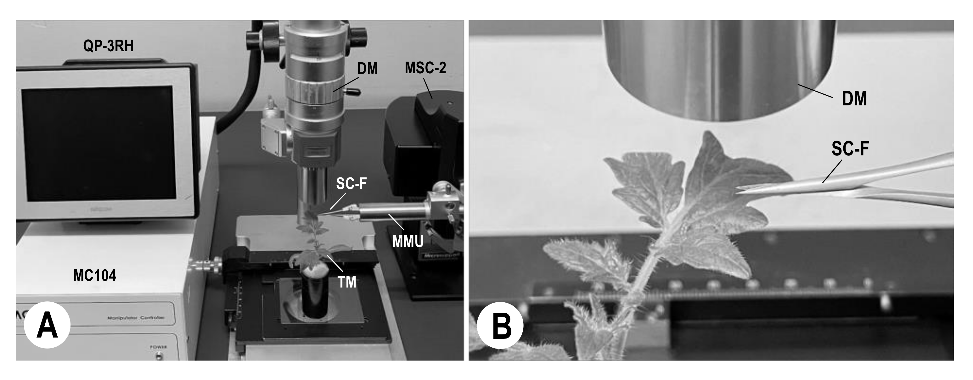
2.5. Collection of Single P. neolycopersici-Inoculated Tomato Leaf Type I Trichomes with Microscissors

Mature KTP-03 conidia (n = 880) were inoculated onto type I trichomes of 14-day-old MM seedlings (n = 880); KTP-03-inoculated MM seedlings were incubated in an LH-240N growth chamber for 0–12 days under the conditions described above. There were eight fungal developmental stages (from conidial attachment to complete cessation of hyphal growth on trichomes): non-germinated conidia, appressorial germ tube-formed conidia, primary appressorium-formed conidia, secondary (one colony-forming) hyphae-possessing conidia, two colony-forming hyphae-possessing conidia, three colony-forming hyphae-possessing conidia, four colony-forming hyphae-possessing conidia, and hyphal growth-ceased conidia. Type I trichomes that had not been inoculated with KTP-03 conidia (i.e., non-inoculated) were prepared as a control condition. Furthermore, all hyphae (including conidia, germ tubes and appressoria) grown on the trichomes were removed using a small glass needle installed in a micromanipulator (as mentioned above); single type I trichomes that possessed only one haustorium or 3–5 haustoria of KTP-03 were prepared. The basal cells of trichomes not inoculated and inoculated with single conidia, as well as trichomes that possessed only haustoria of KTP-03, were cut separately using precision scissors (microscissors) (SC-F; Micro Support, Shizuoka, Japan) inserted into the main microscissors unit and subsequently installed in the manipulator (QuickPro MSC-2; Micro Support). By operating the user interface controller (QP-3RH) and the opening/closing dial in accordance with the instructions of the manufacturer (Micro Support), after powering the motion controller (MC104) (Figure 1A), a single trichome cell was successfully cut with the SC-F microscissors and then collected (Figure 1B).

2.6. Amplification of 5.8S rDNA-ITS Sequence by PCR

To detect ribosomal DNA internal transcribed spacer (rDNA-ITS; AB094991) regions in the genome of P. neolycopersici, the collected single KTP-03-non-inoculated/inoculated and hyphae-removed leaf type I trichomes were directly transferred into 25 µL of TE buffer (10 mM tris aminomethane, 1 mM ethylenediaminetetraacetic acid, pH 8.0) (Nacalai Tesque) in a 1.5-mL Eppendorf tube (As One, Osaka, Japan). One pair of primers was artificially constructed on the basis of their nucleotide sequences in the internal regions of the rDNA-ITS: ITS5/ITS4 [44], as shown in Table 1. The solution (23 µL) was mixed with 2 µL of each primer (10 µM; ITS5 and ITS4) and 25 µL of 2 × Quick TaqTM HS DyeMix (Toyobo, Osaka, Japan) in a single 200-µL Flat 1000 PCR tube (NIPPON Genetics, Osaka, Japan). Genomic PCR was performed using the following protocol: initial denaturation at 94 °C for 2 min, followed by 40 cycles of denaturing at 94 °C for 30 s, annealing at 56 °C for 30 s, extension at 68 °C for 1 min, and a final incubation at 68 °C for 7 min. Amplicons resulting from PCR were run on 2% agarose gels. Experiments were repeated 20 times at each fungal developmental stage (n = 220 type I trichomes). Fragments were sent to Fasmac (Kanagawa, Japan) for sequencing, which was conducted with the same primers used for the amplifications.

2.7. Transcript Amplification of Tomato Plant- and Powdery Mildew Fungus-Derived Genes by suc-PCR (RT-PCR/N-PCR)

Tomato plant- and powdery mildew fungus-derived genes, selected from the cDNA database in the DNA Data Bank of Japan (DDBJ), were used as PCR targets in this study: plasma membrane H+-ATPase (LHA2; AF179442) [46,47] and β-tubulin (TUB2; MK355657), respectively. For amplification of these genes, four pairs of primers were artificially constructed on the basis of their nucleotide sequences in the internal regions of the cDNAs. The primers were lha-1(lpRT-1/lpRT-2) and lha-2(lpN-1/lpN-2) [45] for the LHA2 gene, and tub-1(tbRT-1/tbRT-2) and tub-2 (tbN-1/tbN-2) for the TUB2 gene, as shown in Table 1. Amplification of their gene transcripts was carried out by suc-PCR; the external primer pairs lha-1 and tub-1 were used for primary PCR (RT-PCR; first round of PCR), while the internal primer pairs lha-2 and tub-2 were used for the subsequent N-PCR (second round of PCR). To detect transcripts of the LHA2 gene in leaf type I trichome cells and the TUB2 gene in tomato powdery mildew fungi, the collected single KTP-03-non-inoculated/inoculated and hyphae-removed leaf type I trichomes were directly transferred into 25 µL of TE buffer in a 1.5-mL Eppendorf tube. The solution (20.5 µL) was mixed with 2 µL of each primer (10 µM; lha-1 or tub-1), 2.5 µL of 50 mM Mn(OAc)2 and 25 µL of 2 × RT-PCR Quick Master Mix (Toyobo) in a single 200-µL Flat 1000 PCR tube. For both genes, RT-PCR was performed with reverse transcription at 90 °C for 30 s and 60 °C for 30 min, with initial denaturation at 94 °C for 1 min, 40 cycles of denaturing at 94 °C for 30 s, annealing at 66 °C for 30 s, extension at 72 °C for 1 min, and final incubation at 72 °C for 7 min. A 3-µL aliquot of the reaction mixture was mixed with 2 µL of each primer (10 µM; lha-2 or tub-2), 20 µL of sterile water (H2O) and 25 µL of 2 × Quick TaqTM HS DyeMix in a single 200-µL Flat 1000 PCR tube. For both genes, N-PCR was performed in accordance with the following protocol: initial denaturation at 94 °C for 2 min, followed by 40 cycles of denaturing at 94 °C for 30 s, annealing at 62 °C for 30 s, extension at 68 °C for 1 min, and final incubation at 68 °C for 7 min. The final PCR products were electrophoresed on 2% agarose gels; the lengths of the amplified products were compared to those of the original cDNAs. The experiments were conducted 20 times at each fungal developmental stage (n = 440 type I trichomes). Fragments were sent to Fasmac for sequencing. Sequencing was conducted with the same primers used for the amplifications.

2.8. Simultaneous Transcript Amplification of Tomato Plant- and Powdery Mildew Fungus-Derived Genes by Multiplex RT-PCR/N-PCR

Constitutively expressed genes (LHA2 and TUB2) were used for multiplex RT-PCR/N-PCR amplification. The collected single KTP-03-non-inoculated/inoculated and hyphae-removed leaf type I trichomes were directly transferred into 25 µL of TE buffer in a 1.5-mL Eppendorf tube. The solution (18.5 µL) was mixed with 4 µL of each primer (10 µM; lha-1 and tub-1), 2.5 µL of 50 mM Mn(OAc)2 and 25 µL of 2 × RT-PCR Quick Master Mix in a single 200-µL Flat 1000 PCR tube. The first round of amplification, involving two pairs of RT-PCR primers (lha-1 and tub-1), was performed under the same conditions described above. A 3-µL aliquot of the reaction mixture was mixed with 4 µL of each primer (10 µM; lha-2 and tub-2), 18 µL of sterile water (H2O) and 25 µL of 2 × Quick TaqTM HS DyeMix in a single 200-µL Flat 1000 PCR tube. The second round of amplification, involving two pairs of N-PCR primers (lha-2 and tub-2), was performed under the same conditions mentioned above. The final PCR products were electrophoresed on 2% agarose gels. The experiments were conducted 20 times at each fungal developmental stage (n = 220 type I trichomes).

3. Results

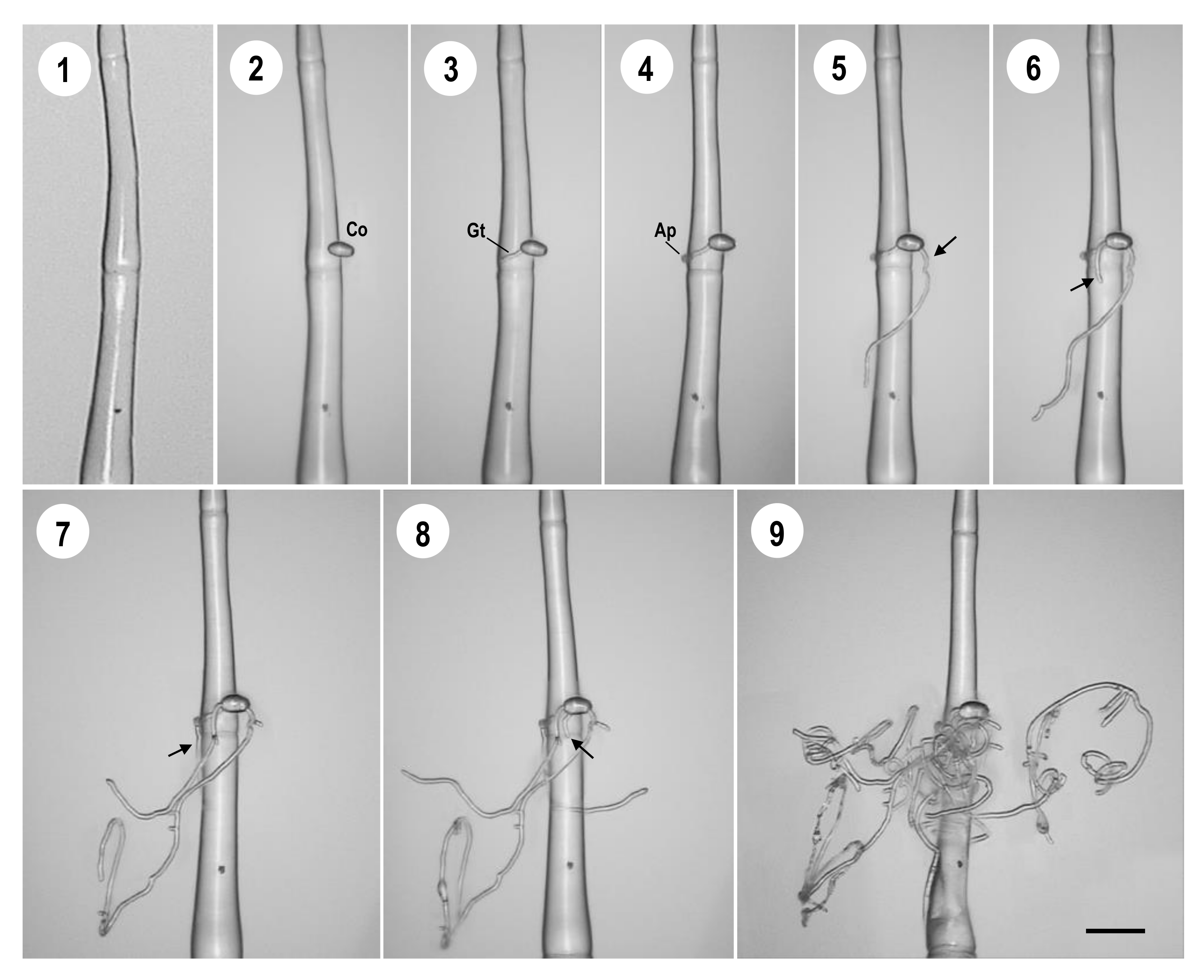
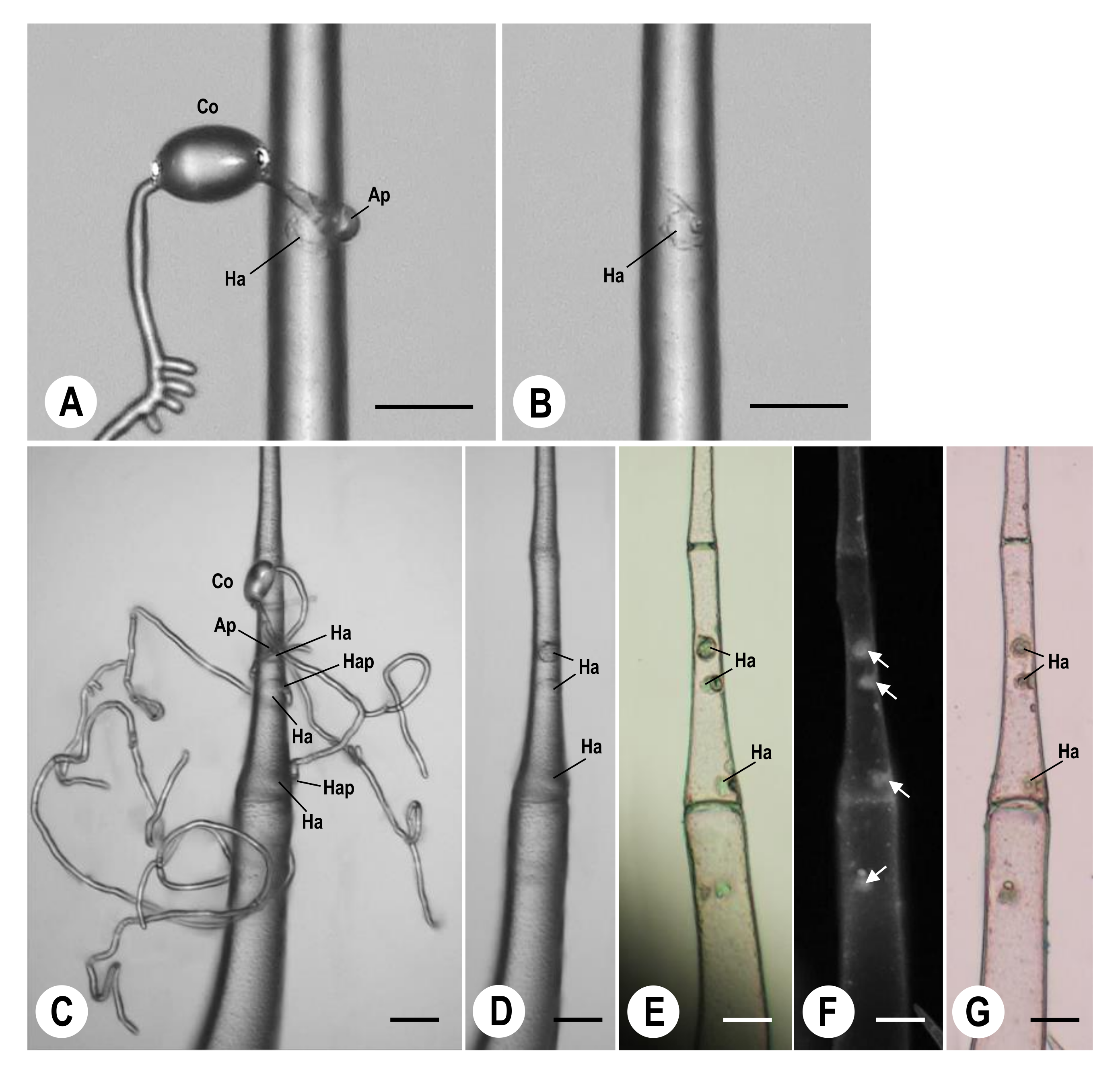
3.1. Analysis of Infection Processes of P. neolycopersici on Leaf Type I Trichome Cells of MM

The infection process of KTP-03 isolates was consecutively monitored on leaf type I trichomes of MM, using a KH-2700 DM (Figure 2). Figure 2—1 shows a single leaf type I trichome of MM. Individual KTP-03 conidia were positioned directly onto the single trichome (Figure 2—2). All conidia (approximately 20) of KTP-03 germinated on the trichomes at 3–5 h (Figure 2—3), and then produced primary appressoria at 6–10 h after inoculation (Figure 2—4). Secondary hyphae were produced from the conidia within 48 h after inoculation (Figure 2—5). In addition, the KTP-03 produced elongating colony-forming hyphae from the conidia: two hyphae at 3 days (Figure 2—6), three hyphae at 4 days (Figure 2—7), and four hyphae at 5 days (Figure 2—8). From a single conidium, a maximum of four hyphae (containing germ tubes) were formed: hyphal development completely ceased at approximately 12 days after inoculation (Figure 2—9). Compared with the infection processes on tomato leaf epidermal cells, no conidiophores were formed on the infected trichomes during the experimental period (Figure 2—9). Haustoria were observed in the trichome cells when using the KH-2700 DM (Figure 3A–D), BX-60 EM (Figure 3E,F) and BX-60 LM (Figure 3G). Figure 3B,D show that one and four haustoria, respectively, formed in single trichome cells after removing all hyphae, including conidia, or after removing germ tubes and appressoria, from the trichome cells at 2 (Figure 3A) and 7 days (Figure 3C) after inoculation with single KTP-03 conidia onto the trichome cells. Using the same samples, we observed haustoria (Ha) and papilla-like structures (white one-sided arrows in Figure 3F) in trichome cells with the BX-60 EM instrument (light, Figure 3E; fluorescence, Figure 3F); we observed pale brown haustoria in trichome cells with the BX-60 LM after histochemical staining (Figure 3G). KTP-03 haustoria were transparent globular structures in the trichome cells surrounded by papillae.

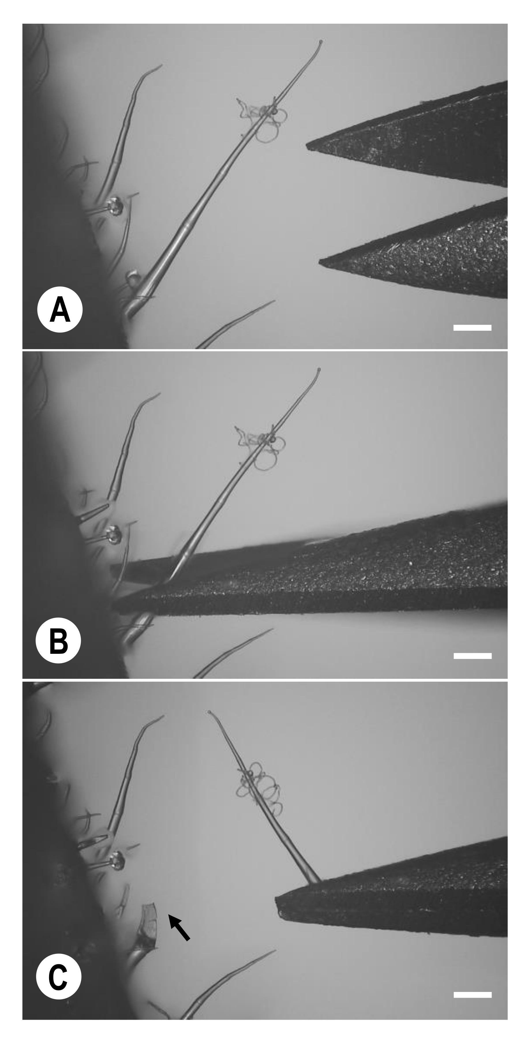
3.2. Collection of Single P. neolycopersici-Inoculated Type I Trichomes Using Microscissors

Using small, pointed scissors (SC-F microscissors) held by the MSC-2 manipulator under a KH-2700 DM (Figure 4), the basal cells of type I trichomes (14-day-old MM seedlings) not inoculated (Figure 2—1) and inoculated with single KTP-03 conidia were collected individually. For the inoculated trichomes, we included cells at various fungal infection stages over 12 days (Figure 2—2–9), and hyphae-removed type I trichomes with a haustorium (Figure 3B) or 3–5 haustoria (Figure 3D). Using the collected trichomes, PCR was directly conducted without DNA or RNA isolation in the next experiment to detect gene expression patterns in single type I trichome cells and fungal cells during each P. neolycopersici infection stage (from conidial attachment to complete cessation of hyphal growth).

3.3. Amplification of Fungal 5.8S rDNA-ITS Sequences by PCR from Single P. neolycopersici-Inoculated Type I Trichome Cells

Amplification of the 5.8S rDNA-ITS region was carried out by genomic PCR (using the ITS5/ITS4 primer pair, Table 1). The PCR amplification success rate was 100% for the 5.8S rDNA-ITS region in 20 conidia inoculated onto trichomes at each fungal infection stage, and in hyphae-removed trichomes that possessed a haustorium or 3–5 haustoria (Table 2). The nucleotide length amplified by PCR was 664 base pairs (bp) for the 5.8S rDNA-ITS region (Figure 5). The nucleotide sequence was examined to confirm that the fragment constituted the 5.8S rDNA-ITS region. The sequence was deposited in Genbank under accession number LC663220. In addition, BLAST analysis indicated that the entire 5.8S rDNA-ITS region of KTP-03 was identical to the tomato powdery mildew fungus P. neolycopersici (MG846017, MH137258 and AB032484 assigned in DDBJ). Overall, genomic PCR successfully amplified the 5.8S rDNA-ITS region from single KTP-03-inoculated leaf type I trichomes and single KTP-03-haustorium-possessing leaf type I trichomes.

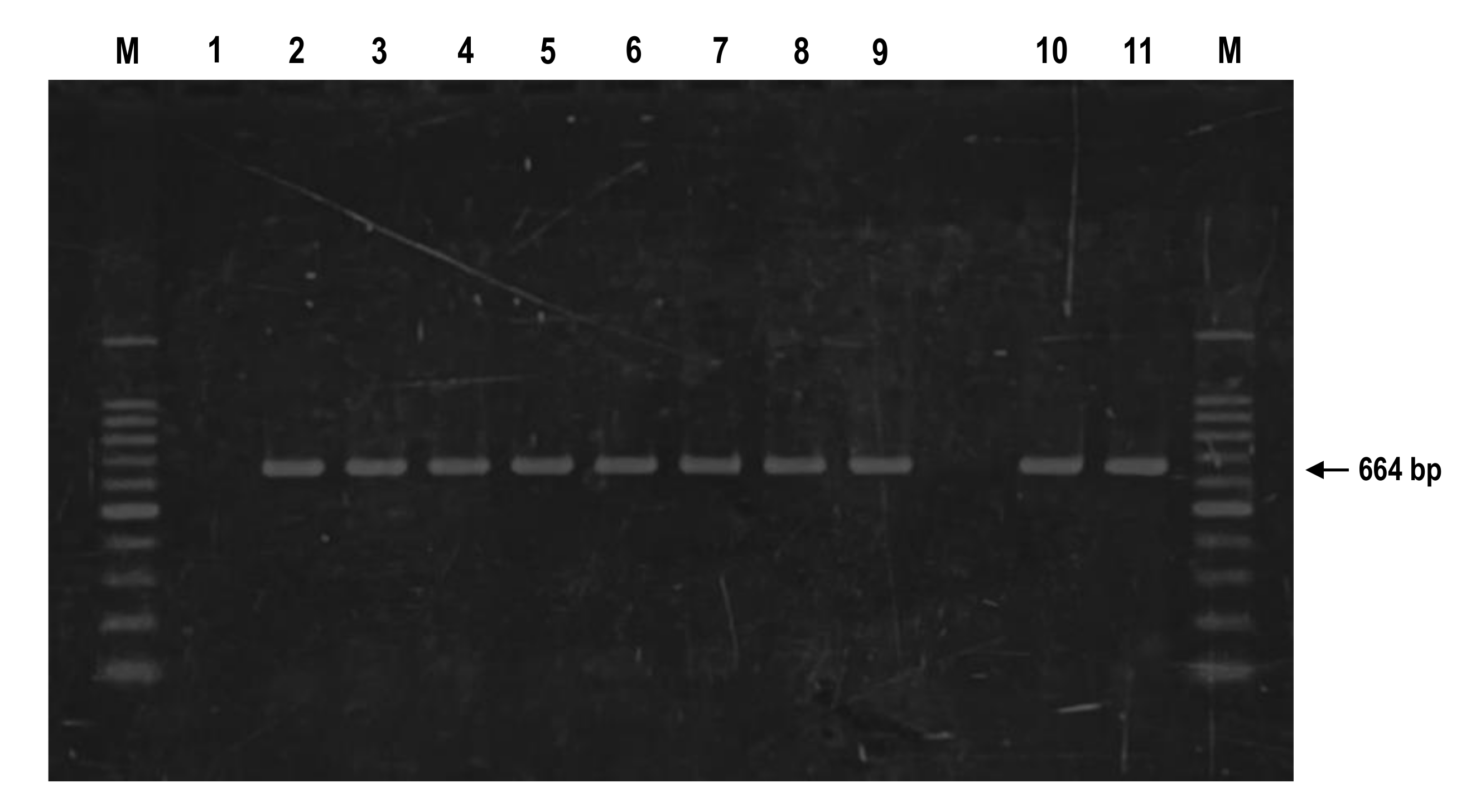
3.4. Transcript Detection of Plant- and Fungus-Derived Genes by suc-PCR (RT-PCR/N-PCR) from Single P. neolycopersici-Inoculated Type I Trichome Cells

Two constitutively expressed genes, LHA2 for tomato genes and TUB2 for powdery mildew fungus genes, were used as PCR targets. Prior to the analyses of the PCR amplification with P. neolycopersici-inoculated type I trichomes, the nucleotide lengths of genomic DNAs of LHA2 and TUB2 were compared to the lengths of their original cDNAs. The LHA2 and TUB2 genes contained three and two intron sequences in the regions amplified using primers lha-2 and tub-2, respectively (Table 1). The nucleotide lengths to be amplified by suc-PCR were 351 bp for LHA2 and 174 bp for TUB2. Successful amplification of LHA2 and TUB2 transcripts was achieved in P. neolycopersici-non-inoculated/inoculated trichome cells (Figure 6A, lanes 1–9) and P. neolycopersici-inoculated trichome cells (Figure 6B, lanes 2–9), at each fungal infection stage. Furthermore, these transcripts were amplified successfully in hyphae-removed trichome cells that possessed a haustorium or 3–5 haustoria (Figure 6A,B, lanes 10 and 11). The success rate of suc-PCR amplification was approximately 97.7% for LHA2 transcripts in 220 trichomes and 94.5% for TUB2 transcripts in 200 conidia inoculated onto trichomes (20 trichomes and 20 conidia at each fungal infection stage), in hyphae-removed trichomes that possessed a haustorium and 3–5 haustoria, respectively (Table 2). Sequences were deposited in GenBank under accession number LC663222 for LHA2 and LC663221 for TUB2. In addition, BLAST analysis indicated that the partial LHA2 gene of MM and partial TUB2 gene of KTP-03 were identical to the tomato S. lycopersicum (NM_001247548 and AF179442) and tomato powdery mildew fungus P. neolycopersici (MK355657 assigned in DDBJ). Overall, intron-spliced mRNAs were successfully amplified by suc-PCR on genetic material from single P. neolycopersici-non-inoculated/inoculated leaf type I trichome cells, and from single trichome cells that possessed only powdery mildew haustoria.

3.5. Simultaneous Transcript Detection of Plant- and Fungus-Derived Genes by Multiplex RT-PCR/N-PCR from Single P. neolycopersici-Inoculated Type I Trichome Cells

To simultaneously detect gene expression patterns in the infected trichomes, we amplified transcripts using mixed primers for multiplex RT-PCR/N-PCR of LHA2 and TUB2 genes. The multiplex RT-PCR/N-PCR method yielded two bands: 351 bp for LHA2 and 174 bp for TUB2 (Figure 6C). Those transcripts were also successfully amplified in P. neolycopersici-inoculated trichome cells at each fungal infection stage (Figure 6C, lanes 2–9), as well as in hyphae-removed trichome cells that possessed a haustorium or 3–5 haustoria (Figure 6C, lanes 10 and 11). The success rate of multiplex RT-PCR/N-PCR amplification was approximately 98.2% for LHA2 transcripts in 220 trichomes and 95.0% for TUB2 transcripts in 200 conidia inoculated onto trichomes (20 samples at each fungal infection stage), in hyphae-removed trichomes that possessed a haustorium and 3–5 haustoria, respectively (Table 2). Overall, these results indicated that intron-spliced mRNAs were simultaneously amplified with multiplex RT-PCR/N-PCR of single P. neolycopersici-non-inoculated/inoculated leaf type I trichome cells, and from single trichome cells that possessed only powdery mildew haustoria.

4. Discussion

Previous studies observed specific steps in the infection processes of P. neolycopersici including appressoria [20,48], haustoria [49,50,51,52], and conidiophores [50,53,54,55] in tomato leaf epidermal cells via histological staining. Such studies typically require chemical treatments (e.g., chlorophyll removal, fixation and staining) to clearly observe morphology (haustoria) and cytological responses (hypersensitive cell death or papillae) in epidermal cells. In our previous and current studies, we focused on the analysis of infection processes of powdery mildew fungi in leaf type I trichome cells of MM plants using the KH-2700 DM without histochemical staining [37]. Our results in Suzuki et al. [37] showed that the cytological responses of leaf trichome cells to non-pathogenic powdery mildew species (Blumeria graminis f. sp. hordei Marchal Race1, Erysiphe trifoliorum Greville and Podosphaera xanthii Pollacci) were identical to the cytological responses of epidermal cells of MM, including papilla-like structures respondes to E. trifoliorum (from red clover), and a hypersensitive cell death response to B. graminis f. sp. hordei (from barley) and P. xanthii (from melon). Using the KH-2700 DM, we could observe functional mature haustoria (haustoria beneath primary appressoria and haustoria beneath hyphal appressoria) in single P. neolycopersici-infected trichome cells. With a field-emission scanning electron microscope (FE-SEM), we further confirmed that the functional mature haustorium contains a haustorial body and several haustorial lobes embedded in an extrahaustorial matrix and surrounded by an extrahaustorial membrane [38].
In the present study, we had three main aims. First, we aimed to clarify the best collection methods for single KTP-03-inoculated trichomes using microscissors. Second, we aimed to identify the developmental processes of KTP-03 (at each fungal infection stage) on trichomes. Third, we aimed to characterise individual and simultaneous gene expression patterns in single trichome cells and/or fungal cells (possessing only haustoria). Consequently, we successfully cut and collected the basal cells of KTP-03-inoculated leaf type I trichomes using microscissors under the KH-2700 DM. The cell content could be used in N-PCR to efficiently amplify target genes. For N-PCR, two sets of primers (external and internal primer pairs for the target sequences) were designed in accordance with the approach of Pepper and Dowd [56]. The first primer pair constituted the external primers for amplification of a trace quantity of target sequences by PCR; the second primer pair constituted the internal primers for subsequent amplification of target sequences by N-PCR, using a higher annealing temperature that permitted amplification via these primer pairs alone. For example, using the primary PCR/N-PCR method, Llop et al. [57] successfully detected a specific DNA fragment of plasmid PEA29 present in trace amounts of bacterial samples (Erwinia amylovora). In addition, Matsuda et al. [58] successfully amplified the entire 5.8S rDNA-ITS region from a single pre- and post-germinated conidium of tomato and red clover powdery mildew fungi. They demonstrated that DNA fragments were amplified in detectable amounts when N-PCR was performed, but not when only a single round of PCR amplification was performed. The use of N-PCR was essential because of the small quantity of primary PCR product obtained from single KTP-03-inoculated or hyphae-removed leaf type I trichome cells. In the second and third experiments, we used suc-PCR (RT-PCR/N-PCR) and multiplex RT-PCR/N-PCR for individual and simultaneously detection of the transcripts of each target gene from plant and/or fungal cells, respectively. Concerning the target genes, Matsuda et al. [59] confirmed LHA2 gene expression through analysis of the intracellular contents of a single tomato leaf type I trichome cell extracted using a microinjection method; Matsuda et al. [58] confirmed β-tubulin (TUB2-ol) gene expression in the intracellular contents of a single P. neolycopersici conidium collected using a micromanipulation method. The LHA2 gene (encoding plasma membrane H+-ATPase; [46,47]) and TUB2 gene (encoding β-tubulin biosynthesis; [60]) are particularly useful because both are constitutively expressed and serve as excellent transcriptional controls. Using suc-PCR for each individual gene and multiplex RT-PCR/N-PCR for both of them, one and two major bands were successfully detected through the amplification of intron-spliced mRNAs of both genes from single KTP-03-inoculated leaf type I trichomes of various fungal infection stages, and from single hyphae-removed trichome cells that possessed only haustoria, respectively. These results indicate that our PCR amplification method is effective for detecting gene expression patterns of intron-containing genes (e.g., LHA2 and TUB2). To our knowledge, this is the first report of a molecular assay method based on suc-PCR and multiplex RT-PCR/N-PCR for detecting the expression patterns of individual and multiple genes, respectively, in the intracellular contents of growing fungal cells (at various fungal infection stages) in single P. neolycopersici-inoculated leaf type I trichomes. We hope that the methods used in this study could be further applied to monitor the expression of both host and powdery mildew genes that are involved in the molecular mechanisms between plants and powdery mildew fungi, such as fungal genes encoding effectors and plant genes for host and non-host defence responses [61,62,63]. Moreover, in future, this employed technique will be available to other plant species and crops, possessing leaf trichome cells, to elucidate the molecular interactions between plants and pathogens.

5. Conclusions

In this study, the infection processes of the KTP-03 isolate were monitored on leaf type I trichomes of MM using a KH-2700 DM. Our results showed that conidia of KTP-03 infect tomato trichomes by forming haustoria. More importantly, we succeed in isolating single basal cells of powdery mildew infected trichomes using small, pointed scissors (microscissors) and using the cell content for detecting expression of genes of both tomato and powdery mildew fungi. Overall, these results indicated that the intron-spliced mRNAs (LHA2 and TUB2) could be individually or simultaneously amplified via suc-PCR and multiplex RT-PCR/N-PCR from single KTP-03-non-inoculated/inoculated leaf type I trichome cells.

Author Contributions

Conceptualization, S.I. and T.N.; methodology, S.I., Y.M. and T.N.; software, S.I. and Y.T.; validation, K.K., Y.M. and T.N.; formal analysis, S.I. and T.S.; investigation, S.I., N.O., Y.K., Y.T. and T.S.; resources, S.I. and T.N.; data curation, S.I. and T.N.; writing—original draft preparation, S.I. and T.N.; writing—review and editing, Y.B. and T.N.; visualization, Y.M., Y.B. and T.N.; supervision, T.N.; project administration, T.N. All authors have read and agreed to the published version of the manuscript.

Funding

This research received no external funding.

Institutional Review Board Statement

Not applicable.

Informed Consent Statement

Not applicable.

Data Availability Statement

Data sets analysed during the current study are available from the current author on reasonable request.

Acknowledgments

This work was supported by a Grant for Agriculture Technology and Innovation Research Institute, Kindai University. In addition, the English in this document has been checked by at least two professional editors, both native speakers of English.

Conflicts of Interest

The authors declare no conflict of interest.

References

  1. Xu, L.; Zhu, L.; Tu, L.; Guo, X.; Long, L.; Sun, L.; Gao, W.; Zhang, X. Differential gene expression in cotton defence response to Verticillium dahlia by SSH. J. Phytopathol. 2011, 159, 684–697. [Google Scholar] [CrossRef]
  2. Mohamed, I.A.; Shalby, N.; El-Badri, A.M.A.; Saleem, M.H.; Khan, M.N.; Nawaz, M.A.; Qin, M.; Agami, R.A.; Kuai, J.; Wang, B. Stomata and xylem vessels traits improved by melatonin application contribute to enhancing salt tolerance and fatty acidcomposition of Brassica napus L. plants. Agronomy 2020, 10, 1186. [Google Scholar] [CrossRef]
  3. Sudisha, J.; Sharathchandra, R.; Amruthesh, K.; Kumar, A.; Shetty, H.S. Pathogenesis related proteins in plant defense response. In Plant Defence: Biological Control; Springer: Berlin/Heidelberg, Germany, 2012; pp. 379–403. [Google Scholar]
  4. Munis, M.F.H.; Tu, L.; Deng, F.; Tan, J.; Xu, L.; Xu, S.; Long, L.; Zhang, X. A thaumatin-like protein gene involved in cotton fiber secondary cell wall development enhances resistance against Verticillium dahliae and other stresses in transgenic tobacco. Biochem. Biophys. Res. Commun. 2010, 393, 38–44. [Google Scholar] [CrossRef] [PubMed]
  5. Ebrahim, S.; Usha, K.; Singh, B. Pathogenesis related (PR) proteins in plant defense mechanism. Sci. Against Microb. Pathog. 2011, 2, 1043–1054. [Google Scholar]
  6. Lawrence, C.B.; Singh, N.P.; Qiu, J.; Gardner, R.G.; Tuzun, S. Constitutive hydrolytic enzymes are associated with polygenic resistance of tomato to Alternaria solani and may function as an elicitor release mechanism. Physiol. Mol. Plant Pathol. 2000, 57, 211–220. [Google Scholar] [CrossRef]
  7. Klarzynski, O.; Plesse, B.; Joubert, J.M.; Yvin, J.C.; Kopp, M.; Kloareg, B.; Fritig, B. Linear β-1,3-glucans are elicitors of defense response in tobacco. Plant Physiol. 2000, 124, 1027–1037. [Google Scholar] [CrossRef] [Green Version]
  8. Hoffmann-Sommergruber, K. Plant allergens and pathogenesis-related proteins. Int. Arch. Allergy Immunol. 2000, 122, 155–166. [Google Scholar] [CrossRef]
  9. Manghwar, H.; Hussain, A.; Ullah, A.; Gul, S.; Shaban, M.; Khan, A.H.; Ali, M.; Sani, S.G.A.S.; Chaudhary, H.J.; Munis, M.F.H. Expression analysis of defense related genes in wheat and maize against Bipolaris sorokiniana. Physiol. Mol. Plant Pathol. 2018, 103, 36–46. [Google Scholar] [CrossRef]
  10. Sunera; Amna; Saqib, S.; Uddin, S.; Zaman, W.; Ullah, F.; Ayaz, A.; Asghar, M.; ur Rehman, S.; Munis, M.F.H.; et al. Characterization and phytostimulatory activity of bacteria isolated from tomato (Lycopersicin esculentum Mill.) rhizosphere. Microb. Pathog. 2020, 140, 103966. [Google Scholar]
  11. Manghwar, H.; Hussain, A.; Ali, Q.; Saleem, M.H.; Abualreesh, M.H.; Alatawi, A.; Ali, S.; Munis, M.F.H. Disease severity, resistance analysis, and expression profiling of pathogenesis-related protein genes after the inoculation of Fusarium equiseti in wheat. Agronomy 2021, 11, 2124. [Google Scholar] [CrossRef]
  12. White, J.F., Jr.; Johnston, S.A.; Wang, C.L.; Chin, C.K. First report of powdery mildew in greenhouse-grown tomatoes in New Jersey. Plant Dis. 1997, 81, 227. [Google Scholar] [CrossRef] [PubMed]
  13. Kiss, L.; Cook, R.T.A.; Saenz, G.S.; Cunnington, J.H.; Takamatsu, S.; Pascoe, I.; Bardin, M.; Nicot, P.C.; Sato, Y.; Rossman, A.Y. Identification of two powdery mildew fungi, Oidium neolycopersici sp. nov. and O. lycopersici, infecting tomato in different parts of the world. Mycol. Res. 2001, 105, 684–697. [Google Scholar] [CrossRef]
  14. Zheng, Z.; Nonomura, T.; Bóka, K.; Matsuda, Y.; Visser, R.G.F.; Toyoda, H.; Kiss, L.; Bai, Y. Detection and quantification of Leveillula taurica growth in pepper leaves. Phytopathology 2013, 103, 623–632. [Google Scholar] [CrossRef] [Green Version]
  15. Braun, U.; Shin, H.D.; Takamatsu, S.; Meeboon, J.; Kiss, L.; Lebeda, A.; Kitner, M.; Götz, M. Phylogeny and taxonomy of Golovinomyces orontii revisited. Mycol. Prog. 2019, 18, 335–357. [Google Scholar] [CrossRef]
  16. Matsuda, Y.; Kashimoto, K.; Takikawa, Y.; Aikami, R.; Nonomura, T.; Toyoda, H. Occurrence of new powdery mildew on greenhouse tomato cultivars. J. Gen. Plant Pathol. 2001, 67, 294–298. [Google Scholar] [CrossRef]
  17. Shimizu, K.; Matsuda, Y.; Nonomura, T.; Ikeda, H.; Tamura, N.; Kusakari, S.; Kimbara, J.; Toyoda, H. Dual protection of hydroponic tomatoes from rhizosphere pathogens Ralstonia solanacearum and Fusarium oxysporum f. sp. radicis-lycopersici and airborne conidia of Oidium neolycopersici with an ozone-generative electrostatic spore precipitator. Plant Pathol. 2007, 56, 987–997. [Google Scholar] [CrossRef]
  18. Nonomura, T.; Matsuda, Y.; Kakutani, K.; Takikawa, Y.; Toyoda, H. Physical control of powdery mildew (Oidium neolycopersici) on tomato leaves by exposure to corona discharge. Can. J. Plant Pathol. 2008, 30, 517–524. [Google Scholar] [CrossRef]
  19. Kashimoto, K.; Matsuda, Y.; Matsutani, K.; Sameshima, T.; Kakutani, K.; Nonomura, T.; Okada, K.; Kusakari, S.; Nakata, K.; Takamatsu, S.; et al. Morphological and molecular characterization for a Japanese isolate of tomato powdery mildew Oidium neolycopersici and its host range. J. Gen. Plant Pathol. 2003, 69, 176–185. [Google Scholar]
  20. Nonomura, T.; Nishitomi, A.; Matsuda, Y.; Soma, C.; Xu, L.; Kakutani, K.; Takikawa, Y.; Toyoda, H. Polymorphic change of appressoria by the tomato powdery mildew Oidium neolycopersici on host tomato leaves reflects multiple unsuccessful penetration attempts. Fungal Biol. 2010, 114, 917–928. [Google Scholar] [CrossRef] [PubMed]
  21. Nonomura, T.; Matsuda, Y.; Yamashita, S.; Akahoshi, H.; Takikawa, Y.; Kakutani, K.; Toyoda, H. Natural woody plant, Mallotus japonicus, as an ecological partner to transfer different pathotypic conidia of Oidium neolycopersici to greenhouse tomatoes. Plant Prot. Sci. 2013, 49, S33–S40. [Google Scholar] [CrossRef] [Green Version]
  22. Seifi, A.; Nonomura, T.; Matsuda, Y.; Toyoda, H.; Bai, Y. An avirulent tomato powdery mildew isolate induces localized acquired resistance to a virulent isolate in a spatiotemporal manner. Mol. Plant-Microbe Interact. 2012, 25, 372–378. [Google Scholar] [CrossRef] [PubMed] [Green Version]
  23. Jones, H.; Whipps, J.M.; Gurr, S.J. The tomato powdery mildew fungus Oidium neolycopersici. Mol. Plant Pathol. 2001, 2, 303–309. [Google Scholar] [CrossRef] [Green Version]
  24. Jacob, D.; David, D.R.; Sztjenberg, A.; Elad, Y. Conditions for development of powdery mildew of tomato caused by Oidium neolycopersici. Phytopathology 2008, 98, 270–281. [Google Scholar] [CrossRef] [Green Version]
  25. Kennedy, G.G.; Sorenson, C.F. Role of glandular trichomes in the resistance of Lycopersicon hirsutum f. glabratum to Colorado potato beetle (Coleoptera: Chrysomelidae). J. Econ. Entomol. 1985, 78, 547–551. [Google Scholar] [CrossRef]
  26. Duffey, S.S. Plant glandular trichomes: Their partial role in defense against insects. In Insects and the Plant Surface; Juniper, B.E., Southwood, T.E., Eds.; Arnold: London, UK, 1986; pp. 151–172. [Google Scholar]
  27. Peter, A.J.; Shanower, T.G. Plant glandular trichomes: Chemical factories with many potential uses. Resonance 1998, 3, 41–45. [Google Scholar] [CrossRef]
  28. Kessler, A.; Baldwin, I.T. Defensive function of herbivore-induced plant volatile emissions in nature. Science 2001, 291, 2141–2144. [Google Scholar] [CrossRef] [PubMed]
  29. Pichersky, E.; Gershenzon, J. The formation and function of plant volatiles: Perfumes for pollinator attraction and defense. Curr. Opin. Plant Biol. 2002, 5, 237–243. [Google Scholar] [CrossRef]
  30. Simmons, A.T.; Gurr, G.M. Trichomes of Lycopersicon species and their hybrids: Effects on pests and natural enemies. Agric. For. Entomol. 2005, 7, 265–276. [Google Scholar] [CrossRef]
  31. Nonomura, T.; Xu, L.; Wada, M.; Kawamura, S.; Miyajima, T.; Nishitomi, A.; Kakutani, K.; Takikawa, Y.; Matsuda, Y.; Toyoda, H. Trichome exudates of Lycopersicon pennellii form a chemical barrier to suppress leaf-surface germination of Oidium neolycopersici conidia. Plant Sci. 2009, 176, 31–37. [Google Scholar] [CrossRef]
  32. Kang, J.-H.; Liu, G.; Shi, F.; Jones, A.D.; Beaudry, R.M.; Howe, G.A. The tomato odorless-2 mutant is defective in trichome-based production of diverse specialized metabolites and broad-spectrum resistance to insect herbivores. Plant Physiol. 2010, 154, 262–272. [Google Scholar] [CrossRef] [Green Version]
  33. Tooker, J.F.; Peiffer, M.; Luthe, D.S.; Felton, G.W. Trichomes as sensors detecting activity on the leaf surface. Plant Signal. Behav. 2010, 5, 73–75. [Google Scholar] [CrossRef] [PubMed]
  34. Lemke, C.A.; Mutschler, M.A. Inheritance of glandular trichomes in crosses between Lycopersicon esculentum and L. pennellii. J. Am. Soc. Hort. Sci. 1984, 109, 592–596. [Google Scholar]
  35. Glover, B.J. Differentiation in plant epidermal cells. J. Exp. Bot. 2000, 51, 497–505. [Google Scholar] [CrossRef] [PubMed]
  36. McDowell, E.T.; Kapteyn, J.; Schmidt, A.; Li, C.; Kang, J.H.; Descour, A.; Shi, F.; Larson, M.; Schilmiller, A.; An, L.; et al. Comparative functional genomic analysis of Solanum glandular trichome types. Plant Physiol. 2011, 155, 524–539. [Google Scholar] [CrossRef] [Green Version]
  37. Suzuki, T.; Murakami, T.; Takizumi, Y.; Ishimaru, H.; Kudo, D.; Takikawa, Y.; Matsuda, Y.; Kakutani, K.; Bai, Y.; Nonomura, T. Trichomes: Interaction sites of tomato leaves with biotrophic powdery mildew pathogens. Eur. J. Plant Pathol. 2018, 150, 115–125. [Google Scholar] [CrossRef]
  38. Suzuki, T.; Nakao, Y.; Ura, R.; Nakaba, M.; Nishiyama, T.; Takikawa, Y.; Matsuda, Y.; Kakutani, K.; Bai, Y.; Nonomura, T. Correlation between total hypha length and haustoria number of Pseudoidium neolycopersici in type I trichome cells of tomato leaves. Austral. Plant Pathol. 2020, 49, 451–460. [Google Scholar] [CrossRef]
  39. Fujita, K.; Matsuda, Y.; Wada, M.; Hirai, Y.; Mori, K.; Moriura, N.; Nonomura, T.; Kakutani, K.; Toyoda, H. Powdery mildew pathogens can suppress the chitinase gene expression induced in detached inner epidermis of barley coleoptile. Plant Cell Rep. 2004, 23, 504–511. [Google Scholar] [CrossRef] [PubMed]
  40. Chandran, D.; Inada, N.; Hather, G.; Kleindt, C.K.; Wildermuth, M.C. Laser microdissection of Arabidopsis cells at the powdery mildew infection site reveals site-specific processes and regulators. Proc. Natl. Acad. Sci. USA 2010, 107, 460–465. [Google Scholar] [CrossRef] [Green Version]
  41. Ayaz, A.; Huang, H.; Zheng, M.; Zaman, W.; Li, D.; Saqib, S.; Zhao, H.; Lü, S. Molecular cloning and functional analysis of GmLACS2-3 reveals its involvement in cutin and suberin biosynthesis along with abiotic stress tolerance. Int. J. Mol. Sci. 2021, 22, 9175. [Google Scholar] [CrossRef]
  42. Nonomura, T.; Matsuda, Y.; Xu, L.; Kakutani, K.; Takikawa, Y.; Toyoda, H. Collection of highly germinatve pseudochain conidia of Oidium neolycopersici from conidiophores by electrostatic attraction. Mycol. Res. 2009, 113, 364–372. [Google Scholar] [CrossRef]
  43. Sameshima, T.; Kashimoto, K.; Kida, K.; Matsuda, Y.; Nonomura, T.; Kakutani, K.; Nakata, K.; Kusakari, S.; Toyoda, H. Cytological events in tomato leaves inoculated with conidia of Blumeria graminis f. sp. hordei and Oidium neolycopersici KTP-01. J. Gen. Plant Pathol. 2004, 70, 7–10. [Google Scholar] [CrossRef]
  44. White, T.J.; Bruns, T.; Lee, S.; Taylor, J. Amplification and direct sequencing of fungal ribosomal RNA genes for phylogenetics. In PCR Protocols: A Guide to Methods and Applications; Innis, M.A., Gelfand, D.H., Sninsky, J.J., White, T.J., Eds.; Academic Press: San Diego, CA, USA, 1990; pp. 315–322. [Google Scholar]
  45. Wada, M.; Matsuda, Y.; Fujita, K.; Nanjo, A.; Nishimura, M.; Nonomura, T.; Kakutani, K.; Toyoda, H. RT-PCR amplification of mRNAs in nuclei or cytosol of single cells of tomato callus after micropipette extraction. Plant Cell Tissue Organ Cult. 2004, 79, 109–114. [Google Scholar] [CrossRef]
  46. Ewing, N.N.; Wimmers, L.E.; Meyer, D.J.; Chetelat, R.T.; Bennett, A.B. Molecular cloning of tomato plasma membrane H+-ATPase. Plant Physiol. 1990, 94, 1874–1881. [Google Scholar] [CrossRef] [PubMed] [Green Version]
  47. Ewing, N.N.; Bennett, A.B. Assessment of the number and expression of P-type H+-ATPase genes in tomato. Plant Physiol. 1994, 106, 547–557. [Google Scholar] [CrossRef] [PubMed] [Green Version]
  48. Lebeda, A.; Mieslerová, B.; Petřivalský, M.; Luhová, L.; Špundová, M.; Sedlářová, M.; Nožková-Hlaváčková, V.; Pink, D.A.C. Resistance mechanisms of wild tomato germplasm to infection of Oidium neolycopersici. Eur. J. Plant Pathol. 2014, 138, 569–596. [Google Scholar] [CrossRef] [Green Version]
  49. Dyki, B.; Staniaszek, M. Infection of tomato by Oidium lycopersicum (Cooke & Massee, emend. Noordeloos & Loerakker). Phytopathol. Pol. 1997, 13, 13–17. [Google Scholar]
  50. Whipps, J.M.; Budge, S.P.; Fenlon, J.S. Characteristics and host range of tomato powdery mildew. Plant Pathol. 1998, 47, 36–48. [Google Scholar] [CrossRef]
  51. LaMondia, J.A.; Smith, V.L.; Douglas, S.M. Host range of Oidium lycopersicum on selected Solanaceous species in Connecticut. Plant Dis. 1999, 83, 341–344. [Google Scholar] [CrossRef]
  52. Li, C.; Faino, L.; Dong, L.; Fan, J.; Kiss, L.; de Giovanni, C.; Lebeda, A.; Scott, J.; Matsuda, Y.; Toyoda, H.; et al. Characterization of polygenic resistance to powdery mildew in tomato at cytological, biochemical and gene expression level. Mol. Plant Pathol. 2012, 13, 148–159. [Google Scholar] [CrossRef]
  53. Dyki, B. Morphogenesis of pathogen causing powdery mildew in Polish cultivation of tomato. Veg. Crop. Res. Bull. 2003, 59, 131–138. [Google Scholar]
  54. Oichi, W.; Matsuda, Y.; Sameshima, T.; Nonomura, T.; Kakutani, K.; Nishimura, H.; Kusakari, S.; Toyoda, H. Consecutive monitoring for conidiogenesis by Oidium neolycopersici on tomato leaves with a high-fidelity digital microscope. J. Gen. Plant Pathol. 2004, 70, 329–332. [Google Scholar] [CrossRef]
  55. Oichi, W.; Matsuda, Y.; Nonomura, T.; Toyoda, H.; Xu, L.; Kusakari, S. Formation of conidial pseudochains by tomato powdery mildew Oidium neolycopersici. Plant. Dis. 2006, 90, 915–919. [Google Scholar] [CrossRef] [Green Version]
  56. Pepper, I.L.; Dowd, S.E. PCR applications for plant and soil microbes. In Manual of Environmental Microbiology; Hurst, C.J., Crawford, R.L., Knudsen, G.R., McInerney, M.J., Stetzenbach, L.D., Eds.; ASM Press: Washington, DC, USA, 2002; pp. 573–582. [Google Scholar]
  57. Llop, P.; Bonaterra, A.; Penalver, J.; Lopez, M.M. Development of a high sensitive nested-PCR procedure using a single closed tube for detection of Erwinia amylovora in asymptomatic plant material. Appl. Environ. Microbiol. 2000, 66, 2071–2078. [Google Scholar] [CrossRef] [Green Version]
  58. Matsuda, Y.; Sameshima, T.; Moriura, N.; Inoue, K.; Nonomura, T.; Kakutani, K.; Nishimura, H.; Kusakari, S.; Takamatsu, S.; Toyoda, H. Identification of individual powdery mildew fungi infecting leaves and direct detection of gene expression by single conidium polymerase chain reaction. Phytopathology 2005, 95, 1137–1143. [Google Scholar] [CrossRef] [Green Version]
  59. Matsuda, Y.; Kakutani, K.; Nishimura, M.; Wada, M.; Nonomura, T.; Toyoda, H. Direct RT-PCR amplification of mature mRNAs in single trichome cells of plant leaves. Recent Res. Dev. Cell Biol. 2003, 1, 145–153. [Google Scholar]
  60. Janke, C.; Magiera, M.M. The tubulin code and its role in controlling microtubule properties and functions. Nat. Rev. 2020, 21, 307–326. [Google Scholar] [CrossRef]
  61. Ellis, J. Insights into nonhost disease resistance: Can they assist disease control in agriculture? Plant Cell 2006, 18, 523–528. [Google Scholar] [CrossRef] [PubMed] [Green Version]
  62. Jones, J.D.G.; Dangl, J.L. The plant immune system. Nature 2006, 444, 323–329. [Google Scholar] [CrossRef] [PubMed] [Green Version]
  63. Schweizer, P. Nonhost resistance of plants to powdery mildew—New opportunities to unravel the mystery. Physiol. Mol. Plant Pathol. 2007, 70, 3–7. [Google Scholar] [CrossRef]
Figure 1. Single KTP-03-inoculated tomato leaf type I trichome collection system combining digital microscopy and micromanipulation techniques. (A) A 14-day-old tomato seedling TM was placed under a KH-2700 digital microscope (DM). Precision scissors (microscissors) (SC-F) inserted into the microscissors main unit (MMU) were installed in the manipulator (MSC-2). After powering the motion controller (MC104), the single trichome cells were cut using SC-F microscissors by operating the user interface controller (QP-3RH) and the opening/closing dial. (B) Enlarged photograph of the devices used to cut and collect single leaf type I trichome cells using SC-F microscissors under a KH-2700 DM.
Agriculture 12 00254 g001
Figure 2. Infection processes of KTP-03 on leaf type I trichome cells of MM observed continuously with a KH-2700 DM. (1) and (2) Micrographs were acquired before (1) and after inoculation with a KTP-03 conidium (Co) onto single leaf type I trichome cells (2). (39) Micrographs were recorded at (3) 3 h, (4) 6 h, (5) 2 days, (6) 3 days, (7) 4 days, (8) 5 days, and (9) 12 days after inoculation with a KTP-03 conidium onto the trichome cell. A KTP-03 conidium formed germ tubes (Gt) on the trichomes at approximately 3 h, primary appressoria (Ap) at approximately 6 h, and haustoria within 2 days after inoculation. Note that the fungus forms colony-forming hyphae (arrows) from a conidial body and continues to vigorously produce elongating hyphae on the trichome after inoculation. As shown in (9), hyphal development completely ceased without conidiophore formation on the trichome at 12 days after inoculation. Bars indicate 40 µm.
Agriculture 12 00254 g002
Figure 3. Digital (AD), light and epifluorescent (E,F), and light (G) micrographs showing haustoria of KTP-03 and papilla-like structures in leaf type I trichome cells of tomato cv. Moneymaker (MM) in the same field of view. (A,C) KTP-03 conidium (Co) formed one haustorium (Ha) beneath the primary appressorium (Ap) (A) and 3–4 haustoria (Ha) beneath the primary appressorium (Ap) and hyphal appressoria (Hap) on the trichome cells (C). (B,D) One (B) and three haustoria (D) located in a trichome cell after the removal of KTP-03 hyphae from the trichome cells shown in (A,C) (at 2 days and 7 days after inoculation of KTP-03 conidia), respectively, using a small glass needle. (E,F) Without chemical treatment, haustoria (Ha) exhibited spherical structures (E) and papilla-like structures (white one-sided arrows) fluoresced in a single trichome cell (F). (G) With chemical treatment, haustoria (Ha) were stained light brown. Micrographs were acquired at 2 days (A) and 7 days (C) and after inoculation with KTP-03 conidia. Bars indicate 20 µm.
Agriculture 12 00254 g003
Figure 4. Procedure for the collection of single KTP-03-non-inoculated/inoculated leaf type I trichome cells of tomato cv. Moneymaker (MM). The basal cells of single trichomes were cut using small, pointed scissors (microscissors) installed in a micromanipulator. The microscissors were placed near the trichome basal cell (A) and directed towards that cell (B). Finally, the KTP-03-inoculated trichomes were cut using the microscissors held by the MSC-2 manipulator under a KH-2700 DM (C). Arrow in (C) indicates the location where a single trichome cell was successfully cut. Bars indicate 150 µm.
Agriculture 12 00254 g004
Figure 5. Electrophoretic detection of the amplified 5.8S rDNA-internal transcribed spacer (ITS) region of KTP-03 conidia inoculated onto single leaf type I trichome cells of tomato cv. Moneymaker (MM) by genomic PCR. Amplification of the 5.8S rDNA-ITS from KTP-03 was conducted using the ITS5/ITS4 external primer pair (Table 1). Electrophoretic products were obtained by 5.8S rDNA-ITS genomic PCR from single KTP-03-non-inoculated/inoculated leaf type I trichomes that had been collected individually using microscissors at various fungal infection stages (shown in Figure 2 and Figure 3). Lanes 1–9: electrophoresis products obtained in P. neolycopersici-non-inoculated/inoculated leaf type I trichomes (correspond to digital micrographs 1–9 in Figure 2). Lanes 10 and 11: electrophoresis products obtained in hyphae-removed leaf type I trichomes (10 correspond to digital micrographs in Figure 3B, and 11 in Figure 3D). Lane M shows the DNA ladder of a molecular weight marker (Toyobo, Osaka, Japan).
Agriculture 12 00254 g005
Figure 6. Electrophoretic detection of LHA2 and TUB2 genes amplified from single KTP-03-non-inoculated/inoculated leaf type I trichomes of tomato cv. Moneymaker (MM) via suc-PCR (RT-PCR/N-PCR) and multiplex RT-PCR/N-PCR, (for the detection of one and two genes, respectively). (A,B) The LHA2 gene from trichome cells (A) and TUB2 gene from KTP-03 (B) were amplified in the first round using an external primer pair (lhaRT-1/lhaRT-2 and tbRT-1/tbRT-2, respectively); this was followed by N-PCR using an internal primer pair, along with lhaN-1/lhaN-2 or tbN-1/tbN-2, respectively. (C) The LHA2 and TUB2 genes were amplified in Figure 1. (lhaRT-2 and tbRT-1/tbRT-2, respectively); this was followed by N-PCR using an internal primer pair (lhaN-1/lhaN-2 and tbN-1/tbN-2, respectively). Electrophoretic products were obtained by RT-PCR/N-PCR of LHA2 and TUB2; multiplex RT-PCR/N-PCR of LHA2 and TUB2 was conducted using single KTP-03-non-inoculated/inoculated leaf type I trichome cells collected individually using microscissors at various fungal infection stages (shown in Figure 2 and Figure 3). Lanes 1–9: electrophoresis products obtained in P. neolycopersici-non-inoculated/inoculated leaf type I trichomes (correspond to digital micrographs 1–9 in Figure 2). Lanes 10 and 11: electrophoresis products obtained in hyphae-removed leaf type I trichomes (10 correspond to digital micrographs in Figure 3B, and 11 in Figure 3D). Lane M shows the DNA ladder of a molecular weight marker.
Agriculture 12 00254 g006
Table 1. Primers used in this study.
PCR TargetsPrimersPrimer Sequences
rDNA-ITSITS-4 a5′-TCCTCCGCTTATTGATATGC-3′
ITS-5 a5′-GGAAGTAAAAGTCGTAACAAGG-3′
LHA2lpRT-1 b5′-GCCAAAGGTGTTGACGCAGATAC-3′
lpRT-2 b5′-CACTCCAAGATTCAAAGCCCTCCT-3′
lpN-1 b5′-AGAACCAGGATGCCATTGAC-3′
lpN-2 b5′-ACAAAGGCAGGAGACCAATG-3′
TUB2tbRT-15′-AACCAAATTGGGGCTGCTTTC-3′
tbRT-25′-CCCAGTTGTTACCAGCCCC-3′
tbN-15′-GCCTTGACGGATCTGGTGT-3′
tbN-25′-GTCGAAAGAGCTGACCGAA-3′
a Primers designed by White et al. [44]. b Primers designed by Wada et al. [45].
Table 2. PCR detection of gene transcripts in the intracellular contents of plant and/or fungal cells with single P. neolycopersici-non-inoculated/inoculated leaf type I trichomes of tomato (Solanum lycopersicum Mill. cv. Moneymaker).
PCR
Methods
Target GenesRates of Gene Expression Detection (%) x
1234567891011 y
PCR/N-PCR5.8S rDNA-ITSND z100.0100.0100.0100.0100.0100.0100.0100.0100.0100.0
RT-PCR/N-PCRLHA2100.0100.0100.0100.095.0100.095.090.0100.095.0100.0
TUB2ND80.085.0100.085.0100.095.0100.0100.0100.0100.0
Multiplex RT-PCR/N-PCRLHA2100.0100.0100.0100.095.0100.095.095.0100.095.0100.0
TUB2ND85.090.0100.095.0100.095.085.0100.0100.0100.0
x Rates of successful PCR amplification of the 5.8S rDNA-ITS region, LHA2 gene, and TUB2 gene using single KTP-03-non-inoculated/inoculated leaf type I trichomes (20 samples per experiment at each infection stage). y Single KTP-03-non-inoculated (1) and -inoculated (lines 2–9) leaf type I trichomes (corresponding to Figure 2) and single hyphae-removed trichomes that possessed one (10) or three (11) haustoria (corresponding to Figure 3). z Not detected.
Publisher’s Note: MDPI stays neutral with regard to jurisdictional claims in published maps and institutional affiliations.

