Article

Effect of Maternal Dietary Condensed Tannins from Sainfoin (Onobrychis viciifolia) on Gut Health and Antioxidant-Immune Crosstalk in Suckling Lambs

by
Jonathan Pelegrin-Valls
1,*,
Javier Álvarez-Rodríguez
1,
María José Martín-Alonso
1,
Gustavo Adolfo Ramírez
1,
Claudia Baila
2,
Sandra Lobon
2,
Margalida Joy
2 and
Beatriz Serrano-Pérez
1,3
1
Departament de Ciència Animal, Universitat de Lleida, Av. Alcalde Rovira Roure 191, 25198 Lleida, Spain
2
Centro de Investigación y Tecnología Agroalimentaria de Aragón, Av. Montaña 930, 50059 Zaragoza, Spain
3
Agrotecnio-Cerca Center, Av. Alcalde Rovira Roure 191, 25198 Lleida, Spain
*
Author to whom correspondence should be addressed.
Agriculture 2022, 12(10), 1694; https://doi.org/10.3390/agriculture12101694
Submission received: 15 September 2022 / Revised: 7 October 2022 / Accepted: 9 October 2022 / Published: 14 October 2022

Abstract

:
Ewes fed sainfoin (a source of condensed tannins “CT”) may influence the homeostasis of the gastrointestinal tract of suckling lambs. This study investigated the effects of CT from sainfoin in the maternal diet on plasma fructosamine, faecal coccidian excretion, and gene expression of immune and antioxidant markers in jejunum and ileum of suckling lambs. Twelve Rasa Aragonesa lambs with their dams were selected. The maternal diet was based on fresh sainfoin (SAINFOIN, n = 6) and sainfoin + polyethylene-glycol (SAINFOIN + PEG, as a CT-binder, n = 6) plus a daily supplement of 200 g barley in both groups. A lower percentage of lambs that shed more than 10 oocysts/g faeces was observed in SAINFOIN compared to the SAINFOIN + PEG group (p = 0.07). Jejunal gene expression of transforming growth factor-β1, tumour necrosis factor-α, and glutathione peroxidase (GPX) 1 and 4 were lower in the SAINFOIN group (p < 0.05). In contrast, ileal catalase and GPX2 expression were increased in the SAINFOIN group (p < 0.05). Overall, the results suggest that the presence of CT in the dams’ diets has a positive effect on reducing excreted coccidian oocysts and favours antioxidant-immune crosstalk at gut level in suckling lambs.

1. Introduction

The production of suckling lambs (around 12 kg of body weight; BW) is based on the mother’s milk until slaughter and is an economically very important system in some regions, such as in the Mediterranean area [1]. However, as in other species, there is a trade-off between ewe and lamb characteristics and immune function that impact on lamb performance [2]. In this context, sustainable animal production requires the use of natural plant resources that diminish predisposition to disease [3,4].
Ovine coccidiosis is a common enteric disease caused by a protozoan parasite from the genus Eimeria. When coccidiosis occurs in young animals, the most common clinical signs are diarrhoea and inefficient weight gain [5], causing an important economic impact. These clinical signs are caused by the destruction of parasitised cells and their subsequent mucosa damage that impairs nutrient absorption [6,7]. In recent years, there has been an increasing interest in alternative nutrients with antiparasitic properties, as a result of the presence of drug residues in food and widespread drug resistance, mainly to chemical coccidiostatics [3,8,9,10]. For this reason, the use of antioxidant-rich natural sources in animal feeding is nowadays considered to improve not only lamb health but also meat’s oxidative stability [11]. Several studies encourage the use of anticoccidials of vegetable origin [8,12,13], such as sainfoin, which integrates condensed tannins (CT; syn. proanthocyanidins) in its structure [3,8,10].
Sainfoin (Onobrychis viciifolia) is a perennial legume extensively used in the Mediterranean area that has been successfully used as a source of CT in ruminants [14,15]. The CT structures present in sainfoin, mostly prodelphinidins (ratio prodelphinidins/procyanidin 75/25) [14], have been associated with changes in the ruminal microbiome, and with a reduction of CH4 emission and protein degradability [16,17]. In this regard, the inclusion of sainfoin in ewes’ diet has been demonstrated to have potential coccidiostatic effects in lactating lambs [8]. Inflammation and oxidative stress are closely related, being the maintenance of redox equilibrium that is essential to avoid impaired animal health [18]. Immunomodulatory effects of CT involve inhibition of inflammatory cytokines and promotion of antioxidant defences through down-regulation of nuclear factor kappa β (NF-κB) activation and increase of nuclear factor erythroid-2-related factor 2 (Nrf2) activity/expression [19,20,21].
Gut homeostasis is dependent on equilibrium between pro- and anti-inflammatory cytokines which help in maintaining tolerance to commensal microbiota and dietary antigens but at the same time coordinate effective immune responses against pathogens [22,23]. The period of transition from pre-ruminant to ruminant occurs from around 3 to 8 weeks of age when the lamb rumen reaches full functionality; however, the pathways preserving gut homeostasis during the oesophageal groove to forestomach function are complex and poorly understood in ruminants [24,25]. At present, little is known about the physiological mechanism through which maternal dietary CT can improve the intestinal environment in suckling lambs with a yet incipient rumen function. For this reason, to test the efficacy of this CT it is necessary to understand the consequences of the effect of CT on ewes’ diet on milk. The present study is a companion paper to a series of investigations performed in lactating ewes fed with fresh sainfoin during the raising period. According to these studies, sainfoin CT did not negatively affect dam dry matter (DM) intake, milk production, and parasitism [26], nor the growth and carcass and meat characteristics of their suckling lambs [27].
The objective of the present study was to examine the effects of CT from sainfoin fed to dams (using polyethylene-glycol-PEG as a blocking agent in a control treatment) on selected markers of gut health and homeostasis of their suckling male lambs. To determine the effects of maternal dietary CT we examined blood fructosamine, coccidian faecal oocyst excretion, and the expression of immune and antioxidant markers in jejunal and ileal tissues of suckling lambs at slaughter. Further anatomopathological analyses were performed.

2. Materials and Methods

2.1. Study Site

The experiment was conducted between April and May 2019 in the experimental facilities of the Centro de Investigación y Tecnología Agroalimentaria de Aragón (Zaragoza, Spain, 41°39′23″ N, 0°52′36″ O; 199 m above sea level). The average monthly temperature for April and May was 14.1 and 17.7 °C, respectively.

2.2. Animals, Diets, and Experimental Design

After lambing, twelve Rasa Aragonesa ewes and their male lambs were selected from a broader study [24]. The ewe–lamb pairs were distributed in two homogeneous groups according to ewe initial BW, body condition score, lambing date, and lamb BW at birth, and were assigned to a different feeding programme: fresh sainfoin (SAINFOIN group, n = 6) and fresh SAINFOIN + PEG group (n = 6). Sainfoin was offered ad libitum with a supplement of barley (200 g/day) distributed in two meals. Before each meal, ewes from the SAINFOIN + PEG group were orally administrated with a solution of PEG to inactivate the effects of the CT from fresh sainfoin (50 g of PEG 4000/100 mL), while ewes from the SAINFOIN group were orally dosed with water.
Lambs were kept permanently with their dams in individual indoor cages (2.2 m2) and nourished mainly by suckling during the whole experimental period. The lambs were slaughtered at a target BW of 10–12 kg. A full description of the animals, feedstuffs, and milk composition depending on the presence of CT, the methodology used to determine the CT content and the week of lactation can be found in Baila et al. [26]. Briefly, the total CT, protein-bound CT, and fibre-bound CT were analysed in freeze-dried sainfoin and barley samples, fractioned, and quantified by the colorimetric HCL-butanol method. Similarly, milk-content polyphenols were determined by the Folin-Ciocalteu method. The CT content of sainfoin was 34.0 ± 6.1 g CT-equivalents sainfoin/kg DM, while the CT of barley grain was 2.1 ± 0.7 g CT-equivalents sainfoin/kg DM. On the other hand, the polyphenols content in milk was 42.3 and 51.8 ± 6.07 µg eq. gallic acid/g of milk for the SAINFOIN and SAINFOIN + PEG group.

2.3. Blood Sample Collection and Fructosamine Analysis

Blood samples were collected individually from lambs in vacuum tubes with heparin (5 mL) (BD Vacutainer, Berkshire, UK) from the jugular vein before the slaughtering. Immediately, the samples were centrifuged in situ to obtain the plasma and stored at −20 °C until analysis.
Plasma concentrations of fructosamine (µmol/L), an indicator of non-enzymatic glycation of circulating proteins, were determined with an automatic analyser (GernonStar, RAL/TRANSASIA, Dabhel, India). The measurement range of the fructosamine was 1 to 1000 µmol/L and intra-assay and inter-assay coefficients of variation were 2.2% and 2.1%, respectively.

2.4. Coccidian Faecal Egg Count

Fresh faecal samples were collected directly from the final rectal portion at slaughter, stored in plastic bags, and kept at 4 °C until coprological analyses were performed at the following day. Faecal samples were examined individually by using the modified McMaster method [28]. Briefly, one gram of faeces was mixed and diluted in 14 mL of zinc sulphate flotation solution (specific gravity, 1.18 g/cm3) and filtered through double cotton gauze. The concentration of oocyst was estimated by screening two complete McMaster flotation chambers under a microscope (10×). The average faecal oocyst counts were multiplied by 100 to obtain the oocysts per gram of faeces (OPG) values. Animals were classified according to OPG values, using arbitrary thresholds of 0, 10, and 500 oocysts/g faeces.

2.5. Histology and RT-qPCR

Jejunal and ileal tissues were aseptically collected immediately after slaughter for histology and Quantitative Real-Time PCR (qPCR) analysis. For histological examination, a 2-cm segment was excised approximately from the middle part of the jejunum, and a 2-cm segment of the ileum was aseptically excised proximal to the ileocecal valve. The samples were then cut open, rinsed with phosphate-buffered saline (PBS) solution and fixed in a 10% formalin solution. Additional sections (<0.5 cm) of mid jejunum and terminal ileum were aseptically excised, rinsed with PBS solution, incubated in RNAlater for 24 h (RNAlater® Solution, Ambion Inc., Austin, TX, USA) and stored at −80 °C for gene expression analysis.

2.6. Histopathological Examination and Microscopic Lesion Scoring

Formalin-fixed tissue samples were trimmed and processed according to standard histological procedures, and sections were stained with haematoxylin–eosin.
Slides were reviewed and scored by a veterinary pathologist in a blinded analysis. Intestinal injury scores were measured using the following aspects of inflammation: inflammatory changes (type of inflammatory infiltrate, intensity, location, fibrin exudation, lymphangiectasis, crypt abscesses and villous blunting), tissue destruction (enterocyte loss, ballooning degeneration, oedema, mucosal atrophy, loss of crypts) and tissue repair (hyperplasia, angiogenesis, granulomas, and fibrosis) adapted from Dommels et al. [29]. The histopathological lesions were scored on a scale from 0 to 3 (0, no change from normal tissue; 1, focal, occasional; 2, numerous (<50%); 3, intense/severe, change involved most areas and all the layers of the intestinal section (≥50%)). The sum of inflammatory lesions, tissue destruction, and tissue reparation score were used to calculate the total histological injury score (HIS) for each tissue according to the adapted method of Dommels et al. [29].

2.7. RT-qPCR Analysis

Total RNA was extracted from 100 mg of jejunal and ileal tissues with E.Z.N.A.® Total RNA Kit (Omega Bio-tek, Norcross, GA, USA) according to the manufacturer’s protocol. RNA purity and quantity were determined spectrophotometrically using NanoDrop 1000 (Thermo Scientific, Waltham, MA, USA). Samples were treated with RNase-free DNase I (Thermo Scientific, Waltham, MA, USA) to eliminate contaminating genomic DNA. First-strand DNA synthesis was carried out with 1 µg of total RNA and random hexamer primers using the Maxima RevertAid H Minus First Strand cDNA Synthesis Kit (Thermo Scientific, Waltham, MA, USA) according to the manufacturer’s recommendations.
The messenger RNA expression was determined by qPCR for target genes: interleukin 10 (IL10), transforming growth factor-β1 (TGFB), tumor necrosis factor-α (TNFA), interferon-γ (IFNG), nuclear factor erythroid-2-related factor 2 (NRF2), nuclear factor kappa β (NFKB), superoxide dismutase 1 and 2 (SOD1 and SOD2), catalase (CAT) and glutathione peroxidase 1, 2, and 4 (GPX1, GPX2, and GPX4). The genes glyceraldehyde-3-phosphate dehydrogenase (GAPDH) and β-actin (ACTB) were used as reference genes. Primer sequences and the origins of the primers are shown in Table 1. The IFNG, NRF2, NFKB, CAT, GPX1, GPX2, and GPX4 primers were designed with the Primer 3 Plus and Primer-Blast tools [30,31] and synthesised by Eurofins Genomics (Eurofins Genomics, Ebersberg, Germany). For each gene, a standard curve was generated by amplifying serial dilutions of a control cDNA to check for linearity between initial template concentration and cycle threshold (Ct) values. Amplification was conducted in an ABI PRISM 7500 sequence detector (Applied Biosystem, Foster City, CA, USA) and performed in triplicate using 3 μL of 30-fold diluted cDNA as a template in a total volume of 8 μL containing 1 × Maxima SYBR Green/ROX qPCR Master Mix (Thermo Scientific, Waltham, MA, USA) as described elsewhere [32]. Data were normalised and analysed by the 2−ΔΔCt method using the mean Ct value obtained for the two reference genes and the Ct values for each target gene [33].

2.8. Statistical Analysis

Statistical analysis was conducted using the statistical package JMP Pro13 (SAS Institute Inc. Cary, NC, USA). The data were analysed with a one-way least square model considering the maternal diet as a fixed effect. When necessary, data were log-transformed to meet the assumption of normality and homoscedasticity. The student’s t-test was used to compare fructosamine, OPG values, and relative mRNA gene expression according to maternal diet. Histopathological injury scoring was analysed with a mixed model considering the fixed effects of maternal diet, intestinal tissue and their interaction. Data are reported as least square means and their standard error. The level of significance was set at 0.05, but tendencies were commented on if the level of significance was below 0.10. Pearson tests on contingency tables were used to evaluate the association between OPG threshold values in lambs and the maternal diet. Spearman correlations between microscopic lesion scores and OPG values were also evaluated.
An additional principal component analysis (PCA) was used as a dimension-reduction technique, as well as an exploratory data analysis tool to study the relationship among the twelve genes’ expression markers in jejunal and ileal tissues. Principal components of log-transformed gene expression relative quantifications were identified by computing the eigenvectors of the covariance matrix of the log-transformed values for relative gene expression quantification. A Bartlett test was applied to perform variance homogeneity tests for each eigenvalue, which represents a partition of the total variation in the multivariate sample. Using the PCA and Varimax rotation method, a factorial analysis was applied to study the relationship among the twelve gene expression markers in jejunal and ileal tissues. The maternal dietary treatment was included as a supplementary variable in PCA.

3. Results

3.1. Lamb Performance and Plasma Fructosamine

The age and slaughter BW did not differ across maternal dietary treatments (31 ± 3.2 days old and 11.2 ± 0.3 kg in the SAINFOIN + PEG group vs. 29.5 ± 3.9 days old and 11.3 ± 0.3 in the SAINFOIN group, respectively, p > 0.05). Regarding to fructosamine values at slaughter, there was no difference between SAIFOIN + PEG and SAINFOIN groups (185.1 vs. 194.7 ± 8.15 µmol/L, respectively, p > 0.05).

3.2. Faecal Oocyst Count

The effects of CT from fresh sainfoin fed to ewes on the level of coccidia in lambs are shown in Table 2. The average excretion of OPG was similar for both groups (p > 0.05). However, a trend toward significance was observed for an increased percentage of lambs that shed more than 10 oocysts/g faeces in the SAINFOIN + PEG group compared to the SAINFOIN group (p = 0.07).

3.3. Histopathological Analysis

Upon histopathological examination, there were no remarkable differences with regards to the average of the total HIS due to the presence of CT in the dams’ diets (Table 3; p > 0.05). No significant association was observed between faecal oocyst counts and average total HIS in jejunum (ρ = 0.23; p = 0.46) and ileum (ρ = 0.24, p = 0.44).
Independently of maternal diet, the jejunum presented higher total HIS scores compared to the ileum (p < 0.001). Jejunal lesions were characterised by mixed cell infiltration (eosinophils, lymphocytes, macrophages, plasma cells), lymphangiectasis, villous blunting, mucosal atrophy, and enterocyte loss. In the case of the ileum, eosinophils were the predominating cell population. In both organs, the histological changes were restricted to the mucosa layer.

3.4. Gene Expression

In jejunal tissues, significantly lower expression of TNFA (p = 0.07, trend), TGFB, GPX1, and GPX4 were observed in the SAINFOIN lambs when compared to SAINFOIN + PEG lambs (Figure 1a; p < 0.05). In ileal tissues, significantly higher expression of GPX2 and CAT were observed in the SAINFOIN lambs compared to the SAINFOIN + PEG lambs (Figure 1b; p ≤ 0.01). The presence of CT in the dams’ diet did not affect jejunal IL10, IFNG, NRF2, NFKB, SOD1, SOD2, CAT, and GPX2 expression (p > 0.10), nor ileal IL10, TGFB, TNFA, IFNG, NRF2, NFKB, SOD1, SOD2, GPX1, and GPX4, expression (p > 0.10).

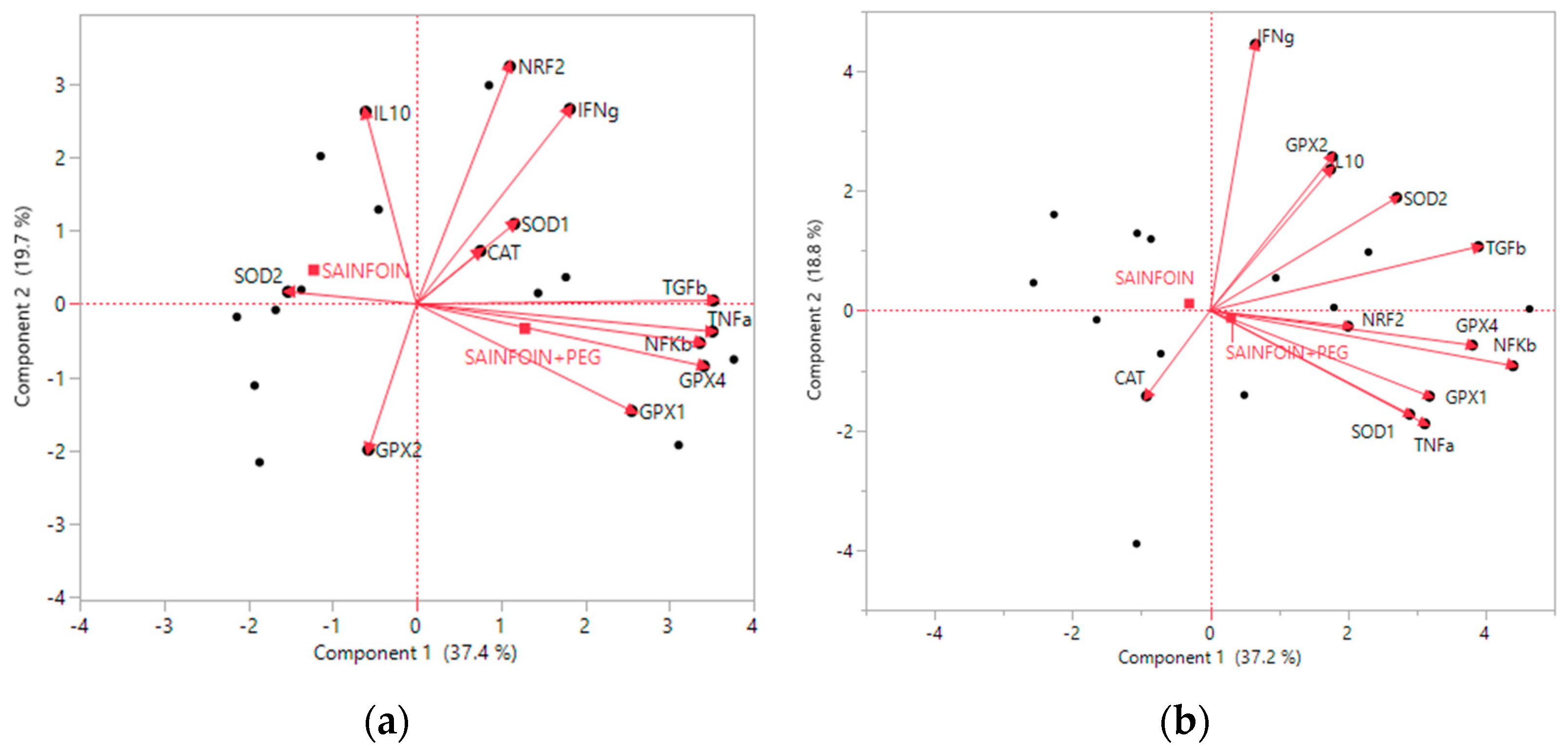
3.5. Principal Components Analysis

Principal components are new variables that are constructed as linear combinations of the initial variables. In jejunal tissues, seven principal components accounted for 95.9% of the variation in the gene expression values, while the total number of components extracted (eigenvalues), based on the amount of variance contributed by each component, was reduced from 4.49 to 0.46. Component 1 accounted for 37.4% of the total variation, while component 2 accounted for 19.7% variation (Figure 2a). Similarly, in ileal tissues, seven principal components accounted for 99.0% of the variation in the gene expression values, while the total number of components extracted (eigenvalues), based on the amount of variance contributed by each component, was reduced from 4.46 to 0.59. Component 1 accounted for 37.2% of the total variation, while component 2 accounted for 18.8% variation (Figure 2b). In both tissues, the left quadrant of component 1 was mainly represented by SAINFOIN individuals, while the right quadrant was mainly represented by lambs whose dams had been fed SAINFOIN + PEG. Likewise, the upper quadrant of component 2 was mainly represented by SAINFOIN individuals, while the bottom quadrant was mainly represented by lambs whose dams had been fed SAINFOIN + PEG.
The PCA identified three clusters based on gene markers in the jejunum. The first cluster was related to the pro-inflammatory marker TNFA (69% out of total cluster variation, 6 gene members), while the second and third cluster were mainly related to NRF2 (54.5% out of total cluster variation; 4 genes) and CAT (74.5% out of total cluster variation; 2 genes) (Table 4).
In ileal tissues, the PCA also identified four clusters. Cluster 1 was mostly represented by NFKB (64.9% out of total cluster variation, 6 gene members), while cluster 2 was mostly represented by IFNG (64.4% out of total cluster variation; 3 genes). The third cluster was mainly represented by the antioxidant enzyme CAT (70.9% out of total cluster variation; 2 genes), and the fourth cluster was represented by a single NRF2 (100% out of total cluster variation; 1 gene) (Table 5).

4. Discussion

In this study, we characterised the effect of maternal dietary CT on coccidiosis and health and antioxidant-immune crosstalk at a gut level in suckling lambs. Our main findings were that: (1) maternal dietary CT tended to reduce faecal oocyst excretion in lambs; (2) the presence of CT in the dams’ diet affected differently the expression of the antioxidant defence system and regulatory immune genes in intestinal tissues; and (3) based on PCA analysis, gut homeostasis in suckling lambs involves a tissue-dependent interaction of pro-inflammatory and antioxidant mediators in both jejunal and ileal tissues.
A large body of published work has demonstrated the antiparasitic properties of CT in gastrointestinal coccidian infection [12,13]. For example, the use of sainfoin delayed the onset of coccidian infections in both pre-weaned [8] and weaned lambs [15,36]. In our study, the percentage of lambs that excreted more than 10 oocysts/g at slaughter age tended to be higher in the SAINFOIN + PEG group than in the SAINFOIN group. This suggests that the chelate used in ewes’ diet of the SAINFOIN + PEG group reduced the protection of CT against coccidia along the intestinal tract of naturally infected lambs. Lambs are infected in early life (with the onset of excretion at 13–15 days) as a result of an oocyst-contaminated environment, however, the presence of large numbers of oocysts in young lambs is not necessarily indicative of disease [37,38]. Clinical coccidiosis seems to be directly linked to the species of coccidian involved, the number of oocysts ingested, significant asexual multiplication in the host, and possible physiological stress [7]. In our study, oocyst excretion was not correlated with the observed microscopic lesion scores in the jejunum and ileum of naturally infected lambs, and both treatments reached the target BW at slaughter with similar age and, thereby, they had similar growth rates [26]. It has been suggested an interplay between passive maternal immunity and active immunisation during the lactation period in infected lambs [39]. On the other hand, solid feed is naturally starting at around 3 weeks of age [40], leading to important anatomical and physiological changes in the gastrointestinal tract. Therefore, the lambs of this study could be familiarising with sainfoin supplied to the dams, and thereby, some other feed components apart of those from ewe milk could be reaching the small intestine. However, the amount of solid feed consumed at the time of slaughter (4–5 weeks of age) may be very low (about 50 g/day) [41]. Furthermore, the jejunum is the part of the intestine where most of the nutrient absorption occurs, so these events could be disrupting the histopathology of the jejunum [42,43]. Nevertheless, as the present study it was conducted under natural coccidian infection, further research on histopathological findings in suckling lambs are needed.
Coccidia cause damage to the intestinal mucosa by activating cellular immune responses [6,44]. However, the activation of an effective immune response can induce a strong competition for the maintenance of growth in parasitised lambs. The protective role of CT has been associated with a direct action on the modulation of specific immune cells and antioxidant activity [45]. In this study, the inclusion of CT from sainfoin in the dams’ diet diminished gene expression of cytosolic GPX1 and phospholipid hydroperoxide GPX4, and anti-inflammatory TGFB and pro-inflammatory TNFA in the jejunum, but increased the gene expression of CAT and gastrointestinal GPX2 in the ileum. Accordingly, PCA revealed that both immune and antioxidant responses are different in lambs from the SAINFOIN group when compared to SAINFOIN + PEG group. These results would indicate that there was a positive transfer of some polyphenols to the milk in the SAINFOIN ewes’ diet, as observed in goats nursing kids [46], due to the CT that may have been absorbed during digestion [14]. Hence, dams’ diet CT may have immunomodulatory effects on the intestinal environment, causing an adverse scenario for the parasites without a negative impact on nutritional well-being.
Plant-derived compounds, as polyphenols, have the ability to prime Nrf2 expression and activity while inhibiting NF-κB activation [19]. However, in this study, we observed a decreased production of both pro- and anti-inflammatory cytokines and antioxidant enzymes in the jejunum. The antioxidant properties of CT are linked to their ability to scavenge free radicals [13,47]. Thus, it is possible that the presence of CT in dams’ diet, and accordingly some of the CT derivatives reaching the gut, helped to reduce local oxidative stress and to diminish immune responses and endogenous antioxidant defences by reducing the production of reactive oxygen species (ROS). This result is in agreement with studies performed in other species, where decreased activities of the oxidative stress-responsive transcription factors NFKB and NRF2 were observed in the duodenal mucosa of pigs supplemented with polyphenol-rich grape by-products [48]. On the other hand, polyphenols also enhance antioxidant defences by increasing the activity of endogenous antioxidant enzymes [49,50]. In mammalian models, marked regional variations of antioxidant capacity in the gastrointestinal tract have been described [51,52]. Thus, the presence of polyphenols in the dams’ diet could help in the detoxifying metabolism of hydroperoxides in suckling lambs by favouring the expression of CAT and GPX2, considered the most important of the four types of GPX for peroxide removal in the intestine [53]. In swine, the 5% grape pomace diet leads to an increase of GPX activity in the colon with respect to the duodenum [52], which was attributed to the presence of the unmetabolised procyanidin trimers. In this trial, the carcass and meat quality of suckling lambs were not affected by the inclusion of CT from sainfoin in the dams’ diet [27]. In agreement with previous studies, plasma concentrations of fructosamine in lambs, as indicator of nutritional status [54], were not affected by the presence of CT in the dams’ diet.
In ruminants, the small intestine is characterised by regional differences in the development of immune responses, probably influenced by selective recruitment of immune cells to the intestinal tissue [55] and gut colonisation [20,56] as a result of different undigested products reaching the intestine. Independently of the maternal experimental diet, PCA analysis defined dynamic/regulatory antioxidant-immune interactions involved in gut growth and development of suckling lambs. In the jejunum, the first cluster was related to the pro-inflammatory marker TNFA that may exert apoptotic and anti-apoptotic pathways [57], while the second and third clusters were mainly related to NRF2, which is involved in redox homeostasis, and CAT, which has H2O2 degrading capacity. In ileal tissues, the first cluster was mostly represented by NFKB, involved in homeostatic and cell survival pathways, while the second cluster was mostly represented by IFNG, involved in cellular immunity. The third cluster was mainly represented by the antioxidant enzyme CAT and the fourth cluster was represented by a single NRF2. Oxidative stress and inflammation are interdependent since an excess of ROS can initiate a cascade of intracellular signalling, that involve NF-κB pathway and pro-inflammatory stimuli. In response to these stresses, it is necessary the activation of the Nrf2 pathway that leads to the activation of various antioxidant enzymes, such as CAT, as observed in the ileum of the current study. Given ROS can induce oxidative injury and also act in redox signalling [58], it has been described as a downregulation of CAT expression by TNFA in order to enhance NF-κB activation, that is stimulated in the presence of H2O2 [57]. This analysis suggests the presence of complex and compartment/region-dependent homeorhetic mechanisms of the intestinal tract in suckling lambs, in agreement with previous studies [25].

5. Conclusions

In conclusion, CT from sainfoin in the diet of ewes tended to reduce coccidian oocyst excretion in their suckling lambs. Maternal diets (SAINFOIN vs. SAINFOIN + PEG) did not affect the histopathological analysis of the jejunum and ileum of suckling lambs. However, the superoxide radical scavenging activity of CT in dams’ diet caused a down-regulation of TNFA and TGFB and GPX1 and GPX4 in jejunal tissue while enhancing the H2O2 degrading capacity of CAT and GPX2 in ileal tissues of lambs from SAINFOIN group. PCA analysis confirmed the presence of complex, and compartment/region-dependent homeorhetic mechanisms mediating the small intestine health in suckling lambs.

Author Contributions

Conceptualisation and supervision, J.Á.-R. and B.S.-P.; methodology, J.P.-V., M.J.M.-A., G.A.R. and B.S.-P.; formal analysis, J.Á.-R.; investigation, J.P.-V., C.B., S.L., M.J. and B.S.-P.; data curation, J.P.-V. and B.S.-P.; writing—original draft preparation, J.P.-V. and B.S.-P.; writing—review and editing, J.P.-V., J.Á.-R., M.J.M.-A., G.A.R., C.B., S.L., M.J. and B.S.-P.; funding acquisition, J.Á.-R. and M.J. All authors have read and agreed to the published version of the manuscript.

Funding

This research was funded by the Ministry of Science, Innovation and Universities of Spain and the European Regional Development Funds, grant number INIA RTA2017-00008-C02-01 and 02.

Institutional Review Board Statement

The Animal Ethics Committee of the Research Centre approved the experimental procedures (protocol code CEEA-03-2014-26), which were in compliance with the European Union Directive 2010/63 on the protection of animals used for experimental and other scientific purposes.

Data Availability Statement

Not applicable.

Acknowledgments

Jonathan Pelegrin-Valls and Claudia Baila are in receipt of an early-stage research staff grant by the Generalitat de Catalunya-European Social Funds (FI_B00376) and by the Spanish government (PRE2018-086670), respectively. The authors thank Beatriz Aquilue for her assistance and valuable advice in coprological analysis.

Conflicts of Interest

The authors declare no conflict of interest. The funders had no role in the design of the study; in the collection, analyses, or interpretation of data; in the writing of the manuscript; or in the decision to publish the results.

References

  1. Sañudo, C.; Sanchez, A.; Alfonso, M. Small ruminant production systems and factors affecting lamb meat quality. Meat Sci. 1998, 49, 29–64. [Google Scholar] [CrossRef]
  2. Lima., E.; Lovatt., F.; Green, M.; Roden, J.; Davies, P.; Kaler, J. Sustainable lamb production: Evaluation of factors affecting lamb growth using hierarchical, cross classified and multiple memberships models. Prev. Vet. Med. 2020, 174, 104822. [Google Scholar] [CrossRef]
  3. Hoste, H.; Torres-Acosta, J.F.J.; Sandoval-Castro, C.A.; Mueller-Harvey, I.; Sotiraki, S.; Louvandini, H.; Thamsborg, S.M.; Terrill, T.H. Tannin containing legumes as a model for nutraceuticals against digestive parasites in livestock. Vet. Parasitol. 2015, 212, 5–17. [Google Scholar] [CrossRef] [PubMed]
  4. Gioxari, A.; Amerikanou, C.; Nestoridi, I.; Gourgari, E.; Pratsinis, H.; Kalogeropoulos, N.; Andrikopoulos, N.K.; Kaliora, A.C. Carob: A Sustainable Opportunity for Metabolic Health. Foods 2022, 11, 2154. [Google Scholar] [CrossRef] [PubMed]
  5. Keeton, S.T.N.; Navarre, C.B. Coccidiosis in large and small ruminants. Vet. Clin. Food Anim. 2017, 34, 201–208. [Google Scholar] [CrossRef] [PubMed]
  6. Bangoura, B.; Bardsley, K.D. Ruminant Coccidiosis. Vet. Clin. Food Anim. 2020, 36, 187–203. [Google Scholar] [CrossRef] [PubMed]
  7. Chartier, C.; Paraud, C. Coccidiosis due to Eimeria in sheep and goats, a review. Small Rumin. Res. 2012, 103, 84–92. [Google Scholar] [CrossRef]
  8. Saratsis, A.; Voutzourakis, N.; Theodosiou, T.; Stefanakis, A.; Sotiraki, S. The effect of sainfoin (Onobrychis viciifolia) and carob pods (Ceratonia silique) feeding regimes on the control of lamb coccidiosis. Parasitol. Res. 2016, 115, 2233–2242. [Google Scholar] [CrossRef] [PubMed]
  9. Roila, R.; Branciari, R.; Pecorelli, I.; Cristofani, E.; Carloni, C.; Ranucci, D.; Fioroni, L. Occurrence and Residue Concentration of Coccidiostats in Feed and Food of Animal Origin; Human Exposure Assessment. Foods 2019, 8, 477. [Google Scholar] [CrossRef] [Green Version]
  10. Muller-Harvey, I.; Bee, G.; Dohme-Meier, F.; Hoste, H.; Karonen, M.; Kölliker, R.; Lüscher, A.; Niderkorn, V.; Pellikaan, W.F.; Salminen, J.P.; et al. Benefits of condensed tannins legumes fed to ruminants: Importance of structure, concentration and diet composition. Crop Sci. 2019, 59, 861–885. [Google Scholar] [CrossRef]
  11. Álvarez-Rodríguez, J.; Urrutia, O.; Lobón, S.; Ripoll, G.; Bertolín, J.R.; Joy, M. Insights into the role of major bioactive dietary nutrients in lamb meat quality: A review. J. Anim. Sci. Biotechnol. 2022, 13, 20. [Google Scholar] [CrossRef] [PubMed]
  12. Blomstrand, B.M.; Enemark, H.L.; Steinshamn, H.; Aasen, I.M.; Johanssen, J.R.E.; Athanasiadou, S.; Thamsborg, S.M.; Sørheim, K.M. Administration of spruce bark (Picea abies) extracts in young lambs exhibits anticoccidial effects but reduces milk intake and body weight gain. Acta Vet. Scand. 2022, 64, 10. [Google Scholar] [CrossRef] [PubMed]
  13. Pérez-Fonseca, A.; Alcala-Canto, Y.; Salem, A.Z.; Alberti-Navarro, A.B. Anticoccidial efficacy of naringenin and a grapefruit peel extract in growing lambs naturally-infected with Eimeria spp. Vet. Parasitol. 2016, 232, 58–65. [Google Scholar] [CrossRef] [PubMed]
  14. Quijada, J.; Drake, C.; Gaudin, E.; El-Korso, R.; Hoste, H.; Muller-Harvey, I. Condensed tannin changes along the digestive tract in lambs fed with sainfoin pellets or hazelnut skins. J. Agric. Food Chem. 2018, 66, 2136–2142. [Google Scholar] [CrossRef]
  15. Rivaroli, D.; Prunier, A.; Meteau, K.; do Prado, I.N.; Prache, S. Tannin-rich sainfoin pellet supplementation reduces fat volatile indoles content and delays digestive parasitism in lambs grazing alfalfa. Animal 2019, 13, 1883–1890. [Google Scholar] [CrossRef]
  16. Niderkorn, V.; Barbier, E.; Macheboeuf, D.; Torrent, A.; Mueller-Harvey, I.; Hoste, H. In vitro rumen fermentation of diets with different types of condensed tannins derived from sainfoin (Onobrychis viciifolia Scop.) pellets and hazelnut (Corylus avellana L.) pericarps. Anim. Feed Sci. Technol. 2020, 259, 114357. [Google Scholar] [CrossRef]
  17. Petrič, D.; Komáromyová, M.; Batťányi, D.; Kozłowska, M.; Filipiak, W.; Łukomska, A.; Ślusarczyk, S.; Szumacher-Strabel, M.; Cieślak, A.; Várady, M.; et al. Effect of Sainfoin (Onobrychis viciifolia) Pellets on Rumen Microbiome and Histopathology in Lambs Exposed to Gastrointestinal Nematodes. Agriculture 2022, 12, 301. [Google Scholar] [CrossRef]
  18. Celi, P.; Gabai, G. Oxidant/Antioxidant Balance in Animal Nutrition and Health: The Role of Protein Oxidation. Front. Vet. Sci. 2015, 2, 48. [Google Scholar] [CrossRef] [Green Version]
  19. Stefanson, A.L.; Bakovic, M. Dietary Regulation of Keap1/Nrf2/ARE Pathway: Focus on Plant-Derived Compounds and Trace Minerals. Nutrients 2014, 6, 3777–3801. [Google Scholar] [CrossRef] [Green Version]
  20. Rajput, S.A.; Sun, L.; Zhang, N.-Y.; Khalil, M.M.; Ling, Z.; Chong, L.; Wang, S.; Rajput, I.R.; Bloch, D.M.; Khan, F.A.; et al. Grape Seed Proanthocyanidin Extract Alleviates AflatoxinB1-Induced Immunotoxicity and Oxidative Stress via Modulation of NF-κB and Nrf2 Signaling Pathways in Broilers. Toxins 2019, 11, 23. [Google Scholar] [CrossRef]
  21. Koudoufio, M.; Feldman, F.; Ahmarani, L.; Delvin, E.; Spahis, S.; Desjardins, Y.; Levy, E. Intestinal protection by proanthocyanidins involves anti-oxidative and anti-inflammatory actions in association with an improvement of insulin sensitivity, lipid and glucose homeostasis. Sci. Rep. 2021, 11, 3878. [Google Scholar] [CrossRef] [PubMed]
  22. Maynard, C.L.; Elson, C.O.; Hatton, R.D.; Weaver, C.T. Reciprocal interactions of the intestinal microbiota and immune system. Nature 2012, 489, 231–241. [Google Scholar] [CrossRef] [PubMed] [Green Version]
  23. Chassaing, B.; Kumar, M.; Baker, M.T.; Singh, V.; Vijay-Kumar, M. Mammalian gut immunity. Biomed. J. 2014, 37, 246–258. [Google Scholar] [CrossRef] [PubMed]
  24. Steele, M.A.; Penner, G.B.; Chaucheyras-Durand, F.; Guan, L.L. Development and physiology of the rumen and the lower gut: Targets for improving gut health. J. Dairy Sci. 2016, 99, 4955–4966. [Google Scholar] [CrossRef] [PubMed] [Green Version]
  25. Bush, S.J.; McCulloch, M.E.B.; Muriuki, C.; Salavati, M.; Davis, G.M.; Farquhar, I.L.; Lisowski, Z.M.; Archibald, A.L.; Hume, D.A.; Clark, E.L. Comprehensive Transcriptional Profiling of the Gastrointestinal Tract of Ruminants from Birth to Adulthood Reveals Strong Developmental Stage Specific Gene Expression. G3 2019, 9, 359–373. [Google Scholar] [CrossRef] [Green Version]
  26. Baila, C.; Joy, M.; Blanco, M.; Casasús, I.; Bertolín, J.R.; Lobón, S. Effects of feeding sainfoin proanthocyanidins to lactating ewes on intake, milk production and plasma metabolites. Animal 2022, 16, 100438. [Google Scholar] [CrossRef]
  27. Baila, C.; Lobón, S.; Blanco, M.; Casasús, I.; Ripoll, G.; Joy, M. Sainfoin in the Dams’ Diet as a Source of Proanthocyanidins: Effect on the Growth, Carcass and Meat Quality of Their Suckling Lambs. Animals 2022, 12, 408. [Google Scholar] [CrossRef]
  28. Bowman, D.D. Georgis’ Parasitology for Veterinarians, 11th ed.; Elsevier Inc.: St. Louis, MO, USA, 2020; pp. 299–300. [Google Scholar] [CrossRef]
  29. Dommels, Y.E.; Butts, C.A.; Zhu, S.; Davy, M.; Martell, S.; Hedderley, D.; Barnett, M.P.; McNabb, W.C.; Roy, N.C. Characterization of intestinal inflammation and identification of related gene expression changes in mdr1a(-/-) mice. Genes Nutr. 2007, 2, 209–223. [Google Scholar] [CrossRef] [Green Version]
  30. Primer3plus. Available online: www.bioinformatics.nl/primer3plus (accessed on 1 February 2019).
  31. Primer-Blast. Available online: www.ncbi.nlm.nih.gov/tools/primer-blast/ (accessed on 1 February 2019).
  32. Pelegrin-Valls, J.; Serrano-Pérez, B.; Villalba, D.; Martín-Alonso, M.J.; Bertolín, J.R.; Joy, M.; Álvarez-Rodríguez, J. Effect of Dietary Crude Protein on Productive Efficiency, Nutrient Digestibility, Blood Metabolites and Gastrointestinal Immune Markers in Light Lambs. Animals 2020, 10, 328. [Google Scholar] [CrossRef]
  33. Schmittgen, T.D.; Livak, K.J. Analysing real-time PCR data by the comparative CT method. Nat. Protoc. 2008, 3, 1101–1108. [Google Scholar] [CrossRef]
  34. Puech, C.; Dedieu, L.; Chantal, I.; Rodrigues, V. Design and evaluation of a unique SYBR Green real-time RT-PCR assay for quantification of five major cytokines in cattle, sheep and goats. BMC Vet. Res. 2015, 11, 65. [Google Scholar] [CrossRef] [PubMed] [Green Version]
  35. Lesage-Padilla, A.; Forde, N.; Poirée, M.; Healey, G.D.; Giraud-Delville, C.; Reinaud, P.; Eozenou, C.; Carvalho, A.V.; Galio, L.; Raliou, M.; et al. Maternal metabolism affects endometrial expression of oxidative stress and FOXL2 genes in cattle. PLoS ONE 2017, 12, e0189942. [Google Scholar] [CrossRef] [PubMed] [Green Version]
  36. Saratsis, A.; Regos, I.; Tzanidakis, N.; Voutzourakis, N.; Stefanakis, A.; Treuter, D.; Joachim, A.; Sotiraki, S. In vivo and in vitro efficacy of sainfoin (Onobrychis viciifolia) against Eimeria spp in lambs. Vet. Parasitol. 2012, 188, 1–9. [Google Scholar] [CrossRef] [PubMed]
  37. Pout, D.D.; Ostler, D.C.; Joyner, L.P.; Norton, C.C. The coccidial population in clinically normal sheep. Vet. Rec. 1966, 78, 455–460. [Google Scholar] [CrossRef] [PubMed]
  38. Saratsis, A.; Joachim, A.; Alexandros, S.; Sotiraki, S. Lamb coccidiosis dynamics in different dairy production systems. Vet. Parasitol. 2011, 181, 131–138. [Google Scholar] [CrossRef]
  39. Reeg, K.J.; Gauly, M.; Bauer, C.; Mertens, C.; Erhardt, G.; Zahner, H. Coccidial infections in housed lambs: Oocyst excretion, antibody levels and genetic influences on the infection. Vet. Parasitol. 2005, 127, 209–219. [Google Scholar] [CrossRef] [PubMed]
  40. Álvarez-Rodríguez, J.; Sanz, A.; Delfa, R.; Revilla, R.; Joy, M. Performance and grazing behaviour of Churra Tensina sheep stocked under different management systems during lactation on Spanish mountain pastures. Livest. Sci. 2007, 107, 152–161. [Google Scholar] [CrossRef]
  41. Faichney, G.J. Consumption of solid feed by lambs during their transition from pre-ruminant to full ruminant function. Appl. Anim. Behav. Sci. 1992, 34, 85–91. [Google Scholar] [CrossRef]
  42. Pond, W.G.; Church, D.C.; Pond, K.R. Basic Animal Nutrition and Feeding, 4th ed.; Wiley: New York, NY, USA, 1995; p. 32. [Google Scholar]
  43. Vi, R.B.; McLeod, K.R.; Klotz, J.L.; Heitmann, R.N. Rumen development, intestinal growth and hepatic metabolism in the pre-and postweaning ruminant. J. Dairy Sci. 2004, 87, 55–65. [Google Scholar] [CrossRef]
  44. Ovington, K.S.; Alleva, L.M.; Kerr, E.A. Cytokines and immunological control of Eimeria spp. Int. J. Parasitol. 1995, 25, 1331–1351. [Google Scholar] [CrossRef]
  45. Yahfoufi, N.; Alsadi, N.; Jambi, M.; Matar, C. The Immunomodulatory and Anti-Inflammatory Role of Polyphenols. Nutrients 2018, 10, 1618. [Google Scholar] [CrossRef] [PubMed] [Green Version]
  46. Jordán, M.J.; Moñino, M.I.; Martíez, C.; Lafuente, A.; Sotomayor, J.A. Introduction of distillate Rosemary leaves into the diets of the Murciano-Granadina goat: Transfer of polyphenolic compounds to goats’ milk and the plasma of suckling goat kids. J. Agric. Food Chem. 2010, 58, 8265–8270. [Google Scholar] [CrossRef] [PubMed]
  47. Bagchi, D.; Garg, A.; Krohn, R.L.; Bagchi, M.; Tran, M.X.; Balmoori, J. Oxygen free radical scavenging abilities of vitamins C and E, and a grape seed proanthocyanidins extract in vitro. Res. Commun. Mol. Pathol. Pharmacol. 1997, 95, 179–189. [Google Scholar] [PubMed]
  48. Gessner, D.K.; Fiesel, A.; Most, E.; Dinges, J.; Wen, G.; Ringseis, R.; Eder, K. Supplementation of a grape seed and grape marc meal extract decreases activities of the oxidative stress-responsive transcription factors NF-κB and Nrf2 in the duodenal mucosa of pigs. Acta Vet. Scand. 2013, 55, 18. [Google Scholar] [CrossRef] [PubMed] [Green Version]
  49. Puiggros, F.; Llópiz, N.; Ardévol, A.; Bladé, C.; Arola, L.; Salvadó, M.J. Grape seed procyanidins prevent oxidative injury by modulating the expression of antioxidant enzyme systems. J. Agric. Food Chem. 2005, 53, 6080–6086. [Google Scholar] [CrossRef] [PubMed]
  50. Mu, C.; Yang, W.; Wang, P.; Zhao, J.; Hao, X.; Zhang, J. Effects of high-concentrate diet supplemented with grape seed proanthocyanidins on growth performance, liver function, meat quality, and antioxidant activity in finishing lambs. Anim. Feed Sci. Technol. 2020, 266, 114518. [Google Scholar] [CrossRef]
  51. Moghadasian, M.H.; Godin, D.V. Ethanol-induced gastrointestinal damage influence of endogenous antioxidant components and gender. Dig. Dis. Sci. 1996, 41, 791–797. [Google Scholar] [CrossRef] [PubMed]
  52. Chedea, V.S.; Palade, L.M.; Marin, D.E.; Pelmus, R.S.; Habeanu, M.; Rotar, M.C.; Gras, M.A.; Pistol, G.C.; Taranu, I. Intestinal Absorption and Antioxidant Activity of Grape Pomace Polyphenols. Nutrients 2018, 10, 588. [Google Scholar] [CrossRef] [PubMed] [Green Version]
  53. Florian, S.; Wingler, K.; Schmehl, K.; Jacobasch, G.; Kreuzer, O.J.; Meyerhof, W.; Brigelius-Flohé, R. Cellular and subcellular localization of gastrointestinal glutathione peroxidase in normal and malignant human intestinal tissue. Free Radic. Res. 2001, 35, 655–663. [Google Scholar] [CrossRef] [PubMed]
  54. Filipović, N.; Stojević, Z.; Mašek, T.; Mikulec, Ž.; Prvanović, N. Relationship between fructosamine with serum protein, albumin and glucose concentrations in dairy ewes. Small Rumin. Res. 2011, 96, 46–48. [Google Scholar] [CrossRef]
  55. Ibeagha-Awemu, E.M.; Bissonnette, N. Do, D.N.; Dudemaine, P.L.; Wang, M.; Facciuolo, A.; Griebel, P. Regionally Distinct Immune and Metabolic Transcriptional Responses in the Bovine Small Intestine and Draining Lymph Nodes during a Subclinical Mycobacterium avium subsp. paratuberculosis Infection. Front. Immunol. 2021, 12, 760931. [Google Scholar] [CrossRef] [PubMed]
  56. Hansen, H.F.; Nielsen, D.S.; Kverka, M.; Zakostelska, Z.; Klimesova, K.; Hudcovic, T.; Tlaskalova-Hogenova, H.; Hansen, A.K. Patterns of Early Gut Colonization Shape Future Immune Responses of the Host. PLoS ONE 2012, 7, e34043. [Google Scholar] [CrossRef] [Green Version]
  57. Lüpertz, R.; Chovolou, Y.; Kampkötter, A.; Wätjen, W.; Kahl, R. Catalase overexpression impairs TNF-alpha induced NF-kappaB activation and sensitizes MCF-7 cells against TNF-alpha. J. Cell. Biochem. 2008, 103, 1497–1511. [Google Scholar] [CrossRef] [PubMed]
  58. Lei, X.G.; Zhu, J.H.; Cheng, W.H.; Bao, Y.; Ho, Y.S.; Reddi, A.R.; Holmgren, A.; Arnér, E.S. Paradoxical Roles of Antioxidant Enzymes: Basic Mechanisms and Health Implications. Physiol. Rev. 2016, 96, 307–364. [Google Scholar] [CrossRef] [PubMed]
Figure 1. Relative quantification of gene transcripts in the jejunum (a) and ileum (b) in suckling lambs according to maternal dietary (SAINFOIN vs. SAINFOIN + PEG). Bars represent least-squares mean values ± SE. The level of significance is expressed as follows: * p ≤ 0.10; ** p ≤ 0.05; *** p ≤ 0.01.
Agriculture 12 01694 g001
Figure 2. Biplot of principal components of gene expression markers in the jejunum (a) and ileum (b).
Agriculture 12 01694 g002
Table 1. Primer Sequences Used for Quantitative Real-Time PCR.
GeneTissue-Function RatioForward and Reverse Primer (5′–3′) 1bp 2Access. No.E (%) 3nM 4Source
GAPDHReference genesF: ATCTCGCTCCTGGAAGATG
R: TCGGAGTGAACGGATTCG
200NM_001190390.11.90600
300
[34]
ACTBReference genesF: CTGGACTTCGAGCAGGAGAT
R: GATGTCGACGTCACACTTC
194NM_0010097841.94600[34]
IL10Anti-inflammatoryF: TTAAGGGTTACCTGGGTTGC
R: TTCACGTGCTCCTTGATGTC
109NM_001009327.11.96200[32]
TGFBAnti-inflammatoryF: TTGACGTCACTGGAGTTGTG
R: CGTTGATGTCCACTTGAAGC
120NM_001009400.22.04200[32]
TNFAPro-inflammatoryF: CAAATAACAAGCCGGTAGCC
R: TGGTTGTCTTTCAGCTCCAC
118NM_001024860.11.96200[32]
IFNGPro-inflammatoryF: AAGTTCTTGAACGGCAGCTC
R: TTGGCGACAGGTCATTCATC
130NM_001009803.11.91500[30,31]
NRF2Redox systemF: GAGCCCAGTCTTCAATGCTC
R: TCAGCCAGCTTGTCATTTTG
171XM_015093345.21.97200[30,31]
NFKBTranscription factorF: CTACACCTTGCCTGTGAGCA
R: AAGGACACCAACAGCTCCAC
173NM_001166184.11.93300[30,31]
SOD1Antioxidant enzymeF: CACCATCCACTTCGAGGCAA
R: GCACTGGTACAGCCTTGTGT
126NM_174615.22.06200[35]
SOD2Antioxidant enzymeF: GGATCCCCTGCAAGGAACAA
R: TGGCCTTCAGATAATCGGGC
110NM_201527.22.03200[35]
CATAntioxidant enzymeF: TTCCGTCCTTTATCCACAGC
R: CCATTGGCATTAACCAGCTT
199XM_004016396.52.04300[30,31]
GPX1Antioxidant enzymeF: GGCATCAGGAAAACGCCAAG
R: GGGGACCACGTGATGAACTT
217XM_004018462.51.98300[30,31]
GPX2Antioxidant enzymeF: ATTGAGAATGTGGCCTCGCT
R: CCAGGGCGGACATACTTGAG
179XM_004010720.52.07300[30,31]
GPX4Antioxidant enzymeF: GGGAGTAATGCGGAGATCAA
R: CCACACAGCCGTTCTTATCA
199XM_027970172.12.04300[30,31]
1 F = forward; R = reverse; 2 bp = amplified product length in base pairs; 3 E (%) = efficiency; 4 nM = optimal primer concentration.
Table 2. Effect of maternal experimental diets (SAINFOIN + PEG vs. SAINFOIN) on faecal oocyst count in suckling lambs at slaughter and percentage of lambs excreting 0, 10 and 500 oocyst/g faeces.
Faecal Oocyst CountSAINFOINSAINFOIN + PEGp-Value
Log-transformed OPG 1
(Log-values ± SE 2 between parenthesis)
19
(1.28 ± 0.65)
219
(2.34 ± 0.65)
0.28
Lambs excreting oocyst (%)
>0 oocysts/g faeces5083.30.21
>10 oocysts/g faeces33.383.30.07
>500 oocysts/g faeces16.733.30.50
1 OPG = oocyst per gram of faeces; 2 SE = standard error.
Table 3. Histopathological injury score of suckling lambs according to maternal experimental diets (SAINFOIN + PEG vs. SAINFOIN) and tissue (jejunal and ileal).
Treatment 2Tissue 2p-Value 3
Item (µm) 1SAINFOINSAINFOIN + PEGJejunalIlealSETreatmentTissue
Total inflammatory4.34.35.13.50.231.0<0.0001
Total tissue destruction2.12.03.30.80.230.61<0.0001
Total Tissue repair0.010.040.040.010.030.320.32
∑ HIS6.46.38.44.30.420.83<0.0001
1 Total inflammatory section = intensity, fibrin exudation, lymphangiectasis, location (layer), crypt abscesses, and villous blunting; total tissue destruction section = enterocyte loss, oedema, mucosal atrophy, loss of crypts; total tissue reparation: hyperplasia, angiogenesis, granulomas and fibrosis; HIS = total histological injury score. 2 Values are expressed as mean ± SE. 3 No interactions between treatment and tissue were observed.
Table 4. Summary of PCA clusters of gene expression markers in the jejunum.
ClusterGene MembersMost Representative VariableProportion of Explained Cluster VariationProportion of Total Variation
1TNFA, TGFB, GPX4, NFKB, GPX1, and SOD2TNFA0.690.35
2NRF2, IFNG, IL10, and SOD1NRF20.550.18
3CAT and GPX2CAT0.750.12
Table 5. Summary of PCA clusters of gene expression markers in the ileum.
ClusterGene MembersMost Representative VariableProportion of Explained Cluster VariationProportion of Total Variation
1NFKB, GPX4, GPX1, TGFB, TNFA, and SOD1NFKB0.650.32
2IFNG, GPX2, and SOD2IFNG0.640.16
3CAT and IL10CAT0.710.12
4NRF2NRF210.08
Publisher’s Note: MDPI stays neutral with regard to jurisdictional claims in published maps and institutional affiliations.

