Review

Progress of m6A Methylation in Lipid Metabolism in Humans and Animals

1
College of Animal Science and Technology, Yangzhou University, Yangzhou 225009, China
2
Shaanxi Key Laboratory of Brain Disorders & Institute of Basic and Translational Medicine, Xi’an Medical University, No.1 Xinwang Road, Xi’an 710021, China
3
Joint International Research Laboratory of Agriculture & Agri-Product Safety, Ministry of Education, Yangzhou University, Yangzhou 225009, China
*
Author to whom correspondence should be addressed.
Agriculture 2022, 12(10), 1683; https://doi.org/10.3390/agriculture12101683
Submission received: 4 September 2022 / Revised: 28 September 2022 / Accepted: 9 October 2022 / Published: 13 October 2022
(This article belongs to the Special Issue Breeding, Genetics and Safety Production of Dairy Cattle)

Abstract

:
N6-methyladenosine (m6A) methylation is a type of methylation modification discovered on RNA molecules, mainly on mRNAs, as well as on other RNAs. Similar to DNA methylation, m6A methylation regulates the post-transcriptional expression level of genes without altering their base sequences. It modulates gene expression mainly by affecting the binding of mRNAs to reader proteins, thereby regulating variable splicing, translation efficiency, and stability of mRNAs. Early in the research, the study of m6A-related biological functions was greatly hindered due to the lack of effective detection methods. As second-generation sequencing and bioinformatics develop, several methods have been available to detect and predict m6A methylation sites in recent years. Moreover, m6A methylation is also closely related to the development of lipid metabolism, as shown in current studies. Combined with recent research, this paper reviews the concept, detection, and prediction means of m6A methylation, especially the relationship between m6A and lipid metabolism, providing a new clue to enrich the molecular mechanism of lipid metabolism.

1. m6A Methylation of RNA

1.1. Concepts Related to m6A Methylation

Early in the 1970s, Desrosiers first proposed RNA-N6-methyladenosine (m6A) methylation as a new type of epigenetic modification [1], that is, a methyl substituent is inserted into the N atom at position 6 of adenosine. This only occurs in a small fraction of fragments containing DRACH (D = A/G/U, R = A/G, H = A/C/U) motif sequences, mainly in the CDS region and 3′UTR region of mRNAs [2]. Numerous studies have shown that m6A methylation plays a crucial role in nearly every stage of RNA’s life cycle, including maturation and degradation. Additionally, it contributes to mRNA processing, splicing, export from the nucleus to the cytoplasm, translation, and decay. M6A methylation modifications are reversible and dynamic in nature, and mainly involve three types of regulators: methyltransferase (writer), demethylase (eraser), and reading protein (reader). Among them, the writer catalyzes the m6A modification of adenosine on mRNAs, the eraser demethylates bases that have undergone m6A modification, and the reader identifies bases that have undergone m6A modification so that they can participate in downstream regulatory pathways, such as RNA translational degradation [3].

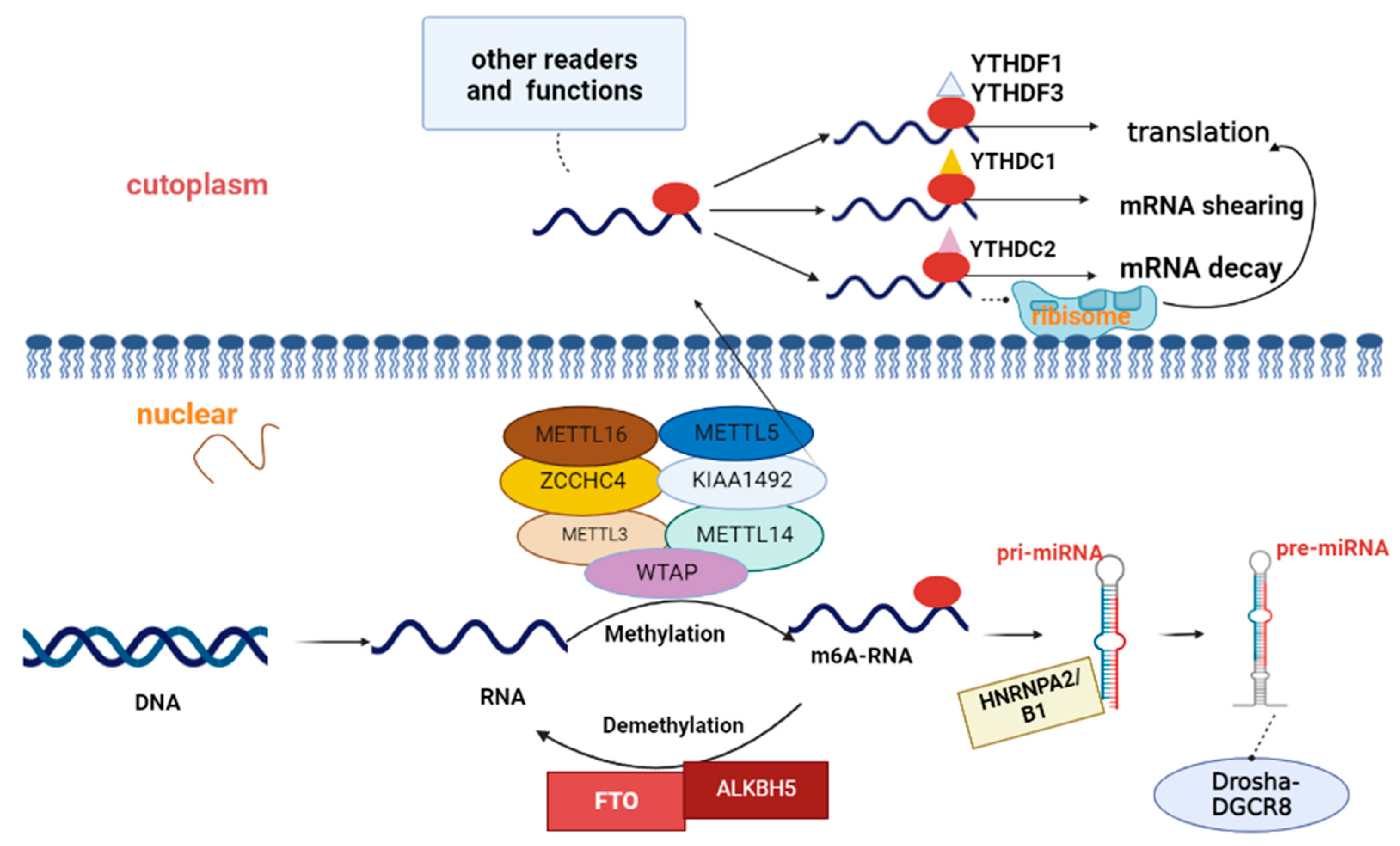
1.1.1. Methyltransferase-Mediated m6A Methylation Modification

Methyltransferase, or “writer,” mainly catalyzes the m6A modification of mRNA bases, and its members include methyltransferase-like 3/14/5/16 (METTL3/14/5/16), Wilms tumor 1-associating protein (WTAP), KIAA1492, and Zinc Finger CCHC-Type Containing 4 (ZCCHC4). METTL3, the first discovered core methyltransferase subunit, plays an important role in m6A methylation. In addition, it acts as a releasing agent in the cytoplasm, promoting the translation of m6A-modified mRNAs, a process independent of its methyltransferase activity [4]. WTAP and KIAA1492 assist in m6A methylation modifications of mRNAs. WTAP helps localize the METTL3/METTL14 complex by interacting with them to bind to the optimal substrates. KIAA1492 recruits methyltransferase components (METTL3, METTL14, and WTAP) and catalyzes them to specific RNA regions [5]. Notably, METTL5, METTL16, and ZCCHC4 all perform independent catalytic functions. For instance, METTL5 can catalyze m6A modification of some structural RNAs, such as 18S rRNA, 28srRNA, and snRNA. In addition to binding U6 snRNA, ncRNAs, and pre-mRNAs [6], METTL16 can directly catalyze the m6A modification of mRNAs in the nucleus, especially nascent mRNAs, such as MALAT1, XIST, and MAT2A [7]. Among the key independent RNA methylation enzymes, ZCCHC4 targets ribosomal subunits and plays an important role in translation as well as cell proliferation. In addition, other members of the writer’s family, including Vir-like m6A methyltransferase-associated (VIRMA), RNA binding motif protein 15 (RBM15), and RNA binding motif protein 15B (RBM15B), have been shown to be capable of methylating modifications of adenosine (Figure 1) [8].
Methyltransferase is also responsible for the methylation of m6A. METTL3 and METTL14 usually form a complex, which is then combined with WTAP to locate. KIAA1492 is another auxiliary catalytic enzyme. ZCCHC4, METTL5, and METTL16 play independent catalytic functions on ribosomal subunits, structural RNA, and snRNA, respectively.
Studies have shown that methyltransferase-mediated m6A modifications, particularly METTL3 and METTL14, are associated with the occurrence of a variety of cancers, including gastric, prostate, breast, colon, pancreatic, kidney, mesothelioma, sarcoma, and leukemia. Among them, gastric cancer (GC) is the fifth most prevalent type of cancer and the third leading cause of cancer-related death worldwide [9]. A recent study showed that the H3K27ac modification of METTL3 in the promoter region is regulated by p300, which in turn induces transcriptional activation of METTL3 and catalyzes the m6A modification of GC-associated HDGF genes. This enables IGF2BP3 to directly bind the m6A site on HDGF, maintaining gene stability and accelerating the progression of GC [4]. Furthermore, METTL3 promotes GC angiogenesis and progression through the m6A-YTHDF2-dependent pathway to reduce the stability of ADAMTS9 mRNA by activating the PI3K/AKT signaling pathway [10].
There is strong evidence that METTL3 also promotes breast cancer cell proliferation and is associated with poor clinical outcomes in breast cancer patients when METTL3 is highly expressed. METTL3 interacts through the hepatitis B virus X protein (HBXIP), which can suppress METTL3 expression by affecting the binding of METTL3’s 3′UTR and miRNA-let-7h, thus promoting cell proliferation. Meanwhile, METTL3 promotes the expression of HBXIP through m6A modification, forming a positive feedback regulatory mechanism among HBXI, let-7h, and METTL3, to promote the proliferation of breast cancer cells [11]. METTL3 has also been suggested as a new potential target for metformin in the treatment of breast cancer. This is because metformin reduces m6A modifications of METTL3 by targeting miR-483-3p, thereby upregulating the expression of p21. In turn, it inhibits the proliferation of breast cancer cells [12]. Wu et al. revealed that the specific binding of Lnc942 to METTL14 protein enhances METTL14 activity. By upregulating the m6A methylation levels of downstream target genes C-X-C Motif Chemokine Receptor 4 (CXCR4) and CYP1B1, the mRNA stability and expression of these genes are enhanced, which ultimately promotes the proliferation and progression of breast cancer cells [13].
As for renal cell carcinoma, METTL3 increases the level of m6A methylation in the 5′UTR of ABCD1, directly enhancing the expression of ABCD1 protein level and promoting the occurrence of renal tumorigenesis. Interestingly, the mRNA levels of ABCD1 do not alter during this process [14]. Chen et al. demonstrated that METTL14 regulates the stability of TRAF1 mRNA through m6A-IGF2BP2, which in turn increases TRAF1 expression levels and induces apoptosis and angiogenesis in cancer cells [15].
METTL3 and METTL14 have also been shown to play potential roles in colorectal cancer (CRC) progression. According to Chen et al., METTL3 catalyzes m6A methylation of BHLHE41, thus promoting expression at the protein level and forming the m6A-BHLHE41-CXCL1/CXCR2 pathway to suppress anti-tumor immunity to promote cancer [16]. In addition, Yu et al. demonstrated that METTL3 can directly bind to the m6A site of the PLAU RNA 3′UTR, form the MAPK/ERK pathway, and upregulate PLAU mRNA through m6A modification, thereby promoting angiogenesis and metastasis of colorectal cancer [17]. METTL3 also serves as a functional clinical oncogene in an m6A-dependent manner to promote cancer progression [18]. Additionally, based on the study of Chen et al., METTL14-dependent m6A methylation regulates DGCR8 processing of pri-miR-375, accelerates maturation of miR-375, and establishes the METTL14-miR-375-YAP1 pathway, thereby inhibiting CRC progression [19].
There are several other methyltransferases associated with cancer. Gong’s group revealed that lncRNA ZNF582-AS1 can inhibit the m6A modification level of mitochondrial MT-RNR1 (DNA12SrRNA) by reducing the expression level of methyltransferase A8K0B9 protein. Accordingly, MT-RNR1 expression is suppressed, leading to mitochondrial dysfunction and inhibiting the growth of renal cell carcinoma, cells, and invasive metastasis [20].

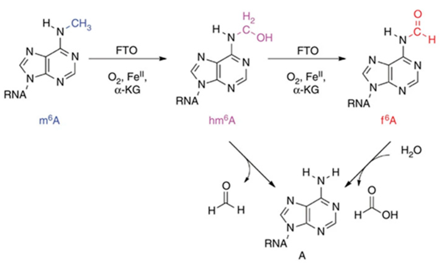
1.1.2. Demethylase-Mediated Methylation Modification

There are only two known demethylases, α-ketone glutarate (α-KG)-dependent dioxygenase and fat mass and obesity-related proteins (FTO) and AlkB Homolog 5 (ALKBH5). Their main function is to remove methylation modifications from m6A that have already occurred (Figure 2). FTO is highly expressed in metabolically active organs such as the brain and fat and affects internal m6A and its modification in the 5′ end cap structure [9]. An increasing number of studies have confirmed that FTO plays an important regulatory role in lipid metabolism. A typical example is that FTO can mediate RNA demethylation, inhibit hepatocyte mitochondrial function, and promote hepatic lipid accumulation [21]. Apart from that, FTO-dependent m6A demethylation promotes adipocyte differentiation and participates in the differentiation and transformation of adipose tissue by downregulating levels of miR-130 and miR-55, thereby upregulating C/EBPβ expression in preadipocytes [22]. Particularly, FTO can directly upregulate C/EBPβ levels to inhibit the differentiation of adipose tissue into brown adipose tissue [23]. According to Lee et al., FTO-dependent demethylation promotes lipogenesis by targeting miR-130 to upregulate the level of peroxisome proliferator-activated receptor γ (PPARγ) [24]. A number of other processes, including lipid metabolism in macrophages and skeletal muscle, are also regulated by FTO (Figure 2) [25]. Based on existing research, we speculate that FTO-mediated m6A modification is closely related to lipid metabolism and is expected to be an important molecular mechanism regulating lipid metabolism. At this stage, however, comprehensive and thorough studies are needed to fully elucidate the specific regulatory mechanisms by which FTO regulates various processes of lipid metabolism. Besides, it is substantiated that FTO-mediated demethylation could be a potential regulatory mechanism in cancer. FTO was found by Niu et al. to be highly expressed in breast cancer and its expression is negatively associated with prognosis [26]. As reported by RUAN et al., under hypoxic conditions, ubiquitination of FTO mediated protein degradation is accelerated and its protein levels are inhibited. Meanwhile, FTO reduces mRNA stability by targeting MTA1, thus inhibiting CRC metastasis [27].
Another identified m6A demethylase, ALKBH5, can regulate mRNA export and RNA metabolism [29]. Numerous studies have shown that ALKBH5 is essential for metastasis of cancer cells. In gastric cancer, ALKBH5 downregulates the expression of PKMYT1 in an m6A-dependent manner, and IGF2BP3 helps stabilize PKMYT1 mRNA by recognizing its m6A modification site, forming the ALKBH5-PKMYT1-IGF2BP3 mechanism of gastric cancer metastasis [30]. Additionally, it promotes invasion and metastasis of GC by reducing the methylation of lncRNA NEAT1 [31]. In breast cancer, ALKBH5 upregulates the expression of NANOG by regulating the m6A site on NANOG, thus promoting metastasis of breast cancer [32]. Furthermore, ALKBH5 was found by TAN et al. to stabilize AURKB RNA in an m6A-dependent manner, thereby exerting an oncogenic effect in renal cell carcinoma [33]. However, studies related to the regulation of lipid metabolism by ALKBH5 have been inadequately explored.

1.1.3. Reading Protein-Mediated m6A Methylation Modifications

The m6A reader proteins are composed of YTH functional domain family proteins (YTHDF1-3), YTH functional domain-containing proteins (YTHDC1-2), insulin-like growth factor 2 mRNA-binding proteins (IGF2PBs), and heterogeneous nuclear ribonucleoproteins (HNRNPs, including HNRNPA2/B1, HNRNPC, and HNRNPG). In the cytoplasm, YTHDF1/YTHDF3 can promote mRNA translation after recruiting translation initiation factors by recognizing m6A, while YTHDF2 promotes mRNA degradation by binding m6A [34]. Additionally, YTHDC1 can regulate the mRNA splicing process by recruiting splicing factor 3 (SRSF13) and blocking the binding of splicing factor 10 (SRSF10) to the nuclear site, which in turn facilitates mRNA translocation from the nucleus to the cytoplasm [35].
YTHDC2, the only protein in the YTH structural domain family that contains a decapping enzyme, regulates the stability of m6A mRNAs by recognizing modifications of methylated m6A and recruiting RNA degradation factors. Moreover, it links m6A-modified mRNAs with ribosomes, facilitating their efficient translation. Unlike YTHDC2, after obtaining m6A modification, HNRNPA2/B1, rather than acting as a direct “reader” of the m6A modification, opens the secondary structure of RNA and promotes recognition of the recognition proteins. In addition, HNRNPA2/B1 recognizes the m6A modification site of pri-miRNAs and accelerates their processing by recruiting the microprocessor complex Drosha-DGCR8 [35]. It is noteworthy that unlike other reader proteins, this process occurs in the nucleus (Figure 3).
It is found that YT521-B homologous structural domain family protein 1 (YTHDF1), an m6A-modified reading protein, plays an essential role in transcription and translation, immune escape, epithelial mesenchymal transition (EMT), and chemoresistance in tumors [36]. In addition to contributing to gastric carcinogenesis by promoting the translation of USP14 and FZD7 proteins in an m6A-dependent manner [37], YTHDF1 has also been testified to promote breast cancer metastasis by recognizing and binding m6A-modified Forkhead Box M1 (FOXM1) mRNAs, thereby accelerating the translation process of FOXM1. In contrast, overexpression of FOXM1 in breast cancer cells could partially reverse the tumor suppressive effect caused by YTHDF1 silencing [38]. In addition, Han et al. found that YTHDF1 recognizes m6A-modified FZD9 and promotes its translation, leading to aberrant activation of Wnt/β-catenin signaling, enhancing the tumorigenicity and stem cell-like activity of CRC [39].
Taken together, m6A methylation regulates the expression of proto-oncogenes or oncogenes, thus affecting cancer development, metastasis, and invasion. The specific process is to first identify the m6A regulators that trigger abnormal methylation levels in cancer and verify their roles in cancer cells. The following steps are combined with the methylation site prediction method to identify target genes and explore their positive or negative regulatory effects on target mRNAs before the final formation of the m6A-regulator-target gene axis. Another conclusion is that there is an interaction between non-coding small molecule RNAs and m6A methylation modifications in cancer. Taking miRNAs as an example, m6A can regulate their processing and maturation to perform cancer-promoting or cancer-suppressing functions. In turn, miRNAs can target m6A regulators to achieve regulation of methylation levels. Moreover, m6A-modified reader proteins (YTH proteins) have been shown to directly regulate lncRNAs’ molecular functions. Similar to miRNAs, lncRNAs can also directly affect m6A levels. Except for this, circRNAs can also serve as targets for m6A methylation modification, although their feedback on m6A modification is less studied. Based on the property that circRNAs act as miRNA sponges in cancer, Ma et al. proposed that circRNAs could be indirectly involved in the regulation of m6A methylation levels, but this still remains to be tested [40]. Previous studies proved that regulators or inhibitors of m6A modification may provide potential therapeutic strategies for cancers.
As a nonsteroidal anti-inflammatory drug, Meclofenamic acid (MA) acts as an FTO inhibitor by competing with the FTO binding site, while MA2, an ethyl ester derivative of MA, increases m6A modification and inhibits tumor progression [41]. SPI1 is a hematopoietic transcription factor that directly inhibits METTL14 expression in malignant hematopoietic cells and may be a potential therapeutic target for acute myeloid leukemia [42]. As an enzyme-specific inhibitor of METTL3, STM2457 shows better oncogenic effects as well as less toxic side effects, revealing promise in the clinical treatment of acute myeloid leukemia [43]. The Wnt signaling program has been shown to promote cancer metastasis. Recent studies have found that in cancer cells with low FTO levels, m6A was enriched in mRNAs belonging to the Wnt signalling program. And through clinical trials, they found that cancer cells or tumours with reduced FTO levels and enhanced Wnt activity were more sensitive to Wnt inhibitors [44]. Collectively, m6A methylation is expected to be a potential target for cancer diagnosis, metastasis identification, and therapy. Meanwhile, the design of small molecules based on m6A regulators also provides new perspectives for cancer intervention, radiotherapy, chemotherapy, and other related fields. Therefore, there is an urgent need for researchers to thoroughly explore the relationship between m6A and cancer, and to improve related strategies to serve cancer diagnosis and clinical treatment.

1.2. Detection of m6A Methylation

Direct detection of m6A methylation bases is difficult because m6A methylation base pairing remains constant and cannot be distinguished from regular bases by reverse transcription. Several tools have been developed successively to predict m6A methylation sites, including the following:
Scilab v1.1SRAMP, software at http://www.cuilab.cn/sramp/ (accessed on 20 February 2016) [45];
Scilab v1.1iRNA-Methyl, software at http://lin.uestc.edu.cn/server/ (accessed on 24 August 2015) [46];
Scilab v1.11 iRNAm5C-PseDNC, software at http://www.jci-bioinfo.cn/iRNAm5CPseDNC (accessed on 20 June 2017) [47];
Scilab v1.1m6A MRFS, software at http://server.malab.cn/M6AMRFS/ (accessed on 25 October 2018) [48];
Scilab v1.1m6APred-EL, software at http://server.malab.cn/M6APred-EL/ (accessed on 7 September 2018) [49];
Scilab v1.1Deep m6A Seq, eq, software at https://github.com/rreybeyb/ (accessed on 31 December 2018) [50];
On the other hand, commonly used techniques for detecting m6A include methylated RNA immunoprecipitation sequencing (MeRIP-seq), m6A individual-nucleotide-resolution crosslinking and immunoprecipitation (miCLIP-seq), liquid chromatography-tandem mass spectrometry (LC-MS/MS), etc.

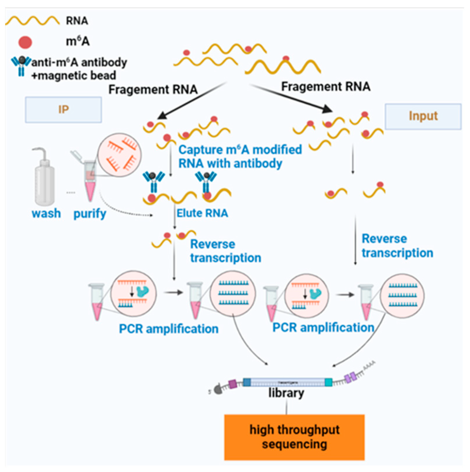
1.2.1. Introduction of MeRIP-Seq

As the most commonly used method for m6A methylation detection, MeRIP-seq identifies m6A methylation levels at the whole transcriptome level. RNA-specific enrichment and interruption are first achieved by RNAs fragmentation reagents (Thermo, Waltham, MA, USA). After enriching the enriched mRNAs with m6A antibodies, their fragments are purified, sequentially reverse transcribed, and PCR amplified to construct a high-throughput sequencing library (IP). Furthermore, a normal transcriptome library (input) is constructed separately as a control to reflect the abundance of the base RNAs. Finally, the two sequencing libraries are sequenced together to obtain an m6A map of the whole transcriptome following biological analysis (Figure 4) [51].
This technique has been extensively used to study m6A modifications because it is easy, fast, and relatively inexpensive. It enables the qualitative analysis of mRNA regions undergoing hypermethylation. Wang et al. detected 9085 m6A methylation peaks by performing MeRIP-seq analysis on the transcriptome of velvet goat skin tissue. The transcriptome and differential peaks were then combined to screen out 19 differentially expressed genes that contain RNA methylation modifications associated with velvet growth [52]. In addition, MeRIP-seq has been widely used as an assay for investigating the mechanisms related to cancer regulation by measuring m6A methylation modifications. Yang et al. identified m6A modifications in the EOC transcriptome of endometrial ovarian cancer by MeRIP-seq for the first time and described methylated m6A to modify differentially expressed genes. This provides a new direction for the underlying molecular mechanisms and signaling pathways of EOC development [53]. During a comprehensive analysis of mRNA m6A modifications in human colorectal cancer, Li et al. conducted a combined analysis by MeRIP-seq and RNA-seq to predict RNA-binding proteins and identify methylation-related genes FMR1, IGF2BP2, and IGF2BP3 that may be involved in the development of CRC [54]. At this stage, MeRIP-seq can only identify regions of m6A hypermethylation but cannot realize single-base resolution sequencing.

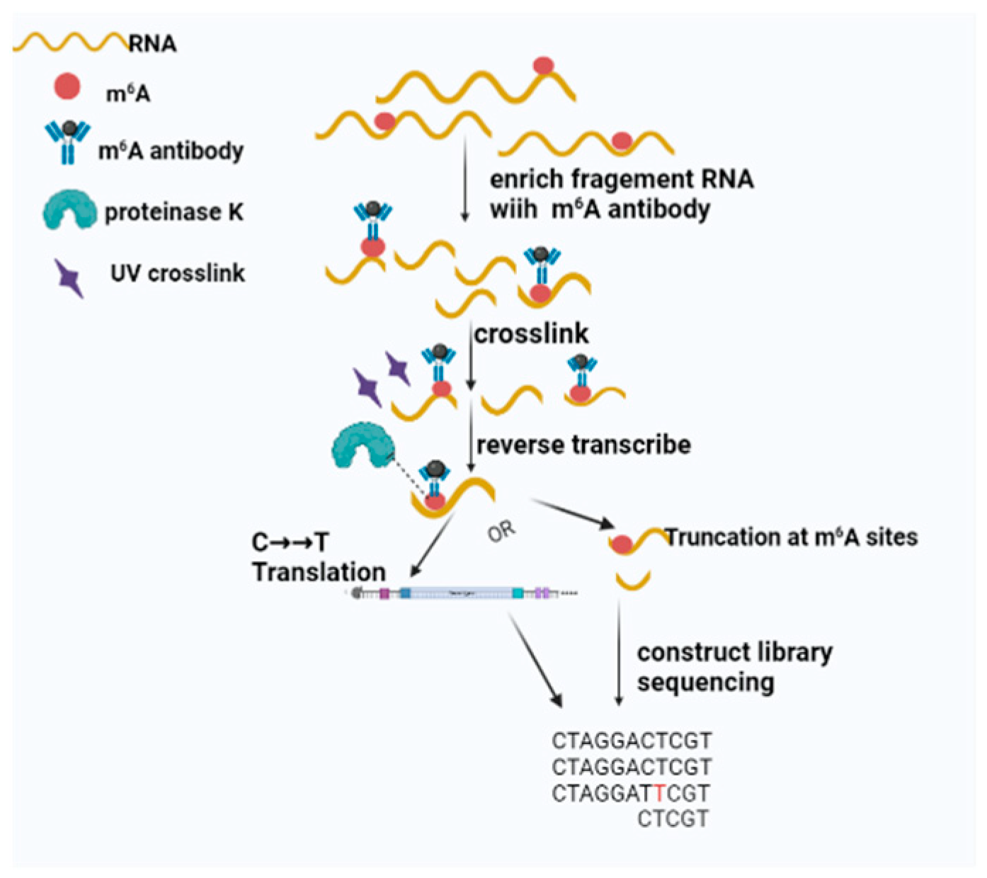
1.2.2. Introduction of miCLIP

MiCLIP induces mutations during reverse transcription by crosslinking single nucleotides that detect methylated bases at the antibody binding sites. As another widely applied m6A detection technology, like MeRIP-seq, it performs immunoprecipitation using UV crosslinking after completing RNA fragmentation and binding of m6A methylation-modified mRNA fragments using antibody immunomagnetic beads. Next, reverse transcription is performed after digestion with K protein. M6A modifications on the RNA result in highly specific mutations or truncations on the corresponding cDNAs. Finally, co-sequencing of the library is achieved to perform single methyl site detection (Figure 5). Furthermore, miCLIP enables high-resolution detection of individual m6A residues and m6A clustering analysis of the entire RNAs. Using miCLIP, combined with RIP-seq, Liu et al. identified a total of eight m6A sites in the genome at single-base resolution, further verifying that SARS-CoV-2 genomic RNA can dynamically modify m6A, making it a negative regulatory RNA in human and monkey cells [54]. This approach was also employed by Zhao and Liu’s team to address and compare the differences in mRNA m6A modifications in temozolomide-sensitive and drug-resistant glioblastoma GBM tissues [55]. They also probed into the interaction mechanism of m6A modifications and histone modifications in regulating drug resistance in glioblastoma.

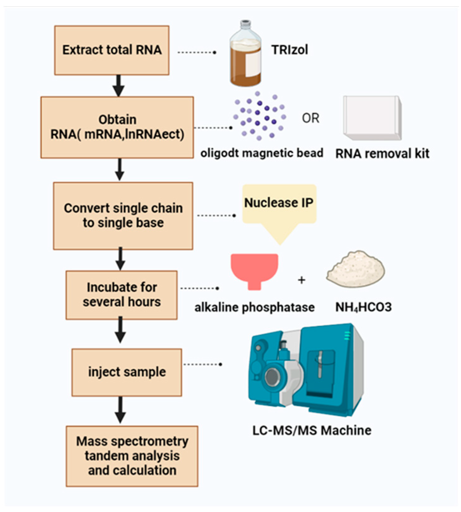
1.2.3. Introduction of LC-MS/MS

LC-MS/MS enables the separation of sample components based on multiple migration rates because various substances interact differently with the stationary and mobile phases in the chromatograph. Specifically, after the extraction of total RNA using the NA extraction reagent TRIzol, oligodT magnetic beads are used to enrich mRNA, or the RNA removal kit is used to obtain RNA including mRNA, lncRNA, and circRNA. In the following step, nuclease P1 is used to digest RNA from single strands to individual bases. After several hours, samples are incubated with alkaline phosphatase and ammonium bicarbonate. Ultimately, following the injection of the sample into the liquid chromatograph, the overall methylation of m6A on the mRNA can be calculated using mass spectrometry tandem analysis based on the ratio of m6A to total adenine (Figure 6) [56].
Compared with the previous two methods, LC-MS/MS is able to detect the overall m6A level of RNA but is more suitable for smaller-scale gene detection and cannot pinpoint methylation sites. ZHANG et al. determined the levels of m6A and A in RNAs by electrospray ionization (ESI) in positive ion multiple reaction monitoring (MRM) mode. They successfully applied this method to detect the level of m6A in RNAs of mouse spleen T cells under different treatment conditions. As reported by SHEN et al., LC-MS/MS analysis of m6A methylation glycolysis in CRC patients demonstrated that levels of m6A methylation are significantly higher in CRC patients who ingested higher levels of fluorodeoxyglucose [18].
In addition to conventional methods, some improved assays are also included. One is m6A-label-seq developed by SHU et al., a method to label RNA m6A sites throughout transcriptome by cellular self-metabolism. Using this method, labeled sites can undergo chemical treatment-induced reverse transcription base mutations, which in turn enables single-base resolution determination [57]. The other is the m6A-SEAl technique created by Wang et al. This is the first technology to achieve m6A chemical labeling using FTO enzyme and has been successfully applied to high-throughput sequencing. Due to its high sensitivity and specificity, it has been validated to be suitable for a small number of m6A samples [58].

2. m6A Methylation in Lipid Metabolism

2.1. m6A Methylation Involved in the Regulation of Human Lipid Metabolism

Lipids are essential components of biological membranes and structural cellular units. Mainly used for energy storage and metabolism, they also play a pivotal role in a variety of cellular activities as signaling molecules. In vivo, lipid metabolism refers to the process of digestion, absorption, synthesis, and decomposition of fat with the help of various related enzymes, including the biosynthesis of saturated fatty acids, the extension of fatty acid carbon chains, and the production of unsaturated fatty acids. This process requires the participation of many enzymes, among which acetyl coenzyme A carboxylase (ACC) and fatty acid synthase (FAS) are the key synthases involved in fat synthesis. DGAT1 is mainly responsible for the synthesis of TAG during fat absorption and storage, while adipose triglyceride lipase (ATGL) and hormone-sensitive lipase (HSL) are mainly involved in fat hydrolysis. In addition, stearoyl coenzyme A desaturase 1 (SCD1) catalyzes the conversion of saturated fatty acids into monounsaturated fatty acids, and peroxisome proliferator-activated receptor (PPAR) and sterol regulatory element binding protein-1 (SREBP-1) regulate lipid metabolism.
Several recent studies have indicated that RNA m6A methylation modifications are closely associated with lipid metabolism and human lipid disorders, and m6A can regulate lipid metabolism-related gene expression. As reported by Liu et al., the zinc finger protein 217 (Zfp217) gene upregulates m6A levels of cell cyclin D1 by increasing the expression of m6A methyltransferase METTL3. As a result, the knockout of Zfp217 prevents the amplification process of mitotic clones and induces inhibition of adipogenesis [59]. As Yang et al. proved, overexpression of METTL3/14 upregulates levels of m6A modification, enhances mRNA stability of ACLY and SCD1, and engenders increasing expression levels of ACLY and SCD1 proteins, thereby promoting FA de novo synthesis and lipid accumulation [60]. According to PENG et al., on the other hand, a novel mechanism is revealed by which m6A mRNA methylation modification affects the lipid metabolism of hepatocytes through the regulation of autophagy. Additionally, YTHDF1 binds to the mRNA of the key autophagy gene rubicon mainly through the m6A motif and inhibits its degradation. When rubicon is expressed at high levels, autophagic vesicles are less likely to fuse with lysosomes, leading to reduced degradation of lipid droplets and increased lipid droplet accumulation in hepatocytes via the lysosomal pathway (Figure 7) [61]. Regarding hepatic lipid metabolism, Li et al. revealed that the METTL3-mediated elevated m6A levels on a high-fat diet exacerbate the disorder of hepatic lipid metabolism, manifested by increased subcutaneous fat, hepatic steatosis, and increased total cholesterol in serum. Further studies showed that M6A methylation achieves the regulation of lipid metabolism by affecting the stability of genes related to lipid metabolism, especially Lpin1 mRNA. Interestingly, in the HFD group, METTL3 is mainly involved in fatty acid catabolism as well as oxidation-related genes. In contrast, METTL3-targeted genes are mainly enriched in the sterol synthesis pathway under normal diet conditions. Therefore, researchers propose that METTL3 can simultaneously participate in the bidirectional regulation of lipid metabolism [62]. Another methyltransferase, WTAP, is also shown to be involved in the regulation of lipid metabolism. The deficiency of WTAP induces the expression and secretion of IGFBP1, thus enhancing lipolysis in eWAT and increasing serum FFA, leading to hepatic steatosis. Furthermore, the hepatic WTAP deficiency increases expression of both CD36 and CCL2, thereby enhancing the hepatic uptake capacity of FFA [63].
FTO functions as a major demethyltransferase that directly regulates lipid metabolism and participates in it by removing m6A methylation modifications. Zfp217 regulates m6A mRNA methylation by activating transcription of the m6A demethylase FTO, and couples gene transcription to m6A mRNA modification to promote adipogenesis [64]. FTO was found by Kang et al. to mediate the reduction in m6A methylation levels and enhance the expression of lipid metabolism-related genes FASN, SCD, and MGAT. Meanwhile, it also inhibits lipid transport-related genes MTTP, APOB, and LIPC, which are involved in fatty acid synthesis as well as triglyceride accumulation [65]. As reported by Zhou et al., FTO-dependent m6A demethylation promotes the expression of key SREBP-1c genes to indirectly increase SREBP1-c expression and promote the formation of hepatocytes and lipid droplets [25]. A subsequent study by Wang et al. further verified that NAPDH binds and enhances FTO expression to promote lipid synthesis in 3T3 cells through m6A demethylation [66]. Adipogenesis is also regulated by FTO-mediated mRNA demethylation, which impacts selective splicing of mRNAs during mitotic clonal amplification (Figure 7) [67]. Sun provided novel insights into the epigenetic mechanism of FTO’s function in hepatic lipid synthesis. Specifically, knockdown of FTO significantly increases m6A levels in FASN mRNA and promotes mRNA decay, thereby decreasing FASN mRNA expression. The protein expression of acetyl-CoA carboxylase and ATP-citrate lyase is further reduced, ultimately inhibiting de novo lipogenesis in HepG2 cells [68]. Another important target gene for the function of m6A methylation on hepatic lipid metabolism was demonstrated by Zhong et al. METTL3 promotes the expression of PPARα by positively regulating the m6A modification of PPARα RNA, while YTHDF2 plays the opposite role. By combining the actions of two regulators, PPARα mRNA remains stable, thus regulating lipid metabolism [69]. Similarly, carboxylesterase 2(CES2), which is involved in the regulation of hepatic lipid metabolism, has also been shown to be regulated by m6A modification. Dual knockdown of methyltransferases METTL3 and METTL14 upregulates CES2 expression and then reduces lipid accumulation. However, knockdown of demethyltransferases FTO or ALKBH5 downregulates CES2 expression and achieves lipid increase in HepaRG and HepG2 cells. Furthermore, the reader protein YTHDC2 reduces CES2 mRNA stability by recognizing m6A in the 5′-UTR of CES2 [70].
In addition, m6A plays a role in long-chain fatty acid uptake, just as Zong et al. revealed. Deletion of methyltransferase METTL3 decreases Traf6 expression in the lipopolysaccharide-mediated IPEC-J2 inflammatory response, thereby inhibiting the downstream NF-kB and MAPK signaling pathways and promoting the uptake of long-chain fatty acids (Figure 7) [71]. Xu et al. found that METTL3 increases levels of caprylic, gamma-linolenic, arachidonic, and docosapentaenoic acid by upregulating the expression of the lipid metabolism regulator CYP4F40, thereby reducing free fatty acid FFA-induced steatosis in hepatocytes. In addition, recent studies have shown that m6A in 18S rRNAs can also promote fatty acid metabolism. Specifically, METTL5-mediated alterations in 18S rRNA m6A modification results in impaired assembly of the 80S ribosome and inhibiting translation of mRNAs involved in fatty acid metabolism. In particular, acyl-CoA synthetase long chain family member 4 (ACSL4), a target gene of METTL5, also enhances the regulatory function of METLT5 in promoting lipid metabolism [72].
The figure shows the regulation of partial m6A on human lipid metabolism. The m6A regulatory factor targets the mRNA of lipid metabolism regulation genes, and the levels of positive or negative regulation genes to achieve the regulation of lipid metabolism.
As shown in the above studies, m6A modification plays a dual role in human lipid metabolism. On the one hand, the discovery of m6A modification is a new direction to regulate lipid metabolism at the molecular level. M6A methylation acts on genes related to lipid metabolism regulation, affects the translation, splicing, transport, and other processes of mRNAs, and realizes the regulation of expression level. On the other hand, m6A levels can be regulated as in Zfp217, thus affecting the role in lipid metabolism. An increasing number of studies have demonstrated that FTO mediates m6A modifications in the regulation of human lipid metabolism, while FTO deficiency triggers the upregulation of m6A modification levels. Ultimately, this leads to browning and thermogenesis in white adipocytes, providing a potential target to combat obesity and metabolic diseases [66]. Nonetheless, there are relatively few studies examining the role of other m6A methylation regulators in lipid metabolism and their specific mechanisms of action.

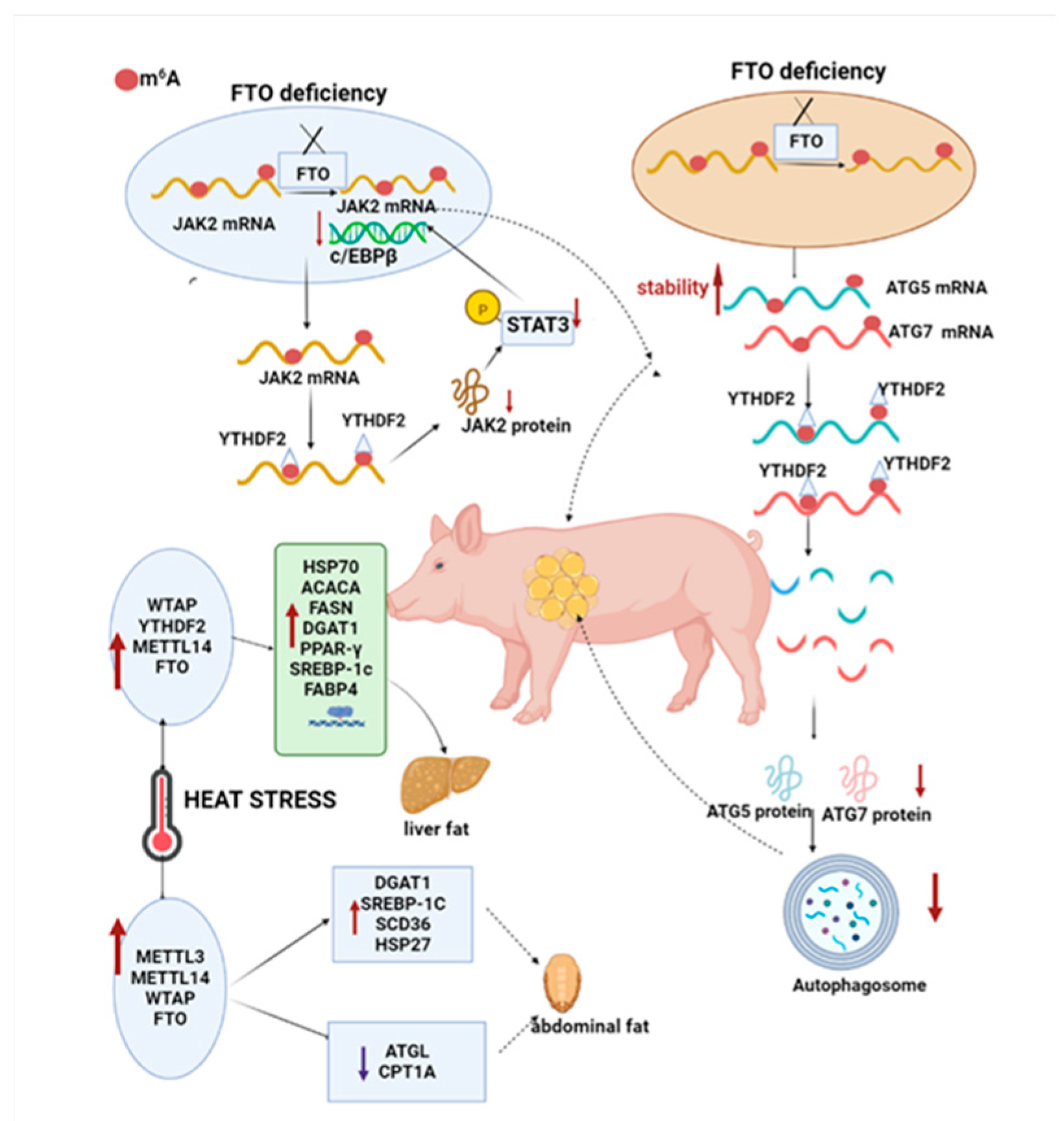
2.2. m6A Methylation Involved in the Regulation of Animal Lipid Metabolism

There is evidence that hypermethylation is primarily involved in cellular responses to peptide hormone stimulation and adipogenesis-related pathways, especially fat metabolism of related tissue in pigs and poultry. In sows subjected to heat stress, methylation regulators FTO, WTAP, YTHDF2, and METTL14 upregulate the expression levels of HSP70 protein and genes, such as ACCAA and FASN that are involved in hepatic fatty acid biosynthesis and fatty acid metabolism. As for abdominal fat, METTL3, METTL4, WTAP, and FTO up-regulate the levels of HSP27 protein as well as fat metabolism-related genes, such as SCD36, but down-regulate the levels of ATGL and CPT1A [73]. Wang’s team revealed that m6A participates in the molecular mechanism of porcine fat deposition through post-transcriptional regulation of the JAK2-STAT3-C/EBPβ signaling pathway. The absence of FTO promotes the recognition and degradation of JAK2 by YTHDF2, which in turn inhibits the expression of JAK2 and the phosphorylation of STAT3, leading to increased difficulty in translocation to the nucleus, thus inhibiting the transcription factor C/EBPβ that is necessary for early adipogenesis. It is also validated that in porcine precursor adipocytes, m6A modifications at the locus of the autophagy-associated protein ATG5 and ATG7 are enhanced by interfering with FTO that is recognized and degraded by YTHDF2, resulting in a reduction in protein expression that inhibits autophagosome formation and autophagic flow occurrence. This ultimately hinders the fat deposition process (Figure 8) [74]. Regarding the process of intramuscular fat formation in pigs, Jiang et al. sequenced m6A in the longest dorsal muscles (LDMs) of Changbai and Jinhua pigs and concluded that m6A modifications may play a regulatory role in intramuscular adipogenesis [75].
An increasing number of studies have suggested that m6A modifications are crucial for adipogenesis and lipid metabolism in poultry [76]. A study by Cheng et al. analyzed the m6A methylationome of chicken abdominal adipose tissue and found that hypermethylation of ACSL1 and FASN mRNAs is associated with increased mRNA stability, thereby promoting TG formation. Through m6A methylation, adipocyte differentiation-associated LPIN1 maintains energy metabolism homeostasis by increasing mRNA expression levels. Besides, researchers also suggested that hypomethylation of LRP4 may promote LRP4 mRNA levels by reducing YTHDF2-mediated mRNA decay, thereby increasing adipocyte size. This conjecture needs to be further tested [77]. According to Hu et al., glucocorticoid (GR)-mediated transactivation of FTO and m6A demethylation at the mRNA level of adipogenic genes SREBP-1, FAS, and SCD facilitate activation of chicken liver adipogenic genes and TG accumulation in primary chicken hepatocyte cells treated with OA/DEX [78]. Zhang et al. have proposed another ALKBH5 as a novel regulator of chicken preadipocyte proliferation and differentiation. ALKBH5 was found to promote chicken preadipocyte differentiation by directly or indirectly enhancing the expression of PPAR γ, FABP, and FAS. The deficiency is that the specific mechanism of action is not clearly explained [79]. N6-methyladenosine demethylase ALKBH5 is a novel regulator of the proliferation and differentiation of chicken preadipocytes. IGF2BP1 is also reported to play a significant role in chicken lipid metabolism. In chicken adipocyte proliferation, cell cycle-related genes, such as CDK1, CCNB3, and so forth, can be induced to promote cell proliferation. Moreover, its overexpression reduces cell population arrest in the G1 phase but increases cell population in the S phase. During adipogenesis, upregulating IGF2BP1 expression-related genes (ACSL5, CPT1APCK1, PPARα, FTO, etc.) could promote chicken adipocyte differentiation, fatty acid metabolism, and lipid droplet accumulation (Figure 9) [80].
Taken together, m6A modification is responsible for regulating lipid metabolism in muscle, viscera, bone, and other body tissues of animals. Research in this field also provides new ideas for improving the growth and breeding of livestock as well as the control of diseases. In addition, recent studies have shown that rumen-protected methionine and lysine in low-protein diets enhance FADS and ACC expression through m6A methylation modifications, affecting polyunsaturated fatty acid synthesis in lamb liver instead of muscle [81]. In exploring the whole transcriptome profile of yak m6A and its potential biological functions in adipocyte differentiation, Zhang et al. presented that several m6A differentially expressed genes, such as KLF9 and FOXO1, are involved in yak adipocyte differentiation through activation of Foxo and Hippo signaling pathways [82].

2.3. m6A Methylation Involved in the Regulation of Milk Fat Metabolism

As economic life continues to develop, people’s demands and quality requirements for dairy products increase. In addition to affecting flavor and nutritional value, milk fat becomes a major economic trait in ruminant reproduction. The process of milk fat regulation is influenced by multiple factors, such as genes and regulatory factors, among which SREBP-1 is one of the important nuclear transcription factors in mammals. In cow mammary tissue, a key function of SREBP-1 is to promote the ab initio synthesis of fatty acids and regulate the composition and content of beneficial fatty acids in milk by activating the expression of key genes and enzymes during milk fat synthesis and secretion. A lactation model established by Wang et al. revealed that METTL3 could promote translation to protein formation by positively regulating the methylation of SREBP1 mRNA, thereby accelerating milk fat synthesis [83]. When exploring the biological functions of m6A methylation in S. aureus-stimulated bovine mammary epithelial cells, Li et al. spotted that m6A hypermethylated genes are significantly associated with fatty acid degradation and adipocytokine signaling pathways, while m6A modification alters the mRNA levels of genes related to milk fat metabolism [84]. It is still necessary to validate the specific mechanism of action. More importantly, this study offers a new proposal for m6A modification to regulate milk fat metabolism.
Currently, m6A modifications have been found to be involved in the regulation of milk fat metabolism, such as SREBP1, which has been shown to be a target factor for m6A methylation. Additionally, genes and signaling pathways related to milk fat metabolism have also been shown to be closely associated with m6A methylation. Furthermore, m6A metabolism-related genes and signaling pathways have also been shown to be closely related to m6A methylation. Excitingly, the mechanism of m6A methylation in the human mammary gland, particularly in breast cancer, has been well established and is expected to provide a reference for research on mammary gland development and milk fat metabolism in ruminants [85].

3. Summary and Outlook

At present, a growing number of researchers are focusing on studying m6A methylation modification, which is leading the public to a deeper understanding of m6A methylation modification. Moreover, m6A methylation modification is a type of epistatic modification that can perform multiple functions, including both molecular and biological functions. At the molecular level, in addition to regulating RNA splicing, translation, transport, and stability, m6A methylation also affects the cleavage, transport, stability, and degradation of non-coding small molecules, such as miRNAs, lncRNAs, and circRNAs. Because of various factors, such as technological limitations, their specific mechanisms of action in ncRNAs, especially with circRNAs molecules, have not yet been clearly elucidated.
In recent years, technologies for m6A detection and methylation site prediction have emerged as a result of the advancement of second-generation sequencing and bioinformatics. In addition to the traditional techniques for detection, such as MeRIP-seq and miCLIPSRAMP, an increasing number of new techniques are being developed. However, new techniques are not as efficient, accurate, convenient, or inexpensive as traditional techniques. Therefore, it is necessary to conduct further research to enhance their applicability and convenience.
Nowadays, m6A is one of the major research hotspots in the field of cancer research, and its regulatory mechanism on cancer is becoming more and more refined, making it a potential target for cancer identification and treatment in the future. In addition, m6A modifications play a regulatory role in lipid metabolism. However, the specific regulatory mechanisms remain unclear due to limited studies, especially in ruminant milk lipid metabolism. The existing studies on the function of m6A in regulating lipid metabolism in organisms mainly focus on the demethyl transferase FTO. It remains to be determined whether other m6A regulators are involved in the regulatory mechanisms and what the specific regulatory mechanisms are. Aside from this, exosomes, as intercellular communication carriers, also play a role in lipid metabolism by interfering with the synthetic transport and degradation processes. According to investigators, melatonin reduces adipocyte-derived exosome resistin levels via m6A RNA demethylation in adipocytes, further reducing hepatic steatosis [86]. In light of this, the authors infer that exosome and m6A might provide new approaches to the regulation of lipid metabolism.

Author Contributions

Writing—original draft preparation, Z.X. and Z.C.; writing—review and editing, T.Z., Q.L. and Z.Y. All authors have read and agreed to the published version of the manuscript.

Funding

Jiangsu Agricultural Science and Technology Independent Innovation Fund (CX(21)3119) Yangzhou University “Blue Project” and Scientific Research Program Funded by the Education Department of Shaanxi Provincial Government (Program No. 21JP110).

Institutional Review Board Statement

Not applicable.

Informed Consent Statement

Not applicable.

Data Availability Statement

Not applicable.

Conflicts of Interest

The authors declare no conflict of interest.

References

  1. Desrosiers, R.; Friderici, K.; Rottman, F. Identification of Methylated Nucleosides in Messenger RNA from Novikoff Hepatoma Cells. Proc. Natl. Acad. Sci. USA 1974, 71, 3971–3975. [Google Scholar] [CrossRef] [Green Version]
  2. Zhang, H.; Shi, X.; Huang, T.; Zhao, X.; Chen, W.; Gu, N.; Zhang, R. Dynamic landscape and evolution of m6A methylation in human. Nucleic Acids Res. 2020, 48, 6251–6264. [Google Scholar] [CrossRef]
  3. Maity, A.; Das, B. N6-methyladenosine modification in mRNA: Machinery, function and implications for health and diseases. FEBS J. 2016, 283, 1607–1630. [Google Scholar] [CrossRef] [PubMed] [Green Version]
  4. Wang, Q.; Chen, C.; Ding, Q.; Zhao, Y.; Wang, Z.; Chen, J.; Jiang, Z.; Zhang, Y.; Xu, G.; Zhang, J.J.G. METTL3-mediated m6A modification of HDGF mRNA promotes gastric cancer progression and has prognostic significance. Gut 2019, 69, 1193–1205. [Google Scholar] [CrossRef] [PubMed]
  5. Jiang, X.; Liu, B.; Nie, Z.; Duan, L.; Xiong, Q.; Jin, Z.; Yang, C.; Chen, Y. The role of m6A modification in the biological functions and diseases. Signal Transduct. Target. Ther. 2021, 6, 74. [Google Scholar] [CrossRef]
  6. Warda, A.S.; Kretschmer, J.; Hackert, P.; Lenz, C.; Urlaub, H.; Höbartner, C.; Sloan, K.E.; Bohnsack, M.T. Human METTL16 is a N6-methyladenosine (m6A) methyltransferase that targets pre-mRNAs and various non-coding RNAs. EMBO Rep. 2017, 18, 2004. [Google Scholar] [CrossRef]
  7. Deng, X.; Su, R.; Weng, H.; Huang, H.; Li, Z.; Chen, J.J.C.R. RNA N 6 -methyladenosine modification in cancers: Current status and perspectives. Cell Res. 2018, 28, 507–517. [Google Scholar] [CrossRef] [Green Version]
  8. Yue, Y.; Liu, J.; Cui, X.; Cao, J.; Luo, G.; Zhang, Z.; Cheng, T.; Gao, M.; Shu, X.; Ma, H.; et al. VIRMA mediates preferential m(6)A mRNA methylation in 3′UTR and near stop codon and associates with alternative polyadenylation. Cell Discov. 2018, 4, 10. [Google Scholar] [CrossRef] [Green Version]
  9. Smyth, E.C.; Nilsson, M.; Grabsch, H.I.; van Grieken, N.C.; Lordick, F. Gastric cancer. Lancet 2020, 396, 635–648. [Google Scholar] [CrossRef]
  10. Wang, N.; Huo, X.; Zhang, B.; Chen, X.; Zhao, S.; Shi, X.; Xu, H.; Wei, X. METTL3-Mediated ADAMTS9 Suppression Facilitates Angiogenesis and Carcinogenesis in Gastric Cancer. Front. Oncol. 2022, 12, 861807. [Google Scholar] [CrossRef]
  11. Cai, X.L.; Wang, X.; Cao, C.; Gao, Y.E.; Zhang, S.Q.; Yang, Z.; Liu, Y.X.; Zhang, X.D.; Zhang, W.Y.; Ye, L.H. HBXIP-elevated methyltransferase METTL3 promotes the progression of breast cancer via inhibiting tumor suppressor let-7g. Cancer Lett. 2018, 415, 11–19. [Google Scholar] [CrossRef]
  12. Cheng, L.; Zhang, X.; Huang, Y.Z.; Zhu, Y.L.; Xu, L.Y.; Li, Z.; Dai, X.Y.; Shi, L.; Zhou, X.J.; Wei, J.F.; et al. Metformin exhibits antiproliferation activity in breast cancer via miR-483-3p/METTL3/m(6)A/p21 pathway. Oncogenesis 2021, 10, 7. [Google Scholar] [CrossRef]
  13. Sun, T.; Wu, Z.; Wang, X.; Wang, Y.; Hu, X.; Qin, W.; Lu, S.; Xu, D.; Wu, Y.; Chen, Q.; et al. LNC942 promoting METTL14-mediated m6A methylation in breast cancer cell proliferation and progression. Oncogene 2020, 39, 5358–5372. [Google Scholar] [CrossRef]
  14. Shi, Y.; Dou, Y.; Zhang, J.; Qi, J.; Xin, Z.; Zhang, M.; Xiao, Y.; Ci, W. The RNA N6-Methyladenosine Methyltransferase METTL3 Promotes the Progression of Kidney Cancer via N6-Methyladenosine-Dependent Translational Enhancement of ABCD1. Front. Cell Dev. Biol. 2021, 9, 737498. [Google Scholar] [CrossRef]
  15. Chen, Y.; Lu, Z.; Qi, C.; Yu, C.; Li, Y.; Huan, W.; Wang, R.; Luo, W.; Shen, D.; Ding, L.; et al. N(6)-methyladenosine-modified TRAF1 promotes sunitinib resistance by regulating apoptosis and angiogenesis in a METTL14-dependent manner in renal cell carcinoma. Mol. Cancer 2022, 21, 111. [Google Scholar] [CrossRef]
  16. Chen, H.; Pan, Y.; Zhou, Q.; Liang, C.; Wong, C.C.; Zhou, Y.; Huang, D.; Liu, W.; Zhai, J.; Gou, H.; et al. METTL3 inhibits anti-tumor immunity by targeting m(6)A-BHLHE41-CXCL1/CXCR2 axis to promote colorectal cancer. Gastroenterology 2022, 163, 891–907. [Google Scholar] [CrossRef]
  17. Yu, T.; Liu, J.; Wang, Y.; Chen, W.; Liu, Z.; Zhu, L.; Zhu, W. METTL3 promotes colorectal cancer metastasis by stabilizing PLAU mRNA in an m6A-dependent manner. Biochem. Biophys. Res. Commun. 2022, 614, 9–16. [Google Scholar] [CrossRef]
  18. Shen, C.; Xuan, B.; Yan, T.; Ma, Y.; Xu, P.; Tian, X.; Zhang, X.; Cao, Y.; Ma, D.; Zhu, X.; et al. m6A-dependent glycolysis enhances colorectal cancer progression. Mol. Cancer 2020, 19, 72. [Google Scholar] [CrossRef] [Green Version]
  19. Chen, X.; Xu, M.; Xu, X.; Zeng, K.; Liu, X.; Sun, L.; Pan, B.; He, B.; Pan, Y.; Sun, H. METTL14 Suppresses CRC Progression via Regulating N6-Methyladenosine-Dependent Primary miR-375 Processing—ScienceDirect. Mol. Ther. 2020, 28, 599–612. [Google Scholar] [CrossRef] [Green Version]
  20. Zhao, W.; Quansah, E.; Yuan, M.; Li, P.; Yi, C.; Cai, X.; Zhu, J. Next-generation sequencing analysis reveals segmental patterns of microRNA expression in yak epididymis. Reprod. Fertil. Dev. 2020, 32, 1067–1083. [Google Scholar] [CrossRef]
  21. He, P.C.; He, C. m6A RNA methylation: From mechanisms to therapeutic potential. EMBO J. 2021, 40, e105977. [Google Scholar] [CrossRef]
  22. Berulava, T.; Rahmann, S.; Rademacher, K.; Klein-Hitpass, L.; Horsthemke, B. N6-adenosine methylation in MiRNAs. PLoS ONE 2015, 10, e0118438. [Google Scholar] [CrossRef]
  23. Kajimura, S.; Seale, P.; Kubota, K.; Lunsford, E.; Frangioni, J.V.; Gygi, S.P.; Spiegelman, B.M. Initiation of myoblast to brown fat switch by a PRDM16-C/EBP-beta transcriptional complex. Nature 2009, 460, 1154–1158. [Google Scholar] [CrossRef] [Green Version]
  24. Lee, E.K.; Lee, M.J.; Abdelmohsen, K.; Kim, W.; Kim, M.M.; Srikantan, S.; Martindale, J.L.; Hutchison, E.R.; Kim, H.H.; Marasa, B.S.; et al. miR-130 Suppresses Adipogenesis by Inhibiting Peroxisome Proliferator-Activated Receptor gamma Expression. Mol. Cell Biol. 2011, 31, 626–638. [Google Scholar] [CrossRef] [Green Version]
  25. Yang, Z.; Yu, G.L.; Zhu, X.; Peng, T.H.; Lv, Y.C. Critical roles of FTO-mediated mRNA m6A demethylation in regulating adipogenesis and lipid metabolism: Implications in lipid metabolic disorders. Genes Dis. 2022, 9, 51–61. [Google Scholar] [CrossRef]
  26. Niu, Y.; Lin, Z.; Wan, A.; Chen, H.; Liang, H.; Sun, L.; Wang, Y.; Li, X.; Xiong, X.-f.; Wei, B.; et al. RNA N6-methyladenosine demethylase FTO promotes breast tumor progression through inhibiting BNIP3. Mol. Cancer 2019, 18, 46. [Google Scholar] [CrossRef] [Green Version]
  27. Ruan, D.-Y.; Li, T.; Wang, Y.-N.; Meng, Q.; Li, Y.; Yu, K.; Wang, M.; Lin, J.-F.; Luo, L.-Z.; Wang, D.-S.; et al. FTO downregulation mediated by hypoxia facilitates colorectal cancer metastasis. Oncogene 2021, 40, 5168–5181. [Google Scholar] [CrossRef]
  28. Fu, Y.; Jia, G.; Pang, X.; Wang, R.N.; Wang, X.; Li, C.J.; Smemo, S.; Dai, Q.; Bailey, K.A.; Nobrega, M.A.; et al. FTO-mediated formation of N6-hydroxymethyladenosine and N6-formyladenosine in mammalian RNA. Nat. Commun. 2013, 4, 1798. [Google Scholar] [CrossRef] [Green Version]
  29. Chen, Y.; Zhao, Y.; Chen, J.; Peng, C.; Zhang, Y.; Tong, R.; Cheng, Q.; Yang, B.; Feng, X.; Lu, Y.; et al. ALKBH5 suppresses malignancy of hepatocellular carcinoma via m6A-guided epigenetic inhibition of LYPD1. Mol. Cancer 2020, 19, 123. [Google Scholar] [CrossRef]
  30. Hu, Y.; Gong, C.; Li, Z.; Liu, J.; Chen, Y.; Huang, Y.; Luo, Q.; Wang, S.; Hou, Y.; Yang, S.; et al. Demethylase ALKBH5 suppresses invasion of gastric cancer via PKMYT1 m6A modification. Mol Cancer 2022, 21, 34. [Google Scholar] [CrossRef]
  31. Zhang, J.; Guo, S.; Piao, H.Y.; Wang, Y.; Wu, Y.; Meng, X.Y.; Yang, D.; Zheng, Z.C.; Zhao, Y. ALKBH5 promotes invasion and metastasis of gastric cancer by decreasing methylation of the lncRNA NEAT1. J. Physiol. Biochem. 2019, 75, 379–389. [Google Scholar] [CrossRef] [Green Version]
  32. Zhang, C.Z.; Samanta, D.; Lu, H.Q.; Bullen, J.W.; Zhang, H.M.; Chen, I.; He, X.S.; Semenza, G.L. Hypoxia induces the breast cancer stem cell phenotype by HIF-dependent and ALKBH5-mediated m(6)A-demethylation of NANOG mRNA. Proc. Natl. Acad. Sci. USA 2016, 113, E2047–E2056. [Google Scholar] [CrossRef] [Green Version]
  33. Tan, L.; Tang, Y.; Li, H.; Li, P.; Ye, Y.; Cen, J.; Gui, C.; Luo, J.; Cao, J.; Wei, J. N6-Methyladenosine Modification of LncRNA DUXAP9 Promotes Renal Cancer Cells Proliferation and Motility by Activating the PI3K/AKT Signaling Pathway. Front. Oncol. 2021, 11, 641833. [Google Scholar] [CrossRef]
  34. Shi, H.; Wang, X.; Lu, Z.; Zhao, B.S.; Ma, H.; Hsu, P.J.; Liu, C.; He, C. YTHDF3 facilitates translation and decay of N6-methyladenosine-modified RNA. Cell Res. 2017, 27, 315–328. [Google Scholar] [CrossRef] [Green Version]
  35. Hsu, P.J.; Zhu, Y.F.; Ma, H.H.; Guo, Y.H.; Shi, X.D.; Liu, Y.Y.; Qi, M.J.; Lu, Z.K.; Shi, H.L.; Wang, J.Y.; et al. Ythdc2 is an N-6-methyladenosine binding protein that regulates mammalian spermatogenesis. Cell Res. 2017, 27, 1115–1127. [Google Scholar] [CrossRef] [Green Version]
  36. Luo, X.; Cao, M.; Gao, F.; He, X. YTHDF1 promotes hepatocellular carcinoma progression via activating PI3K/AKT/mTOR signaling pathway and inducing epithelial-mesenchymal transition. Exp. Hematol. Oncol. 2021, 10, 35. [Google Scholar] [CrossRef]
  37. Chen, D.; Cheung, H.; Lau, H.C.; Yu, J.; Wong, C.C. N(6)-Methyladenosine RNA-Binding Protein YTHDF1 in Gastrointestinal Cancers: Function, Molecular Mechanism and Clinical Implication. Cancers 2022, 14, 3489. [Google Scholar] [CrossRef]
  38. Chen, H.; Yu, Y.; Yang, M.; Huang, H.; Ma, S.; Hu, J.; Xi, Z.; Guo, H.; Yao, G.; Yang, L.; et al. YTHDF1 promotes breast cancer progression by facilitating FOXM1 translation in an m6A-dependent manner. Cell Biosci. 2022, 12, 19. [Google Scholar] [CrossRef]
  39. Han, B.; Yan, S.J.; Wei, S.S.; Xiang, J.; Liu, K.L.; Chen, Z.H.; Bai, R.P.; Sheng, J.H.; Xu, Z.P.; Gao, X.W. YTHDF1-mediated translation amplifies Wnt-driven intestinal stemness. EMBO Rep. 2020, 21, e49229. [Google Scholar] [CrossRef]
  40. Ma, S.; Chen, C.; Ji, X.; Liu, J.; Zhou, Q.; Wang, G.; Yuan, W.; Kan, Q.; Sun, Z. The interplay between m6A RNA methylation and noncoding RNA in cancer. J. Hematol. Oncol. 2019, 12, 121. [Google Scholar] [CrossRef]
  41. Huang, Y.; Yan, J.L.; Li, Q.; Li, J.F.; Gong, S.Z.; Zhou, H.; Gan, J.H.; Jiang, H.L.; Jia, G.F.; Luo, C.; et al. Meclofenamic acid selectively inhibits FTO demethylation of m(6)A over ALKBH5. Nucleic Acids Res. 2015, 43, 373–384. [Google Scholar] [CrossRef] [PubMed]
  42. Weng, H.; Huang, H.; Wu, H.; Qin, X.; Zhao, B.S.; Dong, L.; Shi, H.; Skibbe, J.; Shen, C.; Hu, C.; et al. METTL14 Inhibits Hematopoiet-ic Stem/Progenitor Differentiation and Promotes Leukemogenesis via mRNA m(6)A Modification. Cell Stem Cell 2018, 22, 191–205.e9. [Google Scholar] [CrossRef] [PubMed] [Green Version]
  43. Yanko-va, E.; Blackaby, W.; Albertella, M.; Rak, J.; De Braekeleer, E.; Tsagkogeorga, G.; Pilka, E.S.; Aspris, D.; Leggate, D.; Hendrick, A.G.; et al. Small-molecule inhibition of METTL3 as a strategy against myeloid leukaemia. Nature 2021, 593, 597–601. [Google Scholar] [CrossRef] [PubMed]
  44. Jesch-ke, J.; Collignon, E.; Al Wardi, C.; Krayem, M.; Bizet, M.; Jia, Y.; Garaud, S.; Wimana, Z.; Calonne, E.; Hassabi, B.; et al. Downregula-tion of the FTO m(6)A RNA demethylase promotes EMT-mediated progression of epithelial tumors and sensitivity to Wnt inhibitors. Nat. Cancer 2021, 2, 611–628. [Google Scholar] [CrossRef] [PubMed]
  45. Zhou, Y.; Zeng, P.; Li, Y.H.; Zhang, Z.; Cui, Q. SRAMP: Prediction of mammalian N6-methyladenosine (m6A) sites based on sequence-derived features. Nucleic Acids Res. 2016, 44, e91. [Google Scholar] [CrossRef] [PubMed] [Green Version]
  46. Chen, W.; Feng, P.; Ding, H.; Lin, H.; Chou, K.-C. iRNA-Methyl: Identifying N6-methyladenosine sites using pseudo nucleotide composition. Anal. Biochem. 2015, 490, 26–33. [Google Scholar] [CrossRef]
  47. Qiu, W.R.; Jiang, S.Y.; Xu, Z.C.; Xiao, X.; Chou, K.C. iRNAm5C-PseDNC: Identifying RNA 5-methylcytosine sites by incorporating physical-chemical properties into pseudo dinucleotide composition. Oncotarget 2017, 8, 41178–41188. [Google Scholar] [CrossRef] [Green Version]
  48. Qiang, X.; Chen, H.; Ye, X.; Su, R.; Wei, L. M6AMRFS: Robust Prediction of N6-Methyladenosine Sites with Sequence-Based Features in Multiple Species. Front. Genet. 2018, 9, 495. [Google Scholar] [CrossRef] [Green Version]
  49. Wei, L.; Chen, H.; Su, R. M6APred-EL: A Sequence-Based Predictor for Identifying N6-methyladenosine Sites Using Ensemble Learning. Molecular therapy. Nucleic Acids 2018, 12, 635–644. [Google Scholar] [CrossRef] [Green Version]
  50. Zhang, Y.; Hamada, M. DeepM6ASeq: Prediction and characterization of m6A-containing sequences using deep learning. BMC Bioinform. 2018, 19, 524. [Google Scholar] [CrossRef] [PubMed]
  51. Tao, X.; Chen, J.; Jiang, Y.; Wei, Y.; Chen, Y.; Xu, H.; Zhu, L.; Tang, G.; Li, M.; Jiang, A.; et al. Transcriptome-wide N (6) -methyladeno-si-ne methylome profiling of porcine muscle and adipose tissues reveals a potential mechanism for transcriptional regulation and differential methylation pattern. BMC Genom. 2017, 18, 336. [Google Scholar] [CrossRef] [PubMed] [Green Version]
  52. Wang, Y.; Zheng, Y.; Guo, D.; Zhang, X.; Guo, S.; Hui, T.; Yue, C.; Sun, J.; Guo, S.; Bai, Z.; et al. m6A Methylation Analysis of Differentially Expressed Genes in Skin Tissues of Coarse and Fine Type Liaoning Cashmere Goats. Front. Genet. 2019, 10, 1318. [Google Scholar] [CrossRef]
  53. Yang, L.; Chen, X.; Qian, X.; Zhang, J.J.; Wu, M.J.; Yu, A.J. Comprehensive Analysis of the Transcriptome-Wide m6A Methylome in Endometrioid Ovarian Cancer. Front. Oncol. 2022, 12, 583. [Google Scholar] [CrossRef] [PubMed]
  54. Li, N.; Guo, Q.; Zhang, Q.; Chen, B.J.; Li, X.A.; Zhou, Y. Comprehensive Analysis of Differentially Expressed Profiles of mRNA N6-Methyladenosine in Colorectal Cancer. Front. Cell Dev. Biol. 2022, 9, 760912. [Google Scholar] [CrossRef] [PubMed]
  55. Liu, J.; Xu, Y.P.; Li, K.; Ye, Q.; Zhou, H.Y.; Sun, H.; Li, X.; Yu, L.; Deng, Y.Q.; Li, R.T.; et al. The m(6)A methylome of SARS-CoV-2 in host cells. Cell Res. 2021, 31, 404–414. [Google Scholar] [CrossRef] [PubMed]
  56. Ma-thur, L.; Jung, S.; Jang, C.; Lee, G. Quantitative analysis of m(6)A RNA modification by LC-MS. STAR Protoc. 2021, 2, 100724. [Google Scholar] [CrossRef] [PubMed]
  57. Shu, X.; Cao, J.; Liu, J. m(6)A-label-seq: A metabolic labeling protocol to detect transcriptome-wide mRNA N (6)-methyladenosine (m(6)A) at base resolution. STAR Protoc. 2022, 3, 101096. [Google Scholar] [CrossRef] [PubMed]
  58. Wang, Y.; Xiao, Y.; Dong, S.; Yu, Q.; Jia, G. Antibody-free enzyme-assisted chemical approach for detection of N6-methyladenosine. Nat. Chem. Biol. 2020, 16, 896–903. [Google Scholar] [CrossRef]
  59. Liu, Q.; Zhao, Y.; Wu, R.; Jiang, Q.; Cai, M.; Bi, Z.; Liu, Y.; Yao, Y.; Feng, J.; Wang, Y.; et al. ZFP217 regulates adipogenesis by controlling mitotic clonal expansion in a METTL3-m6A dependent manner. RNA Biol. 2019, 16, 1785–1793. [Google Scholar] [CrossRef] [PubMed]
  60. Yang, Y.; Cai, J.; Yang, X.; Wang, K.; Sun, K.; Yang, Z.; Zhang, L.; Yang, L.; Gu, C.; Huang, X.; et al. Dysregulated m6A modifica-tion promotes lipogenesis and development of non-alcoholic fatty liver disease and hepatocellular carcinoma. Mol. Ther. 2022, 30, 2342–2353. [Google Scholar] [CrossRef]
  61. Peng, Z.S.; Gong, Y.Y.; Wang, X.J.; He, W.M.; Wu, L.T.; Zhang, L.Y.; Xiong, L.; Huang, Y.R.; Su, L.; Shi, P.J.; et al. METTL3-m(6)A-Rubicon axis inhibits autophagy in nonalcoholic fatty liver disease. Mol. Ther. 2022, 30, 932–946. [Google Scholar] [CrossRef] [PubMed]
  62. Li, Y.; Zhang, Q.; Cui, G.; Zhao, F.; Tian, X.; Sun, B.F.; Yang, Y.; Li, W. m(6)A Regulates Liver Metabolic Disorders and Hepatogenous Diabetes. Genom. Proteom. Bioinform. 2020, 18, 371–383. [Google Scholar] [CrossRef] [PubMed]
  63. Li, X.; Ding, K.; Li, X.; Yuan, B.; Wang, Y.; Yao, Z.; Wang, S.; Huang, H.; Xu, B.; Xie, L.; et al. Deficiency of WTAP in hepatocytes induces lipoatrophy and non-alcoholic steatohepatitis (NASH). Nat. Commun. 2022, 13, 4549. [Google Scholar] [CrossRef] [PubMed]
  64. Song, T.; Yang, Y.; Wei, H.; Xie, X.; Lu, J.; Zeng, Q.; Peng, J.; Zhou, Y.; Jiang, S.; Peng, J. Zfp217 mediates m6A mRNA methyla-tion to orchestrate transcriptional and post-transcriptional regulation to promote adipogenic differentiation. Nucleic Acids Res. 2019, 47, 6130–6144. [Google Scholar] [CrossRef] [PubMed] [Green Version]
  65. Merkestein, M.; Laber, S.; McMurray, F.; Andrew, D.; Sachse, G.; Sanderson, J.; Li, M.; Usher, S.; Sellayah, D.; Ashcroft, F.M.; et al. FTO influences adipogenesis by regulating mitotic clonal expansion. Nat. Commun. 2015, 6, 6792. [Google Scholar] [CrossRef] [Green Version]
  66. Wang, L.; Song, C.; Wang, N.; Li, S.; Liu, Q.; Sun, Z.; Wang, K.; Yu, S.-C.; Yang, Q. NADP modulates RNA m6A methylation and adipogenesis via enhancing FTO activity. Nat. Chem. Biol. 2020, 16, 1394–1402. [Google Scholar] [CrossRef] [PubMed]
  67. Liao, X.; Liu, J.; Chen, Y.; Liu, Y.; Chen, W.; Zeng, B.; Liu, Y.; Luo, Y.; Huang, C.; Guo, G.; et al. Metformin combats obesity by targeting FTO in an m6A-YTHDF2-dependent manner. J. Drug Target. 2022, 30, 983–991. [Google Scholar] [CrossRef] [PubMed]
  68. Sun, D.; Zhao, T.; Zhang, Q.; Wu, M.; Zhang, Z. Fat mass and obesity-associated protein regulates lipogenesis via m6A modification in fatty acid synthase mRNA. Cell Biol. Int. 2021, 45, 334–344. [Google Scholar] [CrossRef]
  69. Zhong, X.; Yu, J.; Frazier, K.; Weng, X.; Li, Y.; Cham, C.M.; Dolan, K.; Zhu, X.; Hubert, N.; Tao, Y.; et al. Circadian Clock Regulation of Hepatic Lipid Metabolism by Modulation of m6A mRNA Methylation. Cell Rep. 2018, 25, 1816–1828.e4. [Google Scholar] [CrossRef] [PubMed] [Green Version]
  70. Takemoto, S.; Nakano, M.; Fukami, T.; Nakajima, M. m(6)A modification impacts hepatic drug and lipid metabolism properties by regulating carboxylesterase 2. Biochem. Pharmacol. 2021, 193, 114766. [Google Scholar] [CrossRef] [PubMed]
  71. Zong, X.; Zhao, J.; Wang, H.; Lu, Z.; Wang, F.; Du, H.; Wang, Y. Mettl3 Deficiency Sustains Long-Chain Fatty Acid Absorption through Suppressing Traf6-Dependent Inflammation Response. J. Immunol. 2019, 202, 567–578. [Google Scholar] [CrossRef] [PubMed] [Green Version]
  72. Peng, H.; Chen, B.; Wei, W.; Guo, S.; Han, H.; Yang, C.; Ma, J.; Wang, L.; Peng, S.; Kuang, M.; et al. N6-methyladenosine (m6A) in 18S rRNA promotes fatty acid metabolism and oncogenic transformation. Nat. Metab. 2022, 4, 1041–1054. [Google Scholar] [CrossRef] [PubMed]
  73. Heng, J.H.; Wu, Z.H.; Tian, M.; Chen, J.M.; Song, H.Q.; Chen, F.; Guan, W.T.; Zhang, S.H. Excessive BCAA regulates fat metabolism partially through the modification of m(6)A RNA methylation in weanling piglets. Nutr. Metab. 2020, 17, 10. [Google Scholar] [CrossRef] [Green Version]
  74. Wang, X.; Wu, R.; Liu, Y.; Zhao, Y.; Bi, Z.; Yao, Y.; Liu, Q.; Shi, H.; Wang, F.; Wang, Y. m(6)A mRNA methylation controls autophagy and adipogenesis by targeting Atg5 and Atg7. Autophagy 2020, 16, 1221–1235. [Google Scholar] [CrossRef]
  75. Jiang, Q.; Sun, B.F.; Liu, Q.; Cai, M.; Wu, R.F.; Wang, F.Q.; Yao, Y.X.; Wang, Y.Z.; Wang, X.X. MTCH2 promotes adipogenesis in intramuscular preadipocytes via an m(6)A-YTHDF1-dependent mechanism. FASEB J. 2019, 33, 8690–8691. [Google Scholar] [CrossRef] [Green Version]
  76. Guo, F.; Zhang, Y.; Ma, J.; Yu, Y.; Wang, Q.; Gao, P.; Wang, L.; Xu, Z.; Wei, X.; Jing, M. m(6)A mRNA Methylation Was Associat-ed with Gene Expression and Lipid Metabolism in Liver of Broilers under Lipopolysaccharide Stimulation. Front. Genet. 2022, 13, 818357. [Google Scholar] [CrossRef]
  77. Cheng, B.; Leng, L.; Li, Z.; Wang, W.; Jing, Y.; Li, Y.; Wang, N.; Li, H.; Wang, S. Profiling of RNA N (6)-Methyladenosine Methyla-tion Reveals the Critical Role of m(6)A in Chicken Adipose Deposition. Front. Cell Dev. Biol. 2021, 9, 590468. [Google Scholar] [CrossRef]
  78. Hu, Y.; Feng, Y.; Zhang, L.; Jia, Y.; Cai, D.; Qian, S.B.; Du, M.; Zhao, R. GR-mediated FTO transactivation induces lipid accumulation in hepatocytes via demethylation of m(6)A on lipogenic mRNAs. RNA Biol. 2020, 17, 930–942. [Google Scholar] [CrossRef]
  79. Zhang, Q.; Cheng, B.; Jiang, H.; Zhang, H.; Li, H. N6-methyladenosine demethylase ALKBH5: A novel regulator of prolifera-tion and differentiation of chicken preadipocytes. Acta Biochim. Biophys. Sin. 2022, 54, 55. [Google Scholar] [CrossRef]
  80. Chen, J.; Ren, X.; Li, L.; Lu, S.; Chen, T.; Tan, L.; Liu, M.; Luo, Q.; Liang, S.; Nie, Q.; et al. Integrative Analyses of mRNA Expres-si-on Profile Reveal the Involvement of IGF2BP1 in Chicken Adipogenesis. Int. J. Mol. Sci. 2019, 20, 2923. [Google Scholar] [CrossRef]
  81. Gebeyew, K.; Yang, C.; Mi, H.; Cheng, Y.; Zhang, T.; Hu, F.; Yan, Q.; He, Z.; Tang, S.; Tan, Z. Lipid metabolism and m6A RNA methylation are altered in lambs supplemented rumen-protected methionine and lysine in a low-protein diet. J. Anim. Sci. Biotechnol. 2022, 13, 85. [Google Scholar] [CrossRef] [PubMed]
  82. Zhang, Y.; Liang, C.; Wu, X.; Pei, J.; Guo, X.; Chu, M.; Ding, X.; Bao, P.; Kalwar, Q.; Yan, P. Integrated Study of Transcriptome-wide m6A Methylome Reveals Novel Insights into the Character and Function of m6A Methylation during Yak Adipocyte Differentiation. Front. Cell Dev. Biol. 2021, 9, 689067. [Google Scholar] [CrossRef] [PubMed]
  83. Wang, L.; Qi, H.; Li, D.; Liu, L.; Chen, D.; Gao, X. METTL3 is a key regulator of milk synthesis in mammary epithelial cells. Cell Biol. Int. 2022, 46, 359–369. [Google Scholar] [CrossRef] [PubMed]
  84. Li, T.; Lin, C.; Zhu, Y.; Xu, H.; Yin, Y.; Wang, C.; Tang, X.; Song, T.; Guo, A.; Chen, Y.; et al. Transcriptome Profiling of m(6)A mRNA Modification in Bovine Mammary Epithelial Cells Treated with Escherichia coli. Int. J. Mol. Sci. 2021, 22, 6254. [Google Scholar] [CrossRef] [PubMed]
  85. Zhao, W.; Ahmed, S.; Ahmed, S.; Yangliu, Y.; Wang, H.; Cai, X. Analysis of long non-coding RNAs in epididymis of cattleyak associated with male infertility. Theriogenology 2021, 160, 61–71. [Google Scholar] [CrossRef]
  86. Rong, B.; Feng, R.; Liu, C.; Wu, Q.; Sun, C. Reduced delivery of epididymal adipocyte-derived exo-somal resistin is essential for melatonin ameliorating hepatic steatosis in mice. J. Pineal Res. 2019, 66, e12561. [Google Scholar] [CrossRef]
Figure 1. Mechanism of action and functions of methyltransferases.
Agriculture 12 01683 g001
Figure 2. Mechanism of action of demethylase FTO [28].
Agriculture 12 01683 g002
Figure 3. Mechanism of action of m6A. The m6A methylation by methyltransferase catalytic modification is mainly through the formation of a complex through METTL3, METTL14, and WTAP, and is independently catalyzed by KIAA1429 cofactors, METTL5, METTL16, and ZCCHC4. The m6A modification is removed by the demethylases FTO or ALKBH5. Nuclei m6A readers promote miRNA processing and export. Cytoplasmic readers are involved in RNA translation, splicing, and degradation processes.
Agriculture 12 01683 g003
Figure 4. Specific process of MeRIP-seq. This figure introduces the operation process of MeRIP seq.
Agriculture 12 01683 g004
Figure 5. Specific process of miCLIP. This figure introduces the operation process of miCLIP.
Agriculture 12 01683 g005
Figure 6. Specific process of LC-MS/MS. This figure introduces the operation process of miCLIP.
Agriculture 12 01683 g006
Figure 7. Regulation mechanism of human lipid metabolism by m6A.
Agriculture 12 01683 g007
Figure 8. Regulation mechanism of pig lipid metabolism by m6A. The figure shows the part of the regulation of m6A on pig lipid metabolism. FTO plays an important role in the early stages of porcine adipogenesis, as FTO deletion inhibits JAK2 m6A demethylation modification and thus increases JAK2 mRNA stability YTHDF2 is in-volved in mediating m6A-dependent JAK2 mRNA stability, thereby reducing JAK2 expression and consequently STAT3 phosphorylation levels, making translocation to the nucleus more difficult and thus inhibits the transcription of C/EBPβ, a transcription factor essential for early adipogenesis.In porcine precursor adipocytes, knockdown of the demethylase FTO increased the level of m6A modification of the autophagy-related genes ATG5 and ATG7 and thus enhanced mRNA stability, which was then recognized and degraded by YTHDF2, resulting in a significant decrease in ATG5 and ATG7 protein expression, inhibiting autophagosome formation and autophagic flow, and ul-timately fat deposition.
Agriculture 12 01683 g008
Figure 9. Regulation mechanism of poultry lipid metabolism by m6A. The figure shows the part of regulation of m6A on poultry lipid metabolism.
Agriculture 12 01683 g009
Publisher’s Note: MDPI stays neutral with regard to jurisdictional claims in published maps and institutional affiliations.

