Article

Application of Plant Extracts in Micropropagation and Cryopreservation of Bleeding Heart: An Ornamental-Medicinal Plant Species

Laboratory of Ornamental Plants and Vegetable Crops, Faculty of Agriculture and Biotechnology, UTP University of Science and Technology in Bydgoszcz, Bernardyńska 6, PL-85-029 Bydgoszcz, Poland
*
Author to whom correspondence should be addressed.
Agriculture 2021, 11(6), 542; https://doi.org/10.3390/agriculture11060542
Submission received: 6 May 2021 / Revised: 3 June 2021 / Accepted: 10 June 2021 / Published: 12 June 2021

Abstract

:
Lamprocapnos spectabilis (L.) Fukuhara (bleeding heart) is valued both in the horticultural and pharmaceutical markets. Despite its great popularity, information on the in vitro tissue culture technology in this species is limited. There is also little knowledge on the application of plant extracts in the tissue culture systems of plants other than orchids. The aim of this study is to compare the utility of traditional plant growth regulators (PGRs) and natural extracts—obtained from the coconut shreds, as well as oat, rice, and sesame seeds—in the micropropagation and cryopreservation of L. spectabilis ‘Gold Heart’ and ‘White Gold’. The biochemical analysis of extracts composition is also included. In the first experiment related to micropropagation via axillary buds activation, the single-node explants were cultured for a 10-week-long propagation cycle in the modified Murashige and Skoog medium fortified either with 1.11 µM benzyladenine (BA) and 1.23 µM indole-3-butritic acid (IBA) or with 10% (v/v) plant extracts. A PGRs- and extract-free control was also considered. In the cryopreservation experiment, the same 10% (v/v) extracts were added into the medium during a seven-day preculture in the encapsulation-vitrification cryopreservation protocol. It was found that the impact of natural additives was cultivar- and trait-specific. In the first experiment, the addition of coconut extract favoured the proliferation of shoots and propagation ratio in bleeding heart ‘Gold Heart’. Rice extract, on the other hand, promoted callus formation in ‘White Gold’ cultivar and was more effective in increasing the propagation ratio in this cultivar than the conventional plant growth regulators (4.1 and 2.6, respectively). Sesame extract suppressed the development of the explants in both cultivars analysed, probably due to the high content of polyphenols. As for the second experiment, the addition of plant extracts into the preculture medium did not increase the survival level of the cryopreserved shoot tips (sesame and oat extracts even decreased this parameter). On the other hand, coconut extract, abundant in simple sugars and endogenous cytokinins, stimulated a more intensive proliferation and growth of shoots after rewarming of samples. Analysing the synergistic effect of conventional plant growth regulators and natural extracts should be considered in future studies related to L. spectabilis.

1. Introduction

Ornamental plants are the most profitable sector of the entire horticultural production [1]. They can be used in gardening, landscaping, or as cut and potted flowers. Consequently, the ornamental plant market is worth hundreds of billions of dollars, making it an integral part of the global economy. Its value is constantly increasing, as the international demand for cut and potted plants is rapidly growing, and countless novel cultivars are produced [2]. Among the most popular ornamentals, one can find the bleeding heart; Lamprocapnos spectabilis (L.) Fukuhara.
Lamprocapnos spectabilis, a member of the Papaveraceae family; subfamily Fumarioideae, is an ornamental plant cultivated in parks, gardens, and homes of America, Europe, and Asia. It is a rhizomatous herbaceous perennial, 80–120 cm tall, with 3-lobed compound leaves on fleshy green to pink stems. Bleeding heart forms arching stems with up to 20 heart-shaped pendant red, pink, or white flowers in two planes of symmetry. It has a long season of bloom (3–4-week period) in late spring (April–June)—key months in the floral business [3]. The vase life of cut flowers reaches from eight up to 17 days. The plant is especially popular for Valentine’s and Mother’s Day florist sales [4]. There is also information on the health-stimulating properties of extracts derived from bleeding heart, abundant in alkaloids, useful in treating cancer [5], skin problems [6], bacterial and fungal infections [7], depression, and many other conditions [8,9,10]. Consequently, the species is drawing more and more attention from pharmaceutical manufacturers.
The production and conservation of horticultural and medicinal crops have made tremendous progress in the past few decades [11]. Biotechnology is currently playing an integral part, with the in vitro tissue culture technique being the most basic research tool in plant studies. However, despite the great economic and practical merit, knowledge of tissue culture systems in L. spectabilis is scarce and ought to be better explored.
The most intensively exploited use of tissue culture is micropropagation, i.e., large-scale reproduction of plants under strictly controlled in vitro conditions. This approach allows for producing enormous quantities of high-quality plant material at reduced time and costs [12]. At the advent of the 21st century, propagation by somatic embryogenesis of wild L. spectabilis was described [6,13]. Somatic embryogenesis, however, does not guarantee genetic integrity if a callus phase occurs, or may result in chimera separation if the embryo has a multicellular origin. Moreover, the conversion of somatic embryos into rooted plantlets may be a bottleneck with bleeding heart [13,14]. Therefore, other micropropagation protocols, based on meristematic explants, should also be available.
Another common application of tissue culture is the protection of biodiversity, either under slow-growth conditions or by cryopreservation, i.e., maintenance of plant material at a cryogenic temperature of liquid nitrogen (LN; −196 °C). In vitro medium-term storage in the form of tissue banks has been reported with various plant genera [15,16]. Cryopreservation, on the other hand, has been successfully used in the long-term storage of ornamental [17], vegetable [18], woody [19], medicinal [11], and many other usable and endangered plant species [20]. The protection of genetic resources of bleeding heart should also be considered because, although commercial cultivars are popular worldwide, only a few small endemic populations of L. spectabilis exist [21].
Proper tissue culture conditions are necessary to stimulate efficient growth or regeneration of explants. Among numerous factors affecting the success of an in vitro protocol, the composition of the medium is vital [22]. Synthetic plant growth regulators (PGRs); such as 2,4-dichlorophenoxyacetic acid (2,4-D), benzyladenine (BA), dicamba (DIC), picloram (PIC), thidiazuron (TDZ), and others; are usually used to stimulate the development of explants in vitro. Unfortunately, they are expensive (TDZ, PIC), and may be harmful to the environment (2,4-D) or induce somaclonal variation [23]. Therefore, the application of plant extracts; which can be a cheaper and more natural source of beneficial phytohormones, vitamins, nutrients, phenols, and proteins; should also be considered [24]. Some extracts may also contain natural growth retardants, being an interesting alternative to osmotic agents and synthetic compounds added into the culture medium for slow-growth storage [16].
Several studies related to the application of undefined organic additives, such as yeast and plant extracts, in plant tissue culture and biosynthesis of nanoparticles have been reported [25,26]. The effect of those additives on explant development was usually positive, resulting in improved plant growth and development, as well overproduction of valuable phytochemicals [27,28,29]. For example, coconut water contains zeatin and other minerals, and acts as a physiological buffer, enhancing the rate of shoot multiplication in Olea europaea L. [30,31]. As for Bambusa arundinacea (Retz.) Wild, the highest frequency (95.2%) of axillary bud activation and the maximum number of shoots produced (90.5 per culture) was reported on the medium containing 4% (v/v) coconut water with 4% (w/v) sucrose [32]. Nonetheless, knowledge in the field of natural additives biotechnology is still limited—mainly to germination and micropropagation of orchids [33,34,35], and requires better exploration. Never before have oat, rice, and sesame extracts been used in plant tissue culture. There is also no information on the application of natural extracts in plant cryopreservation.
The aim of this study was to compare, for the first time, the utility of conventional growth regulators and plant extracts, i.e., obtained from the coconut shreds, as well as oat, rice, and sesame seeds, in the micropropagation and cryopreservation of Lamprocapnos spectabilis ‘Gold Heart’ and ‘White Gold’.

2. Materials and Methods
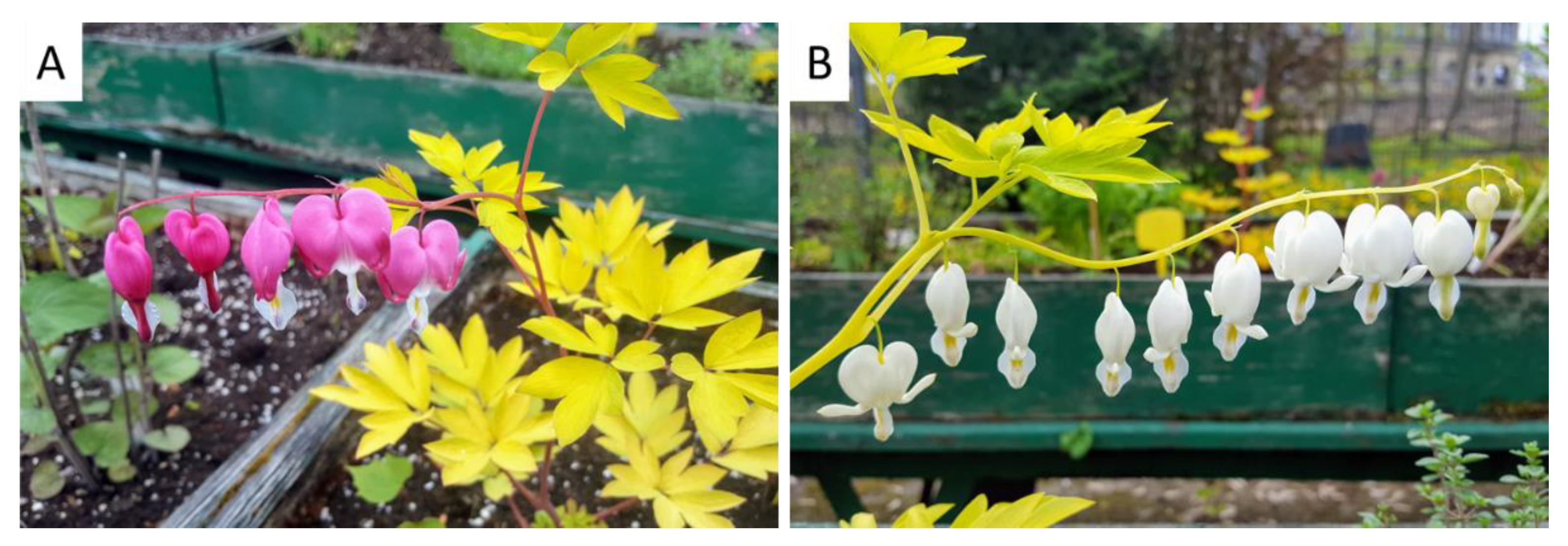
2.1. Plant Material

In vitro-derived Lamprocapnos spectabilis (L.) Fukuhara ‘Gold Heart’ and ’White Gold’ plantlets, 10-week-old, were used as the donor material. Axenic cultures of the two cultivars were obtained from the international commercial plant tissue culture laboratory (Vitroflora, Trzęsacz, Poland). Typical in vivo-grown mature bleeding hearts are shown in Figure 1.

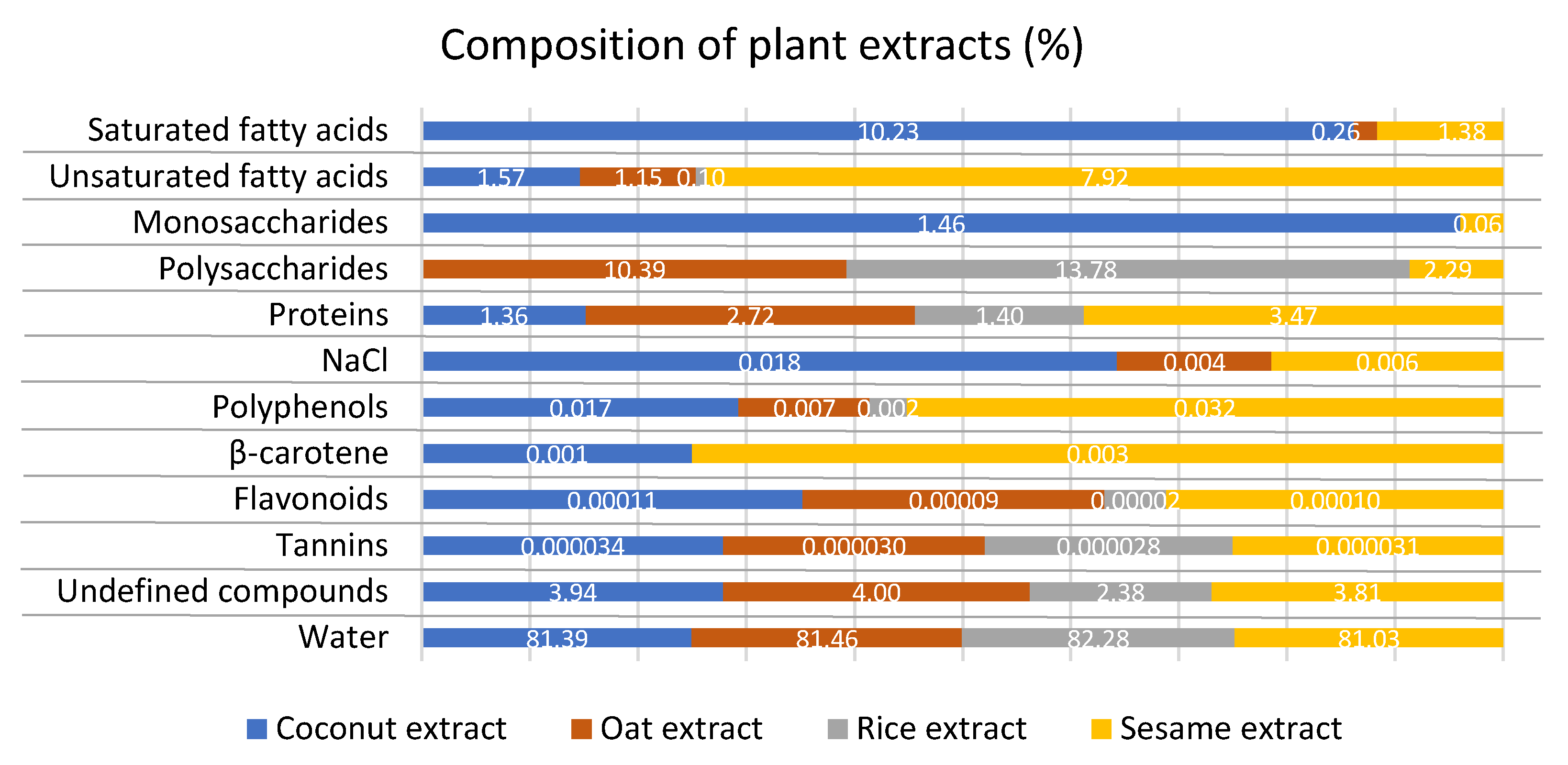
2.2. Preparation of Plant Extracts and Screening of Phytochemicals

Coconut shreds, oat flakes, as well as rice, and sesame seeds purchased from Organic Farma Zdrowia (Warsaw, Poland) were used in the experiments. To eliminate the effect of pesticides and fertilizers, only the plant material from ecological/organic farming was included (certificate of ecological farming: PL-EKO-07 in case of sesame, and PL-EKO-01 in other plants). Approximately 100 g of plant materials were incubated overnight with double distilled water (500 mL) at room temperature and homogenized in a kitchen blender for 2–3 min. Crude extracts were then filtered with gauze to remove the unwanted debris. Fresh extracts were prepared individually for each experimental repetition.
The phytochemical quantification of carbohydrate content was based on the standard anthrone test [36] with minor modifications. Protein content was measured according to the Bradford method [37] with bovine serum albumin (BSA) as the standard. The concentration of carotens and total phenolic content were analysed following Bulda et al. [38] and the Folin–Ciocalteu procedure [39], respectively. The total flavonoid content of the extract was evaluated with a method developed by Brighente et al. [40]. The total tannin content was determined using the Folin–Ciocalteu Phenol reagent as reported by Kabir et al. [41] and a standard tannic acid calibration curve. The spectrophotometric analysis of plant metabolites was performed in a spectrophotometer SmartSpec PlusTM (BioRad, Hercules, CA, USA) at specific wavelengths (λmax): for carbohydrates at 620 nm, for proteins at 595 nm, for β-carotene at 480/495 nm, for phenolics at 765 nm, for flavonoids at 415 nm, and for tannins at 725 nm. The carbohydrate and phenolic contents were calculated using glucose, gallic acid, and quercetin as the calibration standards, respectively. The calibration curve for each phytochemical was drawn by plotting the peak area (y) versus the concentration (x) of each analyte and was fitted to a linear function of type y = ax + b. Chlorides content (NaCl) was analysed argentometrically with Mohr’s method according to the Polish norm [42]. Information on the content of fatty acids (FA) was obtained from Organic Farma Zdrowia (Warsaw, Poland). The result is the mean of three independent replicates, expressed as a mass-volume percentage concentration (% w/v). The composition of plant extracts used in the study is shown in Figure 2.

2.3. Culture Medium and Physical Conditions in the Growth Room

Modified Murashige and Skoog (MS) medium [43] with extra 330 mg·L−1 calcium II chloride (CaCl2·6H2O), 13.9 mg·L−1 iron sulphate (FeSO4), 20.65 mg·L−1 Na2EDTA·2H2O, and 3% (w/v) sucrose (unless otherwise stated), solidified with 0.8% (w/v) agar (Biocorp, Warsaw, Poland) was used in the experiments. The pH was adjusted to 5.8 (with 0.1 M HCl and 0.1 M NaOH) after adding all media components (Chempur, Piekary Śląskie, Poland), including plant extracts (see below), prior to autoclaving at 105 kPa and 121 °C for 20 min. The media (40 mL) were distributed into 350-mL glass jars and sealed with plastic caps. The concentrations of plant extracts and PGRs (provided by Sigma-Aldrich®, St. Louis, MO, USA) are described in the particular experiments.
The cultures were maintained in the growth room at 23 °C ± 1 °C, under 16-h photoperiod conditions and photosynthetic photon flux density (PPFD) of approximately 26.4 µmol·m−2·s−1 provided by standard cool daylight TLD 54/36W fluorescent tubes with a colour temperature of 6200 K (Koninklijke Philips Electronics N.V., Amsterdam, The Netherlands), unless otherwise stated.

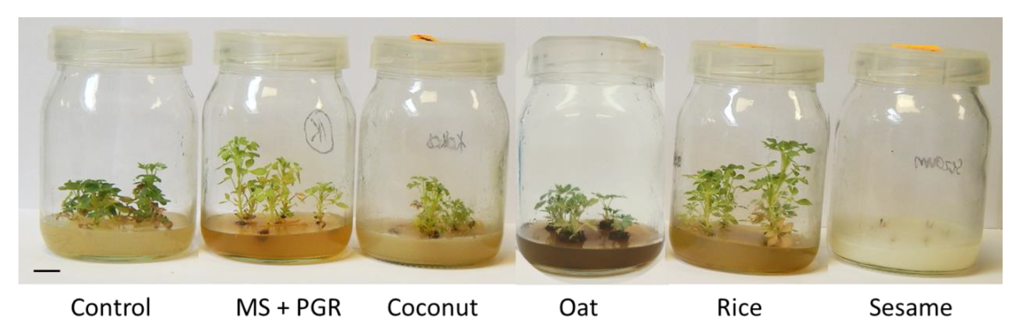
2.4. Application of Plant Extracts in Micropropagation of Bleeding Heart

Single node explants of L. spectabilis were inoculated vertically, six explants per jar, on the modified MS medium with fresh 10% (v/v) coconut or 10% oat or 10% rice or 10% sesame aqueous extract. The modified MS medium devoid of plant extracts or PGRs was considered as a control (MS0). Moreover, modified MS medium supplemented with 1.11 µM (0.25 mg·L−1) BA and 1.23 µM (0.25 mg·L−1) indole-3-butritic acid (IBA) was also included (MS + PGR).

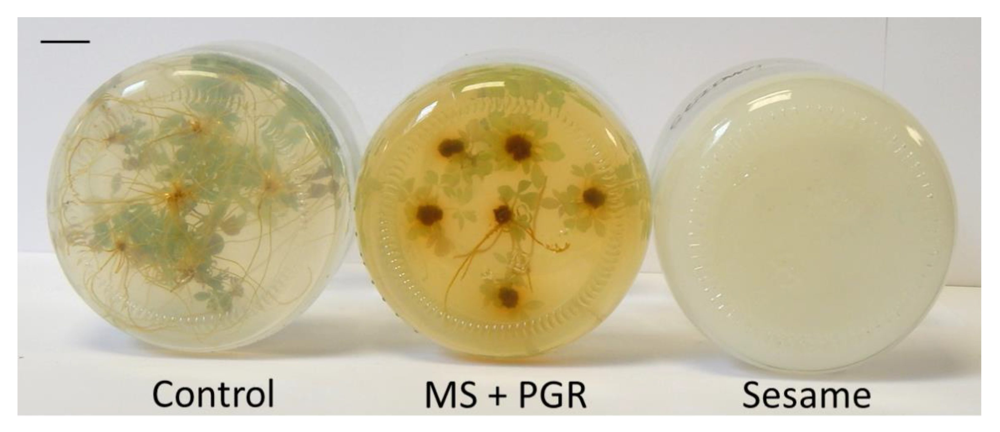
2.5. Application of Plant Extracts in Cryopreservation of Bleeding Heart

The encapsulation-vitrification protocol developed by Kulus [44] was used. The single-node explants were precultured on the modified MS medium with 9% (w/v) sucrose, 4.65 μM (1.0 mg·L−1) kinetin (KIN), 10 µM (2.62 mg·L−1) abscisic acid (ABA), and fresh 10% (v/v) coconut or 10% oat or 10% rice or 10% sesame aqueous extract. A control medium without plant extracts was also considered. Ten explants were placed in a single jar. After one week, shoot tips (1–2 mm-long) were isolated and embedded in 3% calcium alginate. Next, the beads were osmoprotected with the loading solution (2.0 M glycerol and 0.4 M sucrose) for 20 min and dehydrated with plant vitrification solution 3 (PVS3; 50% glycerol and 50% sucrose, w/v) for 150 min at room temperature. Ten beads covered with PVS3 were placed in 2.0-mL sterile polypropylene cryovials and stored in LN (−196 °C) for at least one hour. After storage, the explants were rewarmed and recovered on the modified MS medium with 2.22 μM (0.5 mg·L−1) BA in a 90-mm Petri dish [44].

2.6. Evaluation of Micropropagation and Cryopreservation Efficiency

The regrowth level (%) of the explants was evaluated after 10 weeks of (post-rewarming) in vitro culture. The total number of dissected explants was considered 100%. Moreover, the share of viable explants (%) regenerating spontaneously adventitious roots and callus was determined. The propagation ratio, i.e., number of secondary node explants obtained from a single recovered shoot, number of leaves on a shoot, as well as the number and length (mm) of shoots and roots produced by a single viable explant were assessed. The color of the outer and inner sides of fully-developed leaves in micropropagated shoots was established using the Royal Horticultural Society Colour Chart catalog [45]. The costs of conventional PGRs (BA and IBA) and natural additives used to prepare the multiplication medium were compared based on the market prices (EUR) of those compounds.

2.7. Statistical Analysis

The single-factor experiments were performed in 5 (micropropagation) or 12 (cryopreservation) repetitions for two cultivars independently. One jar/Petri dish with six or ten explants (for micropropagation and cryopreservation, respectively) was considered a single repetition. A total of 360 and 1200 explants were used in the micropropagation and cryopreservation experiments.
For the data expressed as a percentage, the Freeman–Tukey transformation was used. After the normality transformation, the results (completely randomized design) were statistically analyzed with one-way ANOVA (analysis of variance), and the comparisons of means were made with HSD Tukey’s multiple comparison test (p < 0.05) using Statistica 12.0 (StatSoft, Tulsa, OK, USA) and ANALWAR-5.2-FR tools [46]. Tables with results provide real numerical data, while alphabet letters point to homogenous groups, following the statistical calculations based on transformed data.

3. Results

3.1. Micropropagation of Bleeding Heart

The effect of medium composition on the explant development was usually trait- and cultivar-specific (Table 1). No development of shoots was reported in both cultivars tested on the medium supplemented with sesame seed extract. In contrast, 100% regrowth was reported in all other experimental combinations.
As for bleeding heart ‘Gold Heart’, the highest and lowest propagation ratio was found in the medium with coconut (10.5) and rice extracts (5.4), respectively. Coconut extract stimulated a more intense proliferation of shoots (1.8 per explant). The number of leaves per shoot was reduced by oat supplement (5.9) compared to most other treatments (9.6–10.3). The greatest shoot length was found in the MS + PGR medium (44.3 mm), although it was not different from that obtained on the PGRs-free control medium (MS0) and fortified with rice extract (32 mm). The most abundant callus formation (86.7% of explants) was found on the MS + PGR medium (Table 1). Other experimental combinations suppressed its formation entirely or almost entirely (3.3% of explants formed callus in the presence of rice extract).
On the other hand, approximately four new explants could be excised from a single ‘White Gold’ plant produced on the PGRs-free control medium and in the presence of rice extract (Table 1). Those two combinations were also superior in terms of the number of shoots produced (1.5–1.9), number of leaves (8.8–12.0), and shoot length (28–31 mm). Rice extract also stimulated more abundant callus formation (86.7% of explants) and reduced the symptoms of microshoot senescence (Figure 3).
The phenotype analysis of micropropagated plants indicated a change in the content of phytochemicals in bleeding heart ‘White Gold’ (Figure 3). Leaves of plants grown in the control medium and the presence of oat extract had a more intensive, darker colour and could be described by a different RHSSC code than those produced in the other experimental objects (Table 1). As for ‘Gold Heart’ cultivar, no such effect was found.
The medium composition also affected the rhizogenesis efficiency and root parameters in both cultivars tested (Table 2; Figure 4). In bleeding heart ‘Gold Heart’, rice extract suppressed rooting (20.7% of shoots). Likewise, oat and rice additives reduced the number of roots regenerated (1.1–1.7 per shoot) compared to coconut extract which was optimal in terms of this parameter (7.5). In contrast, coconut extract negatively affected the length of the roots. As for ‘White Gold’ cultivar, oat extract had a deleterious effect on the rooting efficiency (33.3%), as well as the number (1.0) and length of roots regenerated (7.7 mm). The longest roots (55.2 mm) were produced in the PGRs-free control medium (Table 2).
Cost analysis of additives used in the preparation of the multiplication medium for bleeding heart showed that conventional PGRs (BA and IBA) are equally expensive as coconut extract (0.053 EUR and 5.29 EUR per 1 and 100 L of the medium, respectively). The cost of preparing 10% (v/v) sesame extract for 1 and 100 L of the medium is 0.035 EUR and 3.45 EUR, respectively, while of oat extract—0.021 EUR and 2.07 EUR. Rice supplement is the cheapest: 0.014 EUR and 1.38 EUR for 1 and 100 L of multiplication medium, accordingly (Figure 5).

3.2. Cryopreservation of Bleeding Heart

The addition of sesame extract into the preculture medium had a negative impact on the regrowth level of LN-derived explants (23.3%) in L. spectabilis ‘Gold Heart’ compared to most other treatments (47.5–72.7%; Table 3; Figure 6). As for ‘White Gold’ cultivar, a higher regrowth level was found with the control and sesame-fortified media (42.5–53.0%) than with oat supplement (8.3%). Coconut extract had a positive effect on the proliferation of shoots (1.9–2.4 per explant) in both cultivars tested and on the number of leaves in bleeding heart ‘White Gold’. On the other hand, the highest number of leaves on the LN-recovered ‘Gold Heart’ plant was found in the oat-fortified combination (12.0), while the lowest with rice and sesame additives (5.9–7.1).
The influence of the tested factor on the shoot length was reported in bleeding heart ‘Gold Heart’ (Table 3). The value of this parameter declined when adding rice extract into the preculture medium (15.8 mm) compared to control and oat combinations (21.7–22.3 mm). This plant supplement also contributed to the lower fresh weight of shoots in ‘Gold Heart’ (135.9 mg), while the highest value of this parameter was reported with oat treatment (391.5 mg). As for ‘White Gold’, the addition of coconut extract into the preculture medium resulted in a higher fresh weight of recovered shoots (338.0 mg) compared to the other combinations (101.6–208.1 mg). The frequency of callus induction was affected by the tested factor in bleeding heart ‘White Gold’; coconut extract was more effective in stimulating its formation (31.7% of explants) compared with the control and rice-fortified media (2.8–8.5%; Table 3).
The shoots recovered after storage in LN produced no roots if sesame (‘Gold Heart’) and oat or rice extracts (‘White Gold’) were added to the preculture medium (Table 3). There was no influence of the experimental factor on the number (1.0–2.4) and length of roots (7.8–13.6 mm) produced in both cultivars tested (data not shown).

4. Discussion

4.1. Application of Plant Extracts in Micropropagation of Bleeding Heart

In the previous studies, several natural supplements, including apple extract, banana pulp, carrot extract, coconut water, potato homogenate and juice, corn extract, date palm syrup, papaya extract, taro extract, tomato juice, and beef extract, were used to promote the development of explants in vitro [26,28,33,34,47,48]. In the study by Swamy et al. [26], 10% coconut water supplemented to MS medium stimulated a better response in all the analysed morphological parameters of Pogostemon cablin Benth. The application of 20% banana extract, 10% carrot extract, 10% papaya extract, and 10% tomato extract increased the production of multiple shoots, their length, and fresh weight. Similarly, 10% of coconut water stimulated the in vitro proliferation of Phalaenopsis violacea H.Witte protocorm-like bodies (PLBs) [24]. In the present study, coconut extract also effectively promoted the multiplication of shoots in bleeding heart ‘Gold Heart’. Rice extract, on the other hand, promoted callogenesis in ‘White Gold’ and was more effective in increasing the propagation ratio in this cultivar than the conventional PGRs (4.1 and 2.6, respectively). The positive impact of coconut extract may result from the high content of natural cytokinins, but also long-chain FA, especially lauric, caprylic, myristic, and palmitic acids [49] (Figure 2), which play key roles in cell membrane structure and function [50]. Moreover, the higher concentration of NaCl in this extract (0.18 ‱), resulting from the halophytic nature of coconut palm, could act positively on the explant development on the principle of hormesis effect. Wrochna et al. [51] demonstrated that the presence of salt in the culture medium stimulated fresh mass accumulation in ornamentals Amaranthus paniculatus L., A. caudatus L., Atriplex hortensis L., and Tamarix tetrandra Pall. ex. M. Bieb. The tested natural supplements, however, neither increased the number of leaves or length of shoots nor the rooting parameters compared to the PGRs-free control or MS medium with BA and IBA. Oat extract even had a deleterious effect on the number of leaves produced (in shoots of both cultivars tested) and root development (in bleeding heart ‘White Gold’). This may be due to lower amounts of carbohydrates (10.39%) in this extract or the presence of some growth inhibitors. The relatively high content of polyphenols present in sesame additive (0.32 ‱) probably arrested the development of explants in both cultivars tested as reported by Kulus and Tymoszuk [52]. Those properties of oat and sesame extracts should be considered in the medium-term conservation of plant germplasm stored in in vitro tissue banks under slow-growth conditions [16]. Plant extracts could become much more preferable than traditional expensive (ABA) and/or environmentally harmful growth retardants such as ancymidol. Especially sesame additive should work even at a low level since in this study, 10% (v/v) concentration resulted in complete inhibition of explant development. The present results underline the varied composition of plant extracts, reported previously also by Swamy et al. [26].
Gnasekaran et al. [24] highlighted the importance of concentration optimization of natural supplements added to the culture medium. In the present study, the most typical 10% (v/v) concentration of plant extracts was applied, however, changing this value (also following the needs of individual cultivars) could contribute to a better micropropagation efficiency in bleeding heart as reported with Corylus avellana L. [27]. Analysing the synergistic effect of conventional PGRs and natural extracts should also be considered in future studies related to the micropropagation of L. spectabilis and other plant species.
Visual observations of the in vitro-produced shootlets suggest physiological changes in bleeding heart ‘White Gold’ as a result of treatment with PGRs and natural extracts. Plants grown in the control medium and in the medium augmented with oat extract probably contained more chlorophyll as they were more intensively green compared to the other experimental objects (Figure 3), although spectral fingerprinting would be required to confirm this. Various other natural additives contributed to higher chlorophyll content, total protein, and total carbohydrate contents in micropropagated Pogostemon cablin Benth. [26]. This may be due to the presence of amino acids in the oat extract that are essential for purine biosynthesis and are a part of the porphyrin ring structure of chlorophyll [53]. This hypothesis is supported by a relatively high content of proteins detected in this extract (Figure 2). It is also worth mentioning that the application of conventional PGRs (BA and IBA) caused a clearly lighter colour of leaves in bleeding heart ‘White Gold’ compared to the non-treated control. This may be due to the synthetic cytokinin BA, which is known to cause certain morpho-physiological, anatomical, and biochemical disorders in micropropagated plants [54,55]. This highlights the need for searching for non-conventional growth regulators in L. spectabilis. Surprisingly, in the present study, no impact of medium composition on the leaf colour was reported with the other cultivar; ‘Gold Heart’. This suggests that the stability of phytochemical profiles in bleeding heart is also cultivar-dependent.
Expenditures on raw materials and chemicals are among the limiting factors of plant tissue culture utilization and can reach 20–40% of total micropropagation costs [1]. Consequently, commercial laboratories are screening for cheaper substitutes [56]. The present study showed that oat and rice extracts not only are more accessible but also significantly cheaper than conventional PGRs, up to four or five times as in the case of rice supplement. Coconut extract is equally expensive as BA and IBA, but more environmentally friendly. This can help to solve the problem of utilization of used media which, after minor processing, could be exploited as fertilizer during and after acclimatisation of plants to ex vitro conditions.

4.2. Application of Plant Extracts in Cryopreservation of Bleeding Heart

Composition of the preculture medium, i.e., the first stage of most cryopreservation procedures, is of crucial importance as it induces the explant resistance to stress related to further dehydration, low-temperature storage, rewarming, and rehydration. Various chemical compounds; such as carbohydrates, sugar alcohols, proline, or growth regulators; can be added into the medium to achieve this goal [17]. The present study addressed for the first time the question of whether plant extracts can be used to increase the explant’s suitability for long-term storage, e.g., by increasing the content of endogenous sugars and proteins. Likewise, in the micropropagation experiment, the results obtained with cryopreservation indicate a cultivar-specific reaction. A similar phenomenon was reported when optimizing the cryostorage procedure with other ornamental and medicinal plant species [17,57], although the genetic factor was irrelevant with the Dianthus genus [58]. To overcome this problem, certain modern approaches of gene manipulation might be necessary [59].
Even though none of the natural supplements improved significantly the survival level of the LN-stored shoot tips or the biometrical parameters of the recovered plantlets, coconut additive improved the proliferation of shoots in Lamprocanos spectabilis ‘Gold Heart’, as well as leaf development, shoot elongation, and callus formation in ‘White Gold’ cultivar. This may result from the high content of easy-to-access simple sugars (1.46%) that are also involved in the formation of the so-called “biological glass”, essential in every cryopreservation protocol [17]. Agampodi and Bimali [60] reported the presence of vitamins and phytohormones (especially zeatin) in coconut aqueous extract, stimulating cell division, activating axillary buds, and affecting the stress-tolerance in plants, which coincides with the present findings.
The varied effect of plant extracts on the cryopreservation effectiveness in bleeding heart may also result from the diverse profile of FA in those supplements. The degree of saturation impacts the physicochemical characteristics of the FA, such as the melting point or the viscosity [50]. This, in turn, directly affects the uptake of cryoprotectants and nutrients from the preculture medium by the explant and its plasticity. Apparently, the higher share of unsaturated FA than of saturated FA in the oat and sesame extracts negatively affects the survivability of shoot tips of bleeding heart ‘Gold Heart’ (sesame and oat extracts) and ‘White Gold’ (oat extract). Oleic and linoleic acids, dominating the lipid composition of sesame and oat grains [61,62], seem to be particularly meaningful.
Future studies should focus on the application of other natural supplements in cryopreservation, such as banana or carrot extracts, as they can be a good source of nitrogen, iron, potassium, as well as vitamins B6 and B12, to promote the regrowth of an LN-recovered explant. It was reported that these constituents increase the leaf size and chlorophyll content during plants grown in a controlled environment, directly affecting their development [63].

5. Conclusions

The stimulatory or inhibitory effect on the morphogenic response in Lamprocapnos spectabilis depends on the type of extract used. Natural supplements tested here, i.e., coconut, oat, rice, and sesame extracts, contain carbohydrates, minerals, proteins, lipids, phenols, phytohormones, and other compounds at various levels. Nonetheless, natural extracts may substitute conventional and more expensive plant growth regulators, such as BA and IBA. Plant extracts containing high levels of saturated fatty acids and low amounts of polyphenols seem especially favourable. Among the tested supplements, coconut and rice additives can be recommended for tissue culture systems of bleeding heart. Sesame extract, on the other hand, could be used in the slow-growth/ medium-term storage of plants. The results obtained here are of significant importance for enterprises interested in the production of this plant species. The development of efficient tissue culture systems in bleeding heart will lead to a better understanding of the biology of this decorative species, reduce the costs of its multiplication, and open new possibilities for the creation of novel cultivars via somaclonal variation, mutation breeding, or genetic transformation.

Author Contributions

Conceptualization, D.K. and N.M.; methodology, D.K. and N.M.; validation, D.K. and N.M.; formal analysis, D.K. and N.M.; investigation, D.K. and N.M.; resources, D.K. and N.M.; data curation, D.K. and N.M.; writing—original draft preparation, D.K.; writing—review and editing, D.K. and N.M.; visualization, D.K. and N.M.; supervision, D.K.; project administration, D.K. and N.M. All authors have read and agreed to the published version of the manuscript.

Funding

This research received no external funding.

Institutional Review Board Statement

Not applicable.

Informed Consent Statement

Not applicable.

Data Availability Statement

Data available by email on reasonable request.

Acknowledgments

The authors wish to acknowledge Dominika Rymarz, Paulina Holub, Pascal Zytkiewicz, and Marta Szankowska (students of UTP in Bydgoszcz) for their technical support in performing the micropropagation experiments.

Conflicts of Interest

The authors declare no conflict of interest.

References

  1. Kulus, D. Selected aspects of ornamental plants micropropagation in Poland and worldwide. Life Sci. 2015, 4, 10–25. [Google Scholar] [CrossRef]
  2. Azadi, P.; Bagheri, H.; Nalousi, A.M.; Nazari, F.; Chandler, S.F. Current status and biotechnological advances in genetic engineering of ornamental plants. Biotechnol. Adv. 2016, 34, 1073–1090. [Google Scholar] [CrossRef]
  3. Hodges, L. Bleeding heart: A review for growers. Hort. Technol. 2012, 22, 517–522. [Google Scholar] [CrossRef] [Green Version]
  4. Roberts, C.M.; Serek, M.; Andersen, A.S. Supplemental irradiance and STS improve the display life of Dicentra species forced as flowering potted plants. Sci. Hortic. 1995, 62, 121–128. [Google Scholar] [CrossRef]
  5. McNulty, J.; Poloczek, J.; Larichev, V.; Werstiuk, N.H.; Griffin, C.; Pandey, S. Discovery of the apoptosis-inducing activity and high accumulation of the butenolides, menisdaurilide and aquilegiolide in Dicentra spectabilis. Planta Med. 2007, 73, 1543–1547. [Google Scholar] [CrossRef] [PubMed]
  6. Lee, D.-H.; Lee, B.-C.; Yoon, E.-J.; Leem, K.-E.; Park, S.-M.; Pyo, H.-B.; Choe, T.-B. Development of effects of plant extracts on the activity and expression of UVA-induced MMPs (matrix metalloproteases). Int. J. Cosmet. Sci. 2004, 26, 317–319. [Google Scholar] [CrossRef]
  7. Ma, W.-G.; Fukuski, Y.; Tahara, S.; Osawa, T. Fungitoxic alkaloids from Hokkaido Papaveraceae. Fitoterapia 2000, 71, 527–534. [Google Scholar] [CrossRef]
  8. Iwasa, K.; Kim, C.-W. Biotransformations of protoberberines in cell cultures of Dicentra spectablis. Phytochemistry 1997, 46, 1359–1363. [Google Scholar] [CrossRef]
  9. Kim, A.H.; Jang, J.H.; Woo, K.W.; Park, J.E.; Lee, K.H.; Jung, H.K.; An, B.; Jung, W.S.; Ham, S.H.; Cho, H.W. Chemical constituents of Dicentra spectabilis and their anti-inflammation effect. J. Appl. Biol. Chem. 2018, 61, 39–46. [Google Scholar] [CrossRef] [Green Version]
  10. Petruczynik, A.; Plech, T.; Tuzimski, T.; Misiurek, J.; Kaproń, B.; Misiurek, D.; Szultka-Młyńska, M.; Buszewski, B.; Waksmundzka-Hajnos, M. Determination of selected isoquinoline alkaloids from Mahonia aquifolia; Meconopsis cambrica; Corydalis lutea; Dicentra spectabilis; Fumaria officinalis; Macleaya cordata extracts by HPLC-DAD and comparison of their cytotoxic activity. Toxins 2019, 11, 575. [Google Scholar] [CrossRef] [PubMed] [Green Version]
  11. Niazian, M. Application of genetics and biotechnology for improving medicinal plants. Planta 2019, 249, 953–973. [Google Scholar] [CrossRef]
  12. Miler, N.; Kulus, D.; Woźny, A.; Rymarz, D.; Hajzer, M.; Wierzbowski, K.; Nelke, R.; Szeffs, L. Application of wide-spectrum light-emitting diodes in micropropagation of popular ornamental plant species: A study on plant quality and cost reduction. In Vitro Cell. Dev. Biol. Plant 2019, 55, 99–108. [Google Scholar] [CrossRef] [Green Version]
  13. Lee, K.P.; Lee, D.W. Somatic embryogenesis and plant regeneration from seeds of wild Dicentra spectabilis (L.) Lem. Plant Cell Rep. 2003, 22, 105–109. [Google Scholar] [CrossRef] [PubMed]
  14. Kulus, D. Influence of growth regulators on the development, quality, and physiological state of in vitro-propagated Lamprocapnos spectabilis (L.) Fukuhara. In Vitro Cell. Dev. Biol. Plant 2020, 56, 447–457. [Google Scholar] [CrossRef]
  15. Kulus, D. Genetic resources and selected conservation methods of tomato. J. Appl. Bot. Food Qual. 2018, 91, 135–144. [Google Scholar] [CrossRef]
  16. Hammond, S.D.; Viehmannova, I.; Zamecnik, J.; Panis, B.; Cepkova, P.H. Efficient slow-growth conservation and assessment of clonal fidelity of Ullucus tuberosus Caldas microshoots. Plant Cell Tissue Organ Cult. 2019, 138, 559–570. [Google Scholar] [CrossRef]
  17. Kulus, D.; Zalewska, M. Cryopreservation as a tool used in long-term storage of ornamental species—A review. Sci. Hortic. 2014, 168, 88–107. [Google Scholar] [CrossRef]
  18. Kulus, D. Managing plant genetic resources using low and ultra-low temperature storage: A case study of tomato. Biodivers. Conserv. 2019, 28, 1003–1027. [Google Scholar] [CrossRef] [Green Version]
  19. Lambardi, M.; Shaarawi, S. Importance of in vitro culture for developing cryopreservation strategies of woody plants. Acta Hortic. 2017, 1187, 177–188. [Google Scholar] [CrossRef]
  20. Matsumoto, T. Cryopreservation of plant genetic resources: Conventional and new methods. Rev. Agric. Sci. 2017, 5, 13–20. [Google Scholar] [CrossRef] [Green Version]
  21. Hammer, K.; Khoshbakht, K. Towards a ’red list’ for crop plant species. Genet. Resour. Crop Evol. 2005, 52, 249–265. [Google Scholar] [CrossRef]
  22. Singh, C.R. Review on problems and its remedy in plant tissue culture. Asian J. Biol. Sci. 2018, 11, 165–172. [Google Scholar] [CrossRef] [Green Version]
  23. Miler, N.; Zalewska, M. Somaclonal variation of chrysanthemum propagated in vitro from different explant types. Acta Sci. Pol. Hort. Cult. 2014, 13, 69–82. [Google Scholar]
  24. Gnasekaran, P.; Rathinam, X.; Sinniah, U.R.; Subramaniyam, S. A study on the use of organic additives on the protocorm-like bodies growth of Phalaenopsis violacea orchid. J. Phytol. 2010, 2, 29–33. [Google Scholar]
  25. Gilaki, M. Biosynthesis of silver nanoparticles using plant extracts. J. Biol. Sci. 2010, 10, 465–467. [Google Scholar] [CrossRef] [Green Version]
  26. Swamy, M.K.; Mohanty, S.K.; Anuradha, M. The effect of plant growth regulators and natural supplements on in vitro propagation of Pogostemon cablin Benth. J. Crop Sci. Biotechnol. 2014, 17, 71–78. [Google Scholar] [CrossRef]
  27. Prando, M.A.S.; Chiavazza, P.; Faggio, A.; Contessa, C. Effect of coconut water and growth regulator supplements on in vitro propagation of Corylus avellana L. Sci. Hortic. 2014, 171, 91–94. [Google Scholar] [CrossRef] [Green Version]
  28. Murdad, R.; Latip, M.A.; Aziz, Z.A.; Ripin, R. Effects of carbon source and potato homogenate on in vitro growth and development of Sabah’s endangered orchid: Phalaenopsis gigantea. Asia Pac. J. Mol. Biol. 2010, 18, 199–202. [Google Scholar]
  29. Raju, R.I.; Roy, S.K. Mass propagation of Bambusa bambos (L.) Voss through in vitro culture. Jahangirnagar Univ. J. Biol. Sci. 2016, 5, 15–26. [Google Scholar] [CrossRef] [Green Version]
  30. Souza, R.A.V.; Braga, F.T.; Setotaw, T.A.; Neto, J.V.; Azevedo, P.H.; Azevedo, V.H.; Cancado, G.M.A. Effect of coconut water on growth of olive embryos cultured in vitro. Cienc. Rural. 2013, 43, 290–296. [Google Scholar] [CrossRef] [Green Version]
  31. Peixe, A.; Raposo, A.; Lourenco, R.; Cardoso, H.; Macedo, E. Coconut water and BAP successfully replaced zeatin in olive (Olea europaea L.) micropropagation. Sci. Hortic. 2017, 113, 1–7. [Google Scholar] [CrossRef] [Green Version]
  32. Venkatachalam, P.; Kalaiarasi, K.; Sreeramanan, S. Influence of plant growth regulators (PGRs) and various additives on in vitro plant propagation of Bambusa arundinacea (Retz.) Wild: A recalcitrant bamboo species. J. Genet. Eng. Biotechnol. 2015, 13, 193–200. [Google Scholar] [CrossRef] [Green Version]
  33. Molnár, Z.; Virág, E.; Ördög, E. Natural substances in tissue culture media of higher plants. Acta Biol. Szeged. 2011, 55, 123–127. [Google Scholar]
  34. Nambiar, N.; Tee, C.S.; Maziah, M. Effects of organic additives and different carbohydrate sources on proliferation of protocorm like bodies in Dendrobium Alya Pink. POJ 2012, 5, 10–18. [Google Scholar]
  35. Utami, E.S.W.; Hariyanto, S.; Manuhara, Y.S.W. In vitro propagation of the endangered medicinal orchid, Dendrobium lasianthera J.J.Sm through mature seed culture. As. Pac. J. Tropic. Biomed. 2017, 7, 406–410. [Google Scholar] [CrossRef]
  36. Trevelyan, W.E.; Forrest, R.S.; Harrison, J.S. Determination of yeast carbohydrates with the anthrone reagent. Nature 1952, 170, 626–627. [Google Scholar] [CrossRef] [PubMed]
  37. Bradford, M.M. A rapid and sensitive method for the quantitation of microgram quantities of protein utilizing the principle of protein-dye binding. Anal. Biochem. 1976, 72, 248–254. [Google Scholar] [CrossRef]
  38. Bulda, O.V.; Rassadina, V.V.; Alekseichuk, H.N.; Laman, N.A. Spectrophotometric measurement of carotenes, xanthophylls, and chlorophylls in extracts from plant seeds. Russ. J. Plant. Physiol. 2008, 55, 544. [Google Scholar] [CrossRef]
  39. Waterhouse, A.L. Determination of total phenolics. In Current Protocols in Food Analytical Chemistry; Wrolstad, R.E., Ed.; John Wiley & Sons: New York, NY, USA, 2001; pp. I1.1.1–I1.1.8. [Google Scholar] [CrossRef]
  40. Brighente, I.; Dias, M.; Verdi, G.; Pizzolatti, G. Antioxidant activity and total phenolic content of some Brazilian species. Pharm. Biol. 2007, 45, 156–161. [Google Scholar] [CrossRef]
  41. Kabir, M.S.H.; Hossain, M.M.; Kabir, I.; Rahman, M.; Hasanat, A.; Emran, T.B.; Rahman, A. Phytochemical screening, antioxidant, thrombolytic, α-amylase inhibition and cytotoxic activities of ethanol extract of Steudnera colocasiifolia K. Koch leaves. J. Young Pharm. 2016, 8, 391–397. [Google Scholar] [CrossRef] [Green Version]
  42. FSSAI. PN-90/A-75101.10+Az1:2002. Fruit and Vegetable Preserves—Sample Preparation and Physicochemical Test Methods—Determination of Total Acidity; FSSAI: New Delhi, India, 2015.
  43. Murashige, T.; Skoog, F. A revised medium for rapid growth and bio assays with tobacco tissue cultures. Physiol. Plant 1962, 15, 473–497. [Google Scholar] [CrossRef]
  44. Kulus, D. Shoot tip cryopreservation of Lamprocapnos spectabilis (L.) Fukuhara using different approaches and evaluation of stability on the molecular, biochemical, and plant architecture levels. Int. J. Mol. Sci. 2020, 21, 3901. [Google Scholar] [CrossRef]
  45. RHSCC. The Royal Horticultural Society Colour Chart; RHSCC: London, UK, 1966. [Google Scholar]
  46. Haynes, W. Tukey’s Test. In Encyclopedia of Systems Biology; Dubitzky, W., Wolkenhauer, O., Cho, K.H., Yokota, H., Eds.; Springer: New York, NY, USA, 2013. [Google Scholar] [CrossRef]
  47. Islam, M.O.; Rahman, A.R.M.M.; Matsui, S.; Prodhan, A.K.M.A. Effects of complex organic extracts on callus growth and PLB regeneration through embryogenesis in the Doritaenopsis orchid. Jpn. Agric. Res. Quart. 2003, 37, 229–235. [Google Scholar] [CrossRef]
  48. Sudipta, K.M.; Swamy, M.K.; Anuradha, M. Influence of various carbon sources and organic additives on in vitro growth and morphogenesis of Leptadenia reticulata (Wight & Arn), a valuable medicinal plant of India. Int. J. Pharm. Sci. Rev. Res. 2013, 21, 174–179. [Google Scholar]
  49. Boateng, L.; Ansong, R.; Owusu, W.B.; Steiner-Asiedu, M. Coconut oil and palm oil’s role in nutrition, health and national development: A review. Ghana Med. J. 2016, 50, 189–196. [Google Scholar] [CrossRef] [PubMed]
  50. Meï, C.; Michaud, M.; Cussac, M.; Albrieux, C.; Gros, V.; Maréchal, E.; Block, M.A.; Jouhet, J.; Rébeillé, F. Levels of polyunsaturated fatty acids correlate with growth rate in plant cell cultures. Sci. Rep. 2015, 5, 15207. [Google Scholar] [CrossRef] [Green Version]
  51. Wrochna, M.; Gawrońska, H.; Gawroński, S.W. Effect of salt stress on fresh and dry matter production and accumulation of Na+, K+, Ca2+, Mg2+, Cl- ions in selected species of ornamental plants. Acta Agrophys. 2006, 7, 775–785. [Google Scholar]
  52. Kulus, D.; Tymoszuk, A. Induction of callogenesis, organogenesis, and embryogenesis in non-meristematic explants of bleeding heart and evaluation of chemical diversity of key metabolites from callus. Int. J. Mol. Sci. 2020, 21, 5826. [Google Scholar] [CrossRef] [PubMed]
  53. Paul, K.A.; Karunakaran, S.S.; Joseph, J.; Ramaiah, D. Amino acid-porphyrin conjugates: Synthesis and study of their photophysical and metal ion recognition properties. Phytochem. Photobiol. 2015, 91, 1348–1355. [Google Scholar] [CrossRef] [PubMed]
  54. Bidabadi, S.S.; Mohan Jain, S. Cellular, molecular, and physiological aspects of in vitro plant regeneration. Plants 2020, 9, 702. [Google Scholar] [CrossRef] [PubMed]
  55. Manokari, M.; Priyadharshini, S.; Jogam, P.; Dey, A.; Shekhawat, M.S. Meta-topolin and liquid medium mediated enhanced micropropagation via ex vitro rooting in Vanilla planifolia Jacks. ex Andrews. Plant Cell. Tissue Organ Cult. 2021, 1–14. [Google Scholar] [CrossRef]
  56. Biswas, K.; Biswas, R.; Negi, P. Novel low-cost culture media “KFA and KFA Plus” for micropropagation of Mentha spp. Int. J. Curr. Microbiol. Appl. Sci. 2014, 3, 172–182. [Google Scholar]
  57. Rajasekharan, P.E.; Prakashkumar, R. Cryopreservation of medicinal plant systems: Progress, problems and prospects. IUP J. Genet. Evol. 2010, 3, 57–83. [Google Scholar]
  58. Sekizawa, K.; Yamamoto, S.; Rafique, T.; Fukui, K.; Niino, T. Cryopreservation of in vitro-grown shoot tips of carnation (Dianthus caryophyllus L.) by vitrification method using aluminium cryo-plates. Plant Biotechnol. 2011, 28, 401–405. [Google Scholar] [CrossRef] [Green Version]
  59. Afroz, A.; Chaudhry, Z.; Rashid, U.; Khan, M.R.; Ali, G.M. Enhanced regeneration in explants of tomato (Lycopersicon esculentum L.) with the treatment of coconut water. Afr. J. Biotechnol. 2010, 9, 3634–3644. [Google Scholar]
  60. Agampodi, V.A.; Bimali, J. Effect of coconut (Cocos nucifera L.) water extracts on adventitious root development in vegetative propagation of Dracaena purplecompacta L. Acta Physiol. Plant. 2009, 31, 271–284. [Google Scholar] [CrossRef]
  61. Krasilnikov, V.N.; Batalova, G.A.; Popov, V.S.; Sergeyeva, S.S. Fatty acid composition of lipids in naked oat grain of domestic varieties. Russ. Agric. Sci. 2018, 44, 406–408. [Google Scholar] [CrossRef]
  62. Wacal, C.; Ogata, N.; Basalirwa, D.; Sasagawa, D.; Kato, M.; Handa, T.; Masunaga, T.; Yamamoto, S.; Nishihara, E. Fatty acid composition of sesame (Sesamum indicum L.) seeds in relation to yield and soil chemical properties on continuously monocropped upland fields converted from paddy fields. Agronomy 2019, 9, 801. [Google Scholar] [CrossRef] [Green Version]
  63. Zhao, D.; Oosterhuis, D.M.; Bednarz, C.W. Influence of potassium deficiency on photosynthesis, chlorophyll content and chloroplast ultrastructure of cotton plants. Photosynthetica 2001, 39, 103–109. [Google Scholar] [CrossRef]
Figure 1. Lamprocapnos spectabilis (bleeding heart) ‘Gold Heart’ (A) and ‘White Gold’ (B).
Agriculture 11 00542 g001
Figure 2. Profile of various organic and inorganic compounds (%) in plant extracts used in the study. Length of the bars presents the proportion between the concentration of the same component (%) within various extracts.
Agriculture 11 00542 g002
Figure 3. Influence of medium composition on the development of shoots in L. spectabilis ‘White Gold’ after 10 weeks of culture; bar = 1 cm.
Agriculture 11 00542 g003
Figure 4. Influence of medium composition on the development of roots in L. spectabilis ‘White Gold’ after 10 weeks of culture; bar = 1 cm.
Agriculture 11 00542 g004
Figure 5. Comparison of costs (EUR) of conventional plant growth regulators (PGRs: BA and IBA) and natural additives used for the preparation of 100 L of multiplication medium.
Agriculture 11 00542 g005
Figure 6. Development of L. spectabilis ‘Gold Heart’ shoots after storage in liquid nitrogen and earlier preculture on the MS medium with various plant extracts: (a) Control; (b) Coconut; (c) Oat; (d) Rice; (e) Sesame; (f) Complete microshoot with roots recovered from the control treatment after 10 weeks of culture; bar = 1 cm.
Agriculture 11 00542 g006
Table 1. Influence of medium composition on the explant regrowth level (%), propagation ratio, number, and length (mm) of shoots produced by a single explant, number and colour (RHSCC) of leaves, as well as the share (%) of explants forming callus after 10 weeks of culture.
MediumRegrowth
(%)
Propagation RatioNo. of ShootsNo. of LeavesShoot Length (mm)Color CodeOuter/InnerCallus
(%)
Gold Heart
MS0100 a8.5 ± 0.2 ab1.1 ± 0.1 b9.6 ± 0.4 a32.5 ± 4.9 ab131A/133D0.0 b
MS + PGR100 a9.8 ± 1.5 ab1.2 ± 0.1 b10.3 ± 1.2 a44.3 ± 2.2 a131A/133D86.7 a
coconut100 a10.5 ± 1.8 a1.8 ± 0.2 a7.8 ± 0.6 ab20.1 ± 0.9 b131A/133D0.0 b
oat100 a7.7 ± 0.4 ab1.4 ± 0.1 ab5.9 ± 0.3 b28.3 ± 0.9 b131A/133D0.0 b
rice100 a5.4 ± 0.5 b1.0 ± 0.0 b9.7 ± 0.8 a32.0 ± 3.6 ab131A/133D3.3 b
sesame0.0 bn.a.n.a.n.a.n.a.n.a.n.a.
White Gold
MS0100 a4.0 ± 0.3 a1.9 ± 0.2 a12.0 ± 1.5 a31.0 ± 1.8 a145A/145C0.0 c
MS + PGR100 a2.6 ± 0.3 b1.2 ± 0.1 b7.8 ± 0.9 bc26.7 ± 3.3 ab149C/150D40.0 b
coconut100 a2.7 ± 0.2 b1.4 ± 0.1 b7.8 ± 0.4 bc18.8 ± 1.1 b149C/150D0.0 c
oat100 a3.4 ± 0.3 ab1.2 ± 0.1 b5.0 ± 0.2 c19.8 ± 1.8 b145B/145C0.0 c
rice100 a4.1 ± 0.5 a1.5 ± 0.1 ab8.8 ± 0.7 ab28.0 ± 2.3 a149C/150D86.7 a
sesame0.0 bn.a.n.a.n.a.n.a.n.a.n.a. 1
1 Means ± SE (standard errors) marked with the same letter do not differ significantly at p < 0.05 according to Tukey’s test; RHSCC—Royal Horticultural Society Colour Chart [45]; n.a.—not available due to no recovery of explants.
Table 2. Influence of medium composition on the rooting rate (%), number, and length (mm) of roots produced by a single shoot in L. spectabilis after 10 weeks of culture.
MediumRooting (%)No. of RootsRoot Length (mm)
Gold Heart
MS091.7 a4.3 ± 0.4 b44.9 ± 12.2 a
MS+PGR63.3 ab3.7 ± 1.3 b39.9 ± 6.6 ab
coconut50.0 ab7.5 ± 1.6 a7.7 ± 1.4 b
oat63.3 ab1.7 ± 0.3 c22.1 ± 6.2 ab
rice20.7 b1.1 ± 0.7 c16.0 ± 9.3 ab
sesamen.a.n.a.n.a.
White Gold
MS093.3 a7.9 ± 1.0 a55.2 ± 6.4 a
MS+PGR100 a4.2 ± 0.9 a-c17.9 ± 2.3 bc
coconut88.0 a3.4 ± 0.7 bc13.9 ± 2.6 bc
oat33.3 b1.0 ± 0.5 c7.7 ± 2.4 c
rice76.7 a6.1 ± 1.3 ab28.4 ± 2.7 b
sesamen.a.n.a.n.a. 1
1 Means ± SE marked with the same letter do not differ significantly at p < 0.05 according to Tukey’s test.
Table 3. Influence of preculture medium composition on the explant regrowth level (%), number of shoots and leaves produced by a single viable explant, shoot length (mm) and fresh weight (mg), as well as the share (%) of viable explants forming callus and roots in L. spectabilis after 10 weeks of post-rewarming culture.
MediumRegrowth
(%)
No. of ShootsNo. of LeavesShoot Length (mm)Shoot Weight
(mg)
Callus
(%)
Rooting (%)
Gold Heart
control72.7 ± 3.0 a1.6 ± 0.1 b10.1 ± 1.2 ab22.3 ± 2.4 a361.6 ± 46.8 ab25.8 a33.3 a
coconut47.5 ± 5.5 b2.4 ± 0.2 a10.0 ± 1.2 ab20.8 ± 2.2 ab320.3 ± 92.3 ab29.7 a12.5 ab
oat45.9 ± 5.1 bc2.2 ± 0.4 ab12.0 ± 1.7 a21.7 ± 2.2 a391.5 ± 104.2 a29.9 a29.4 ab
rice65.1 ± 6.0 ab1.6 ± 0.1 b7.1 ± 0.8 b15.8 ± 1.0 b135.9 ± 20.7 b36.4 a7.1 ab
sesame23.3 ± 3.6 c1.6 ± 0.2 b5.9 ± 0.8 b19.0 ± 2.0 ab187.3 ± 59.1 ab22.9 a0.0 b
White Gold
control53.0 ± 8.6 a1.7 ± 0.1 ab6.1 ± 0.5 b10.3 ± 0.8 a208.1 ± 48.5 b8.5 b20.0 a
coconut40.5 ± 4.0 ab1.9 ± 0.2 a8.7 ± 1.2 a13.5 ± 1.2 a338.0 ± 50.7 a31.7 a16.7 a
oat8.3 ± 2.7 b1.7 ± 0.2 ab4.7 ± 1.3 b11.8 ± 2.2 a101.6 ± 55.6 b25.0 ab0.0 a
rice30.0 ± 7.5 ab1.2 ± 0.1 b5.5 ± 0.6 b12.8 ± 1.3 a184.0 ± 37.4 b2.8 b0.0 a
sesame42.5 ± 7.6 a1.4 ± 0.1 ab6.2 ± 0.7 b11.4 ± 1.2 a161.7 ± 36.0 b15.1 ab10.0 a 1
1 Means ± SE marked with the same letter do not differ significantly at p < 0.05 according to Tukey’s test.
Publisher’s Note: MDPI stays neutral with regard to jurisdictional claims in published maps and institutional affiliations.

