
Optical Coherence Tomography Angiography in CRB1-Associated Retinal Dystrophies
 , , ,
, , ,  , and
, and
Abstract
:1. Introduction
2. Methods
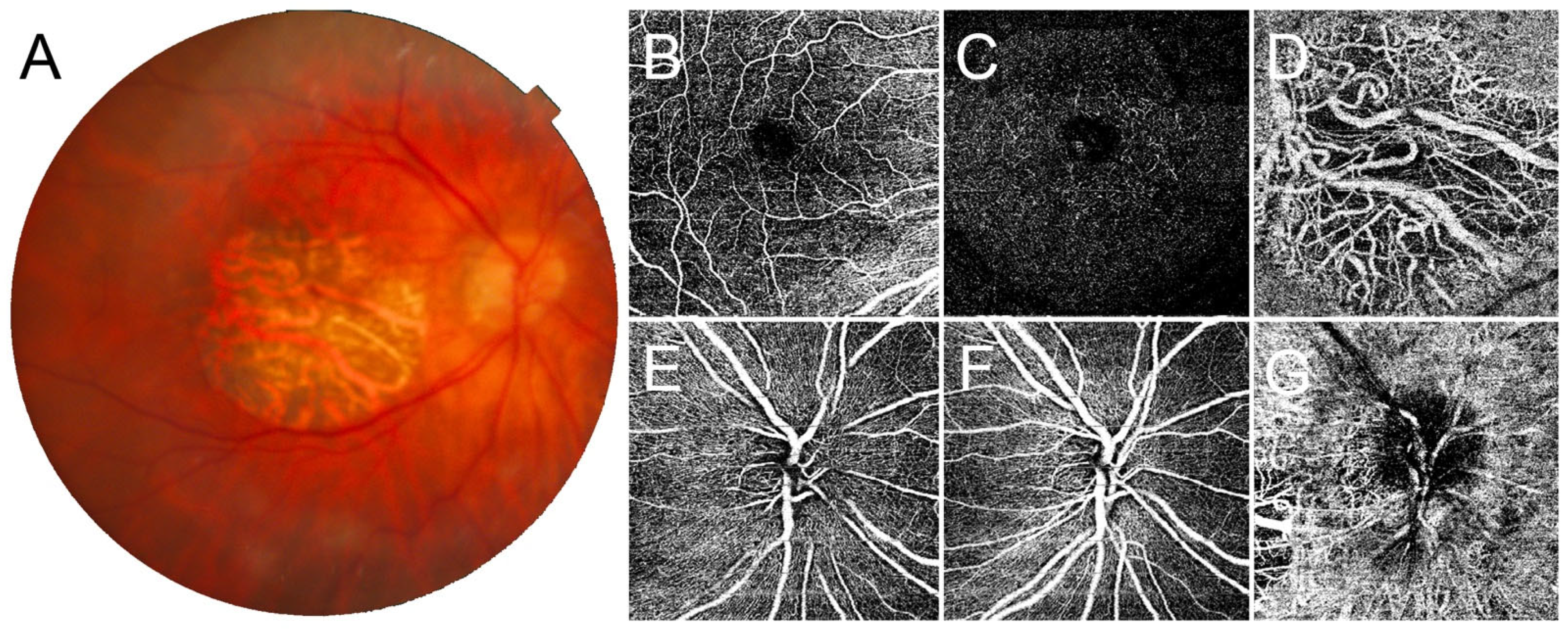
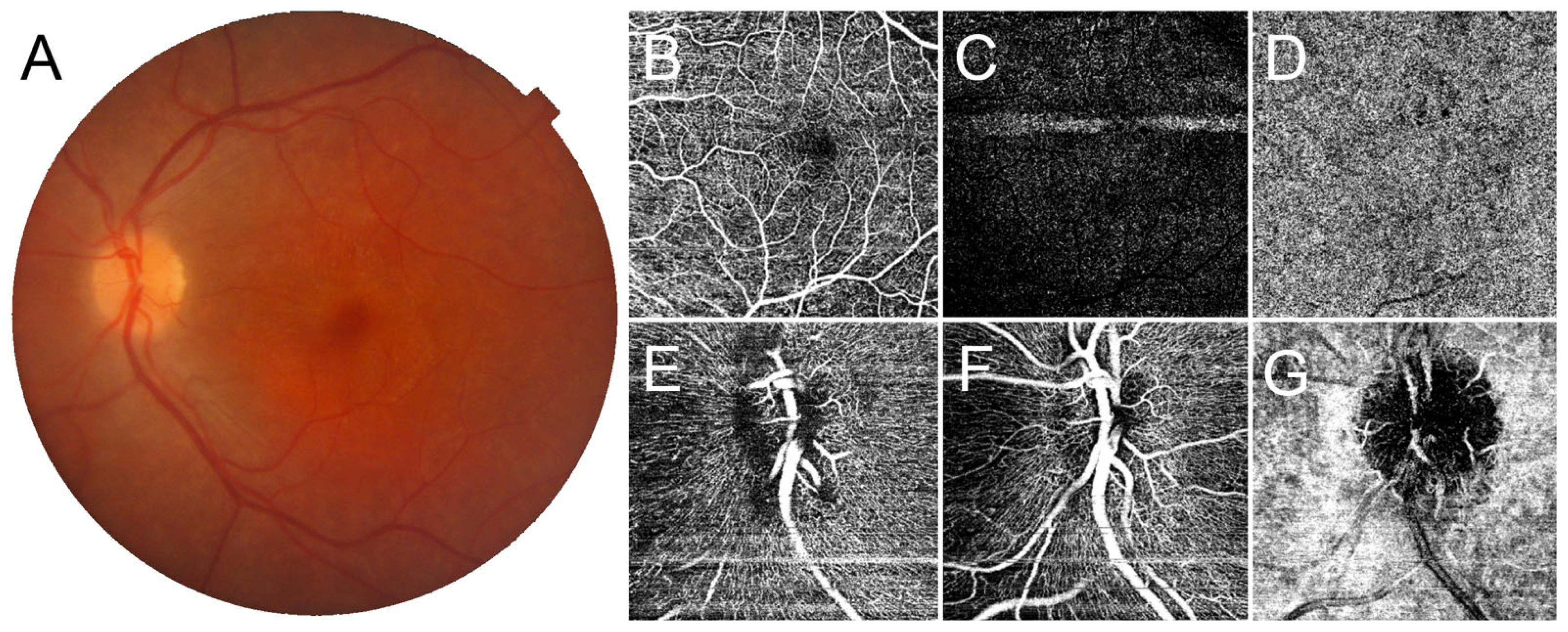
3. Results
4. Discussion
Author Contributions
Funding
Institutional Review Board Statement
Informed Consent Statement
Data Availability Statement
Conflicts of Interest
References
- Hollander, A.I.D.; Brink, J.B.T.; de Kok, Y.J.; van Soest, S.; Born, L.I.V.D.; van Driel, M.A.; van de Pol, D.J.; Payne, A.M.; Bhattacharya, S.S.; Kellner, U.; et al. Mutations in a human homologue of Drosophila crumbs cause retinitis pigmentosa (RP12). Nat. Genet. 1999, 23, 217–221. [Google Scholar] [CrossRef]
- Khan, K.N.; Robson, A.; Mahroo, O.A.R.; Arno, G.; Inglehearn, C.F.; Armengol, M.; Waseem, N.; Holder, G.E.; Carss, K.J.; Raymond, L.F.; et al. A clinical and molecular characterisation of CRB1-associated maculopathy. Eur. J. Hum. Genet. 2018, 26, 687–694. [Google Scholar] [CrossRef] [Green Version]
- Roshandel, D.; Thompson, J.A.; Jeffery, R.C.H.; Sampson, D.M.; Chelva, E.; McLaren, T.L.; Lamey, T.M.; De Roach, J.N.; Durkin, S.R.; Chen, F.K. Multimodal Retinal Imaging and Microperimetry Reveal a Novel Phenotype and Potential Trial End Points in CRB1-Associated Retinopathies. Transl. Vis. Sci. Technol. 2021, 10, 38. [Google Scholar] [CrossRef] [PubMed]
- Henderson, R.H.H.; Mackay, D.; Li, Z.; Moradi, P.; Sergouniotis, P.; Russell-Eggitt, I.; Thompson, D.; Robson, A.; Holder, G.E.; Webster, A.R.; et al. Phenotypic variability in patients with retinal dystrophies due to mutations in CRB1. Br. J. Ophthalmol. 2010, 95, 811–817. [Google Scholar] [CrossRef] [PubMed]
- Wang, Y.; Sun, W.; Xiao, X.; Li, S.; Jia, X.; Wang, P.; Zhang, Q. Clinical and Genetic Analysis of 63 Families Demonstrating Early and Advanced Characteristic Fundus as the Signature of CRB1 Mutations. Am. J. Ophthalmol. 2021, 223, 160–168. [Google Scholar] [CrossRef]
- Varela, M.D.; Georgiou, M.; Alswaiti, Y.; Kabbani, J.; Fujinami, K.; Fujinami-Yokokawa, Y.; Khoda, S.; Mahroo, O.A.; Robson, A.G.; Webster, A.R.; et al. CRB1-Associated Retinal Dystrophies: Genetics, Clinical Characteristics, and Natural History. Am. J. Ophthalmol. 2022, 246, 107–121. [Google Scholar] [CrossRef]
- Bujakowska, K.; Audo, I.; Mohand-Saïd, S.; Lancelot, M.-E.; Antonio, A.; Germain, A.; Léveillard, T.; Letexier, M.; Saraiva, J.-P.; Lonjou, C.; et al. CRB1 mutations in inherited retinal dystrophies. Hum. Mutat. 2011, 33, 306–315. [Google Scholar] [CrossRef] [PubMed] [Green Version]
- Talib, M.; Van Cauwenbergh, C.; De Zaeytijd, J.; Van Wynsberghe, D.; De Baere, E.; Boon, C.J.F.; Leroy, B.P. CRB1-associated retinal dystrophies in a Belgian cohort: Genetic characteristics and long-term clinical follow-up. Br. J. Ophthalmol. 2021, 106, 696–704. [Google Scholar] [CrossRef]
- Jacobson, S.G.; Cideciyan, A.V.; Aleman, T.S.; Pianta, M.; Sumaroka, A.; Schwartz, S.B.; Smilko, E.E.; Milam, A.H.; Sheffield, V.; Stone, E.M. Crumbs homolog 1 (CRB1) mutations result in a thick human retina with abnormal lamination. Hum. Mol. Genet. 2003, 12, 1073–1078. [Google Scholar] [CrossRef] [Green Version]
- Talib, M.; van Schooneveld, M.J.; van Genderen, M.M.; Wijnholds, J.; Florijn, R.J.; Brink, J.B.T.; Schalij-Delfos, N.E.; Dagnelie, G.; Cremers, F.P.; Wolterbeek, R.; et al. Genotypic and Phenotypic Characteristics of CRB1 -Associated Retinal Dystrophies. Ophthalmology 2017, 124, 884–895. [Google Scholar] [CrossRef]
- Mathijssen, I.B.; Florijn, R.J.; Born, L.I.V.D.; Zekveld-Vroon, R.C.; Brink, J.B.T.; Plomp, A.S.; Baas, F.; Meijers-Heijboer, H.; Bergen, A.A.B.; van Schooneveld, M.J. Long-Term Follow-Up of Patients with Retinitis Pigmentosa Type 12 Caused by Crb1 Mutations: A Severe Phenotype with Considerable Interindividual Variability. Retina 2017, 37, 161–172. [Google Scholar] [CrossRef]
- Arrigo, A.; Romano, F.; Aragona, E.; di Nunzio, C.; Sperti, A.; Bandello, F.; Parodi, M.B. OCTA-Based Identification of Different Vascular Patterns in Stargardt Disease. Transl. Vis. Sci. Technol. 2019, 8, 26. [Google Scholar] [CrossRef] [Green Version]
- Toto, L.; Parodi, M.B.; D’Aloisio, R.; Mercuri, S.; Senatore, A.; Di Antonio, L.; Di Marzio, G.; Di Nicola, M.; Mastropasqua, R. Cone Dystrophies: An Optical Coherence Tomography Angiography Study. J. Clin. Med. 2020, 9, 1500. [Google Scholar] [CrossRef]
- Battaglia Parodi, M.; Romano, F.; Cicinelli, M.V.; Rabiolo, A.; Arrigo, A.; Pierro, L.; Iacono, P.; Bandello, F. Retinal Vascular Impairment in Best Vitelliform Macular Dystrophy Assessed by Means of Optical Coherence Tomography Angiography. Am. J. Ophthalmol. 2018, 187, 61–70. [Google Scholar] [CrossRef]
- Mastropasqua, R.; Toto, L.; Di Antonio, L.; Parodi, M.B.; Sorino, L.; Antonucci, I.; Stuppia, L.; Di Nicola, M.; Mariotti, C. Optical Coherence Tomography Angiography Findings in X-Linked Retinoschisis. Ophthalmic Surg. Lasers Imaging Retin. 2018, 49, e20–e31. [Google Scholar] [CrossRef]
- Arrigo, A.; Romano, F.; Parodi, M.B.; Issa, P.C.; Birtel, J.; Bandello, F.; MacLaren, R.E. Reduced vessel density in deep capillary plexus correlates with retinal layer thickness in choroideremia. Br. J. Ophthalmol. 2021, 105, 687–693. [Google Scholar] [CrossRef]
- Bianco, L.; Arrigo, A.; Antropoli, A.; Carrera, P.; Spiga, I.; Patricelli, M.G.; Bandello, F.; Parodi, M.B. Multimodal imaging evaluation of occult macular dystrophy associated with a novel RP1L1 variant. Am. J. Ophthalmol. Case Rep. 2022, 26, 101550. [Google Scholar] [CrossRef]
- Parodi, M.B.; Arrigo, A.; Rajabian, F.; Mansour, A.; Mercuri, S.; Starace, V.; Bordato, A.; Manitto, M.P.; Martina, E.; Bandello, F. Multimodal imaging in Schubert-Bornschein congenital stationary night blindness. Ophthalmic Genet. 2022, 1–6. [Google Scholar] [CrossRef]
- Arrigo, A.; Bordato, A.; Romano, F.; Aragona, E.; Grazioli, A.; Bandello, F.; Parodi, M.B. Choroidal Patterns in Retinitis Pigmentosa: Correlation with Visual Acuity and Disease Progression. Transl. Vis. Sci. Technol. 2020, 9, 17. [Google Scholar] [CrossRef]
- Arrigo, A.; Romano, F.; Albertini, G.; Aragona, E.; Bandello, F.; Parodi, M.B. Vascular Patterns in Retinitis Pigmentosa on Swept-Source Optical Coherence Tomography Angiography. J. Clin. Med. 2019, 8, 1425. [Google Scholar] [CrossRef]
- Montemagni, M.; Arrigo, A.; Parodi, M.B.; Bianco, L.; Antropoli, A.; Malegori, A.; Bandello, F.; Tranfa, F.; Costagliola, C. Optical coherence tomography angiography in Bietti crystalline dystrophy. Eur. J. Ophthalmol. 2022. [Google Scholar] [CrossRef] [PubMed]
- Arrigo, A.; Aragona, E.; Parodi, M.B.; Bandello, F. Quantitative approaches in multimodal fundus imaging: State of the art and future perspectives. Prog. Retin. Eye Res. 2023, 92, 101111. [Google Scholar] [CrossRef] [PubMed]
- Hollander, A.I.D.; Heckenlively, J.R.; Born, L.I.V.D.; de Kok, Y.J.; van der Velde-Visser, S.D.; Kellner, U.; Jurklies, B.; van Schooneveld, M.J.; Blankenagel, A.; Rohrschneider, K.; et al. Leber Congenital Amaurosis and Retinitis Pigmentosa with Coats-like Exudative Vasculopathy Are Associated with Mutations in the Crumbs Homologue 1 (CRB1) Gene. Am. J. Hum. Genet. 2001, 69, 198–203. [Google Scholar] [CrossRef] [PubMed] [Green Version]
- Magliyah, M.; Alshamrani, A.A.; Schatz, P.; Taskintuna, I.; Alzahrani, Y.; Nowilaty, S.R. Clinical spectrum, genetic associations and management outcomes of Coats-like exudative retinal vasculopathy in autosomal recessive retinitis pigmentosa. Ophthalmic Genet. 2021, 42, 178–185. [Google Scholar] [CrossRef]
- Hasan, S.M.; Azmeh, A.; Mostafa, O.; Megarbane, A. Coat’s like vasculopathy in leber congenital amaurosis secondary to homozygous mutations in CRB1: A case report and discussion of the management options. BMC Res. Notes 2016, 9, 91. [Google Scholar] [CrossRef] [PubMed] [Green Version]
- Alves, H.A.; Pellissier, L.P.; Wijnholds, J. The CRB1 and adherens junction complex proteins in retinal development and maintenance. Prog. Retin. Eye Res. 2014, 40, 35–52. [Google Scholar] [CrossRef]
- Den Hollander, A.I.; Ghiani, M.; de Kok, Y.J.; Wijnholds, J.; Ballabio, A.; Cremers, F.P.; Broccoli, V. Isolation of Crb1, a mouse homologue of Drosophila crumbs, and analysis of its expression pattern in eye and brain. Mech Dev. 2002, 110, 203–207. [Google Scholar] [CrossRef]
- Ray, T.A.; Cochran, K.; Kozlowski, C.; Wang, J.; Alexander, G.; Cady, M.A.; Spencer, W.J.; Ruzycki, P.A.; Clark, B.S.; Laeremans, A.; et al. Comprehensive identification of mRNA isoforms reveals the diversity of neural cell-surface molecules with roles in retinal development and disease. Nat. Commun. 2020, 11, 3328. [Google Scholar] [CrossRef]
- Mairot, K.; Smirnov, V.; Bocquet, B.; Labesse, G.; Arndt, C.; Defoort-Dhellemmes, S.; Zanlonghi, X.; Hamroun, D.; Denis, D.; Picot, M.-C.; et al. CRB1-Related Retinal Dystrophies in a Cohort of 50 Patients: A Reappraisal in the Light of Specific Müller Cell and Photoreceptor CRB1 Isoforms. Int. J. Mol. Sci. 2021, 22, 12642. [Google Scholar] [CrossRef]


| Gender | BCVA (logMAR) | CRT (μm) | SFCT (μm) | CRB1 Variants | |
|---|---|---|---|---|---|
| Patient 1 | M | 0.4 | 173 | 465 | c.772_779delinsG; c.498_506del |
| 0.4 | 163 | 340 | |||
| Patient 2 | M | 0 | 223 | 274 | c.498_506del |
| 0.3 | 210 | 285 | |||
| Patient 3 | M | 0.2 | 220 | 239 | c.1584C>A; c.498_506del |
| 0.3 | 158 | 251 | |||
| Patient 4 | F | 0.2 | 189 | 74 | c.614T>C |
| 0.7 | 29 | 56 | |||
| Patient 5 | M | 0.7 | 151 | 290 | c.2549G>T; c.4176_4177delAA |
| 0.7 | 131 | 238 | |||
| Patient 6 | F | 0.4 | 178 | 276 | c.614T>C |
| 0.8 | 110 | 280 |
| mSCP | mDCP | mCC | nRCP | nSCP | nDCP | nCC | |
|---|---|---|---|---|---|---|---|
| CRB1 | 0.405 ± 0.013 | 0.360 ± 0.031 | 0.482 ± 0.014 | 0.398 ± 0.024 | 0.395 ± 0.037 | 0.305 ± 0.038 | 0.513 ± 0.042 |
| Controls | 0.413 ± 0.012 | 0.434 ± 0.005 | 0.500 ± 0.006 | 0.443 ± 0.007 | 0.426 ± 0.010 | 0.402 ± 0.019 | 0.542 ± 0.031 |
| p-value | 0.13352 | 4.46 × 10⁻⁹ * | 0.000138 * | 4.71 × 10⁻⁷ * | 0.005277 * | 1.70 × 10⁻⁸ * | 0.060817 |
Disclaimer/Publisher’s Note: The statements, opinions and data contained in all publications are solely those of the individual author(s) and contributor(s) and not of MDPI and/or the editor(s). MDPI and/or the editor(s) disclaim responsibility for any injury to people or property resulting from any ideas, methods, instructions or products referred to in the content. |
© 2023 by the authors. Licensee MDPI, Basel, Switzerland. This article is an open access article distributed under the terms and conditions of the Creative Commons Attribution (CC BY) license (https://creativecommons.org/licenses/by/4.0/).
Share and Cite
Rajabian, F.; Arrigo, A.; Bianco, L.; Antropoli, A.; Manitto, M.P.; Martina, E.; Bandello, F.; Chhablani, J.; Battaglia Parodi, M. Optical Coherence Tomography Angiography in CRB1-Associated Retinal Dystrophies. J. Clin. Med. 2023, 12, 1095. https://doi.org/10.3390/jcm12031095
Rajabian F, Arrigo A, Bianco L, Antropoli A, Manitto MP, Martina E, Bandello F, Chhablani J, Battaglia Parodi M. Optical Coherence Tomography Angiography in CRB1-Associated Retinal Dystrophies. Journal of Clinical Medicine. 2023; 12(3):1095. https://doi.org/10.3390/jcm12031095
Chicago/Turabian StyleRajabian, Firuzeh, Alessandro Arrigo, Lorenzo Bianco, Alessio Antropoli, Maria Pia Manitto, Elisabetta Martina, Francesco Bandello, Jay Chhablani, and Maurizio Battaglia Parodi. 2023. "Optical Coherence Tomography Angiography in CRB1-Associated Retinal Dystrophies" Journal of Clinical Medicine 12, no. 3: 1095. https://doi.org/10.3390/jcm12031095