Review

Pathophysiology of Early Brain Injury and Its Association with Delayed Cerebral Ischemia in Aneurysmal Subarachnoid Hemorrhage: A Review of Current Literature

1
Department of Neurology, University of Tennessee Health Science Center, Memphis, TN 38163, USA
2
Neurological Service, SS Annunziata Hospital, Sulmona, 67039 L’Aquila, Italy
3
Department of Neurology, University of Mississippi Medical Center, Jackson, MS 39216, USA
4
Department of Neurology, University of New Mexico, Albuquerque, NM 87131, USA
5
Department of Neurology, The Ohio State University Wexner Medical Center, Columbus, OH 43210, USA
6
Department of Neurology, University of North Carolina, Chapel Hill, NC 27599, USA
7
Department of Advanced Medical and Surgical Sciences, University of Campania, 80138 Naples, Italy
8
Department of Human Genetics, Punjabi University, Patiala 147002, India
9
Unidad de Cuidados Intensivos, Sanatorio de la Trinidad San Isidro, Buenos Aires 1640, Argentina
10
Stroke Division, Department of Neurology, Beth Israel Deaconess Medical Center, Harvard Medical School, Boston, MA 02115, USA
*
Author to whom correspondence should be addressed.
J. Clin. Med. 2023, 12(3), 1015; https://doi.org/10.3390/jcm12031015
Submission received: 30 December 2022 / Revised: 25 January 2023 / Accepted: 25 January 2023 / Published: 28 January 2023
(This article belongs to the Special Issue Clinical Advances in Cerebral Aneurysm Treatment)

Abstract

:
Background: Delayed cerebral ischemia (DCI) is a common and serious complication of aneurysmal subarachnoid hemorrhage (aSAH). Though many clinical trials have looked at therapies for DCI and vasospasm in aSAH, along with reducing rebleeding risks, none have led to improving outcomes in this patient population. We present an up-to-date review of the pathophysiology of DCI and its association with early brain injury (EBI). Recent Findings: Recent studies have demonstrated that EBI, as opposed to delayed brain injury, is the main contributor to downstream pathophysiological mechanisms that play a role in the development of DCI. New predictive models, including advanced monitoring and neuroimaging techniques, can help detect EBI and improve the clinical management of aSAH patients. Summary: EBI, the severity of subarachnoid hemorrhage, and physiological/imaging markers can serve as indicators for potential early therapeutics in aSAH. The microcellular milieu and hemodynamic pathomechanisms should remain a focus of researchers and clinicians. With the advancement in understanding the pathophysiology of DCI, we are hopeful that we will make strides toward better outcomes for this unique patient population.

1. Introduction

Aneurysmal subarachnoid hemorrhage (aSAH) (Figure 1) is a devastating stroke subtype with high morbidity and mortality, accounting for approximately 5% of all strokes. A systematic review of population-based studies showed that the crude incidence of aSAH in 2010 in North America was 6.9 per 100,000 person-years with an annual decline of 0.7% since 1955 [1]. However, despite significant advances in diagnosis and management of aSAH, the overall mortality remains high at 35% (range 20–67%) [2,3] and approximately 20% of survivors experience significant morbidity [4], causing a substantial economic and social burden on the patients, families, and healthcare systems [5]. Delayed cerebral ischemia (DCI) is a feared complication of aSAH, increasing the risk of morbidity and mortality, specifically in patients with a severe clinical presentation at ictus and high SAH burden [6,7]. Published data suggests DCI affects approximately 20–40% of aSAH patients and is an independent poor prognostic factor in this population [8,9,10]. DCI is classically defined as a development of a new neurological deficit(s), impaired consciousness, or infarct on imaging that commences 3 to 4 days after the initial insult and peaks around 7 to 8 days post-bleed [11]. An earlier onset of DCI prior to day 7 has been associated with higher mortality and greater infarct load [12]. Several treatment modalities targeting different pathophysiological pathways causing DCI have been studied. However, there has been minimal change in outcomes due to either DCI prevention or treatment.
The definition and pathophysiology of this complex and multifactorial process have been debated and studied extensively. In 2007, a study with over 3500 patients [13] evaluated independent variables associated with unfavorable outcomes in aSAH. Authors defined the term “symptomatic vasospasm” as a ≥2 point decrease in the Glasgow Coma Scale (GCS) or a ≥2 point increase in the motor score of the National Institute of Health Stroke Scale (NIHSS) lasting >8 h, which has since fallen out of favor due to the lack of clear correlation with outcomes. Interestingly, both “symptomatic vasospasm” and cerebral infarction noted on computer tomography (CT) post-operatively showed a significant association with poor functional outcomes at 3 months, with cerebral infarction showing the highest association with unfavorable outcomes (OR = 5.38, p < 0.0001) [13]. In 2010, Vergouwen et al. [14] developed a uniform definition of DCI, incorporating clinical deterioration and cerebral infarction as important DCI characteristics that have been well adopted since (see Table 1). Following the definition standardization, in an observational study from Finland that included 340 aSAH patients, 37.1% fulfilled the consensus definition of clinical deterioration secondary to DCI and were strongly associated with poor outcomes at discharge (OR = 2.65, p < 0.001) [15]. This has provided an objective, homogenous, and reliable definition of DCI.
For years, large vessel vasospasm was the recognized complication of DCI, leading to the assumption that vasospasm prevention and treatment were paramount in managing aSAH after securing the ruptured aneurysm. However, clazosentan, which was found to treat and reverse vasospasm successfully, failed to improve outcomes. This is possibly because cerebral infarction, secondary to DCI, often occurs in vascular territories outside of the vasoconstricted territories [8,16,17,18]. Furthermore, DCI has been reported among patients with none or only mild angiographic evidence of vasospasm. Though cerebral vasospasm has been found to occur in 50–70% of aSAH patients, only 20–40% develop DCI [8,13]. Because of this, efforts in the last two decades have been placed on establishing and understanding the pathophysiology of DCI. Hence, the focus has shifted to early brain injury (EBI) and its association with autoregulatory failure, neuroinflammation, and eventually delayed injury (also defined as DCI), with the thought that the treatment of large vessel vasospasms alone might be reactionary and does not address the inciting mechanisms of injury. This review will discuss the current knowledge of pathophysiological mechanisms surrounding EBI and its association with DCI and clinical outcomes after aSAH. In addition, we will discuss the future directions of clinical practice and research to accurately identify various risk factors and strategize therapeutic strategies to improve outcomes in this population.

2. Risk Factors Associated with aSAH and DCI

DCI entails modifiable and non-modifiable pre-morbid risk factors. Though a comprehensive discussion of pre-morbid risk factors is outside this review’s scope, Ya et al. [19] noted that smoking was a modifiable risk factor for DCI after aSAH in a meta-analysis. The exact mechanism of this correlation is not well understood, and the role of nicotine is unclear.
Other modifiable and non-modifiable risk factors have been identified in multiple studies. A large multicenter cohort of over 500 patients with aSAH found that female sex, pre-morbid diabetes mellitus, and poor grade aSAH were independent risk factors for DCI [20]. Conversely, Crobeddu et al. [21] reported that older patients (>68 years) with good clinical and radiographic grades were less likely to develop DCI.
Genetic studies have described the role of inheritance patterns in intracranial aneurysm cases, ensuing rupture, and the development of post-rupture complications [22,23]. It has been observed that an individual with a family history of aSAH has a 10% chance of aSAH [24]. A large twins cohort study revealed that the estimated heritability of aSAH is 41% suggesting substantial genetic involvement in its pathogenesis [25]. Candidate gene association studies, meta-analysis, genome-wide association studies (GWAS), and epigenome-wide association studies (EWAS) have exposed several risk variants for unruptured and post-rupture of intracranial aneurysms [26,27]. Interestingly, most (if not all) of the genetic variants observed are unrelated in different studies with discordant inferences. For instance, three meta-analyses have been conducted so far to identify genetic variants responsible for cerebral vasospasm and DCI post-aSAH. The first meta-analysis [28] comprising 16 studies and four genes (endothelial nitric oxide synthase [eNOS], apolipoprotein E [APOE], haptoglobin [Hp], and ryanodine-1 [RYY-1]) demonstrated that carriers of Hp2-2 genotype are at higher risk of developing cerebral vasospasm and DCI. In contrast, another meta-analysis [29] has refuted this claim and showed that Hp2-2 was not associated with either cerebral vasospasm or DCI. The third meta-analysis has suggested that Hp1-2 and Hp1-1 subtypes confer protective effects for cerebral vasospasm and DCI [30]. The possible reasons for such inconsistent results are different sample sizes of the studies deriving variable statistical power, lack of true source of heterogeneity, presence of population stratification, unforeseen epistasis, and miscalculating gene-environmental interactions [31]. A GWAS examining the gene expression array of blood cells post-aSAH has revealed that the angiogenic and pro-inflammatory gene, neuregulin 1 (NRG1), expresses in patients with DCI and predicts DCI with excellent statistics (AUROC: 0.96) [32]. An EWAS on methylation profiles of patients with DCI has revealed that the hypermethylation of two genes (INSR, CDHR5) at cg00441765 and cg11464053 sites is significantly associated with reduced mRNA expression [33]. Another study has examined DNA methylation vis-à-vis cerebral vasospasm and DCI along with an unfavorable Glasgow Outcome Scale (1–3). The results revealed that methylation site cg25713625 of the STEAP3 gene is significantly associated with unfavorable outcomes and can be used as a therapeutic target for the clinical management of cerebral vasospasm and DCI post-aSAH [34].
Genetic studies are used in clinical practice for decision-making and preventing the worst outcomes of complex diseases [35]. Several genetic variants interact with modifiable risk factors to exhibit heterogeneous effects in complex pathologies such as cerebral vasospasm and DCI. Developing polygenic risk scores (PRS) has ignited a ray of hope that individuals can be risk-stratified on their personalized propensity of the disease [36,37]. A study has utilized the effect estimates for generating weighted PRS (wPRS) by involving 125 intracranial aneurysms and 222 acute ischemic stroke patients in a validation study with 296 matched controls [38]. The risk scoring included 29 GWAS (p < 5 × 10⁻⁸) and two already investigated genetic variants (p < 0.01), which have shown excellent predictability for intracranial aneurysms (AUROC: 0.95, 95%CI: 0.93–0.97) and acute ischemic stroke (AUROC: 0.842, 95%CI: 0.81–0.88) with a wPRSIA demonstrating a clinical sensitivity of 0.73, specificity of 0.96, and predictability of 0.93 after stratification. This model was able to discriminate intracranial aneurysm patients from controls with an accuracy of 85.5% [38]. Similar future studies will help formulate a quantifiable risk score to improve the clinical decision-making for the prognosis, diagnosis, and therapeutic management in aSAH with formidable complications of cerebral vasospasm and DCI.

3. Pathophysiologic and Hemodynamic Factors Associated with aSAH
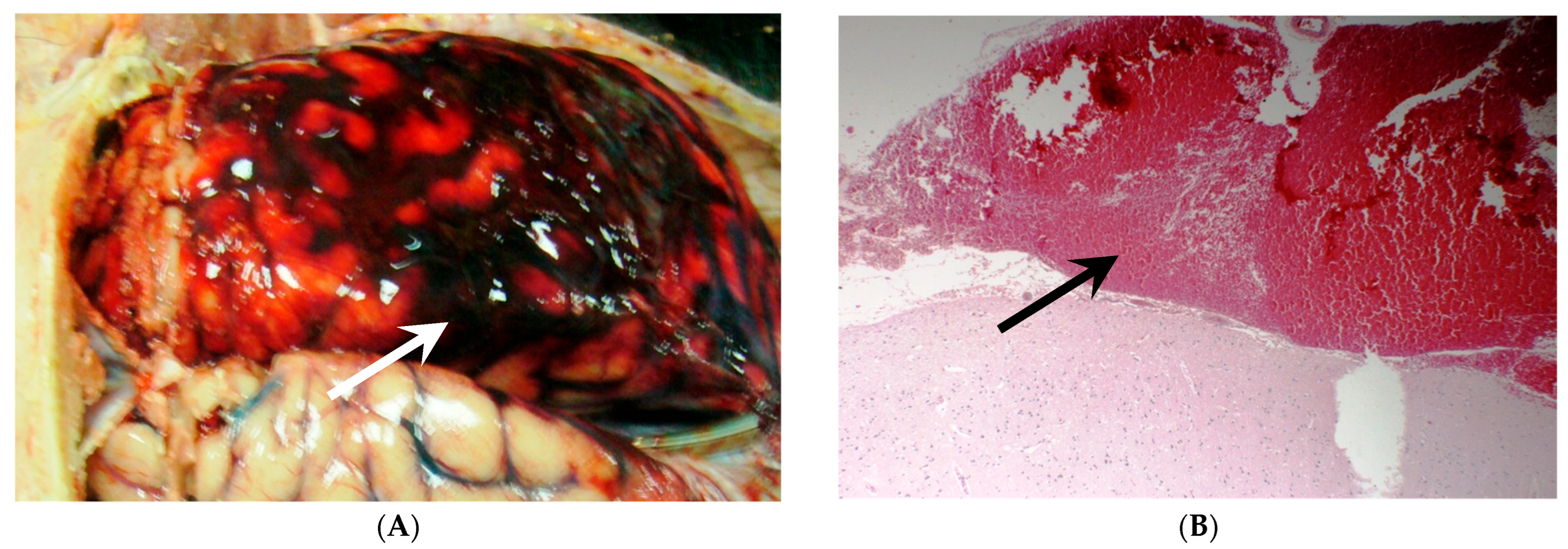
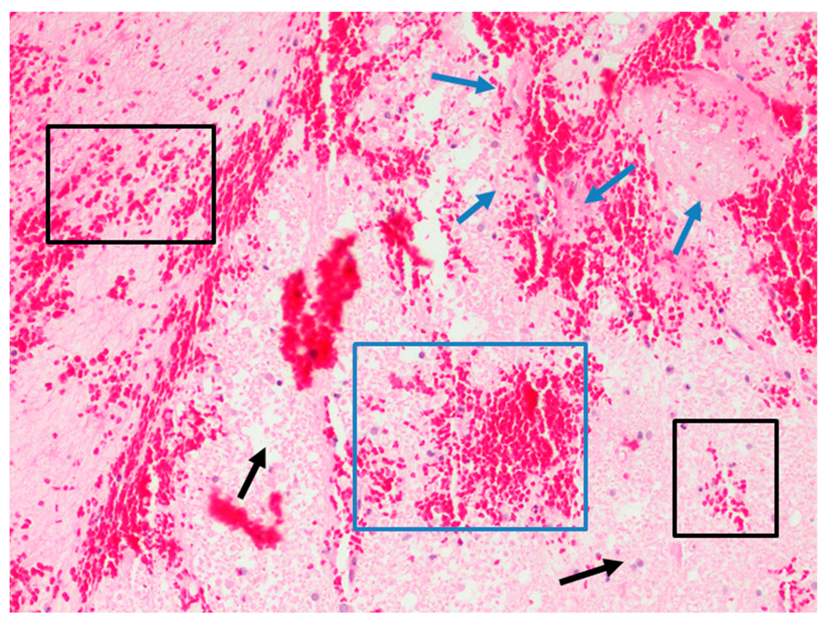
Arterial blood enters the subarachnoid space, cisterns, and often into the ventricular system at the time of initial aneurysm rupture (ictus), leading to a higher-pressure system. As the intracranial pressure (ICP) rises, the cerebral perfusion pressure (CPP) is decreased (CPP = MAP–ICP). In a normal physiological condition, a drop in CPP can sustain cerebral blood flow (CBF) to a limited extent prior to autoregulatory dysfunction and failure. However, in a sudden and large increase in ICP, such as in high-grade aSAH, CBF is markedly decreased, leading to the vasodilation of distal cerebral arterioles with the concomitant rise in arterial blood pressure that causes an increase in CBF, further increasing the ICP, until a complete cessation of CBF occurs. This leads to a phenomenon known as “transient global cerebral ischemia” [39]. Early or ictal infarcts can occur during the initial insult where early MRI may be helpful to differentiate this from DCI [11]. Weimer et al. [40] compared the whole brain apparent diffusion coefficient (ADC) on MRI in aSAH patients with controls to quantify the degree of early cytotoxic and vasogenic edema and observed significantly higher ADC values in the aSAH group, providing evidence of early brain edema as a possible mechanism for EBI.
The drop in CPP due to elevated ICP described at ictus is followed closely by a sympathetic catecholamine surge. Injury to the hypothalamus and brainstem, particularly the medulla, due to increased pressure leads to the release of catecholamines mediated by the rostral ventrolateral medulla and the paraventricular nucleus in the hypothalamus [41]. In 1963, Crompton [42] described this phenomenon after aSAH as related to necrosis or hemorrhage of the hypothalamus or damage or vasoconstriction of the arteries that feed it. More recently, this effect was well documented using the diffusion tensor imaging tractography technique showing a decrement in fractional anisotropy and an increase in ADC [43]. The injuries associated with the initial rise in ICP are thought to lead to the accumulation of carbon dioxide and glutamate, which gives rise to reactive astrocytes, and sympathetic activation, followed by secondary activation of the renin-angiotensin-aldosterone system in the kidney [44,45,46]. This sympathetic reflex has traditionally been recognized as leading to the well-known Cushing response from sympathetic over-excitation due to elevated ICP. The Cushing reflex occurs during prolonged ICP elevation with Lundberg A or plateau waves [47]. However, there has been evidence of an early Cushing-like response, thought to occur in the sudden and early, rather than sustained, rise in ICP after brain injury; this is associated with an intracranial baroreflex and sympatho-excitatory response to raise systemic blood pressure in an attempt to maintain CPP and CBF [48]. Moreover, the catecholamine surge can also lead to endothelin activation, thought to play a role in vasospasm, as well as cytokine release that leads to hyperglycemia, hypokalemia, and leukocytosis. This pathophysiology is the cause of cardiac, lung, renal, and other systemic complications, with a proven dramatic increase in plasma norepinephrine in the setting of cardiac injury in these patients [44,49]. In addition, the insular cortex stimulation with blood spilled due to SAH is realized in paroxysmal sympathetic hyperactivity syndrome [50]. These processes can cause hypotension and hypoxia, which further potentiates brain injury. Though this process starts at the ictus, it continues past the initial injury, causing the process known as EBI.
The prevention of worsening from initial ischemia is of paramount importance post-aSAH. Management of early complications, like rebleeding and hydrocephalus, is an important first step in managing aSAH. Cortical tissue irritation from subarachnoid blood can induce clinical seizures at the onset, most commonly occurring within the first 24 h. This complication occurs in over a quarter of patients, contributing to further increases in cerebral metabolic demand and a secondary increase in ICP due to cerebral blood flow elevations during the ictal period, potentiating ischemia and causing deleterious effects [51]. To manage cerebral autoregulatory failure, bedside blood pressure management is most commonly used, and the maintenance of mean arterial pressure is a mainstay of aSAH management.
In addition to the aforementioned pathophysiological mechanisms, molecular changes contribute to cerebral edema and DCI development. Macrophage migratory inhibitory factor (MIF), a homotrimer protein, has been demonstrated to function as a proinflammatory cytokine capable of activating inflammatory responses in the central nervous system [52,53]. In experimental studies, MIF is released from activated glial cells, which further activate the release of inflammatory mediators from astrocytes leading to brain injury by promoting neuronal cell death [54,55,56]. In recent studies, serum MIF concentrations have been shown to be associated with severity and long-term outcomes in patients with acute ischemic stroke, traumatic brain injury, and spontaneous intracerebral hemorrhage [57,58,59]. Similarly, MIF has also been studied as a marker of the severity of brain injury in patients with aSAH. In a prospective study by Chen et al. [53], serum MIF concentrations were found to be significantly higher in patients with aSAH as compared to controls (30.1 ng/mL vs. 7.9 ng/mL; p < 0.001). After adjusting for confounding variables (World Federation of Neurosurgical Societies [WFNS] and Fisher scores), serum MIF was identified as an independent predictor for 6-month unfavorable outcomes (Glasgow Outcome Scale Extended, 1–4) in aSAH patients [53]. In another study by Yang et al. [60], higher serum MIF levels were seen in patients who developed DCI as compared to patients without DCI (26.4 [IQR: 22.6–32.4] ng/mL vs. 20.4 [IQR:16.4–24.6] ng/mL, p < 0.001). Furthermore, MIF showed a greater discriminatory ability for DCI than the APACHE-II score, C-reactive protein, and interleukin 6 (IL-6) with an AUC of 0.780 (95% CI, 0.710–0.849).
Upregulation of vascular endothelial growth factor and aquaporin-4, increased levels of bradykinin, cytokine release of IL-6, and matrix metalloproteinase-9 are possible targets for treatment, although studies have shown mixed results [51,61]. Drier et al. [62] described that spreading depolarization in the gray matter led to cytotoxic edema, which resulted in ischemia, shown microscopically in Figure 2, leading to neuronal cell death and DCI. Predictive models and prognostication of early edema in aSAH are vast research topics. Wartenberg et al. [63] studied 21 high-grade aSAH patients with early MRI, noting that early ADC diffusion restriction correlates in regions not seen on initial CT. Of those patients, 71% died, and only 12% had a satisfactory outcome. This study proposed that infarcts on CT imaging provided a small picture, and that early MRI could help in noticing and managing early ischemia. In 2018, Ahn et al. [64] evaluated the presence of early edema and noted it as a possible surrogate for EBI and predictor of DCI by designing the Subarachnoid Hemorrhage Early Brain Edema Score (SEBES) that has been recognized as an independent predictor of DCI and is described later in this review. Aside from survival and functional outcome, cerebral edema has also been noted as a predictor of poor cognitive outcomes [61].

Early Brain Injury

EBI is believed to occur in the first 72 h following ictus and is a precursor for DCI. Impaired autoregulation is also most prominent in the first 72 h, associated with the increased risk of DCI [65]. The catecholamine surge at ictus continues up to 7 to 10 days post-rupture [44]. As previously mentioned, glutamate accumulation and impaired uptake are part of this process. This causes NMDA receptor activation leading to calcium ion flooding the cell, inducing apoptosis and cell necrosis, as well as the breakdown of the blood-brain barrier (BBB), which all contribute to EBI [65].
Endothelial dysfunction is thought to be a major contributor to vasoconstriction. Oxy- and deoxyhemoglobin damage healthy neurons that secrete neuronal nitric oxide synthase in the brain vessels and parenchyma. The decrease in nitric oxide level needed for vasodilation leads to vasoconstriction. The normal reactionary increase in endothelial nitric oxide synthase is also impaired, which causes continued vasoconstriction until nitric oxide is restored to a normal level [65]. This is also implicated in BBB breakdown due to associated apoptosis and increased permeability, especially in microvessels [65]. Activation of calcium channels and enhanced membrane depolarization are thought to lead to further microvessel spasms [66].
Platelet aggregation is another important contributor to EBI. In animal models, microthrombi from platelet aggregates have been noted as early as 10 min following aSAH. Punctate infarcts on MRI could be associated with microthrombi and have been shown to predict worse outcomes [67]. Another study that evaluated MRI diffusion restriction in the first four days after poor grade aSAH demonstrated that 86% of patients had diffusion restriction and ADC correlates that were not seen on CT [63].
Cellular loss of ionic gradient leads to cortical spreading depolarization, which is seen as a negative direct current, noting differences in depolarization. Sodium and water enter the depolarized cell, leading to cellular swelling and necrosis. This sustained neuronal depolarization presents as slow-moving and propagating waves, detected using subdural electrodes. In severe cases, this can lead to spreading depolarization-induced spreading depression, which is a decline in the amplitude of the alternating current, or loss of spontaneous electrical activity in the brain [62]. This is associated with injury in a bimodal fashion, with both early and delayed brain injury after aSAH [17,51]. In a recently published pilot clinical trial on depolarizations in ischemia after subarachnoid hemorrhage (DISCHARGE-1) [68], the primary outcome was the peak total depression time from spreading depolarization that was indicative of DCI. The authors hypothesized that a 60-min cutoff of spreading depolarization-induced depression would predict delayed infarction with high sensitivity (>0.6) and specificity (>0.8). However, the study’s results revealed that this time interval was too short of reaching the desired specificity of 0.80 (specificity was 0.59), though it reached higher than desired sensitivity (0.76). A 180-min cutoff was found to have lower sensitivity (0.62), but higher specificity (0.83). However, the 60-min cutoff had a higher sensitivity and specificity for predicting DCI than the defined cerebral infarction. In their conclusion, Dreier et al. [68] found that spreading depolarizations were likely an independent marker of brain injury post-aSAH. One episode of spreading depolarization alone was shown to significantly affect the risk of poor outcomes in their studied population. In another study of 19 subjects, Owen et al. [69] reviewed cerebral autoregulation and spreading depolarizations simultaneously through multimodal monitoring. Spreading depolarization was associated with impaired cerebral autoregulation, proposing a negative feedback phenomenon from impaired cerebral autoregulation that leads to decreased CBF and spreading depolarization, leading back to further worsening of cerebral autoregulation. Though there is no consensus, given these findings, it is plausible that cortical spreading depression provokes ischemia. These important studies further support the use of advanced neuromonitoring, especially in high-grade aSAH that also has the potential to provide us with a therapeutic target. Carlson et al. [70] published a pilot study of ketamine to suppress spreading depolarizations in traumatic brain injury and aSAH patients, with a subsequent study to look at an alternative N-methyl-D-aspartate receptor antagonist, memantine, which seems to be a promising alternative [71].

4. Recent Developments

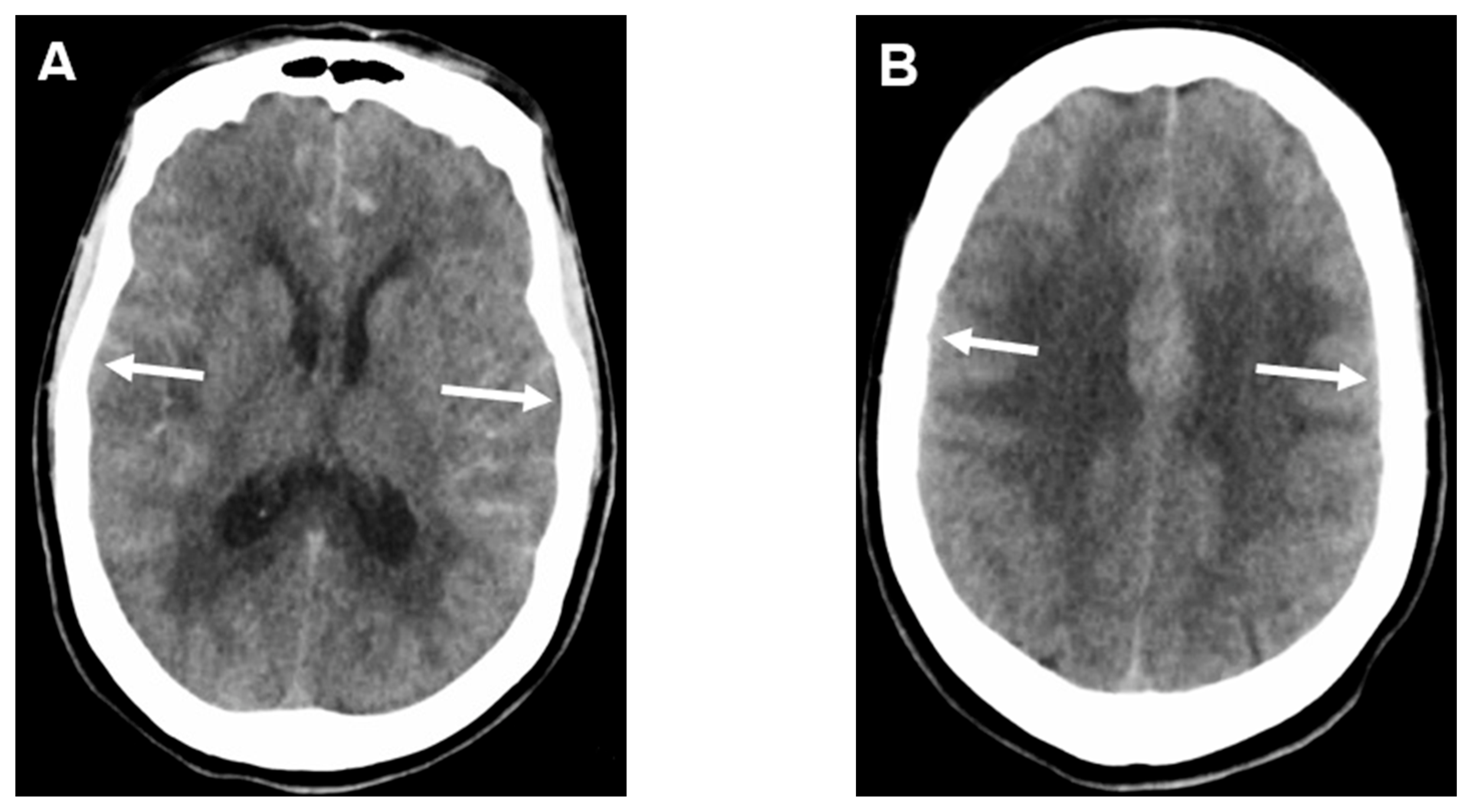
4.1. Subarachnoid Hemorrhage Early Brain Edema Score (SEBES)

The SEBES score is a semi-quantitative CT grading scale that was developed as a novel radiographic marker of EBI, also helpful in predicting DCI and unfavorable outcomes. The score is based on a scale of 0–4 (where 4 presents the worst) and calculated similarly to the Alberta Stroke Program Early CT Score (ASPECTS) [72] in acute ischemic stroke, at two separate slices: Slice 1: the level of the insular cortex, showing the thalamus and basal ganglia, and Slice 2: the level of the centrum semiovale above the level of the lateral ventricle. A point is assigned for each side with effacement of the sulci, at each level, as seen in Figure 3. In a prospective study that included 164 patients, high-grade Hunt and Hess (HH) and higher WFNS grade were identified as predictors of high-grade SEBES score (defined as a score of 3 or 4). After adjusting for covariates, SEBES was identified as an independent predictor of DCI (OR = 2.24, 95% CI: 1.58–3.17) and unfavorable outcome (OR = 3.45, 95% CI: 1.95–6.07) [64]. In another study by Said et al. [73], independent associations were found between higher SEBES scores (3 or 4) and the need for medical management of ICP, decompressive craniectomy, development of cerebral infarction, as well as unfavorable outcomes. In an observational cohort, Rass et al. [74] used the SEBES score repeatedly to follow the course of edema. They concluded that the score could identify patients with delayed resolution of early brain edema. As stated, aggressive control of cerebral edema, and the factors that play a role in the late resolution, should be part of the initial management of these patients using the SEBES score.

4.2. Volumetric Analysis of Edema

Dhar and his team have recently studied volumetric analysis of global cerebral edema (GCE) as a predictor of poor outcomes after aSAH [75], where the selective sulcal volume (SSV) and the CSF volume above the lateral ventricles were measured using an automated machine learning algorithm and studied as a quantitative biomarker of GCE. In this study, an SSV cutoff below 5 mL accurately identified GCE in patients with aSAH. Furthermore, early SSV (i.e., the lowest SSV within 72 h of ictus) was noted to be an independent predictor of poor outcomes with the strongest impact on the prediction of GCE in younger patients (<70 years old).

4.3. Multimodal Monitoring, Predictive Models, and Diagnosis of Dci

Multimodal monitoring (Table 2) is becoming more widely used in this patient population. It has been shown to aid in detecting DCI, especially in patients with limited reliable secondary brain injury examination [76,77]. Lazaridis’s summary of cerebral shock explains different pathophysiologic profiles associated with these monitoring techniques, summarized in Table 3 [78]. Brain tissue oxygen (PbtO2) and cerebral microdialysis can be used for individualized patient care vis-à-vis the above mechanisms of injury from local and regional hypoxia. Trend analysis of PbtO2 helps distinguish early from delayed injury, where a drop in PbtO2 has been shown to correlate with vasospasm and DCI [10]. Cerebral microdialysis can give data on the brain’s metabolic profile that can be affected days before the irreversible injury [10]. The lactate-to-pyruvate ratio (LPR) has been studied as a potential biomarker in aSAH and other neurologic injuries. An increase in the LPR can be associated with ischemia and poor patient outcomes [79]. A recent study [80] used a multimodal approach to create a useful diagnostic tool to detect patients at risk for ischemia. A fraction of inspired oxygen (FiO2) challenge was completed that would normally increase the PbtO2. The investigators noted that patients with higher cerebral lactate had less increase in PbtO2 and concluded that a less-than-expected increase in PbtO2 vis-à-vis FiO2 challenge could be a surrogate for elevated cerebral lactate and risk for ischemia. Megjhani et al. [81] measured CPP in association with PbtO2. A non-linear relationship was found, but it showed that hypoperfusion and less than optimal values of CPP were associated with decreased PbtO2 and regional brain hypoxia. Because of a nonstandard approach related to these modalities, studies of invasive multimodal monitoring have produced mixed results. The ability to interpret results, recognize, and treat patients based on those results are user and training-dependent. However, these tools demonstrate the focus on microcellular milieu data, contrasted with the previous thought of management based on large vessel vasospasm alone.
Near-infrared spectroscopy (NIRS) is a non-invasive brain tissue oxygenation monitoring device, but its use is limited due to the ability to monitor mainly the frontal lobe. De Courson et al. looked at the correlation between PbtO2 and NIRS measurements among 51 SAH patients and found no correlation [82]. Pupillometry has been well studied as a tool for early detection of potential worsening in brain injury cases. Regarding aSAH, Aoun et al. [83] looked at 56 patients with transcranial doppler and Neurological Pupil index (NPi) measurements in combination. In this study, sonographic vasospasm was not significantly associated with the NPi measurement, but sonographic vasospasm and abnormal decreases in NPi were each associated with DCI.
Cortical spreading depolarization and depression can be monitored along with continuous electroencephalogram (cEEG) to detect changes such as decreasing relative alpha variability, decreasing alpha-delta ratio, worsening focal slowing, or late-appearing epileptiform abnormalities that are considered EEG alarms [84,85,86]. This approach [85,86] could predict DCI days before the actual event, which is an exciting advancement and also shows this as a dynamic pathophysiologic process.
Vasospasm does occur around the period of DCI and cerebral infarction, though not always found to correlate with location or degree of ischemia. Transcranial Doppler is a non-invasive tool that can detect possible vasospasm. Mean flow velocities in the middle and anterior cerebral arteries are typically considered abnormal for >120 cm/s, with >200 cm/s consistent with severe vasospasm [10]. CT angiogram is another valuable non-invasive imaging modality that can be used for diagnosing cerebral vasospasm. However, catheter-based angiography remains the gold standard, giving both the ability to diagnose and treat. CT perfusion (CTP) imaging has been studied as a potential tool in detecting DCI. In a systematic review that included 345 patients, aSAH patients with perfusion deficits on CTP imaging demonstrated an approximately 23-fold higher likelihood of developing DCI than patients with normal CTP results [87]. Further, a mean transit time (MTT) exceeding 6.4 s, or regional CBF below 25 mL/100 g/min correlated with the development of DCI in the study [88]. In another study by Cremers et al. [89], aSAH patients with assessed perfusion deficit with CTP at the time of clinical deterioration were more often noted to develop infarction on follow-up imaging (88%) as compared with patients without a perfusion deficit (38%). CTP imaging can provide detailed information on microvascular circulation. Therefore, its combination with CT angiography or digital subtraction angiography can provide a complete view of cerebral circulation [90,91,92,93]. However, due to high variability in post-processing methods and equipment used, along with inconsistency with quantitative variables and their thresholds for detecting DCI, the question of the sensitivity of CTP is a question of clinicians [94]. Further prospective studies are required before the wide acceptance of CTP as a promising tool in detecting DCI.

5. Future Directions

Clinical and Risk Factor Assessment

Several studies have resulted in different grading systems based on clinical or radiological factors to predict DCI or outcomes to guide treatment [95]. The GCS, HH, and WFNS are the widely used clinical grading scales, focusing on signs and symptoms; however, these grading structures do not take into account the quantity and severity of bleeding. With the improved CT techniques, some radiographic scales have been further developed. Quantifying thickness and area of subarachnoid blood on CT scans, the Fisher Scale and the modified Fisher Scale can predict the incidence of cerebral vasospasm and DCI [7]. The SEBES is a new scoring tool that displays the degree of EBI to predict the incidence of DCI. However, further research is needed to determine its accuracy and effectiveness [64]. The main issue with radiological scales is the absence of clinical symptoms. Thus, a few combined grading systems have been promoted, including VASOGRADE (VG) [96] and the HAIR [97] scales described in Table 4, to predict DCI and in-hospital mortality respectively [97]. However, those grading structures do not consider the importance of EBI. New studies have recognized that the incidence of DCI is associated with the severity of EBI after SAH [98]. Other scores include combining multiple variables of admission data [99] and more dynamic scores to give clinicians insight into the possibility of current DCI [100].
Multiple predictive models have been recommended for DCI. Coated platelets, a subset of platelet that keeps the prothrombotic substance attached to the platelet and a marker for thrombogenicity, was studied to look at DCI prediction. The trend of coated platelets after initial ictus was found to be a reliable predictor of DCI [101]. Machine-learning capabilities for better prediction have been shown to be beneficial in multiple studies and are promising roads for future research [102,103].

6. Necessity for Ongoing Research

Further reliable predictive models and markers to determine patients at high risk for DCI and other complications following aSAH are an important continued endeavor. As mentioned previously regarding genetic predisposition to DCI, the potential of polymorphisms associated with nitric oxide synthase (NOS), specifically the neuronal type, has shown promising bench research results. Endothelial NOS was not significantly shown to increase DCI or vasospasm risk in a meta-analysis [28]. Although challenging due to the unavailability of either pre- or post-morbid genetic testing, further prospective studies may shed a better light on the role of endothelial NOS.
The oxidative stress associated with the initial catecholamine surge at the ictus has been studied vis-à-vis possible mechanisms to address the complications arising from that excitatory response. Renal denervation was investigated to stabilize the sympathetic response by deactivating efferent sympathetic nerves in the periphery. This reduced angiotensin-II and endothelin-1 in animal models and offered hope for inhibiting vasoconstriction and vasospasm [45]. Although previous studies have proven that treatment of vasospasm may not improve outcomes, the idea of continued research to address the ictal sympathetic surge is possible.
A multicenter study with a large sample size and standardized variables is needed regarding aSAH to find the best generalizable and reproducible results to lead to a change in clinical practice. Using relevant preclinical models is an essential part of conducting translational studies [104]. This is necessary to address the limited treatment options that currently exist for aSAH and DCI with a significant degree of poor outcomes.

7. Conclusions

The primary goal of neurointensivists in clinical practice is to ameliorate the burden of morbidity and mortality associated with aSAH. DCI is a complex multifactorial pathophysiological process that starts early post-aSAH, with risk factors present before and at ictus. The pathophysiology of EBI and the mechanisms mentioned earlier act as a substrate for the development of DCI. This includes the well-known mechanism of vasoconstriction and spasm, focusing on the sympathetic surge and cytokine release, microthrombosis, and BBB breakdown. Early focus on EBI and global cerebral edema are essential in managing aSAH. Our research goals should continue to change and attempt to address these newer pathomechanisms in a search for improved outcomes that can be translated into routine clinical practice.

Funding

This research received no external funding.

Institutional Review Board Statement

Not applicable.

Informed Consent Statement

Not applicable.

Data Availability Statement

Data sharing not applicable.

Acknowledgments

Divani’s studies are funded by the UNM Center for Brain Recovery and Repair Center of Biomedical Research Excellence through Grant Number NIH P20GM109089 (Pilot PI), W81XWH-17-2-0053 (PI), and 1R21NS130423-01 (PI).

Conflicts of Interest

The authors declare no conflict of interest.

Abbreviations

aSAHAneurysmal subarachnoid hemorrhage
ADCApparent diffusion coefficient
APOEApolipoprotein E
CBFCerebral blood flow
CPPCerebral perfusion pressure
CTComputed tomography
DCIDelayed cerebral ischemia
EBIEarly brain injury
EEGElectroencephalogram
eNOSEndothelial nitric oxide synthase
EWASEpigenome wide association studies
FiO2Fraction of inspired oxygen
GCSGlasgow Coma Scale
GWASgenome wide association studies
HpHaptoglobin
ICPIntracranial pressure
IL-6Interleukin 6
MAPMean arterial pressure
MIFMacrophage migratory inhibitory factor
MRIMagnetic resonance imaging
NIHSSNational Institute of Health Stroke Scale
NIRSNear-infrared spectroscopy
NOSNitric oxide synthase
NpiNeurological Pupil index
NRG1Neuregulin 1
OR Odds ratio
PbtO2Brain tissue oxygenation
PRSPolygenic risk scores
RYY-1Ryanodine-1
SEBESSubarachnoid hemorrhage early brain edema score
wPRSWeighted PRS

References

  1. Etminan, N.; Chang, H.S.; Hackenberg, K.; de Rooij, N.K.; Vergouwen, M.D.I.; Rinkel, G.J.E.; Algra, A. Worldwide Incidence of Aneurysmal Subarachnoid Hemorrhage According to Region, Time Period, Blood Pressure, and Smoking Prevalence in the Population: A Systematic Review and Meta-analysis. JAMA Neurol. 2019, 76, 588–597. [Google Scholar] [CrossRef]
  2. De Oliveira Manoel, A.L.; Mansur, A.; Silva, G.S.; Germans, M.R.; Jaja, B.N.; Kouzmina, E.; Marotta, T.R.; Abrahamson, S.; Schweizer, T.A.; Spears, J.; et al. Functional Outcome After Poor-Grade Subarachnoid Hemorrhage: A Single-Center Study and Systematic Literature Review. Neurocritical Care 2016, 25, 338–350. [Google Scholar] [CrossRef] [PubMed]
  3. Nieuwkamp, D.J.; Setz, L.E.; Algra, A.; Linn, F.H.; de Rooij, N.K.; Rinkel, G.J. Changes in case fatality of aneurysmal subarachnoid haemorrhage over time, according to age, sex, and region: A meta-analysis. Lancet Neurol. 2009, 8, 635–642. [Google Scholar] [CrossRef]
  4. Springer, M.V.; Schmidt, J.M.; Wartenberg, K.E.; Frontera, J.A.; Badjatia, N.; Mayer, S.A. Predictors of global cognitive impairment 1 year after subarachnoid hemorrhage. Neurosurgery 2009, 65, 1043–1050. [Google Scholar] [CrossRef]
  5. Geraghty, J.R.; Testai, F.D. Delayed Cerebral Ischemia after Subarachnoid Hemorrhage: Beyond Vasospasm and Towards a Multifactorial Pathophysiology. Curr. Atheroscler. Rep. 2017, 19, 50. [Google Scholar] [CrossRef]
  6. Adams, H.P., Jr.; Kassell, N.F.; Torner, J.C.; Haley, E.C., Jr. Predicting cerebral ischemia after aneurysmal subarachnoid hemorrhage: Influences of clinical condition, CT results, and antifibrinolytic therapy. A report of the Cooperative Aneurysm Study. Neurology 1987, 37, 1586–1591. [Google Scholar] [CrossRef]
  7. Frontera, J.A.; Claassen, J.; Schmidt, J.M.; Wartenberg, K.E.; Temes, R.; Connolly, E.S., Jr.; MacDonald, R.L.; Mayer, S.A. Prediction of symptomatic vasospasm after subarachnoid hemorrhage: The modified fisher scale. Neurosurgery 2006, 59, 21–27. [Google Scholar] [CrossRef] [Green Version]
  8. Francoeur, C.L.; Mayer, S.A. Management of delayed cerebral ischemia after subarachnoid hemorrhage. Crit. Care 2016, 20, 277. [Google Scholar] [CrossRef] [PubMed] [Green Version]
  9. Saripalli, M.; Tan, D.; Chandra, R.V.; Lai, L.T. Predictive Relevance of Early Temperature Elevation on the Risk of Delayed Cerebral Ischemia Development Following Aneurysmal Subarachnoid Hemorrhage. World Neurosurg. 2021, 150, e474–e481. [Google Scholar] [CrossRef] [PubMed]
  10. Rass, V.; Helbok, R. How to diagnose delayed cerebral ischaemia and symptomatic vasospasm and prevent cerebral infarction in patients with subarachnoid haemorrhage. Curr. Opin. Crit. Care 2021, 27, 103–114. [Google Scholar] [CrossRef]
  11. Ikram, A.; Javaid, M.A.; Ortega-Gutierrez, S.; Selim, M.; Kelangi, S.; Anwar, S.M.H.; Torbey, M.T.; Divani, A.A. Delayed Cerebral Ischemia after Subarachnoid Hemorrhage. J. Stroke Cerebrovasc. Dis. 2021, 30, 106064. [Google Scholar] [CrossRef]
  12. Schmidt, T.P.; Weiss, M.; Hoellig, A.; Nikoubashman, O.; Schulze-Steinen, H.; Albanna, W.; Clusmann, H.; Schubert, G.A.; Veldeman, M. Revisiting the Timeline of Delayed Cerebral Ischemia After Aneurysmal Subarachnoid Hemorrhage: Toward a Temporal Risk Profile. Neurocritical Care 2022, 37, 735–743. [Google Scholar] [CrossRef] [PubMed]
  13. Rosengart, A.J.; Schultheiss, K.E.; Tolentino, J.; Macdonald, R.L. Prognostic factors for outcome in patients with aneurysmal subarachnoid hemorrhage. Stroke J. Cereb. Circ. 2007, 38, 2315–2321. [Google Scholar] [CrossRef] [Green Version]
  14. Vergouwen, M.D.; Vermeulen, M.; van Gijn, J.; Rinkel, G.J.; Wijdicks, E.F.; Muizelaar, J.P.; Mendelow, A.D.; Juvela, S.; Yonas, H.; Terbrugge, K.G.; et al. Definition of delayed cerebral ischemia after aneurysmal subarachnoid hemorrhage as an outcome event in clinical trials and observational studies: Proposal of a multidisciplinary research group. Stroke J. Cereb. Circ. 2010, 41, 2391–2395. [Google Scholar] [CrossRef] [PubMed] [Green Version]
  15. Raatikainen, E.; Vahtera, A.; Kuitunen, A.; Junttila, E.; Huhtala, H.; Ronkainen, A.; Pyysalo, L.; Kiiski, H. Prognostic value of the 2010 consensus definition of delayed cerebral ischemia after aneurysmal subarachnoid hemorrhage. J. Neurol. Sci. 2021, 420, 117261. [Google Scholar] [CrossRef]
  16. Etminan, N.; Vergouwen, M.D.; Macdonald, R.L. Angiographic vasospasm versus cerebral infarction as outcome measures after aneurysmal subarachnoid hemorrhage. Acta Neurochir. Suppl. 2013, 115, 33–40. [Google Scholar] [CrossRef]
  17. Foreman, B. The Pathophysiology of Delayed Cerebral Ischemia. J. Clin. Neurophysiol. 2016, 33, 174–182. [Google Scholar] [CrossRef] [PubMed]
  18. Macdonald, R.L.; Higashida, R.T.; Keller, E.; Mayer, S.A.; Molyneux, A.; Raabe, A.; Vajkoczy, P.; Wanke, I.; Bach, D.; Frey, A.; et al. Clazosentan, an endothelin receptor antagonist, in patients with aneurysmal subarachnoid haemorrhage undergoing surgical clipping: A randomised, double-blind, placebo-controlled phase 3 trial (CONSCIOUS-2). Lancet Neurol. 2011, 10, 618–625. [Google Scholar] [CrossRef]
  19. Ya, X.; Zhang, C.; Zhang, S.; Zhang, Q.; Cao, Y.; Wang, S.; Zhao, J. The Relationship Between Smoking and Delayed Cerebral Ischemia After Intracranial Aneurysm Rupture: A Systematic Review and Meta-Analysis. Front. Neurol. 2021, 12, 625087. [Google Scholar] [CrossRef]
  20. Duan, W.; Pan, Y.; Wang, C.; Wang, Y.; Zhao, X.; Wang, Y.; Liu, L.; CNSR Investigators. Risk Factors and Clinical Impact of Delayed Cerebral Ischemia after Aneurysmal Subarachnoid Hemorrhage: Analysis from the China National Stroke Registry. Neuroepidemiology 2018, 50, 128–136. [Google Scholar] [CrossRef]
  21. Crobeddu, E.; Mittal, M.K.; Dupont, S.; Wijdicks, E.F.; Lanzino, G.; Rabinstein, A.A. Predicting the lack of development of delayed cerebral ischemia after aneurysmal subarachnoid hemorrhage. Stroke J. Cereb. Circ. 2012, 43, 697–701. [Google Scholar] [CrossRef] [PubMed] [Green Version]
  22. Bakker, M.K.; Ruigrok, Y.M. Genetics of Intracranial Aneurysms. Stroke J. Cereb. Circ. 2021, 52, 3004–3012. [Google Scholar] [CrossRef]
  23. Gaastra, B.; Alexander, S.; Bakker, M.K.; Bhagat, H.; Bijlenga, P.; Blackburn, S.; Collins, M.K.; Dore, S.; Griessenauer, C.; Hendrix, P.; et al. Genome-Wide Association Study of Clinical Outcome After Aneurysmal Subarachnoid Haemorrhage: Protocol. Transl. Stroke Res. 2022, 13, 565–576. [Google Scholar] [CrossRef] [PubMed]
  24. Rinkel, G.J. Intracranial aneurysm screening: Indications and advice for practice. Lancet Neurol. 2005, 4, 122–128. [Google Scholar] [CrossRef] [PubMed]
  25. Korja, M.; Silventoinen, K.; McCarron, P.; Zdravkovic, S.; Skytthe, A.; Haapanen, A.; de Faire, U.; Pedersen, N.L.; Christensen, K.; Koskenvuo, M.; et al. Genetic epidemiology of spontaneous subarachnoid hemorrhage: Nordic Twin Study. Stroke J. Cereb. Circ. 2010, 41, 2458–2462. [Google Scholar] [CrossRef] [Green Version]
  26. Bakker, M.K.; van der Spek, R.A.A.; van Rheenen, W.; Morel, S.; Bourcier, R.; Hostettler, I.C.; Alg, V.S.; van Eijk, K.R.; Koido, M.; Akiyama, M.; et al. Genome-wide association study of intracranial aneurysms identifies 17 risk loci and genetic overlap with clinical risk factors. Nat. Genet. 2020, 52, 1303–1313. [Google Scholar] [CrossRef]
  27. Theodotou, C.B.; Snelling, B.M.; Sur, S.; Haussen, D.C.; Peterson, E.C.; Elhammady, M.S. Genetic associations of intracranial aneurysm formation and sub-arachnoid hemorrhage. Asian J. Neurosurg. 2017, 12, 374–381. [Google Scholar] [CrossRef] [Green Version]
  28. Solodovnikova, Y.; Ivaniuk, A.; Marusich, T.; Son, A. Meta-analysis of associations of genetic polymorphisms with cerebral vasospasm and delayed cerebral ischemia after aneurysmal subarachnoid hemorrhage. Acta Neurol. Belg. 2021, 122, 1547–1556. [Google Scholar] [CrossRef]
  29. Gaastra, B.; Ren, D.; Alexander, S.; Bennett, E.R.; Bielawski, D.M.; Blackburn, S.L.; Borsody, M.K.; Dore, S.; Galea, J.; Garland, P.; et al. Haptoglobin genotype and aneurysmal subarachnoid hemorrhage: Individual patient data analysis. Neurology 2019, 92, e2150–e2164. [Google Scholar] [CrossRef] [Green Version]
  30. Gaastra, B.; Glazier, J.; Bulters, D.; Galea, I. Haptoglobin Genotype and Outcome after Subarachnoid Haemorrhage: New Insights from a Meta-Analysis. Oxid. Med. Cell. Longev. 2017, 2017, 6747940. [Google Scholar] [CrossRef] [Green Version]
  31. McInnes, M.D.; Bossuyt, P.M. Pitfalls of Systematic Reviews and Meta-Analyses in Imaging Research. Radiology 2015, 277, 13–21. [Google Scholar] [CrossRef] [PubMed]
  32. Baumann, A.; Devaux, Y.; Audibert, G.; Zhang, L.; Bracard, S.; Colnat-Coulbois, S.; Klein, O.; Zannad, F.; Charpentier, C.; Longrois, D.; et al. Gene expression profile of blood cells for the prediction of delayed cerebral ischemia after intracranial aneurysm rupture: A pilot study in humans. Cerebrovasc. Dis. 2013, 36, 236–242. [Google Scholar] [CrossRef]
  33. Kim, B.J.; Kim, Y.; Youn, D.H.; Park, J.J.; Rhim, J.K.; Kim, H.C.; Kang, K.; Jeon, J.P. Genome-wide blood DNA methylation analysis in patients with delayed cerebral ischemia after subarachnoid hemorrhage. Sci. Rep. 2020, 10, 11419. [Google Scholar] [CrossRef] [PubMed]
  34. Heinsberg, L.W.; Weeks, D.E.; Alexander, S.A.; Minster, R.L.; Sherwood, P.R.; Poloyac, S.M.; Deslouches, S.; Crago, E.A.; Conley, Y.P. Iron homeostasis pathway DNA methylation trajectories reveal a role for STEAP3 metalloreductase in patient outcomes after aneurysmal subarachnoid hemorrhage. Epigenetics Commun. 2021, 1, 4. [Google Scholar] [CrossRef]
  35. Guttmacher, A.E.; Porteous, M.E.; McInerney, J.D. Educating health-care professionals about genetics and genomics. Nat. Rev. Genet. 2007, 8, 151–157. [Google Scholar] [CrossRef] [PubMed]
  36. Torkamani, A.; Wineinger, N.E.; Topol, E.J. The personal and clinical utility of polygenic risk scores. Nat. Rev. Genet. 2018, 19, 581–590. [Google Scholar] [CrossRef]
  37. Slunecka, J.L.; van der Zee, M.D.; Beck, J.J.; Johnson, B.N.; Finnicum, C.T.; Pool, R.; Hottenga, J.J.; de Geus, E.J.C.; Ehli, E.A. Implementation and implications for polygenic risk scores in healthcare. Hum. Genom. 2021, 15, 46. [Google Scholar] [CrossRef]
  38. Hong, E.P.; Youn, D.H.; Kim, B.J.; Lee, J.J.; Na, D.; Ahn, J.H.; Park, J.J.; Rhim, J.K.; Kim, H.C.; Jeon, H.J.; et al. Genome-wide polygenic risk impact on intracranial aneurysms and acute ischemic stroke. PLoS ONE 2022, 17, e0265581. [Google Scholar] [CrossRef]
  39. Sehba, F.A.; Pluta, R.M.; Macdonald, R.L. Brain injury after transient global cerebral ischemia and subarachnoid hemorrhage. Stroke Res. Treat. 2013, 2013, 827154. [Google Scholar] [CrossRef] [Green Version]
  40. Weimer, J.M.; Jones, S.E.; Frontera, J.A. Acute Cytotoxic and Vasogenic Edema after Subarachnoid Hemorrhage: A Quantitative MRI Study. AJNR Am. J. Neuroradiol. 2017, 38, 928–934. [Google Scholar] [CrossRef] [Green Version]
  41. Hasegawa, Y.; Uchikawa, H.; Kajiwara, S.; Morioka, M. Central sympathetic nerve activation in subarachnoid hemorrhage. J. Neurochem. 2022, 160, 34–50. [Google Scholar] [CrossRef]
  42. Crompton, M.R. Hypothalamic lesions following the rupture of cerebral berry aneurysms. Brain 1963, 86, 301–314. [Google Scholar] [CrossRef]
  43. Lee, S.J.; Jang, S.H. Hypothalamic injury in spontaneous subarachnoid hemorrhage: A diffusion tensor imaging study. Clin. Auton. Res. 2021, 31, 321–322. [Google Scholar] [CrossRef]
  44. Naredi, S.; Lambert, G.; Eden, E.; Zall, S.; Runnerstam, M.; Rydenhag, B.; Friberg, P. Increased sympathetic nervous activity in patients with nontraumatic subarachnoid hemorrhage. Stroke J. Cereb. Circ. 2000, 31, 901–906. [Google Scholar] [CrossRef] [PubMed] [Green Version]
  45. Takemoto, Y.; Hasegawa, Y.; Hayashi, K.; Cao, C.; Hamasaki, T.; Kawano, T.; Mukasa, A.; Kim-Mitsuyama, S. The Stabilization of Central Sympathetic Nerve Activation by Renal Denervation Prevents Cerebral Vasospasm after Subarachnoid Hemorrhage in Rats. Transl. Stroke Res. 2020, 11, 528–540. [Google Scholar] [CrossRef] [PubMed]
  46. Kawakita, F.; Kanamaru, H.; Asada, R.; Suzuki, Y.; Nampei, M.; Nakajima, H.; Oinaka, H.; Suzuki, H. Roles of glutamate in brain injuries after subarachnoid hemorrhage. Histol. Histopathol. 2022, 37, 1041–1051. [Google Scholar] [CrossRef]
  47. Wan, W.H.; Ang, B.T.; Wang, E. The Cushing Response: A case for a review of its role as a physiological reflex. J. Clin. Neurosci. 2008, 15, 223–228. [Google Scholar] [CrossRef]
  48. Schmidt, E.A.; Czosnyka, Z.; Momjian, S.; Czosnyka, M.; Bech, R.A.; Pickard, J.D. Intracranial baroreflex yielding an early cushing response in human. Acta Neurochir. Suppl. 2005, 95, 253–256. [Google Scholar] [CrossRef]
  49. Chen, S.; Li, Q.; Wu, H.; Krafft, P.R.; Wang, Z.; Zhang, J.H. The harmful effects of subarachnoid hemorrhage on extracerebral organs. Biomed. Res. Int. 2014, 2014, 858496. [Google Scholar] [CrossRef] [Green Version]
  50. Meyfroidt, G.; Baguley, I.J.; Menon, D.K. Paroxysmal sympathetic hyperactivity: The storm after acute brain injury. Lancet Neurol. 2017, 16, 721–729. [Google Scholar] [CrossRef] [PubMed]
  51. Osgood, M.L. Aneurysmal Subarachnoid Hemorrhage: Review of the Pathophysiology and Management Strategies. Curr. Neurol. Neurosci. Rep. 2021, 21, 50. [Google Scholar] [CrossRef] [PubMed]
  52. Calandra, T.; Bucala, R. Macrophage Migration Inhibitory Factor (MIF): A Glucocorticoid Counter-Regulator within the Immune System. Crit. Rev. Immunol. 2017, 37, 359–370. [Google Scholar] [CrossRef]
  53. Chen, Y.H.; Cheng, Z.Y.; Shao, L.H.; Shentu, H.S.; Fu, B. Macrophage migration inhibitory factor as a serum prognostic marker in patients with aneurysmal subarachnoid hemorrhage. Clin. Chim. Acta 2017, 473, 60–64. [Google Scholar] [CrossRef]
  54. Koda, M.; Nishio, Y.; Hashimoto, M.; Kamada, T.; Koshizuka, S.; Yoshinaga, K.; Onodera, S.; Nishihira, J.; Moriya, H.; Yamazaki, M. Up-regulation of macrophage migration-inhibitory factor expression after compression-induced spinal cord injury in rats. Acta Neuropathol. 2004, 108, 31–36. [Google Scholar] [CrossRef]
  55. Su, Y.; Wang, Y.; Zhou, Y.; Zhu, Z.; Zhang, Q.; Zhang, X.; Wang, W.; Gu, X.; Guo, A.; Wang, Y. Macrophage migration inhibitory factor activates inflammatory responses of astrocytes through interaction with CD74 receptor. Oncotarget 2017, 8, 2719–2730. [Google Scholar] [CrossRef] [Green Version]
  56. Inacio, A.R.; Ruscher, K.; Leng, L.; Bucala, R.; Deierborg, T. Macrophage migration inhibitory factor promotes cell death and aggravates neurologic deficits after experimental stroke. J. Cereb. Blood Flow Metab. 2011, 31, 1093–1106. [Google Scholar] [CrossRef] [Green Version]
  57. Li, Y.S.; Chen, W.; Liu, S.; Zhang, Y.Y.; Li, X.H. Serum macrophage migration inhibitory factor levels are associated with infarct volumes and long-term outcomes in patients with acute ischemic stroke. Int. J. Neurosci. 2017, 127, 539–546. [Google Scholar] [CrossRef] [PubMed]
  58. Yang, D.B.; Yu, W.H.; Dong, X.Q.; Zhang, Z.Y.; Du, Q.; Zhu, Q.; Che, Z.H.; Wang, H.; Shen, Y.F.; Jiang, L. Serum macrophage migration inhibitory factor concentrations correlate with prognosis of traumatic brain injury. Clin. Chim. Acta 2017, 469, 99–104. [Google Scholar] [CrossRef]
  59. Lin, Q.; Cai, J.Y.; Lu, C.; Sun, J.; Ba, H.J.; Chen, M.H.; Chen, X.D.; Dai, J.X.; Lin, J.H. Macrophage migration inhibitory factor levels in serum from patients with acute intracerebral hemorrhage: Potential contribution to prognosis. Clin. Chim. Acta 2017, 472, 58–63. [Google Scholar] [CrossRef] [PubMed]
  60. Yang, X.; Peng, J.; Pang, J.; Wan, W.; Zhong, C.; Peng, T.; Bao, K.; Jiang, Y. The Association Between Serum Macrophage Migration Inhibitory Factor and Delayed Cerebral Ischemia After Aneurysmal Subarachnoid Hemorrhage. Neurotox. Res. 2020, 37, 397–405. [Google Scholar] [CrossRef]
  61. Hayman, E.G.; Wessell, A.; Gerzanich, V.; Sheth, K.N.; Simard, J.M. Mechanisms of Global Cerebral Edema Formation in Aneurysmal Subarachnoid Hemorrhage. Neurocritical Care 2017, 26, 301–310. [Google Scholar] [CrossRef] [Green Version]
  62. Dreier, J.P.; Lemale, C.L.; Kola, V.; Friedman, A.; Schoknecht, K. Spreading depolarization is not an epiphenomenon but the principal mechanism of the cytotoxic edema in various gray matter structures of the brain during stroke. Neuropharmacology 2018, 134, 189–207. [Google Scholar] [CrossRef] [PubMed]
  63. Wartenberg, K.E.; Sheth, S.J.; Michael Schmidt, J.; Frontera, J.A.; Rincon, F.; Ostapkovich, N.; Fernandez, L.; Badjatia, N.; Sander Connolly, E.; Khandji, A.; et al. Acute ischemic injury on diffusion-weighted magnetic resonance imaging after poor grade subarachnoid hemorrhage. Neurocritical Care 2011, 14, 407–415. [Google Scholar] [CrossRef]
  64. Ahn, S.H.; Savarraj, J.P.; Pervez, M.; Jones, W.; Park, J.; Jeon, S.B.; Kwon, S.U.; Chang, T.R.; Lee, K.; Kim, D.H.; et al. The Subarachnoid Hemorrhage Early Brain Edema Score Predicts Delayed Cerebral Ischemia and Clinical Outcomes. Neurosurgery 2018, 83, 137–145. [Google Scholar] [CrossRef]
  65. Sehba, F.A.; Hou, J.; Pluta, R.M.; Zhang, J.H. The importance of early brain injury after subarachnoid hemorrhage. Prog. Neurobiol. 2012, 97, 14–37. [Google Scholar] [CrossRef] [PubMed] [Green Version]
  66. Pluta, R.M.; Hansen-Schwartz, J.; Dreier, J.; Vajkoczy, P.; Macdonald, R.L.; Nishizawa, S.; Kasuya, H.; Wellman, G.; Keller, E.; Zauner, A.; et al. Cerebral vasospasm following subarachnoid hemorrhage: Time for a new world of thought. Neurol. Res. 2009, 31, 151–158. [Google Scholar] [CrossRef] [Green Version]
  67. Frontera, J.A.; Provencio, J.J.; Sehba, F.A.; McIntyre, T.M.; Nowacki, A.S.; Gordon, E.; Weimer, J.M.; Aledort, L. The Role of Platelet Activation and Inflammation in Early Brain Injury Following Subarachnoid Hemorrhage. Neurocritical Care 2017, 26, 48–57. [Google Scholar] [CrossRef] [PubMed]
  68. Dreier, J.P.; Winkler, M.K.L.; Major, S.; Horst, V.; Lublinsky, S.; Kola, V.; Lemale, C.L.; Kang, E.J.; Maslarova, A.; Salur, I.; et al. Spreading depolarizations in ischaemia after subarachnoid haemorrhage, a diagnostic phase III study. Brain 2022, 145, 1264–1284. [Google Scholar] [CrossRef]
  69. Owen, B.; Vangala, A.; Fritch, C.; Alsarah, A.A.; Jones, T.; Davis, H.; Shuttleworth, C.W.; Carlson, A.P. Cerebral Autoregulation Correlation with Outcomes and Spreading Depolarization in Aneurysmal Subarachnoid Hemorrhage. Stroke J. Cereb. Circ. 2022, 53, 1975–1983. [Google Scholar] [CrossRef]
  70. Carlson, A.P.; Abbas, M.; Alunday, R.L.; Qeadan, F.; Shuttleworth, C.W. Spreading depolarization in acute brain injury inhibited by ketamine: A prospective, randomized, multiple crossover trial. J. Neurosurg. 2018, 130, 1513–1519. [Google Scholar] [CrossRef] [Green Version]
  71. Reinhart, K.M.; Humphrey, A.; Brennan, K.C.; Carlson, A.P.; Shuttleworth, C.W. Memantine Improves Recovery After Spreading Depolarization in Brain Slices and can be Considered for Future Clinical Trials. Neurocritical Care 2021, 35, 135–145. [Google Scholar] [CrossRef] [PubMed]
  72. Barber, P.A.; Demchuk, A.M.; Zhang, J.; Buchan, A.M. Validity and reliability of a quantitative computed tomography score in predicting outcome of hyperacute stroke before thrombolytic therapy. ASPECTS Study Group. Alberta Stroke Programme Early CT Score. Lancet 2000, 355, 1670–1674. [Google Scholar] [CrossRef] [PubMed]
  73. Said, M.; Gumus, M.; Herten, A.; Dinger, T.F.; Chihi, M.; Darkwah Oppong, M.; Deuschl, C.; Wrede, K.H.; Kleinschnitz, C.; Sure, U.; et al. Subarachnoid Hemorrhage Early Brain Edema Score (SEBES) as a radiographic marker of clinically relevant intracranial hypertension and unfavorable outcome after subarachnoid hemorrhage. Eur. J. Neurol. 2021, 28, 4051–4059. [Google Scholar] [CrossRef] [PubMed]
  74. Rass, V.; Ianosi, B.A.; Wegmann, A.; Gaasch, M.; Schiefecker, A.J.; Kofler, M.; Lindner, A.; Addis, A.; Almashad, S.S.; Rhomberg, P.; et al. Delayed Resolution of Cerebral Edema Is Associated with Poor Outcome After Nontraumatic Subarachnoid Hemorrhage. Stroke J. Cereb. Circ. 2019, 50, 828–836. [Google Scholar] [CrossRef]
  75. Yuan, J.Y.; Chen, Y.; Kumar, A.; Zlepper, Z.; Jayaraman, K.; Aung, W.Y.; Clarke, J.V.; Allen, M.; Athiraman, U.; Osbun, J.; et al. Automated Quantification of Reduced Sulcal Volume Identifies Early Brain Injury After Aneurysmal Subarachnoid Hemorrhage. Stroke J. Cereb. Circ. 2021, 52, 1380–1389. [Google Scholar] [CrossRef]
  76. Veldeman, M.; Albanna, W.; Weiss, M.; Conzen, C.; Schmidt, T.P.; Schulze-Steinen, H.; Wiesmann, M.; Clusmann, H.; Schubert, G.A. Invasive neuromonitoring with an extended definition of delayed cerebral ischemia is associated with improved outcome after poor-grade subarachnoid hemorrhage. J. Neurosurg. 2020, 134, 1527–1534. [Google Scholar] [CrossRef]
  77. Veldeman, M.; Albanna, W.; Weiss, M.; Park, S.; Hoellig, A.; Clusmann, H.; Helbok, R.; Temel, Y.; Alexander Schubert, G. Invasive Multimodal Neuromonitoring in Aneurysmal Subarachnoid Hemorrhage: A Systematic Review. Stroke J. Cereb. Circ. 2021, 52, 3624–3632. [Google Scholar] [CrossRef]
  78. Lazaridis, C. Brain Shock-Toward Pathophysiologic Phenotyping in Traumatic Brain Injury. Crit. Care Explor. 2022, 4, e0724. [Google Scholar] [CrossRef]
  79. Zahra, K.; Gopal, N.; Freeman, W.D.; Turnbull, M.T. Using Cerebral Metabolites to Guide Precision Medicine for Subarachnoid Hemorrhage: Lactate and Pyruvate. Metabolites 2019, 9, 245. [Google Scholar] [CrossRef] [Green Version]
  80. Hosmann, A.; Schnackenburg, P.; Rauscher, S.; Hopf, A.; Bohl, I.; Engel, A.; Brugger, J.; Graf, A.; Plochl, W.; Reinprecht, A.; et al. Brain Tissue Oxygen Response as Indicator for Cerebral Lactate Levels in Aneurysmal Subarachnoid Hemorrhage Patients. J. Neurosurg. Anesthesiol. 2022, 34, 193–200. [Google Scholar] [CrossRef]
  81. Megjhani, M.; Weiss, M.; Ford, J.; Terilli, K.; Kastenholz, N.; Nametz, D.; Kwon, S.B.; Velazquez, A.; Agarwal, S.; Roh, D.J.; et al. Optimal Cerebral Perfusion Pressure and Brain Tissue Oxygen in Aneurysmal Subarachnoid Hemorrhage. Stroke J. Cereb. Circ. 2023, 54, 189–197. [Google Scholar] [CrossRef]
  82. De Courson, H.; Proust-Lima, C.; Tuaz, E.; Georges, D.; Verchere, E.; Biais, M. Relationship Between Brain Tissue Oxygen and Near-Infrared Spectroscopy in Patients with Nontraumatic Subarachnoid Hemorrhage. Neurocritical Care 2022, 37, 620–628. [Google Scholar] [CrossRef]
  83. Aoun, S.G.; Stutzman, S.E.; Vo, P.N.; El Ahmadieh, T.Y.; Osman, M.; Neeley, O.; Plitt, A.; Caruso, J.P.; Aiyagari, V.; Atem, F.; et al. Detection of delayed cerebral ischemia using objective pupillometry in patients with aneurysmal subarachnoid hemorrhage. J. Neurosurg. 2019, 132, 27–32. [Google Scholar] [CrossRef]
  84. Yu, Z.; Wen, D.; Zheng, J.; Guo, R.; Li, H.; You, C.; Ma, L. Predictive Accuracy of Alpha-Delta Ratio on Quantitative Electroencephalography for Delayed Cerebral Ischemia in Patients with Aneurysmal Subarachnoid Hemorrhage: Meta-Analysis. World Neurosurg. 2019, 126, e510–e516. [Google Scholar] [CrossRef]
  85. Baang, H.Y.; Chen, H.Y.; Herman, A.L.; Gilmore, E.J.; Hirsch, L.J.; Sheth, K.N.; Petersen, N.H.; Zafar, S.F.; Rosenthal, E.S.; Westover, M.B.; et al. The Utility of Quantitative EEG in Detecting Delayed Cerebral Ischemia After Aneurysmal Subarachnoid Hemorrhage. J. Clin. Neurophysiol. 2022, 39, 207–215. [Google Scholar] [CrossRef]
  86. Rosenthal, E.S.; Biswal, S.; Zafar, S.F.; O’Connor, K.L.; Bechek, S.; Shenoy, A.V.; Boyle, E.J.; Shafi, M.M.; Gilmore, E.J.; Foreman, B.P.; et al. Continuous electroencephalography predicts delayed cerebral ischemia after subarachnoid hemorrhage: A prospective study of diagnostic accuracy. Ann. Neurol. 2018, 83, 958–969. [Google Scholar] [CrossRef]
  87. Cremers, C.H.; van der Schaaf, I.C.; Wensink, E.; Greving, J.P.; Rinkel, G.J.; Velthuis, B.K.; Vergouwen, M.D. CT perfusion and delayed cerebral ischemia in aneurysmal subarachnoid hemorrhage: A systematic review and meta-analysis. J. Cereb. Blood Flow Metab. 2014, 34, 200–207. [Google Scholar] [CrossRef] [Green Version]
  88. Mir, D.I.; Gupta, A.; Dunning, A.; Puchi, L.; Robinson, C.L.; Epstein, H.A.; Sanelli, P.C. CT perfusion for detection of delayed cerebral ischemia in aneurysmal subarachnoid hemorrhage: A systematic review and meta-analysis. AJNR Am. J. Neuroradiol. 2014, 35, 866–871. [Google Scholar] [CrossRef] [Green Version]
  89. Cremers, C.H.; Vos, P.C.; van der Schaaf, I.C.; Velthuis, B.K.; Vergouwen, M.D.; Rinkel, G.J.; Dankbaar, J.W. CT perfusion during delayed cerebral ischemia after subarachnoid hemorrhage: Distinction between reversible ischemia and ischemia progressing to infarction. Neuroradiology 2015, 57, 897–902. [Google Scholar] [CrossRef] [Green Version]
  90. Greenberg, E.D.; Gobin, Y.P.; Riina, H.; Johnson, C.E.; Tsiouris, A.J.; Comunale, J.; Sanelli, P.C. Role of CT perfusion imaging in the diagnosis and treatment of vasospasm. Imaging Med. 2011, 3, 287–297. [Google Scholar] [CrossRef] [Green Version]
  91. Taran, S.; Mandell, D.M.; McCredie, V.A. CT Perfusion for the Detection of Delayed Cerebral Ischemia in the Presence of Neurologic Confounders. Neurocritical Care 2020, 33, 317–322. [Google Scholar] [CrossRef]
  92. Shi, D.; Jin, D.; Cai, W.; Zhu, Q.; Dou, X.; Fan, G.; Shen, J.; Xu, L. Serial low-dose quantitative CT perfusion for the evaluation of delayed cerebral ischaemia following aneurysmal subarachnoid haemorrhage. Clin. Radiol. 2020, 75, 131–139. [Google Scholar] [CrossRef] [PubMed]
  93. Dong, L.; Zhou, Y.; Wang, M.; Yang, C.; Yuan, Q.; Fang, X. Whole-brain CT perfusion on admission predicts delayed cerebral ischemia following aneurysmal subarachnoid hemorrhage. Eur. J. Radiol. 2019, 116, 165–173. [Google Scholar] [CrossRef] [PubMed]
  94. Allen, J.W.; Prater, A.; Kallas, O.; Abidi, S.A.; Howard, B.M.; Tong, F.; Agarwal, S.; Yaghi, S.; Dehkharghani, S. Diagnostic Performance of Computed Tomography Angiography and Computed Tomography Perfusion Tissue Time-to-Maximum in Vasospasm Following Aneurysmal Subarachnoid Hemorrhage. J. Am. Heart. Assoc. 2022, 11, e023828. [Google Scholar] [CrossRef] [PubMed]
  95. Rajajee, V. Grading scales in subarachnoid hemorrhage—Many options, but do we have a winner? Eur. J. Neurol. 2018, 25, 207–208. [Google Scholar] [CrossRef]
  96. De Oliveira Manoel, A.L.; Jaja, B.N.; Germans, M.R.; Yan, H.; Qian, W.; Kouzmina, E.; Marotta, T.R.; Turkel-Parrella, D.; Schweizer, T.A.; Macdonald, R.L.; et al. The VASOGRADE: A Simple Grading Scale for Prediction of Delayed Cerebral Ischemia After Subarachnoid Hemorrhage. Stroke J. Cereb. Circ. 2015, 46, 1826–1831. [Google Scholar] [CrossRef] [Green Version]
  97. Lee, V.H.; Ouyang, B.; John, S.; Conners, J.J.; Garg, R.; Bleck, T.P.; Temes, R.E.; Cutting, S.; Prabhakaran, S. Risk stratification for the in-hospital mortality in subarachnoid hemorrhage: The HAIR score. Neurocritical Care 2014, 21, 14–19. [Google Scholar] [CrossRef]
  98. Cahill, J.; Calvert, J.W.; Zhang, J.H. Mechanisms of early brain injury after subarachnoid hemorrhage. J. Cereb. Blood Flow Metab. 2006, 26, 1341–1353. [Google Scholar] [CrossRef] [Green Version]
  99. Liu, H.; Xu, Q.; Li, A. Nomogram for predicting delayed cerebral ischemia after aneurysmal subarachnoid hemorrhage in the Chinese population. J. Stroke Cerebrovasc. Dis. 2020, 29, 105005. [Google Scholar] [CrossRef]
  100. Megjhani, M.; Terilli, K.; Weiss, M.; Savarraj, J.; Chen, L.H.; Alkhachroum, A.; Roh, D.J.; Agarwal, S.; Connolly, E.S., Jr.; Velazquez, A.; et al. Dynamic Detection of Delayed Cerebral Ischemia: A Study in 3 Centers. Stroke J. Cereb. Circ. 2021, 52, 1370–1379. [Google Scholar] [CrossRef]
  101. Ray, B.; Pandav, V.M.; Mathews, E.A.; Thompson, D.M.; Ford, L.; Yearout, L.K.; Bohnstedt, B.N.; Chaudhary, S.; Dale, G.L.; Prodan, C.I. Coated-Platelet Trends Predict Short-Term Clinical OutcomeAfter Subarachnoid Hemorrhage. Transl. Stroke Res. 2018, 9, 459–470. [Google Scholar] [CrossRef]
  102. Ramos, L.A.; van der Steen, W.E.; Sales Barros, R.; Majoie, C.; van den Berg, R.; Verbaan, D.; Vandertop, W.P.; Zijlstra, I.; Zwinderman, A.H.; Strijkers, G.J.; et al. Machine learning improves prediction of delayed cerebral ischemia in patients with subarachnoid hemorrhage. J. Neurointerventional Surg. 2019, 11, 497–502. [Google Scholar] [CrossRef]
  103. Savarraj, J.P.J.; Hergenroeder, G.W.; Zhu, L.; Chang, T.; Park, S.; Megjhani, M.; Vahidy, F.S.; Zhao, Z.; Kitagawa, R.S.; Choi, H.A. Machine Learning to Predict Delayed Cerebral Ischemia and Outcomes in Subarachnoid Hemorrhage. Neurology 2021, 96, e553–e562. [Google Scholar] [CrossRef]
  104. Goursaud, S.; de Lizarrondo, S.M.; Grolleau, F.; Chagnot, A.; Agin, V.; Maubert, E.; Gauberti, M.; Vivien, D.; Ali, C.; Gakuba, C. Delayed Cerebral Ischemia After Subarachnoid Hemorrhage: Is There a Relevant Experimental Model? A Systematic Review of Preclinical Literature. Front. Cardiovasc. Med. 2021, 8, 752769. [Google Scholar] [CrossRef]
Figure 1. Subarachnoid hemorrhage. The arrows indicate both macroscopic (A) and microscopic (B) large bleeding (H&E × 10).
Jcm 12 01015 g001
Figure 2. H&E section (×20) showing distribution pattern of erythrocyte extravasations in cerebral ischemia. Intraparenchymal zone in which erythrocyte extravasations are partly diffused (black box), partly wider (blue box), in a context of ischemic necrosis (blue arrow) and edema with aspects of hypoxic cell damage (black arrow).
Jcm 12 01015 g002
Figure 3. SEBES score calculation from non-contrast CT of the head. (A) Slice 1 with 2 points for bilateral effacement of sulci. (B) Slice 2 of the same CT with 2 points for bilateral sulcal effacement. The total SEBES score for this scan is 4.
Jcm 12 01015 g003
Table 1. Consensus definition of clinical deterioration and cerebral infarction due to DCI.
Clinical Deterioration Due to DCIOccurrence of focal neurological impairment (such as hemiparesis, aphasia, apraxia, hemianopia, or neglect), or a decrease of at least 2 points on the GCS (either on the total score or on one of its individual components [eye, motor on either side, verbal]). This should last for at least 1 h, is not apparent immediately after aneurysm occlusion, and cannot be attributed to other causes by means of clinical assessment, CT or MRI scanning of the brain, and appropriate laboratory studies.
Cerebral Infarction Due to DCIPresence of cerebral infarction on CT or MR scan of the brain within 6 weeks after SAH, or on the latest CT or MR scan made before death within 6 weeks, or proven at autopsy, not present on the CT or MR scan between 24 and 48 h after early aneurysm occlusion, and not attributable to other causes such as surgical clipping or endovascular treatment. Hypodensities on CT imaging resulting from ventricular catheter or intraparenchymal hematoma should not be regarded as cerebral infarctions from DCI.
Table 2. Multimodal Advanced Neuromonitoring.
Neurophysiological ParametersSubtypes and Notes
Cerebral Autoregulation
  • Relies on multiple modalities to maintain preferred CPP
  • Helpful to have an arterial line and advanced hemodynamic monitoring system
Cerebral Blood Flow
  • Transcranial Doppler
  • Thermal Diffusion Flowmetry (Thermal Clearance Method)
  • CT or MR perfusion
  • Positron emission tomography—rarely used in practice
Cerebral Oxygenation
  • PbtO2
  • NIRS
  • Jugular venous oximetry
Intracranial Pressure
  • External ventricular drain—benefit of draining CSF as well as monitoring ICP
  • Parenchymal probe
  • Subdural ICP monitor
Electroencephalography (EEG)
  • Conventional scalp EEG—non-invasive technique, easy to remove and replace
  • Subdural or depth electrodes
Pupillometry
  • Abnormal decreases in NPi may be an early sign of DCI
Cerebral Microdialysis
  • Clinical utility unclear
Table 3. Pathophysiologic Types of Cerebral Shock (adapted from Brain Shock-Toward Pathophysiologic Phenotyping in Traumatic Brain Injury [78]). Brain tissue oxygen (PbtO2); lactate/pyruvate ratio (LPR); cerebral blood flow (CBF); cerebral perfusion pressure (CPP); intracranial pressure (ICP); cortical spreading depression (CSD). If a neuromonitoring result is not listed, it is either unchanged or unpredictable.
Pathophysiologic TypeNeuromonitoring ResultUnderlying Pathophysiology and Management
Flow Dependent ↓: PbtO2, glucose, pyruvate
↑: lactate, LPR
Suboptimal CBF → optimize hemodynamic parameters and CPP
Flow Independent
Oxygen Diffusion Limitation
↓: PbtO2
↑: lactate, LPR
Intracellular/interstitial edema → appropriately manage cerebral edema
Flow Independent
Energy Production (Mitochondrial) Failure
↓: lactate, LPR, possibly pyruvate
↑: glucose
Management is unclear
Flow Independent
Microvascular Shunting
↓: PbtO2 (from ↑CBF)
↑: glucose, lactate
Microvascular shunting → appropriately manage ICP
Low Extraction ↓: PbtO2, pyruvate
↑: lactate, LPR
Hypoxemic, anemic or high-affinity hypoxia → treat appropriate underlying cause to improve oxygenation
Hypermetabolic ↓: PbtO2, glucose, pyruvate
↑: lactate, LPR
Similar profile to flow dependent
Increase in metabolic demand → Avoid hyperthermia, seizures, CSD; consider sedation if appropriate
Table 4. VASOGRADE and HAIR scores, from respective studies, for aneurysmal subarachnoid hemorrhage. Modified Fisher scale (mF), World Federation of Neurosurgical Societies scale (WFNS); Hunt and Hess score (HH); Intraventricular hemorrhage (IVH).
ScorePurposeScore componentsFindings
VASOGRADE
(N = 746)
DCI risk stratificationGreen: mF 1–2 AND WFNS 1–2
Yellow: mF 3–4 AND WFNS 1–3
Red: WFNS 4–5 regardless of mF grade
Yellow: tendency to DCI compared to Green
Red: 3-fold increased risk of DCI compared to Green
HAIR
(N = 400–score development)
(N = 302 –score validation)
In-hospital mortality risk stratificationScore 0–8 total
HH: 1–3 = 0 pts; 4 = 1 pt; 5 = 4 pts
Age: <60 = 0 pts; 60–80 = 1 pt; ≥80 = 2 pts
IVH: No = 0 pts; Yes = 1 pt
Re-bleed (within 24hrs): No = 0 pts; Yes = 1 pt
Increase in HAIR score increase in rate of in-hospital mortality
Disclaimer/Publisher’s Note: The statements, opinions and data contained in all publications are solely those of the individual author(s) and contributor(s) and not of MDPI and/or the editor(s). MDPI and/or the editor(s) disclaim responsibility for any injury to people or property resulting from any ideas, methods, instructions or products referred to in the content.

