
From Structural to Functional Hypertension Mediated Target Organ Damage—A Long Way to Heart Failure with Preserved Ejection Fraction
 , , ,
, , ,
Abstract
:1. Introduction
2. Left Ventricular Hypertrophy
3. Coronary Microvascular Dysfunction
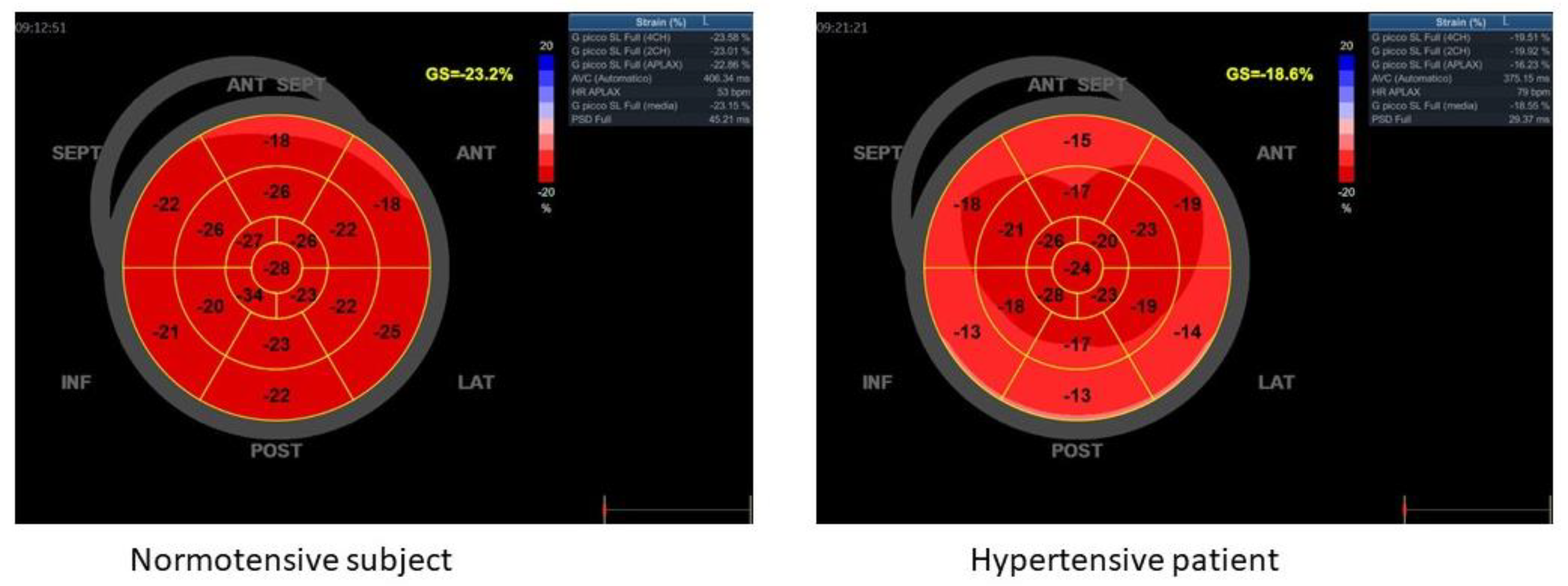
4. Systolic Dysfunction
5. Abnormal Renal Function and Microalbuminuria
6. Treatment Strategies for HFPEF
7. Conclusions
Author Contributions
Funding
Data Availability Statement
Conflicts of Interest
References
- Williams, B.; Mancia, G.; Spiering, W.; Agabiti Rosei, E.; Azizi, M.; Burnier, M.; Clement, D.; Coca, A.; De Simone, G.; Dominiczak, A.; et al. 2018 Practice Guidelines for the management of arterial hypertension of the European Society of Hypertension and the European Society of Cardiology: ESH/ESC Task Force for the Management of Arterial Hypertension. J. Hypertens. 2018, 36, 2284–2309. [Google Scholar] [CrossRef] [PubMed]
- Ruilope, L.M.; Schmieder, R.E. Left ventricular hypertrophy and clinical outcomes in hypertensive patients. Am. J. Hypertens. 2008, 21, 500–508. [Google Scholar] [CrossRef] [PubMed]
- Carpinella, G.; Pagano, G.; Buono, F.; Petitto, M.; Guarino, G.; Orefice, G.; Rengo, G.; Trimarco, B.; Morisco, C. Prognostic value of combined target-organ damage in patients with essential hypertension. Am. J. Hypertens. 2015, 28, 127–134. [Google Scholar] [CrossRef] [PubMed]
- Cao, G.; Chen, C.; Lin, Q.; Chen, Y.; Zhen, Z.; Zou, Y.; Liu, J.; Wu, M.; Wang, R.; Liu, M.; et al. Prevalence, clinical characteristics and echocardiography parameters of non-resistant, resistant and refractory hypertension in Chinese. Postgrad. Med. 2017, 129, 187–192. [Google Scholar] [CrossRef] [PubMed]
- Mancusi, C.; Gerdts, E.; De Simone, G.; Abdelhai, Y.M.; Lonnebakken, M.T.; Boman, K.; Wachtell, K.; Dahlof, B.; Devereux, R.B. Impact of isolated systolic hypertension on normalization of left ventricular structure during antihypertensive treatment (the LIFE study). Blood Press. 2014, 23, 206–212. [Google Scholar] [CrossRef] [PubMed]
- Celentano, A.; Palmieri, V.; Di Palma Esposito, N.; Pietropaolo, I.; Arezzi, E.; Mureddu, G.F.; de Simone, G. Relations of pulse pressure and other components of blood pressure to preclinical echocardiographic abnormalities. J. Hypertens. 2002, 20, 531–537. [Google Scholar] [CrossRef]
- Muiesan, M.L.; Pasini, G.; Salvetti, M.; Calebich, S.; Zulli, R.; Castellano, M.; Rizzoni, D.; Bettoni, G.; Cinelli, A.; Porteri, E.; et al. Cardiac and vascular structural changes. Prevalence and relation to ambulatory blood pressure in a middle-aged general population in northern Italy: The Vobarno Study. Hypertension 1996, 27, 1046–1052. [Google Scholar] [CrossRef]
- Mancusi, C.; Trimarco, V.; Losi, M.A.; Canciello, G.; Morisco, C.; Manzi, M.V.; Arnone, M.I.; Fucile, I.; de Simone, G.; Izzo, R.; et al. Impact of visit-to-visit blood pressure variability on hypertensive-mediated target organ damage and future cardiovascular events: The Campania salute network. J. Hypertens. 2021, 39, 1852–1858. [Google Scholar] [CrossRef]
- Manzi, M.V.; Mancusi, C.; Trimarco, V.; Izzo, R.; Franco, D.; Barbato, E.; Morisco, C.; Trimarco, B. The intergated approach to the management of arterial hypertension: The Campania Salute Network. Panminerva Med. 2021, 63, 451–457. [Google Scholar] [CrossRef]
- Kuznetsova, T.; Haddad, F.; Tikhonoff, V.; Kloch-Badelek, M.; Ryabikov, A.; Knez, J.; Malyutina, S.; Stolarz-Skrzypek, K.; Thijs, L.; Schnittger, I.; et al. Impact and pitfalls of scaling of left ventricular and atrial structure in population-based studies. J. Hypertens. 2016, 34, 1186–1194. [Google Scholar] [CrossRef]
- de Simone, G.; Mancusi, C.; Esposito, R.; De Luca, N.; Galderisi, M. Echocardiography in Arterial Hypertension. High Blood Press. Cardiovasc. Prev. 2018, 25, 159–166. [Google Scholar] [CrossRef]
- de Simone, G.; Pasanisi, F.; Ferrara, A.L.; Roman, M.J.; Lee, E.T.; Contaldo, F.; Howard, B.V.; Devereux, R.B. Relative fat-free mass deficiency and left ventricular adaptation to obesity: The Strong Heart Study. Int. J. Cardiol. 2013, 168, 729–733. [Google Scholar] [CrossRef]
- de Simone, G.; Daniels, S.R.; Devereux, R.B.; Meyer, R.A.; Roman, M.J.; de Divitiis, O.; Alderman, M.H. Left ventricular mass and body size in normotensive children and adults: Assessment of allometric relations and impact of overweight. J. Am. Coll. Cardiol. 1992, 20, 1251–1260. [Google Scholar] [CrossRef]
- de Simone, G.; Devereux, R.B.; Maggioni, A.P.; Gorini, M.; de Divitiis, O.; Verdecchia, P.; Group, M.S. Different normalizations for body size and population attributable risk of left ventricular hypertrophy: The MAVI study. Am. J. Hypertens. 2005, 18, 1288–1293. [Google Scholar] [CrossRef]
- Lonnebakken, M.T.; Izzo, R.; Mancusi, C.; Gerdts, E.; Losi, M.A.; Canciello, G.; Giugliano, G.; De Luca, N.; Trimarco, B.; de Simone, G. Left Ventricular Hypertrophy Regression During Antihypertensive Treatment in an Outpatient Clinic (the Campania Salute Network). J. Am. Heart Assoc. 2017, 6, e004152. [Google Scholar] [CrossRef]
- Izzo, R.; Losi, M.A.; Stabile, E.; Lonnebakken, M.T.; Canciello, G.; Esposito, G.; Barbato, E.; De Luca, N.; Trimarco, B.; de Simone, G. Development of Left Ventricular Hypertrophy in Treated Hypertensive Outpatients: The Campania Salute Network. Hypertension 2017, 69, 136–142. [Google Scholar] [CrossRef] [PubMed]
- Buono, F.; Crispo, S.; Pagano, G.; Rengo, G.; Petitto, M.; Grieco, F.; Trimarco, B.; Morisco, C. Determinants of left ventricular hypertrophy in patients with recent diagnosis of essential hypertension. J. Hypertens. 2014, 32, 166–173. [Google Scholar] [CrossRef] [PubMed]
- Mancusi, C.; Gerdts, E.; Losi, M.A.; D’Amato, A.; Manzi, M.V.; Canciello, G.; Trimarco, V.; De Luca, N.; de Simone, G.; Izzo, R. Differential effect of obesity on prevalence of cardiac and carotid target organ damage in hypertension (the Campania Salute Network). Int. J. Cardiol. 2017, 244, 260–264. [Google Scholar] [CrossRef]
- Lonnebakken, M.T.; Mancusi, C.; Losi, M.A.; Gerdts, E.; Izzo, R.; Manzi, M.V.; De Luca, N.; de Simone, G.; Trimarco, B. Weight loss facilitates reduction of left ventricular mass in obese hypertensive patients: The Campania Salute Network. Nutr. Metab. Cardiovasc. Dis. NMCD 2019, 29, 185–190. [Google Scholar] [CrossRef] [PubMed]
- Mancusi, C.; Angeli, F.; Verdecchia, P.; Poltronieri, C.; de Simone, G.; Reboldi, G. Echocardiography in Low-Risk Hypertensive Patients. J. Am. Heart Assoc. 2019, 8, e013497. [Google Scholar] [CrossRef] [PubMed]
- Heinzel, F.R.; Hohendanner, F.; Jin, G.; Sedej, S.; Edelmann, F. Myocardial hypertrophy and its role in heart failure with preserved ejection fraction. J. Appl. Physiol. 2015, 119, 1233–1242. [Google Scholar] [CrossRef] [PubMed]
- Paulus, W.J. H2FPEF Score: At Last, a Properly Validated Diagnostic Algorithm for Heart Failure with Preserved Ejection Fraction. Circulation 2018, 138, 871–873. [Google Scholar] [CrossRef] [PubMed]
- Pieske, B.; Tschope, C.; de Boer, R.A.; Fraser, A.G.; Anker, S.D.; Donal, E.; Edelmann, F.; Fu, M.; Guazzi, M.; Lam, C.S.P.; et al. How to diagnose heart failure with preserved ejection fraction: The HFA-PEFF diagnostic algorithm: A consensus recommendation from the Heart Failure Association (HFA) of the European Society of Cardiology (ESC). Eur. J. Heart Fail. 2020, 22, 391–412. [Google Scholar] [CrossRef] [PubMed]
- Petrie, M.C.; Caruana, L.; Berry, C.; McMurray, J.J. “Diastolic heart failure” or heart failure caused by subtle left ventricular systolic dysfunction? Heart 2002, 87, 29–31. [Google Scholar] [CrossRef]
- Mohammed, S.F.; Hussain, S.; Mirzoyev, S.A.; Edwards, W.D.; Maleszewski, J.J.; Redfield, M.M. Coronary microvascular rarefaction and myocardial fibrosis in heart failure with preserved ejection fraction. Circulation 2015, 131, 550–559. [Google Scholar] [CrossRef]
- Paulus, W.J.; Tschope, C. A novel paradigm for heart failure with preserved ejection fraction: Comorbidities drive myocardial dysfunction and remodeling through coronary microvascular endothelial inflammation. J. Am. Coll. Cardiol. 2013, 62, 263–271. [Google Scholar] [CrossRef]
- Shah, N.; Badheka, A.O.; Grover, P.M.; Patel, N.J.; Chothani, A.; Mehta, K.; Hoosien, M.; Singh, V.; Savani, G.T.; Deshmukh, A.; et al. Influence of left ventricular remodeling on atrial fibrillation recurrence and cardiovascular hospitalizations in patients undergoing rhythm-control therapy. Int. J. Cardiol. 2014, 174, 288–292. [Google Scholar] [CrossRef]
- Oliver, W.; Matthews, G.; Ayers, C.R.; Garg, S.; Gupta, S.; Neeland, I.J.; Drazner, M.H.; Berry, J.D.; Matulevicius, S.; de Lemos, J.A. Factors Associated with Left Atrial Remodeling in the General Population. Circ. Cardiovasc. Imaging 2017, 10, e005047. [Google Scholar] [CrossRef]
- Losi, M.A.; Izzo, R.; Canciello, G.; Giamundo, A.; Manzi, M.V.; Strisciuglio, T.; Stabile, E.; De Luca, N.; de Simone, G.; Trimarco, B. Atrial Dilatation Development in Hypertensive Treated Patients: The Campania-Salute Network. Am. J. Hypertens. 2016, 29, 1077–1084. [Google Scholar] [CrossRef]
- Thomas, L.; Abhayaratna, W.P. Left Atrial Reverse Remodeling: Mechanisms, Evaluation, and Clinical Significance. JACC Cardiovasc. Imaging 2017, 10, 65–77. [Google Scholar] [CrossRef]
- Galderisi, M. Diagnosis and management of left ventricular diastolic dysfunction in the hypertensive patient. Am. J. Hypertens. 2011, 24, 507–517. [Google Scholar] [CrossRef] [PubMed]
- Piskorz, D.; Keller, L.; Citta, L.; Mata, L.; Citta, N.; Bongarzoni, L.; Citta, P. Ventricular-Arterial Uncoupling and Hypertension Mediated Diastolic Dysfunction. High Blood Press. Cardiovasc. Prev. 2022, 29, 361–366. [Google Scholar] [CrossRef] [PubMed]
- Schelbert, E.B.; Fridman, Y.; Wong, T.C.; Abu Daya, H.; Piehler, K.M.; Kadakkal, A.; Miller, C.A.; Ugander, M.; Maanja, M.; Kellman, P.; et al. Temporal Relation Between Myocardial Fibrosis and Heart Failure with Preserved Ejection Fraction: Association with Baseline Disease Severity and Subsequent Outcome. JAMA Cardiol. 2017, 2, 995–1006. [Google Scholar] [CrossRef] [PubMed]
- Thakker, R.A.; Rodriguez Lozano, J.; Rodriguez Lozano, P.; Motiwala, A.; Rangasetty, U.; Khalife, W.; Chatila, K. Coronary Microvascular Disease. Cardiol. Ther. 2022, 11, 23–31. [Google Scholar] [CrossRef]
- Chen, C.; Wei, J.; AlBadri, A.; Zarrini, P.; Bairey Merz, C.N. Coronary Microvascular Dysfunction- Epidemiology, Pathogenesis, Prognosis, Diagnosis, Risk Factors and Therapy. Circ. J. 2016, 81, 3–11. [Google Scholar] [CrossRef]
- Mangiacapra, F.; Bressi, E.; Di Gioia, G.; Pellicano, M.; Di Serafino, L.; Peace, A.J.; Bartunek, J.; Morisco, C.; Wijns, W.; De Bruyne, B.; et al. Coronary microcirculation and peri-procedural myocardial injury during elective percutaneous coronary intervention. Int. J. Cardiol. 2020, 306, 42–46. [Google Scholar] [CrossRef]
- Niccoli, G.; Scalone, G.; Lerman, A.; Crea, F. Coronary microvascular obstruction in acute myocardial infarction. Eur. Heart J. 2016, 37, 1024–1033. [Google Scholar] [CrossRef]
- Camici, P.G.; Tschope, C.; Di Carli, M.F.; Rimoldi, O.; Van Linthout, S. Coronary microvascular dysfunction in hypertrophy and heart failure. Cardiovasc. Res. 2020, 116, 806–816. [Google Scholar] [CrossRef]
- Bellis, A.; Di Gioia, G.; Mauro, C.; Mancusi, C.; Barbato, E.; Izzo, R.; Trimarco, B.; Morisco, C. Reducing Cardiac Injury during ST-Elevation Myocardial Infarction: A Reasoned Approach to a Multitarget Therapeutic Strategy. J. Clin. Med. 2021, 10, 2968. [Google Scholar] [CrossRef]
- Di Serafino, L.; Magliulo, F.; Barbato, E.; Cirillo, P.; Esposito, M.; Serino, F.; Ziviello, F.; Stabile, E.; Franzone, A.; Piccolo, R.; et al. ADDED Index or Percentage Diameter of Residual Coronary Stenosis to Risk-Stratify Patients Presenting with STEMI. Cardiovasc. Revasc. Med. 2022, 34, 92–98. [Google Scholar] [CrossRef]
- Vancheri, F.; Longo, G.; Vancheri, S.; Henein, M. Coronary Microvascular Dysfunction. J. Clin. Med. 2020, 9, 2880. [Google Scholar] [CrossRef]
- Colaiori, I.; Izzo, R.; Barbato, E.; Franco, D.; Di Gioia, G.; Rapacciuolo, A.; Bartunek, J.; Mancusi, C.; Losi, M.A.; Strisciuglio, T.; et al. Severity of Coronary Atherosclerosis and Risk of Diabetes Mellitus. J. Clin. Med. 2019, 8, 1069. [Google Scholar] [CrossRef] [PubMed]
- Wong, T.Y.; Mitchell, P. The eye in hypertension. Lancet 2007, 369, 425–435. [Google Scholar] [CrossRef]
- Konst, R.E.; Guzik, T.J.; Kaski, J.C.; Maas, A.; Elias-Smale, S.E. The pathogenic role of coronary microvascular dysfunction in the setting of other cardiac or systemic conditions. Cardiovasc. Res. 2020, 116, 817–828. [Google Scholar] [CrossRef] [PubMed]
- Ungvari, Z.; Toth, P.; Tarantini, S.; Prodan, C.I.; Sorond, F.; Merkely, B.; Csiszar, A. Hypertension-induced cognitive impairment: From pathophysiology to public health. Nat. Rev. Nephrol. 2021, 17, 639–654. [Google Scholar] [CrossRef] [PubMed]
- Antony, I.; Nitenberg, A.; Foult, J.M.; Aptecar, E. Coronary vasodilator reserve in untreated and treated hypertensive patients with and without left ventricular hypertrophy. J. Am. Coll. Cardiol. 1993, 22, 514–520. [Google Scholar] [CrossRef]
- Erdogan, D.; Yildirim, I.; Ciftci, O.; Ozer, I.; Caliskan, M.; Gullu, H.; Muderrisoglu, H. Effects of normal blood pressure, prehypertension, and hypertension on coronary microvascular function. Circulation 2007, 115, 593–599. [Google Scholar] [CrossRef]
- Shah, S.J.; Lam, C.S.P.; Svedlund, S.; Saraste, A.; Hage, C.; Tan, R.S.; Beussink-Nelson, L.; Ljung Faxen, U.; Fermer, M.L.; Broberg, M.A.; et al. Prevalence and correlates of coronary microvascular dysfunction in heart failure with preserved ejection fraction: PROMIS-HFpEF. Eur. Heart J. 2018, 39, 3439–3450. [Google Scholar] [CrossRef]
- Lanza, G.A.; Morrone, D.; Pizzi, C.; Tritto, I.; Bergamaschi, L.; De Vita, A.; Villano, A.; Crea, F. Diagnostic approach for coronary microvascular dysfunction in patients with chest pain and no obstructive coronary artery disease. Trends Cardiovasc. Med. 2021. [Google Scholar] [CrossRef]
- Toyama, T.; Sato, C.; Koyama, K.; Kasama, S.; Murakami, J.; Yamashita, E.; Kawaguchi, R.; Adachi, H.; Hoshizaki, H.; Oshima, S. Olmesartan improves coronary flow reserve of hypertensive patients using coronary magnetic resonance imaging compared with amlodipine. Cardiology 2012, 122, 230–236. [Google Scholar] [CrossRef]
- van den Heuvel, A.F.; Blanksma, P.K.; Siebelink, H.M.; van Wijk, L.M.; Boomsma, F.; Vaalburg, W.; Crijns, H.J.; van Veldhuisen, D.J. Impairment of myocardial blood flow reserve in patients with asymptomatic left ventricular dysfunction: Effects of ACE-inhibition with perindopril. Int. J. Cardiovasc. Imaging 2001, 17, 353–359. [Google Scholar] [CrossRef]
- Wilck, N.; Marko, L.; Balogh, A.; Kraker, K.; Herse, F.; Bartolomaeus, H.; Szijarto, I.A.; Gollasch, M.; Reichhart, N.; Strauss, O.; et al. Nitric oxide-sensitive guanylyl cyclase stimulation improves experimental heart failure with preserved ejection fraction. JCI Insight 2018, 3. [Google Scholar] [CrossRef]
- Pieske, B.; Maggioni, A.P.; Lam, C.S.P.; Pieske-Kraigher, E.; Filippatos, G.; Butler, J.; Ponikowski, P.; Shah, S.J.; Solomon, S.D.; Scalise, A.V.; et al. Vericiguat in patients with worsening chronic heart failure and preserved ejection fraction: Results of the SOluble guanylate Cyclase stimulatoR in heArT failurE patientS with PRESERVED EF (SOCRATES-PRESERVED) study. Eur. Heart J. 2017, 38, 1119–1127. [Google Scholar] [CrossRef] [PubMed]
- Adingupu, D.D.; Gopel, S.O.; Gronros, J.; Behrendt, M.; Sotak, M.; Miliotis, T.; Dahlqvist, U.; Gan, L.M.; Jonsson-Rylander, A.C. SGLT2 inhibition with empagliflozin improves coronary microvascular function and cardiac contractility in prediabetic ob/ob(−/−) mice. Cardiovasc. Diabetol. 2019, 18, 16. [Google Scholar] [CrossRef] [PubMed]
- Wan, S.H.; Vogel, M.W.; Chen, H.H. Pre-clinical diastolic dysfunction. J. Am. Coll. Cardiol. 2014, 63, 407–416. [Google Scholar] [CrossRef]
- Sorrentino, R.; Esposito, R.; Santoro, C.; Vaccaro, A.; Cocozza, S.; Scalamogna, M.; Lembo, M.; Luciano, F.; Santoro, A.; Trimarco, B.; et al. Practical Impact of New Diastolic Recommendations on Noninvasive Estimation of Left Ventricular Diastolic Function and Filling Pressures. J. Am. Soc. Echocardiogr. 2020, 33, 171–181. [Google Scholar] [CrossRef] [PubMed]
- Lembo, M.; Sicari, R.; Esposito, R.; Rigo, F.; Cortigiani, L.; Lo Iudice, F.; Picano, E.; Trimarco, B.; Galderisi, M. Association Between Elevated Pulse Pressure and High Resting Coronary Blood Flow Velocity in Patients with Angiographically Normal Epicardial Coronary Arteries. J. Am. Heart Assoc. 2017, 6, e005710. [Google Scholar] [CrossRef] [PubMed]
- Tufano, A.; Lembo, M.; Di Minno, M.N.; Nardo, A.; Esposito, R.; Santoro, C.; Buonauro, A.; Cerbone, A.M.; Di Minno, G.; Galderisi, M. Left ventricular diastolic abnormalities other than valvular heart disease in antiphospholipid syndrome: An echocardiographic study. Int. J. Cardiol. 2018, 271, 366–370. [Google Scholar] [CrossRef]
- Petitto, M.; Esposito, R.; Sorrentino, R.; Lembo, M.; Luciano, F.; De Roberto, A.M.; La Mura, L.; Pezzullo, E.; Maffei, S.; Galderisi, M.; et al. Sex-specific echocardiographic reference values: The women’s point of view. J. Cardiovasc. Med. 2018, 19, 527–535. [Google Scholar] [CrossRef]
- Otterstad, J.E. Measuring left ventricular volume and ejection fraction with the biplane Simpson’s method. Heart 2002, 88, 559–560. [Google Scholar] [CrossRef]
- Cameli, M.; Lembo, M.; Sciaccaluga, C.; Bandera, F.; Ciccone, M.M.; D’Andrea, A.; D’Ascenzi, F.; Esposito, R.; Evola, V.; Liga, R.; et al. Identification of cardiac organ damage in arterial hypertension: Insights by echocardiography for a comprehensive assessment. J. Hypertens. 2020, 38, 588–598. [Google Scholar] [CrossRef] [PubMed]
- Lembo, M.; Esposito, R.; Santoro, C.; Lo Iudice, F.; Schiano-Lomoriello, V.; Fazio, V.; Grimaldi, M.G.; Trimarco, B.; de Simone, G.; Galderisi, M. Three-dimensional echocardiographic ventricular mass/end-diastolic volume ratio in native hypertensive patients: Relation between stroke volume and geometry. J. Hypertens. 2018, 36, 1697–1704. [Google Scholar] [CrossRef] [PubMed]
- Lembo, M.; Santoro, C.; Sorrentino, R.; Canonico, M.E.; Fazio, V.; Trimarco, B.; Tadic, M.; Galderisi, M.; Esposito, R. Interrelation between midwall mechanics and longitudinal strain in newly diagnosed and never-treated hypertensive patients without clinically defined hypertrophy. J. Hypertens. 2020, 38, 295–302. [Google Scholar] [CrossRef] [PubMed]
- de Simone, G.; Devereux, R.B.; Koren, M.J.; Mensah, G.A.; Casale, P.N.; Laragh, J.H. Midwall left ventricular mechanics. An independent predictor of cardiovascular risk in arterial hypertension. Circulation 1996, 93, 259–265. [Google Scholar] [CrossRef] [PubMed]
- Losi, M.A.; Izzo, R.; Mancusi, C.; Wang, W.; Roman, M.J.; Lee, E.T.; Howard, B.V.; Devereux, R.B.; de Simone, G. Depressed Myocardial Energetic Efficiency Increases Risk of Incident Heart Failure: The Strong Heart Study. J. Clin. Med. 2019, 8, 1044. [Google Scholar] [CrossRef]
- Cioffi, G.; Mancusi, C.; de Simone, G.; Ognibeni, F.; Orsolini, G.; Dalbeni, A.; Gatti, D.; Fassio, A.; Adami, G.; Rossini, M.; et al. Predictors and prognostic role of low myocardial mechano-energetic efficiency in chronic inflammatory arthritis. J. Hypertens. 2021, 39, 53–61. [Google Scholar] [CrossRef]
- de Simone, G.; Izzo, R.; Losi, M.A.; Stabile, E.; Rozza, F.; Canciello, G.; Mancusi, C.; Trimarco, V.; De Luca, N.; Trimarco, B. Depressed myocardial energetic efficiency is associated with increased cardiovascular risk in hypertensive left ventricular hypertrophy. J. Hypertens. 2016, 34, 1846–1853. [Google Scholar] [CrossRef]
- Mancusi, C.; Losi, M.A.; Izzo, R.; Canciello, G.; Manzi, M.V.; Sforza, A.; De Luca, N.; Trimarco, B.; de Simone, G. Effect of diabetes and metabolic syndrome on myocardial mechano-energetic efficiency in hypertensive patients. The Campania Salute Network. J. Hum. Hypertens. 2017, 31, 395–399. [Google Scholar] [CrossRef]
- Manzi, M.V.; Mancusi, C.; Lembo, M.; Esposito, G.; Rao, M.A.E.; de Simone, G.; Morisco, C.; Trimarco, V.; Izzo, R.; Trimarco, B. Low mechano-energetic efficiency is associated with future left ventricular systolic dysfunction in hypertensives. ESC Heart Fail. 2022, 9, 2291–2300. [Google Scholar] [CrossRef]
- Lembo, M.; Manzi, M.V.; Mancusi, C.; Morisco, C.; Rao, M.A.E.; Cuocolo, A.; Izzo, R.; Trimarco, B. Advanced imaging tools for evaluating cardiac morphological and functional impairment in hypertensive disease. J. Hypertens. 2022, 40, 4–14. [Google Scholar] [CrossRef]
- Tadic, M.; Cuspidi, C.; Majstorovic, A.; Kocijancic, V.; Celic, V. The relationship between left ventricular deformation and different geometric patterns according to the updated classification: Findings from the hypertensive population. J. Hypertens. 2015, 33, 1954–1961, discussion 1961. [Google Scholar] [CrossRef] [PubMed]
- Lembo, M.; Esposito, R.; Lo Iudice, F.; Santoro, C.; Izzo, R.; De Luca, N.; Trimarco, B.; de Simone, G.; Galderisi, M. Impact of pulse pressure on left ventricular global longitudinal strain in normotensive and newly diagnosed, untreated hypertensive patients. J. Hypertens. 2016, 34, 1201–1207. [Google Scholar] [CrossRef] [PubMed]
- Galderisi, M.; Trimarco, B. Global longitudinal strain: A novel hallmark of cardiac risk in arterial hypertension. J. Hypertens. 2016, 34, 1050–1051. [Google Scholar] [CrossRef] [PubMed]
- Saito, M.; Khan, F.; Stoklosa, T.; Iannaccone, A.; Negishi, K.; Marwick, T.H. Prognostic Implications of LV Strain Risk Score in Asymptomatic Patients with Hypertensive Heart Disease. JACC Cardiovasc. Imaging 2016, 9, 911–921. [Google Scholar] [CrossRef]
- Lembo, M.; Santoro, C.; Sorrentino, R.; Fazio, V.; Canonico, M.E.; Chiariello, L.; Galderisi, M.; Esposito, R. Prominent basal and middle strain longitudinal involvement in newly-diagnosed and never treated hypertensive patients without clear-cut hypertrophy. Int. J. Cardiol. 2020, 304, 179–184. [Google Scholar] [CrossRef]
- Rudolph, A.; Abdel-Aty, H.; Bohl, S.; Boye, P.; Zagrosek, A.; Dietz, R.; Schulz-Menger, J. Noninvasive detection of fibrosis applying contrast-enhanced cardiac magnetic resonance in different forms of left ventricular hypertrophy relation to remodeling. J. Am. Coll. Cardiol. 2009, 53, 284–291. [Google Scholar] [CrossRef]
- Kouzu, H.; Yuda, S.; Muranaka, A.; Doi, T.; Yamamoto, H.; Shimoshige, S.; Hase, M.; Hashimoto, A.; Saitoh, S.; Tsuchihashi, K.; et al. Left ventricular hypertrophy causes different changes in longitudinal, radial, and circumferential mechanics in patients with hypertension: A two-dimensional speckle tracking study. J. Am. Soc. Echocardiogr. 2011, 24, 192–199. [Google Scholar] [CrossRef]
- Aurigemma, G.P.; Zile, M.R.; Gaasch, W.H. Contractile behavior of the left ventricle in diastolic heart failure: With emphasis on regional systolic function. Circulation 2006, 113, 296–304. [Google Scholar] [CrossRef]
- Gonzalez, A.; Ravassa, S.; Lopez, B.; Moreno, M.U.; Beaumont, J.; San Jose, G.; Querejeta, R.; Bayes-Genis, A.; Diez, J. Myocardial Remodeling in Hypertension. Hypertension 2018, 72, 549–558. [Google Scholar] [CrossRef]
- Messerli, F.H.; Rimoldi, S.F.; Bangalore, S. The Transition from Hypertension to Heart Failure: Contemporary Update. JACC Heart Fail. 2017, 5, 543–551. [Google Scholar] [CrossRef]
- Rame, J.E.; Ramilo, M.; Spencer, N.; Blewett, C.; Mehta, S.K.; Dries, D.L.; Drazner, M.H. Development of a depressed left ventricular ejection fraction in patients with left ventricular hypertrophy and a normal ejection fraction. Am. J. Cardiol. 2004, 93, 234–237. [Google Scholar] [CrossRef] [PubMed]
- Drazner, M.H.; Rame, J.E.; Marino, E.K.; Gottdiener, J.S.; Kitzman, D.W.; Gardin, J.M.; Manolio, T.A.; Dries, D.L.; Siscovick, D.S. Increased left ventricular mass is a risk factor for the development of a depressed left ventricular ejection fraction within five years: The Cardiovascular Health Study. J. Am. Coll. Cardiol. 2004, 43, 2207–2215. [Google Scholar] [CrossRef] [PubMed]
- Krishnamoorthy, A.; Brown, T.; Ayers, C.R.; Gupta, S.; Rame, J.E.; Patel, P.C.; Markham, D.W.; Drazner, M.H. Progression from normal to reduced left ventricular ejection fraction in patients with concentric left ventricular hypertrophy after long-term follow-up. Am. J. Cardiol. 2011, 108, 997–1001. [Google Scholar] [CrossRef] [PubMed]
- Ovchinnikov, A.; Belyavskiy, E.; Potekhina, A.; Ageev, F. Asymptomatic Left Ventricular Hypertrophy Is a Potent Risk Factor for the Development of HFpEF but Not HFrEF: Results of a Retrospective Cohort Study. J. Clin. Med. 2022, 11, 3885. [Google Scholar] [CrossRef]
- Tadic, M.; Cuspidi, C.; Vukomanovic, V.; Ilic, S.; Obert, P.; Kocijancic, V.; Celic, V. Layer-specific deformation of the left ventricle in uncomplicated patients with type 2 diabetes and arterial hypertension. Arch. Cardiovasc. Dis. 2018, 111, 17–24. [Google Scholar] [CrossRef]
- Kim, D.; Shim, C.Y.; Hong, G.R.; Park, S.; Cho, I.; Chang, H.J.; Ha, J.W.; Chung, N. Differences in left ventricular functional adaptation to arterial stiffness and neurohormonal activation in patients with hypertension: A study with two-dimensional layer-specific speckle tracking echocardiography. Clin. Hypertens. 2017, 23, 21. [Google Scholar] [CrossRef]
- Lee, W.H.; Liu, Y.W.; Yang, L.T.; Tsai, W.C. Prognostic value of longitudinal strain of subepicardial myocardium in patients with hypertension. J. Hypertens. 2016, 34, 1195–1200. [Google Scholar] [CrossRef]
- Navarini, S.; Bellsham-Revell, H.; Chubb, H.; Gu, H.; Sinha, M.D.; Simpson, J.M. Myocardial Deformation Measured by 3-Dimensional Speckle Tracking in Children and Adolescents with Systemic Arterial Hypertension. Hypertension 2017, 70, 1142–1147. [Google Scholar] [CrossRef]
- Galderisi, M.; Esposito, R.; Schiano-Lomoriello, V.; Santoro, A.; Ippolito, R.; Schiattarella, P.; Strazzullo, P.; de Simone, G. Correlates of global area strain in native hypertensive patients: A three-dimensional speckle-tracking echocardiography study. Eur. Heart J. Cardiovasc. Imaging 2012, 13, 730–738. [Google Scholar] [CrossRef]
- Shah, A.M.; Claggett, B.; Sweitzer, N.K.; Shah, S.J.; Anand, I.S.; Liu, L.; Pitt, B.; Pfeffer, M.A.; Solomon, S.D. Prognostic Importance of Impaired Systolic Function in Heart Failure with Preserved Ejection Fraction and the Impact of Spironolactone. Circulation 2015, 132, 402–414. [Google Scholar] [CrossRef] [Green Version]
- Kraigher-Krainer, E.; Shah, A.M.; Gupta, D.K.; Santos, A.; Claggett, B.; Pieske, B.; Zile, M.R.; Voors, A.A.; Lefkowitz, M.P.; Packer, M.; et al. Impaired systolic function by strain imaging in heart failure with preserved ejection fraction. J. Am. Coll. Cardiol. 2014, 63, 447–456. [Google Scholar] [CrossRef] [PubMed]
- Schiffrin, E.L.; Lipman, M.L.; Mann, J.F. Chronic kidney disease: Effects on the cardiovascular system. Circulation 2007, 116, 85–97. [Google Scholar] [CrossRef] [PubMed]
- Whelton, P.K.; Carey, R.M.; Aronow, W.S.; Casey, D.E., Jr.; Collins, K.J.; Dennison Himmelfarb, C.; DePalma, S.M.; Gidding, S.; Jamerson, K.A.; Jones, D.W.; et al. 2017 ACC/AHA/AAPA/ABC/ACPM/AGS/APhA/ASH/ASPC/NMA/PCNA Guideline for the Prevention, Detection, Evaluation, and Management of High Blood Pressure in Adults: A Report of the American College of Cardiology/American Heart Association Task Force on Clinical Practice Guidelines. Hypertension 2018, 71, e13–e115. [Google Scholar] [CrossRef]
- Mule, G.; Castiglia, A.; Cusumano, C.; Scaduto, E.; Geraci, G.; Altieri, D.; Di Natale, E.; Cacciatore, O.; Cerasola, G.; Cottone, S. Subclinical Kidney Damage in Hypertensive Patients: A Renal Window Opened on the Cardiovascular System. Focus on Microalbuminuria. Adv. Exp. Med. Biol. 2017, 956, 279–306. [Google Scholar] [CrossRef] [PubMed]
- Bohm, M.; Thoenes, M.; Danchin, N.; Bramlage, P.; La Puerta, P.; Volpe, M. Association of cardiovascular risk factors with microalbuminuria in hypertensive individuals: The i-SEARCH global study. J. Hypertens. 2007, 25, 2317–2324. [Google Scholar] [CrossRef]
- Mancusi, C.; Izzo, R.; di Gioia, G.; Losi, M.A.; Barbato, E.; Morisco, C. Insulin Resistance the Hinge Between Hypertension and Type 2 Diabetes. High Blood Press. Cardiovasc. Prev. 2020, 27, 515–526. [Google Scholar] [CrossRef]
- Bigazzi, R.; Bianchi, S.; Baldari, D.; Campese, V.M. Microalbuminuria predicts cardiovascular events and renal insufficiency in patients with essential hypertension. J. Hypertens. 1998, 16, 1325–1333. [Google Scholar] [CrossRef]
- Jensen, J.S.; Feldt-Rasmussen, B.; Strandgaard, S.; Schroll, M.; Borch-Johnsen, K. Arterial hypertension, microalbuminuria, and risk of ischemic heart disease. Hypertension 2000, 35, 898–903. [Google Scholar] [CrossRef]
- Meccariello, A.; Buono, F.; Verrengia, E.; Orefice, G.; Grieco, F.; Romeo, F.; Trimarco, B.; Morisco, C. Microalbuminuria predicts the recurrence of cardiovascular events in patients with essential hypertension. J. Hypertens. 2016, 34, 646–653. [Google Scholar] [CrossRef]
- Sander, D.; Weimar, C.; Bramlage, P.; Brandt, T.; Rosin, L.; Siebler, M. Microalbuminuria indicates long-term vascular risk in patients after acute stroke undergoing in-patient rehabilitation. BMC Neurol. 2012, 12, 102. [Google Scholar] [CrossRef] [Green Version]
- Schlaich, M.P. Microalbuminuria—An important marker of residual risk: Evidence from a primary care setting. J. Hypertens. 2016, 34, 627–628. [Google Scholar] [CrossRef] [PubMed]
- Vernooij, J.W.; van der Graaf, Y.; Nathoe, H.M.; Bemelmans, R.H.; Visseren, F.L.; Spiering, W. Hypertensive target organ damage and the risk for vascular events and all-cause mortality in patients with vascular disease. J. Hypertens. 2013, 31, 492–499, discussion 499–500. [Google Scholar] [CrossRef] [PubMed]
- Chen, L.; Jin, C.; Chen, L.; Li, M.; Zhong, Y.; Xu, Y. Value of microalbuminuria in the diagnosis of heart failure with preserved ejection fraction. Herz 2021, 46, 215–221. [Google Scholar] [CrossRef] [PubMed]
- Alatas, O.D.; Biteker, M.; Demir, A.; Yildirim, B.; Acar, E.; Gokcek, K.; Gokcek, A. Microalbuminuria and its Prognostic Significance in Patients with Acute Heart Failure with Preserved, Mid-Range, and Reduced Ejection Fraction. Arq. Bras. Cardiol. 2022, 118, 703–709. [Google Scholar] [CrossRef]
- De Luca, M.R.; Sorriento, D.; Massa, D.; Valente, V.; De Luise, F.; Barbato, E.; Morisco, C. Effects of inhibition of the renin-angiotensin system on hypertension-induced target organ damage: Clinical and experimental evidence. Monaldi Arch. Chest Dis. 2021, 91. [Google Scholar] [CrossRef]
- Valente, V.; Izzo, R.; Manzi, M.V.; De Luca, M.R.; Barbato, E.; Morisco, C. Modulation of insulin resistance by renin angiotensin system inhibitors: Implications for cardiovascular prevention. Monaldi Arch. Chest Dis. 2021, 91. [Google Scholar] [CrossRef]
- Heidenreich, P.A.; Bozkurt, B.; Aguilar, D.; Allen, L.A.; Byun, J.J.; Colvin, M.M.; Deswal, A.; Drazner, M.H.; Dunlay, S.M.; Evers, L.R.; et al. 2022 AHA/ACC/HFSA Guideline for the Management of Heart Failure: A Report of the American College of Cardiology/American Heart Association Joint Committee on Clinical Practice Guidelines. Circulation 2022, 145, e895–e1032. [Google Scholar] [CrossRef]
- Cleland, J.G.; Tendera, M.; Adamus, J.; Freemantle, N.; Polonski, L.; Taylor, J.; Investigators, P.-C. The perindopril in elderly people with chronic heart failure (PEP-CHF) study. Eur. Heart J. 2006, 27, 2338–2345. [Google Scholar] [CrossRef]
- Yusuf, S.; Pfeffer, M.A.; Swedberg, K.; Granger, C.B.; Held, P.; McMurray, J.J.; Michelson, E.L.; Olofsson, B.; Ostergren, J.; Investigators, C.; et al. Effects of candesartan in patients with chronic heart failure and preserved left-ventricular ejection fraction: The CHARM-Preserved Trial. Lancet 2003, 362, 777–781. [Google Scholar] [CrossRef]
- Massie, B.M.; Carson, P.E.; McMurray, J.J.; Komajda, M.; McKelvie, R.; Zile, M.R.; Anderson, S.; Donovan, M.; Iverson, E.; Staiger, C.; et al. Irbesartan in patients with heart failure and preserved ejection fraction. N. Engl. J. Med. 2008, 359, 2456–2467. [Google Scholar] [CrossRef] [Green Version]
- Pitt, B.; Pfeffer, M.A.; Assmann, S.F.; Boineau, R.; Anand, I.S.; Claggett, B.; Clausell, N.; Desai, A.S.; Diaz, R.; Fleg, J.L.; et al. Spironolactone for heart failure with preserved ejection fraction. N. Engl. J. Med. 2014, 370, 1383–1392. [Google Scholar] [CrossRef] [PubMed]
- Anker, S.D.; Butler, J.; Filippatos, G.; Ferreira, J.P.; Bocchi, E.; Bohm, M.; Brunner-La Rocca, H.P.; Choi, D.J.; Chopra, V.; Chuquiure-Valenzuela, E.; et al. Empagliflozin in Heart Failure with a Preserved Ejection Fraction. N. Engl. J. Med. 2021, 385, 1451–1461. [Google Scholar] [CrossRef] [PubMed]
- Solomon, S.D.; McMurray, J.J.V.; Anand, I.S.; Ge, J.; Lam, C.S.P.; Maggioni, A.P.; Martinez, F.; Packer, M.; Pfeffer, M.A.; Pieske, B.; et al. Angiotensin-Neprilysin Inhibition in Heart Failure with Preserved Ejection Fraction. N. Engl. J. Med. 2019, 381, 1609–1620. [Google Scholar] [CrossRef] [PubMed] [Green Version]


| Problem | Inaccuracy-Related Conditions | Potential Solution |
|---|---|---|
| Geometry reliance | LBBB, significant abnormalities of wall motion, off-axis imaging | 3D imaging, geometry-independent approaches |
| Load reliance | Extreme afterload values and mitral regurgitation | Pressure volume loops, pre-ejection markings |
| High vs. low HR | Heart block and tachycardias | None |
| Endocardial shortening marker | LVH | Mid-myocardial shortening |
| Insensitivity to slight modifications | Prognostic value close to EF 50% | Methods besides EF for evaluating subclinical dysfunction |
| Expertise | Common usage of EF | Quantification |
| HFpEF | HFrEF | |
|---|---|---|
| LV Structure/Function | ||
| End-diastolic volume | ≃ | ↑ |
| End-systolic volume | ≃ | ↑ |
| Wall thickness | ↑ | ≃ |
| Mass | ↑ | ↑ |
| Mass/volume ratio | ↑ | ↓ |
| Remodeling | Concentric | Eccentric |
| Ejection fraction | ≃ | ↓ |
| Stroke work | ≃ | ↓ |
| End-systolic elastance | ≃ | ↓ |
| End-diastolic stiffness | ↑ | ↓ |
| LV Ultrastructure | ||
| Myocyte diameter | ↑ | ≃ |
| Myocyte length | ≃ | ↑ |
| Myocyte remodeling | Concentric | Eccentric |
| Fibrosis | Interstitial/reactive | Focal/replacement |
Publisher’s Note: MDPI stays neutral with regard to jurisdictional claims in published maps and institutional affiliations. |
© 2022 by the authors. Licensee MDPI, Basel, Switzerland. This article is an open access article distributed under the terms and conditions of the Creative Commons Attribution (CC BY) license (https://creativecommons.org/licenses/by/4.0/).
Share and Cite
Mancusi, C.; Lembo, M.; Manzi, M.V.; Basile, C.; Fucile, I.; Morisco, C. From Structural to Functional Hypertension Mediated Target Organ Damage—A Long Way to Heart Failure with Preserved Ejection Fraction. J. Clin. Med. 2022, 11, 5377. https://doi.org/10.3390/jcm11185377
Mancusi C, Lembo M, Manzi MV, Basile C, Fucile I, Morisco C. From Structural to Functional Hypertension Mediated Target Organ Damage—A Long Way to Heart Failure with Preserved Ejection Fraction. Journal of Clinical Medicine. 2022; 11(18):5377. https://doi.org/10.3390/jcm11185377
Chicago/Turabian StyleMancusi, Costantino, Maria Lembo, Maria Virginia Manzi, Christian Basile, Ilaria Fucile, and Carmine Morisco. 2022. "From Structural to Functional Hypertension Mediated Target Organ Damage—A Long Way to Heart Failure with Preserved Ejection Fraction" Journal of Clinical Medicine 11, no. 18: 5377. https://doi.org/10.3390/jcm11185377