Effects of Advanced Age, Pituitary Pars Intermedia Dysfunction and Insulin Dysregulation on Serum Antioxidant Markers in Horses

 , , , , and
Abstract
:1. Introduction
2. Materials and Methods
2.1. Animals
2.2. Blood Sampling
2.3. Hormones
2.4. Protein Concentration
2.5. Antioxidants
2.5.1. Superoxide Dismutase (Total SOD, CuZnSOD, MnSOD)
2.5.2. Lipofuscin (LPS)
2.5.3. Ceruloplasmin (CER)
2.5.4. Total Oxidant Status (TOS)
2.5.5. Total Antioxidant Capacity (TAC)
2.5.6. Lipid Peroxidation
2.5.7. Serum Thiols (Containing Sulfhydryl Group -SH)
2.6. Statistical Analysis
3. Results
4. Discussion
5. Conclusions
Author Contributions
Funding
Conflicts of Interest
References
- McFarlane, D.; Cribb, A.E. Systemic and pituitary pars intermedia antioxidant capacity associated with pars intermedia oxidative stress and dysfunction in horses. Am. J. Vet. Res. 2005, 66, 2065–2072. [Google Scholar] [CrossRef]
- McFarlane, D.; Dybdal, N.; Donaldson, M.T.; Miller, L.; Cribb, A.E. Nitration and Increased α-Synuclein Expression Associated With Dopaminergic Neurodegeneration In Equine Pituitary Pars Intermedia Dysfunction. J. Neuroendocrinol. 2005, 17, 73–80. [Google Scholar] [CrossRef]
- Horohov, D.W.; Adams, A.A.; Chambers, T.M. Immunosenescence of the Equine Immune System. J. Comp. Pathol. 2010, 142, 78–84. [Google Scholar] [CrossRef] [PubMed]
- Pamplona, R.; Portero-Otin, M.; Riba, D.; Ruiz, C.; Prat, J. Mitochondrial membrane peroxidizability index is inversely related to maximum life span in mammals. J. Lipid Res. 1998, 39, 1989–1994. [Google Scholar] [PubMed]
- Adiga, U.; Adiga, S. Total Antioxidant Activity in Old age. Biomed. Res. 2008, 19, 185–186. [Google Scholar]
- Czech, A.; Kiesz, M.; Ognik, K.; Różański, P. Influence of the manner of use, age and sex on the biochemical and antioxidant blood parameters of Malopolski horses. Med. Weter. 2016, 72, 652–655. [Google Scholar] [CrossRef] [Green Version]
- Górecka, R.; Sitarska, E.; Klucińska, W. Antioxidant Parameters of Horses According to Age, Sex, Breed and Environment. Pol. J. Vet. Sci. 2002, 5, 209–216. [Google Scholar] [PubMed]
- Firląg, M.; Kamaszewski, M.; Gaca, K.; Bałasińska, B. Age-Related Changes in the Central Nervous System in Selected Domestic Mammals and Primates. Postepy Hig. Med. Dosw. 2013, 67, 269–275. [Google Scholar] [CrossRef]
- McFarlane, D.; Donaldson, M.T.; Saleh, T.M.; Cribb, A.E. The role of dopaminergic neurodegeneration in equine pituitary pars intermedia dysfunction (equine Cushing’s disease). In Proceedings of the 49th Annual Convention of the American Association of Equine Practitioners, New Orleans, LA, USA, 21–25 November 2003; pp. 233–237. [Google Scholar]
- McFarlane, D. Equine Pituitary Pars Intermedia Dysfunction. Vet. Clin. North Am. Equine Pr. 2011, 27, 93–113. [Google Scholar] [CrossRef]
- Keen, J.A.; McLaren, M.; Chandler, K.J.; McGorum, B.C. Biochemical indices of vascular function, glucose metabolism and oxidative stress in horses with Cushing’s disease. Equine Vet. J. 2004, 36, 226–229. [Google Scholar] [CrossRef]
- Equine Endocrinology Group; Frank, N.; Andrews, F.; Durham, A.; Kritchevsky, J.; McFarlane, D.; Schott, H. Recommendations for the Diagnosis and Treatment of Pituitary Pars Intermedia Dysfunction (PPID). 2015. Available online: https://sites.tufts.edu/equineendogroup/files/2015/12/2015-10-16_EEG-2015-recommendations.pdf (accessed on 20 May 2020).
- McFarlane, D.; Donaldson, M.T.; McDonell, S.M.; Cribb, A.E. Effects of season and sample handling on measurement of plasma alpha-melanocyte-stimulating hormone concentrations in horses and ponies. Am. J. Res. 2004, 65, 1463–1468. [Google Scholar]
- Zak, A.; Siwinska, N.; Elzinga, S.; Barker, V.D.; Stefaniak, T.; Schanbacher, B.J.; Place, N.J.; Niedzwiedz, A.; Adams, A.A. Effects of advanced age and pituitary pars intermedia dysfunction on inflammation and acute-phase markers in horses. Domest. Anim. Endocrinol. 2020, 72, 106476. [Google Scholar] [CrossRef] [PubMed]
- Zak, A.; Siwinska, N.; Elzinga, S.; Barker, V.D.; Stefaniak, T.; Schanbacher, B.J.; Place, N.J.; Niedzwiedz, A.; Adams, A.A. Effects of equine metabolic syndrome (EMS) on inflammation and acute-phase markers in horses. Domest. Anim. Endocrinol. 2020, 72, 106448. [Google Scholar] [CrossRef] [PubMed]
- Lowry, O.H.; Rosebrough, N.J.; Farr, A.L.; Randall, R.J. Protein measurement with the Folin phenol reagent. J. Biol. Chem. 1951, 193, 265–275. [Google Scholar]
- Oyanagui, Y. Reevaluation of assay methods and establishment of kit for superoxide Dismutase activity. Anal. Biochem. 1984, 142, 290–296. [Google Scholar] [CrossRef]
- Tsuchida, M.; Miura, T.; Mizutani, K.; Aibara, K. Fluorescent substances in mouse and human serum as a parameter of vivo lipid peroxidation. Biochim. Biophys. Acta 1985, 834, 196–204. [Google Scholar]
- Richterich, R. Clinical Chemistry; PZWL: Warsaw, Poland, 1971. (In Polish) [Google Scholar]
- Erel, O. A novel automated direct measurement method for total antioxidant capacity using a new generation, more stable ABTS radical cation. Clin. Biochem. 2004, 37, 277–285. [Google Scholar] [CrossRef]
- Ohkawa, H.; Ohishi, N.; Yagi, K. Assay for lipid peroxides in animal tissues by thiobarbituric acid reaction. Anal. Biochem. 1979, 95, 351–358. [Google Scholar] [CrossRef]
- Koster, J.F.; Biemond, P.; Swaak, A.J. Intracellular and extracellular sulphydryl levels in rheumatoid arthritis. Ann. Rheum. Dis. 1986, 45, 44–46. [Google Scholar] [CrossRef] [Green Version]
- McGorum, B.C.; Wilson, R.; Pirie, R.S.; Mayhew, I.G.; Kaur, H.; Aruoma, O.I. Systemic concentrations of antioxidants and biomarkers of macromolecular oxidative damage in horses with grass sickness. Equine Vet. J. 2003, 35, 121–126. [Google Scholar] [CrossRef]
- Niedzwiedz, A.; Jaworski, Z. Oxidant-antioxidant Status in the Blood of Horses with Symptomatic Recurrent Airway Obstruction (RAO). J. Vet. Intern. Med. 2014, 28, 1845–1852. [Google Scholar] [CrossRef] [PubMed] [Green Version]
- Kirschvink, N.; Smith, N.; Fievez, L.; Bougnet, V.; Art, T.; Degand, G.; Marlin, D.; Roberts, C.; Geenicot, B.; Lindsey, P.; et al. Effect of chronic airway inflammation and exercise on pulmonary and systemic antioxidant status of healthy and heaves affected horses. Equine Vet. J. 2002, 34, 563–571. [Google Scholar] [CrossRef]
- McGorum, B.C.; Fry, S.C.; Wallace, G.; Coenen, K.; Robb, J.; Williamson, G.; Aruoma, O.I. Properties of herbage in relation to equine dysautonomia: Biochemical composition and antioxidant and prooxidant actions. Equine Vet. J. 2000, 48, 2346–2352. [Google Scholar] [CrossRef] [PubMed]
- McGorum, B.C.; Mayew, I.G.; Amory, H.; Deprez, P.; Gillies, L.; Green, K.; Mair, T.S.; Nollet, H.; Wjinberg, I.D.; Hahn, C.N. Horses on pasture may be affected by equine motor neuron disease. Equine Vet. J. 2006, 38, 47–51. [Google Scholar] [CrossRef] [PubMed]
- Valentine, B.A.; Hintz, H.F.; Freels, K.M.; Reynolds, A.J.; Thompson, K.N. Dietary control of experimental rhabdomyolysis in horses. JAVMA 1998, 212, 1588–1593. [Google Scholar] [PubMed]
- Dimock, A.N.; Siciliano, P.D.; McIlwraith, C.W. Evidence supporting an increased presence of reactive oxygen species in the diseased equine joint. Equine Vet. J. 2000, 32, 439–443. [Google Scholar] [CrossRef]
- Williams, C.A.; Gordon, M.E.; Betros, C.L.; McKeever, K.H. Apoptosis and antioxidant status are influenced by age and exercise training in horses. J. Anim. Sci. 2008, 86, 576–583. [Google Scholar] [CrossRef]
- Smarch, D.N.; Williams, C.A. Oxidative Stress and Antioxidant Status in Standardbreds: Effect of Age and Acute Phase Exercise Before and After Training. J. Equine Vet. Sci 2016, 47, 92–106. [Google Scholar] [CrossRef]
- De Moffarts, B.; Kirschvink, N.; Van Erck, E.; Art, T.; Pincemail, J.; Lekeux, P. Assessment of the oxidant–antioxidant blood balance in a field exercise test in Standardbredand eventing horses. Equine Comp. Exerc. Physiol. 2005, 2, 253–261. [Google Scholar] [CrossRef]
- Niedzwiedz, A.; Kubiak, K.; Nicpon, J. Plasma total antioxidant status in horses after 8-h of road transportation. Acta Vet. Scand. 2013, 55, 58. [Google Scholar] [CrossRef] [Green Version]
- Soffler, C. Oxidative stress. Vet. Clin. North. Am. Equine Pract. 2007, 23, 135–157. [Google Scholar] [CrossRef] [PubMed]
- Kirschvink, N.; De Moffarts, B.; Farnir, F.; Pincemail, J.; Lekeux, P. Investigation of blood oxidant/antioxidant markers in healthy competition horses of different breeds. Equine Vet. J. 2006, 38, 239–244. [Google Scholar] [CrossRef] [PubMed]
- Mochizuki, M.; Minowa, F.; Ishimoto, C.; Gin, A.; Ishioka, K.; Okubo, K. The effect of aging on biochemical markers in equine serum. J. Equine Vet. Sci. 2016, 42, 1–6. [Google Scholar] [CrossRef]
- Suresh, D.; Kumaran, S.; Annam, V.; Veena, H. Age related changes in malondialdehyde: Total antioxidant capacity ratio—A novel marker of oxidative stress. Int. J. Pharma Bio Sci. 2010, 1, 1–6. [Google Scholar]
- Giustarini, D.; Dalle-Donne, I.; Lorenzini, S.; Milzani, A.; Rossi, R. Age-Related Influence on Thiol, Disulfide, and Protein-Mixed Disulfide Levels in Human Plasma. J. Gerontol. 2006, 61, 1030–1038. [Google Scholar] [CrossRef] [PubMed] [Green Version]
- Dröge, W. Aging-related changes in the thiol/disulfide redox state: Implications for the use of thiol antioxidants. Exp. Gerontol. 2002, 37, 1333–1345. [Google Scholar] [CrossRef]
- Radi, R.; Bush, K.M.; Cosgrove, T.P.; Freeman, B.A. Reaction of Xanthine Oxidase-Derived Oxidants with Lipid and Protein of Human Plasma. Arch. Biochem. Biophys. 1991, 286, 117–125. [Google Scholar] [CrossRef]
- Glover, C.M.; Miller, L.M.; Dybdal, N.O.; Lopez, A.; Duckett, W.M.; McFarlane, D. Extrapituitary and pituitary pathological findings in horses with pituitary pars intermedia dysfunction: A retrospective study. J. Equine Vet. Sci. 2009, 29, 146–153. [Google Scholar] [CrossRef]
- Andersen, J.K. Oxidative stress in neurodegeneration: Cause or consequence? Nat. Med. 2004, 10, 18–25. [Google Scholar] [CrossRef]
- McFarlane, D. Advantages and limitations of the equine disease, pituitary pars intermedia dysfunction as a model of spontaneous dopaminergic neurodegenerative disease. Ageing Res. Rev. 2007, 6, 54–63. [Google Scholar] [CrossRef]
- Yuan, R.Y.; Wu, M.Y.; Hu, S.P. Antioxidant status in patients with Parkinson’s disease. Nutr. Res. 2000, 20, 647–652. [Google Scholar] [CrossRef]
- De Laat, M.A.; McGowan, C.M.; Sillence, M.N.; Pollitt, C. Equine laminitis: Induced by 48 h hyperinsulinaemia in Standardbred horses. Equine Vet. J. 2010, 42, 129–135. [Google Scholar] [CrossRef] [PubMed]
- Asplin, K.E.; Sillence, M.N.; Pollitt, C.C.; McGovan, C.M. Induction of laminitis by prolonged hyperinsulinaemia in clinically normal ponies. Vet. J. 2007, 174, 530–535. [Google Scholar] [CrossRef] [PubMed]
- Johnson, P.J.; Messer, N.T.; Ganjam, V.K. Cushing’s syndromes, insulin resistance and endocrinopathic laminitis. Equine Vet. J. 2004, 36, 194–198. [Google Scholar] [CrossRef] [PubMed]
- Treiber, K.; Carter, R.; Gay, L.; Williams, C.; Geor, R. Inflammatory and redox status of ponies with a history of pastured- associated laminitis. Vet. Immunol. Immunopathol. 2009, 129, 216–220. [Google Scholar] [CrossRef] [PubMed]
- Kenez, A.; Warnken, T.; Feige, K.; Huber, K. Lower plasma trans-4-hydroxyproline and methionine sulfoxide levels are associated with insulin dysregulation in horses. BMC Vet. Res. 2018, 14, 146. [Google Scholar] [CrossRef] [Green Version]
- Houstis, N.; Rosen, E.D.; Lander, E.S. Reactive oxygen species have a causal role in multiple forms of insulin resistance. Nature 2006, 440, 944–948. [Google Scholar] [CrossRef]
- Becuwe, P.; Ennen, M.; Klotz, R.; Barbieux, C.; Grandemange, S. Manganese superoxide dismutase in breast cancer: From molecular mechanisms of gene regulation to biological and clinical significance. Free Radic. Biol. Med. 2014, 77, 139–151. [Google Scholar] [CrossRef]
- Hoehn, K.L.; Salmon, A.B.; Hohnen-Behrens, C.; Turner, N.; Hoy, A.J.; Maghzal, G.J.; Stocker, R.; Van Remmen, H.; Kraegen, E.W.; Cooney, G.J.; et al. Insulin resistance is a cellular antioxidant defense mechanism. Proc. Natl. Acad. Sci. USA 2009, 106, 17787–17792. [Google Scholar] [CrossRef] [Green Version]


| Group | n | Age (Mean ± SD) | ACTH T0 [pg/mL] (Median, Q1–Q3) | ACTH T10 [pg/mL] (Median, Q1–Q3) | Insulin T0 [µU/mL] (Median, Q1–Q3) | Insulin T60 [µU/mL] (Median, Q1–Q3) |
|---|---|---|---|---|---|---|
| Young | 7 | 12.1 ± 2.7 | 19.5 (18.1–21.1) | 32.9 (27.3–44.1) | 16.9 (11.3–23.9) | 40.9 (21.5–41.9) |
| Geriatric | 6 | 22.0 ± 2.2 | 26.0 (18.2–34.1) | 49.3 (35.6–57.9) | 21.5 (17.2–26.0) | 28.8 (22.8–39.4) |
| PPID | 19 | 23.6 ± 4.6 | 51.4 (25.3–91.3) | 524.0 (129.0–829.0) | 22.7 (19.8–52.4) | 56.2 (21.5–129.5) |
| PPID ID+ | 10 | 21.7 ± 4.9 | 38.4 (19.9–59.9) | 416.0 (166.0–707.0) | 49.6 (28.6–86.9) | 120.2 (66.2–149.1) |
| PPID ID− | 9 | 25.7 ± 3.1 | 77.4 (46.2–188.0) | 631.0 (129.0–1195.0) | 19.8 (15.4–22.3) | 21.5 (19.6–39.2) |
| Antioxidative Markers | Young n = 7 | Geriatric n = 6 | PPID n =19 | p(ANOVA) | p Young vs. Geriatric | p Young vs. PPID | p Geriatric vs. PPID |
|---|---|---|---|---|---|---|---|
| Protein (g/L) | 71.0 ± 4.4 | 66.7 ± 2.7 | 68.9 ±3.9 | 0.146 | – | – | – |
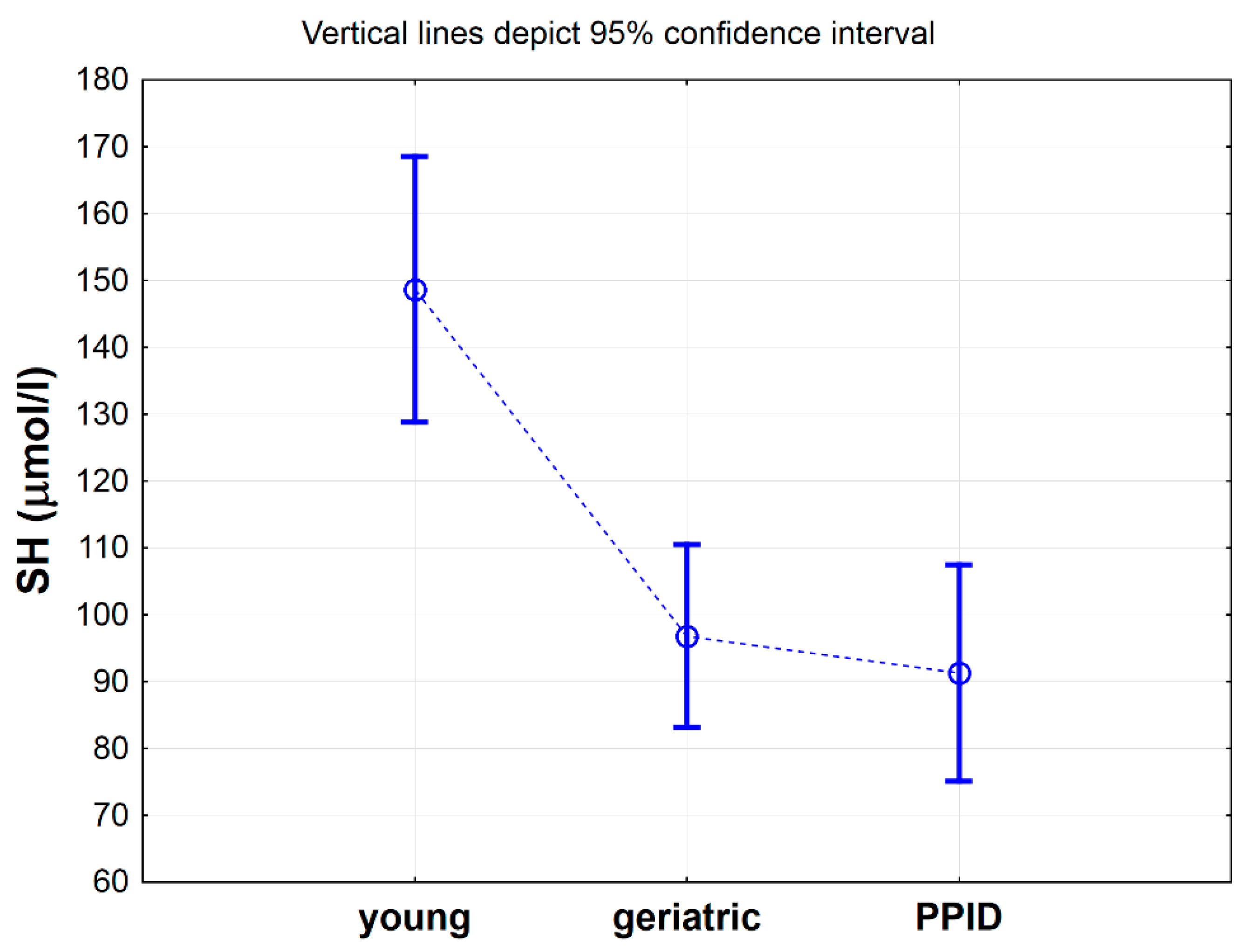
| SH (µmol/L) | 148.7 ± 21.5 | 96.8 ± 13.0 | 91.3 ± 33.5 | <0.001 | <0.01 | <0.01 | 0.940 |
| CER (mg/dL) | 17.0 ± 4.1 | 16.6 ± 3.9 | 13.9 ± 2.9 | <0.05 | 0.970 | 0.163 | 0.303 |
| TAC (mmol/L) | 0.67 ± 0.05 | 0.63 ± 0.04 | 0.63 ± 0.04 | 0.102 | – | – | – |
| TOS (µmol/L) | 3.8 ± 2.2 | 4.3 ± 1.7 | 7.2 ± 4.1 | 0.055 | – | – | – |
| Total SOD (NU/mL) | 20.1 ±1.2 | 19.8 ± 1.0 | 21.1 ± 1.1 | <0.05 | 0.896 | 0.208 | 0.117 |
| MnSOD (NU/mL) | 13.3 ± 1.1 | 12.3 ± 1.3 | 12.8 ± 0.9 | 0.217 | – | – | – |
| CuZnSOD (NU/mL) | 6.8 ± 1.7 | 7.5 ± 0.5 | 8.3 ± 1.4 | <0.05 | 0.632 | 0.111 | 0.579 |
| LPS RF | 1.6 ± 0.3 | 1.5 ± 0.3 | 1.6 ± 0.3 | 0.714 | – | – | – |
| MDA (µmol/L) | 203.2 ± 46.8 | 215.3 ± 31.1 | 175.6 ± 34.0 | <0.05 | 0.833 | 0.395 | 0.157 |
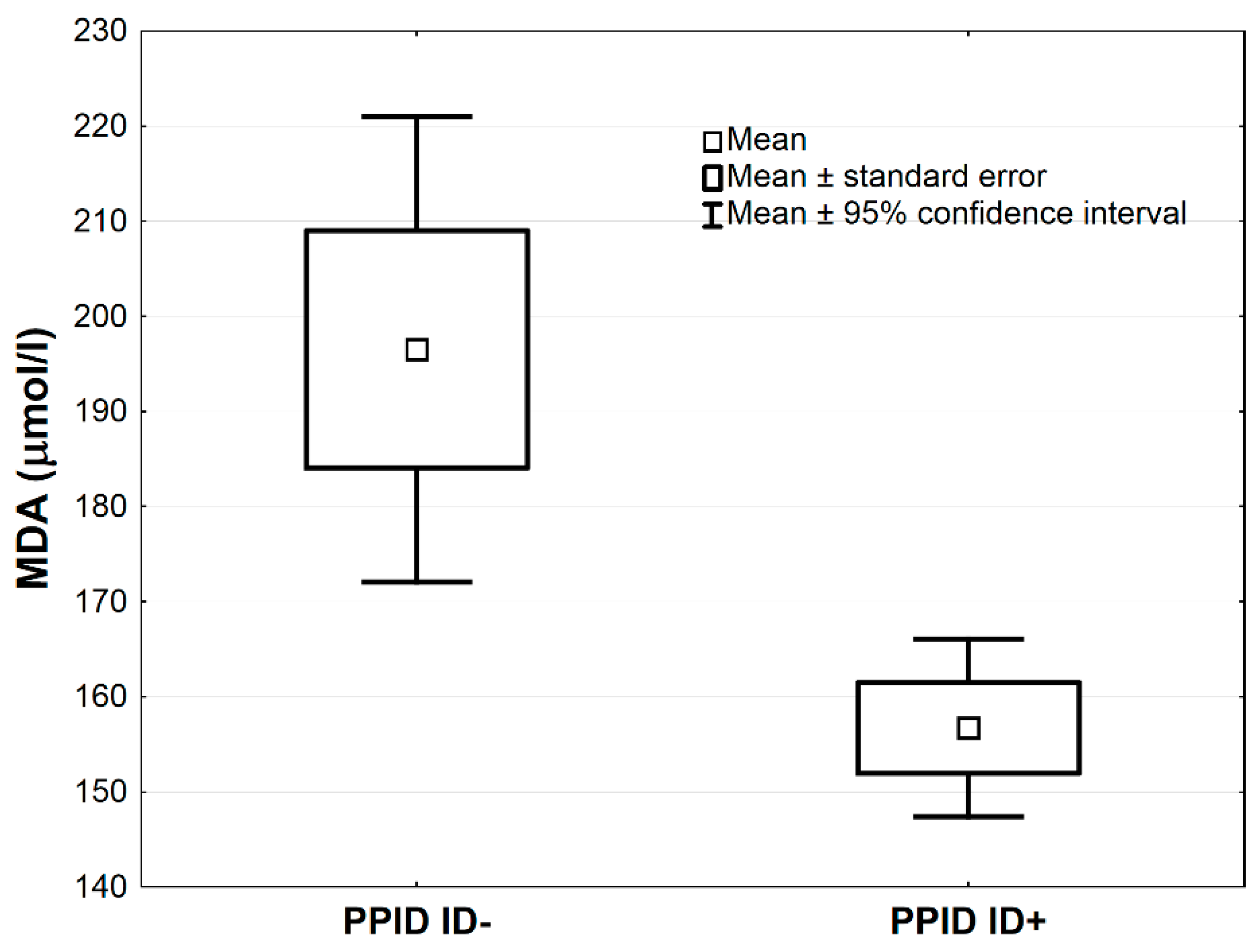
| Antioxidative Markers | PPID ID− n = 9 | PPID ID+ n=10 | t | p |
|---|---|---|---|---|
| Protein (g/L) | 69.1 ± 4.7 | 68.8 ± 4.7 | 0.197 | 0.846 |
| SH (µmol/L) | 78.9 ± 21.3 | 102.4 ± 39.4 | 1.585 | 0.131 |
| CER (mg/dL) | 14.3 ± 3.0 | 13.1 ± 2.7 | 0.944 | 0.359 |
| TAC (mmol/L) | 0.63 ± 0.03 | 0.64 ± 0.04 | 0.806 | 0.431 |
| TOS (µmol/L) | 6.0 ± 2.6 | 7.0 ± 3.4 | 0.691 | 0.499 |
| SOD (NU/mL) | 21.1 ± 1.2 | 21.1 ± 1.0 | 0.104 | 0.918 |
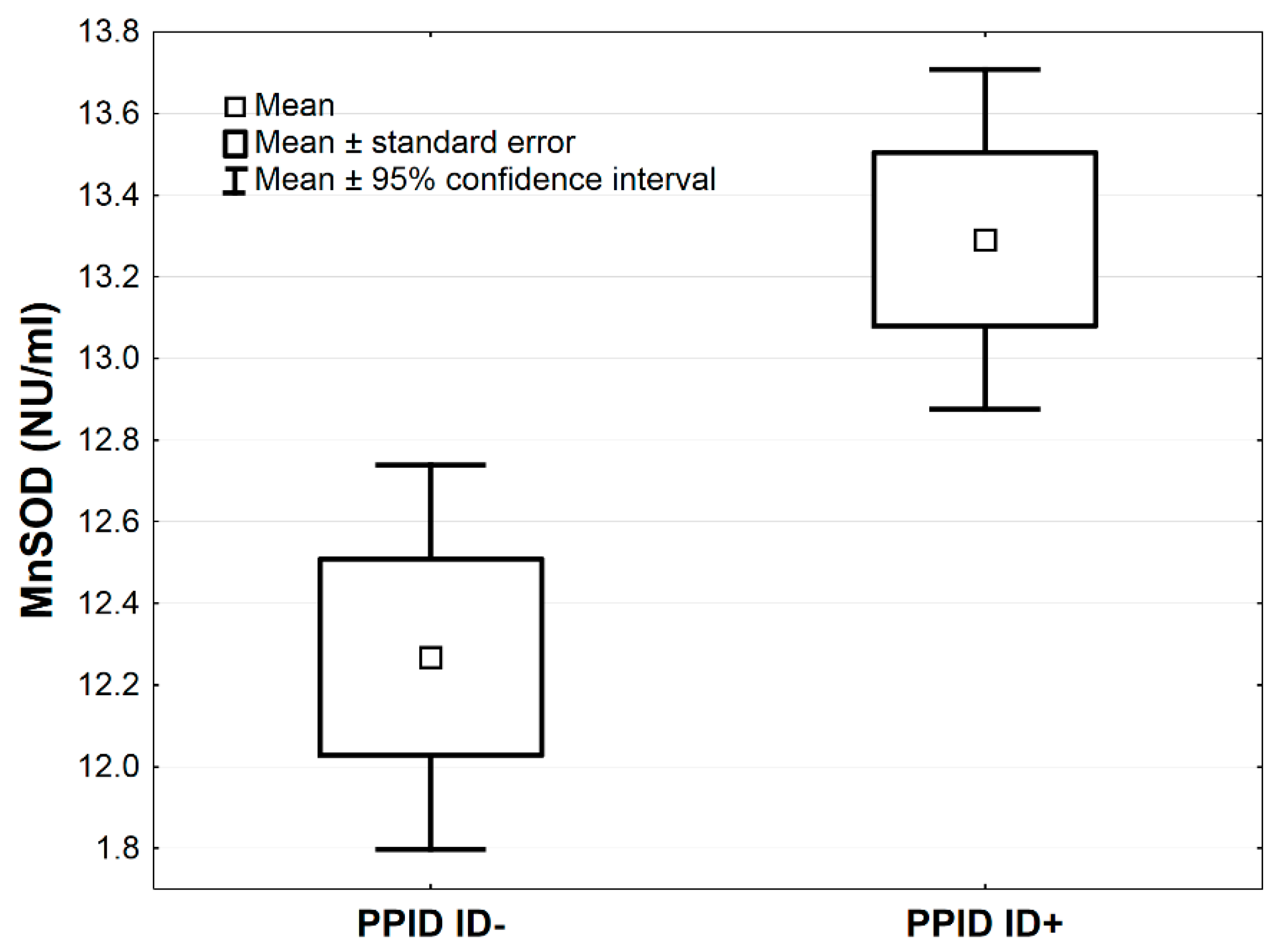
| MnSOD (NU/mL) | 12.3 ± 0.7 | 13.3 ± 0.7 | 3.205 | <0.01 |
| CuZnSOD (NU/mL) | 8.8 ± 1.1 | 7.7 ± 1.5 | 1.826 | 0.086 |
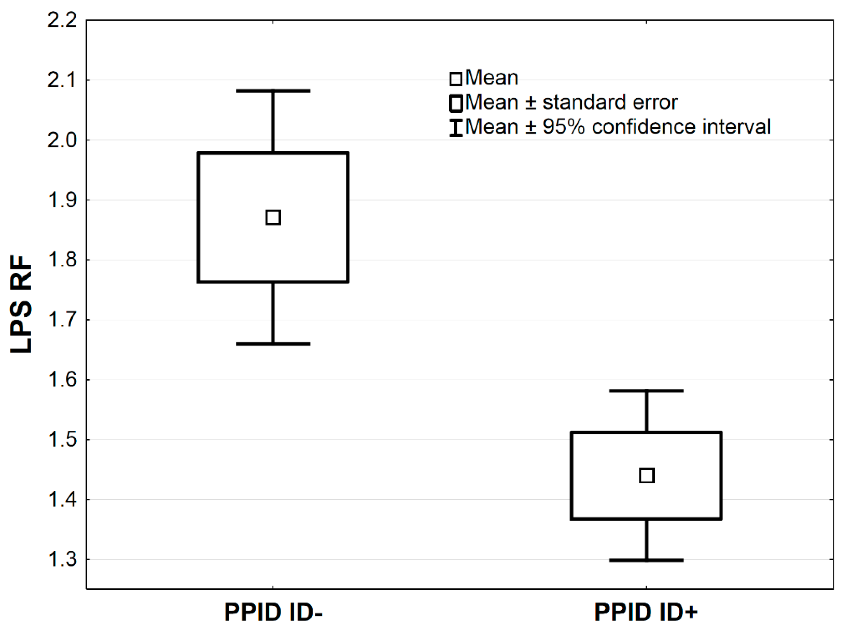
| LPS RF | 1.9 ± 0.3 | 1.4 ± 0.2 | 3.382 | <0.01 |
| MDA (µmol/L) | 196.5 ± 37.5 | 156.7 ± 15.1 | 2.981 | <0.05 |
© 2020 by the authors. Licensee MDPI, Basel, Switzerland. This article is an open access article distributed under the terms and conditions of the Creative Commons Attribution (CC BY) license (http://creativecommons.org/licenses/by/4.0/).
Share and Cite
Żak, A.; Siwińska, N.; Chełmecka, E.; Bażanów, B.; Romuk, E.; Adams, A.; Niedźwiedź, A.; Stygar, D. Effects of Advanced Age, Pituitary Pars Intermedia Dysfunction and Insulin Dysregulation on Serum Antioxidant Markers in Horses. Antioxidants 2020, 9, 444. https://doi.org/10.3390/antiox9050444
Żak A, Siwińska N, Chełmecka E, Bażanów B, Romuk E, Adams A, Niedźwiedź A, Stygar D. Effects of Advanced Age, Pituitary Pars Intermedia Dysfunction and Insulin Dysregulation on Serum Antioxidant Markers in Horses. Antioxidants. 2020; 9(5):444. https://doi.org/10.3390/antiox9050444
Chicago/Turabian StyleŻak, Agnieszka, Natalia Siwińska, Elżbieta Chełmecka, Barbara Bażanów, Ewa Romuk, Amanda Adams, Artur Niedźwiedź, and Dominika Stygar. 2020. "Effects of Advanced Age, Pituitary Pars Intermedia Dysfunction and Insulin Dysregulation on Serum Antioxidant Markers in Horses" Antioxidants 9, no. 5: 444. https://doi.org/10.3390/antiox9050444