Article

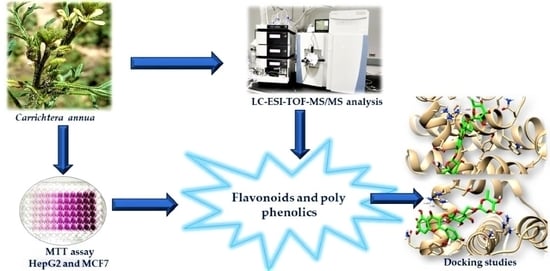
Chemical Profiling, Antioxidant, Cytotoxic Activities and Molecular Docking Simulation of Carrichtera annua DC. (Cruciferae)

1
Department of Pharmacognosy, Faculty of Pharmacy, Suez Canal University, Ismailia 41522, Egypt
2
Department of Natural Products, Faculty of Pharmacy, King Abdulaziz University, Jeddah 21589, Saudi Arabia
3
Ismailia Health Affairs Directorate, Ismailia 41525, Egypt
4
Department of Pharmacy Practice, Faculty of Pharmacy, King Abdulaziz University, Jeddah 21589, Saudi Arabia
5
Department of Chemistry, Faculty of Science, Suez Canal University, Ismailia 41522, Egypt
*
Author to whom correspondence should be addressed.
These authors have equal contribution.
Antioxidants 2020, 9(12), 1286; https://doi.org/10.3390/antiox9121286
Submission received: 24 November 2020 / Revised: 13 December 2020 / Accepted: 14 December 2020 / Published: 16 December 2020
(This article belongs to the Special Issue Antioxidant and Biological Properties of Plant Extracts)

Abstract

:
Our investigation intended to analyze the chemical composition and the antioxidant activity of Carrichtera annua and to evaluate the antiproliferative effect of C. annua crude and phenolics extracts by MTT assay on a panel of cancerous and non-cancerous breast and liver cell lines. The total flavonoid and phenolic contents of C. annua were 47.3 ± 17.9 mg RE/g and 83.8 ± 5.3 mg respectively. C. annua extract exhibited remarkable antioxidant capacity (50.92 ± 5.64 mg GAE/g) in comparison with BHT (74.86 ± 3.92 mg GAE/g). Moreover, the extract exhibited promising reduction ability (1.17 mMol Fe+2/g) in comparison to the positive control (ascorbic acid with 2.75 ± 0.91) and it displayed some definite radical scavenging effect on DPPH (IC50 values of 211.9 ± 3.7 µg/mL). Chemical profiling of C. annua extract was achieved by LC-ESI-TOF-MS/MS analysis. Forty-nine hits mainly polyphenols were detected. Flavonoid fraction of C. annua was more active than the crude extract. It demonstrated selective cytotoxicity against the MCF-7 and HepG2 cells (IC50 = 13.04 and 19.3 µg/mL respectively), induced cell cycle arrest at pre-G1 and G2/M-phases and displayed apoptotic effect. Molecular docking studies supported our findings and revealed that kaempferol-3,7-O-bis-α-L-rhamnoside and kaempferol-3-rutinoside were the most active inhibitors of Bcl-2. Therefore, C. annua herb seems to be a promising candidate to further advance anticancer research. In extrapolation, the intake of C. annua phenolics might be adventitious for alleviating breast and liver malignancies and tumoral proliferation in humans.

Graphical Abstract

1. Introduction

At the moment, cancer is the second cause of mortality and morbidity worldwide [1,2]. It accounts for 9.6 million cases of deaths in 2018 as estimated by WHO [3,4]. Based on the incident rate, the most common neoplasms in women are breast, cervical, colorectal, thyroid and lung tumors. On the other hand, liver, prostate, colorectal, stomach and lung are the most common types of cancer in men [4]. The rapid creation and growth of abnormal cells in neoplasms is correlated to the un-controlled cell hyper-proliferation which has several hallmarks, such as resistance to apoptosis, insensitivity to antigrowth signals, enabling angiogenesis, activation of tissue invasion and metastasis [5,6,7]. Metastases are the major cause of death [5]. Due to the increase of cancer cases and consequently the rise in mortality rates, Cancer therapy is an ongoing challenge with better treatment protocols needed. Until now, surgery combined with radio and chemotherapies are the most impactful treatment approach. Regrettably, resistance to various chemotherapies and a lack of drug selectivity and toxicity are also problematic. Hence, there is an increasing need for new treatment strategies and more effectual antineoplastic agents to combat malignant tumors [2,3].
Natural products are promising candidates as anticancer agents for being more available and less harmful [1,2]. Another possibility is the combination of existing chemotherapeutics with plant polyphenols [3]. Polyphenols have emerged as one of the most abundant natural products with a relevant antioxidant activity therefore, they oppose ROS formation and attenuate oxidative DNA damage and mitochondrial dysfunction by acting as chemo-preventive agents. Moreover, several polyphenols have reported to induce cell cycle arrest in different malignant cell lines [1]. Plant derived flavonoids have been validated to be efficient chemotherapeutic candidates against numerous cancers via modulation of apoptosis. The main mechanisms involved are the intrinsic and extrinsic activation of apoptotic proteins, and induction of DNA damage besides their interference with multiple signal transduction in the process of carcinogenesis and consequently obstruct proliferation, angiogenesis and metastasis [1,2].
Family Brassicaceae (Cruciferae) is composed of 350 genera including about 3500 species [8]. Species of family Brassicaceae are considered as a good source of food, vegetable oils and spices [9]. Additionally, Family Brassicaceae accumulates different groups of phytochemicals such as phenolics [10,11], flavonoids [12], alkaloids [13] and glucosinolates [14]. These phytochemicals contribute to the reported antioxidant, antimicrobial, anti-inflammatory [15], anticancer [6], and cardiovascular protective activities [10]. Carrichtera annua L. is a plant belonging to family Brassicaceae. Despite several Brassicaceae species have been extensively subjected to phytochemical studies and investigations on their medicinal value in ameliorating human and animal diseases [16], there are limited reports concerning C. annua. Indeed, as far as we know, there are few studies on the chemical constituents of C. annua and a number of flavonoid derivatives were reported [17,18,19,20,21] and only article concerning the biological activity of C. annua where the anticomplement activity of the plant was reported [21]. On the basis of the aforementioned considerations, the present work involves the estimation of total phenolic and flavonoid contents, and antioxidant activity of C. annua. In addition, the whole plant is investigated for its chemical constituents using LC-ESI-TOF-MS/MS technique. Moreover, the antiproliferative effect of C. annua extract as well as its flavonoid rich fraction was assessed. The molecular docking tool was utilized to determine the most active compounds by inspecting their interaction with active cavities of the target receptors.

2. Materials and Methods

2.1. Plant Material

Carrichtera annua (L.) DC Cruicferae (Brassicaceae) parts were collected from Sinai, Egypt. Authentication of the plant was done by Prof. Dr Elsayeda M. Gamal El-Din, Department of Botany, Faculty of Science, Suez Canal University. A voucher specimen of the plant was placed in the Herbarium of Pharmacognosy Department, Faculty of Pharmacy, Suez Canal University, Ismailia, Egypt (under registration number SAA-159). The collected plant was dried at room temperature and then pulverized.

2.2. Preparation of Plant Extract

Two hundred and fifty grams of powdered aerial part of C. annua were extracted with ethanol till exhaustion. The extracts were combined, dried under vacuum to give 10.94 g of brownish-green residue.

2.3. Determination of Total Phenolic Content

Quantification of total phenolics of the C. annua (L.) DC extract was achieved spectrophotometrically following the method described by the Saeed research team [22] with slight modification. A 200 μg/mL solution of the C. annua in methanol was prepared. The extract solution (0.5 mL) and 10-fold diluted Folin–Ciocalteu reagent (2.5 mL) were mixed together. Then 75 mg/mL sodium carbonate solution (2 mL) was added. The reaction mixture was kept for 10 min at 50 °C. Using gallic acid as a standard, the UV absorbance was recorded at λ 630 nm against blank using (Milton Roy, Spectronic 1201, Houston, TX, USA). The result was obtained as gallic acid equivalents (mg∙GAE/g dry extract)

2.4. Estimation of Total Flavonoid Content

Total flavonoids were estimated by AlCl3 method as mentioned by Saeed and coworker [22]. A solution of final concentration 1 mg/mL was prepared by dissolving the crude extract in methanol. 0.3 mL of the extract solution was diluted with 3.4 mL of methanol (30% v/v) and mixed with 0.15 mL 0.5 M NaNO2 and 0.3 M AlCl3.6H2O (0.15 mL of each). The mixture was subjected to vigorous shaking and incubated for 5 min. at 20 °C. Then 1 mL of 1M NaOH solution was added. By applying rutin as a standard, the UV absorption was recorded spectrophotometrically at λ 510 nm using (Milton Roy, Spectronic 1201, Houston, TX, USA). The result was obtained as rutin equivalent (mg∙RE/g dry extract).

2.5. Evaluation of Antioxidant Activity

2.5.1. DPPH Free Radical Scavenging Activity

The free radical-scavenging activity of C. annua crude extract was evaluated by using the method reported by Fuochi and coworkers [23]. In short, a 100 µM solution of 2,2-diphenyl-1-picrylhydrazyl (DPPH) radical freshly prepared in methanol then stored at 10 °C in dark. The extract under investigation was prepared (at the various concentrations). An aliquot of 70 uL of the extract solution was added to DPPH solution (3 mL). The reaction mixture was incubated in dark for 30 min.at room temperature. The absorbance of the mixture was recorded at λ 515 nm with a UV-visible spectrophotometer (Milton Roy, Spectronic 1201, Houston, TX, USA). The control absorbance (only DPPH radical solution) and butylated hydroxytoluene (BHT) as a standard were also estimated. The measurements were calculated as the average of three replicates. The inhibition % (PI) of the DPPH radical was determined as previously reported from the formula:
PI = [{(AC−AT)/AC} × 100]
where AC represents the control absorbance at and AT represents sample + DPPH absorbance.
The 50% inhibitory concentration (IC50) was calculated from the dose/response curve using Graphpad Prism software (San Diego, CA, USA).

2.5.2. Ferric Reducing Antioxidant Power (FRAP) Assay

The FRAP of C. annua ethanol extract was determined using the procedure described by Nsimba in 2008 [24]. The mechanism of the assay based on electron transfer process at low pH, where the colourless complex (Fe3+-TPTZ) is reduced to the blue colored complex (Fe2+-tripyridyltriazine). The reaction was monitored by measuring the change in absorbance at λ 593 nm using (Milton Roy, Spectronic 1201, Houston, TX, USA). 40 µL of the extract solution was diluted with 0.2 mL of distilled water and mixed with 1.8 mL of warm freshly prepared FRAP reagent prepared as previously described [24]. The results were illustrated as the concentration of antioxidants having ferric reducing ability equivalent to that of 1 mM FeSO4, expressed as m Mol Fe+2 equivalent/g dry sample. The utilized positive controls were ascorbic acid and Butylated hydroxytoluene (BHT; Sigma-Aldrich, St. Louis, MO, USA).

2.5.3. Total Antioxidant Capacity (TAC) Assay

The total extract of C. annua were evaluated for total antioxidant capacity (TAC) by spectrophotometrical determination using phosphomolybdenum assay. The procedure was executed as previously described by Saeed and his research team [22] with some modification. In Eppendorf tube; 0.1 mL of a 1 mg/mL extract (as methanolic solution) was added to reagent solution (1 mL). The tubes were capped, incubated in a water bath at 95 °C (for 90 min). The reaction mixture was then cooled to room temperature. Measurement of the absorbance was performed by UV-visible spectrophotometer using (Milton Roy, Spectronic 1201, Houston, TX, USA) at λ 695 nm against blank. TAC results were calculated (mg/g of dry sample) and expressed as gallic acid equivalents. Butylated hydroxytoluene (BHT; Sigma-Aldrich, St. Louis, MO, USA) was used as a reference compound.

2.6. Preparation of Phenolics Extract of C. annua

Phenolic compounds of C. annua were extracted using the method described in El-Shaer and coworkers [25]. In brief, one hundred and fifty grams of powdered aerial parts of C. annua were treated with an aqueous solution of 5% Na2CO3. After one hour, the mixture was filtered and washed with distilled water to ensure complete extraction. The filtrate was diluted with distilled water and neutralized by HCl then partitioned between ethyl acetate and n-butanol. The obtained extracts were combined together then concentrated under reduced pressure to afford 4.03 g of C. annua total phenolics extract.

2.7. Preparation of the Sample and LC-HRMS Analysis

As previously described [26], the mobile phase working solution consisted of DI-Water: Acetonitrile: Methanol in a ratio of 50:25:25. Fifty mg of weighted dry methanolic extract was dissolved in one ml of MP-WS then vortex for 2 min. After that, ultra-sonication for 10 min followed by centrifugation for 10 min at 10,000 rpm were performed. 20 µL of the stock (50/1000 µL) was diluted with reconstitution solvent (1000 µL). At last, the concentration used for injection was 1 µg/µL where 25 µLs were injected in both positive and negative modes. Additionally, 25 µL of mobile phase working solution was used for injection as a blank sample. The used mobile phases consisted of: (A) 5 mM ammonium formate buffer pH 3 containing 1% methanol and used for positive TOF MS mode; (B) 5 mM ammonium formate buffer pH8 containing 1% methanol used for negative TOF MS mode; in addition to mobile phase (C) composed of 100% acetonitrile used for positive and negative modes. The pre column used was In-Line filter disks (Phenomenex, 0.5 µm × 3.0 mm) whereas, the column was X select HSS T3 (Waters, 2.5 µm, 2.1 × 150 mm) and the flow rate was 0.3 mL/min. Data processing was performed via MS-DIAL3.52. Feature (peaks) extraction from total ion chromatogram was achieved using Master view software, according to the following criteria: features intensities of the sample-to-blank should be more than 5 and features should have Signal-to-Noise not less than 5 (Non-targeted analysis). Identification of compounds was achieved via accurate mass measurements, MS/MS data, exploration of specific spectral libraries and public repositories for MS-based metabolomic analysis (MassBank NORMAN, MassBank MoNA, PubChem), retention times as well as data comparison with the literature.

2.8. Biological Assays

2.8.1. Cell Culture Treatment

A panel of cancerous and non-cancerous breast and liver cell lines; MCF-7, MDA_MB-231, MCF-10A, HepG2 and THLE2 were maintained in RPMI-1640/DMEM (Sigma-Aldrich, St. Louis, MO, USA). Both types of media were supplemented with 2mML-glutamine (Lonza, Belgium) and 10% FBS (Sigma, St. Louis, MO, USA), 1% Penicillin/Streptomycin (Lonza, Belgium). Incubation was performed for all cells at 37 °C in 5% CO2 atmosphere (NuAire, Lane Plymouth, MN 55447, USA) according to the routine tissue culture procedure [27].

2.8.2. Cytotoxicity Using the MTT Assay

Cells were plated at a density of 5000 cells in triplicates in a 96-well plate. On the next day, treatment of the cells was performed with the indicated compound/s at the specified concentrations in 100 μL media as a final volume. Cell viability was considered after 72 h using MTT solution (Promega, Madison, WI, USA) [28]. The reagent (20 μLs) was added to each well followed by incubation of the plate for 3 h and fluorescence was subsequently measured (570 nm) using a plate reader, then IC50 values were assessed using the GraphPad prism 7 [29,30].

2.8.3. Annexin V/PI and Cell Cycle Analysis

Apoptosis rate in cells was quantified using annexin V-FITC (BD Pharmingen, San Diego, CA, USA). Cells were seeded into 6-well culture plates (3 × 10⁵ cells/well); overnight incubation was done at 37 °C, under 5% CO2. Cells were then treated with indicated compounds for 48h. Next, media supernatants and cells were collected, followed by washing with ice-cold PBS. Next, cells were suspended in 100 µL of annexin binding buffer solution (25 mM CaCl2, 1.4 M NaCl, and 0.1 M Hepes/NaOH, pH 7.4). This was followed by incubation for 30 min in the dark with annexin V-FITC solution (1:100) and PI at 10 µg/mL concentration for 30 min. Stained cells were then acquired by Cytoflex FACS machin. Data was analyzed using cytExpert software. This assay was carried out as previously published in [31,32,33].

2.8.4. RT-PCR

Treatment of MCF-7 cells with phenolics extract of C. annua (IC50 = 13.04 μg/mL) was done for 72 h. After completion of the treatment, collection of the cells and extraction of total RNA were performed using Rneasy® Mini Kit (Qiagen, Hilden, Germany) as instructed by manufacturer. cDNA synthesis was executed using 500 ng of RNA using i-Script cDNA synthesis kit (BioRad, Hercules, CA, USA) according to the instructions of the manufacturer. Real-time RT-PCR reactions composed of 25 µL Fluocycle®II SYBR® (Euroclone, Milan, Italy), 1.5 µL of both 10 µM forward and reverse primers, 3 µL cDNA, and 19 µL of H2O. Performance of the reactions was done for 35 cycles using temperature profiles as follows: for initial denaturation (95 °C for 5 m); for denaturation (95 °C for 15 min); Annealing (55 °C for 30 min), and finally extension (72 °C for 30 min) [31,32,33]. At the end, the Ct values were collected, and the relative folds of changes between all the samples. Primer used were listed in Table 1.

2.9. Simulated Molecular Docking

All identified derivatives were screened for their binding activities towards the “B-cell lymphoma 2 (Bcl-2)” protein, its crystal structure with the co-crystallized ligand was freely accessible from the protein data bank. Processes concerning preparation of the protein, optimization of the ligands and software validation are implemented following the regular work as published by Nafie et al., 2019 [34]. The “MOE-2019 software” was used for molecular docking study. Each ligand-receptor complex was tested for binding interaction analysis with the binding energy (Kcal/mol), 3D images were performed using Chimera as a visualizing software [35].

3. Results and Discussion

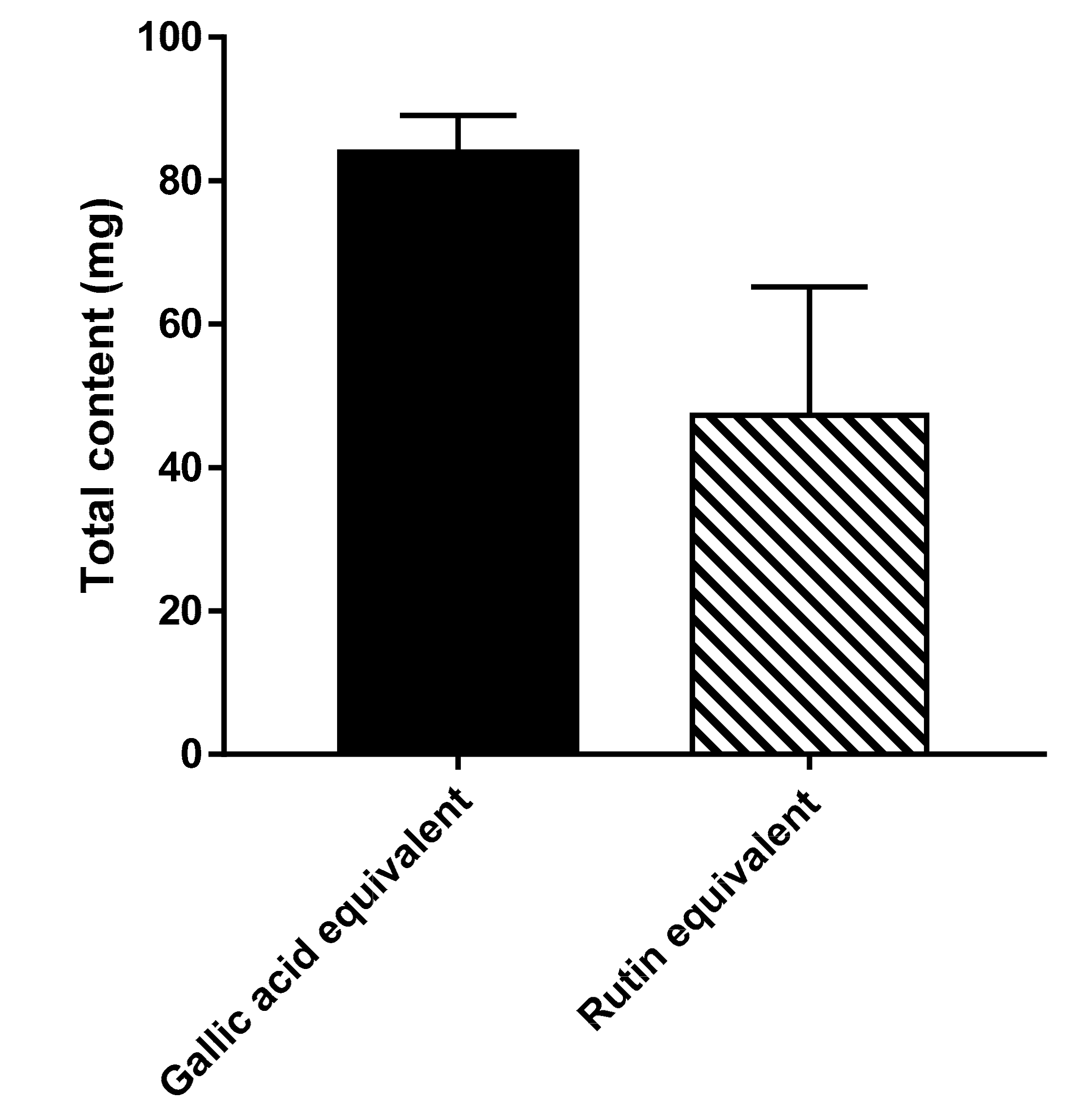
3.1. Total Phenolic and Flavonoid Content

Phenolic compounds are widespread phytoconstituents and their main sources in human diet are fruits and vegetables. The same bioactive polyphenols, such as hydroxycinnamic acid derivatives, flavonoids and proanthocyanidins are also obtainable from forest trees [22]. Therefore, determination of the polyphenols content in the extract; the total phenols and total flavonoids is reasonable, in order to estimate the potential antioxidant capacity of C. annua crude extract. Total phenolic content of C. annua extract was estimated spectrophotometrically using Folin–Ciocalteu reagent. Based on the calibration curve of gallic acid, the obtained linear equation obtained was Y= 0.0011X + 0.0131 (R2 = 0.9946). The total phenolic content of C. annua methanolic extract was 83.8 ± 5.3 mg GAE/g of plant extract. Total flavonoid content in C. annua extract was obtained spectrophotometrically using AlCl3 reagent and rutin as standard. Derived from the calibration curve of rutin, the obtained linear equation was; Y = 0.0011X + 0.0131 (R2 = 0.9946). The total flavonoids content of C. annua methanolic extract estimated from the above equation was 47.3 ± 17.9 mgRE/g of plant extract (see Figure 1).

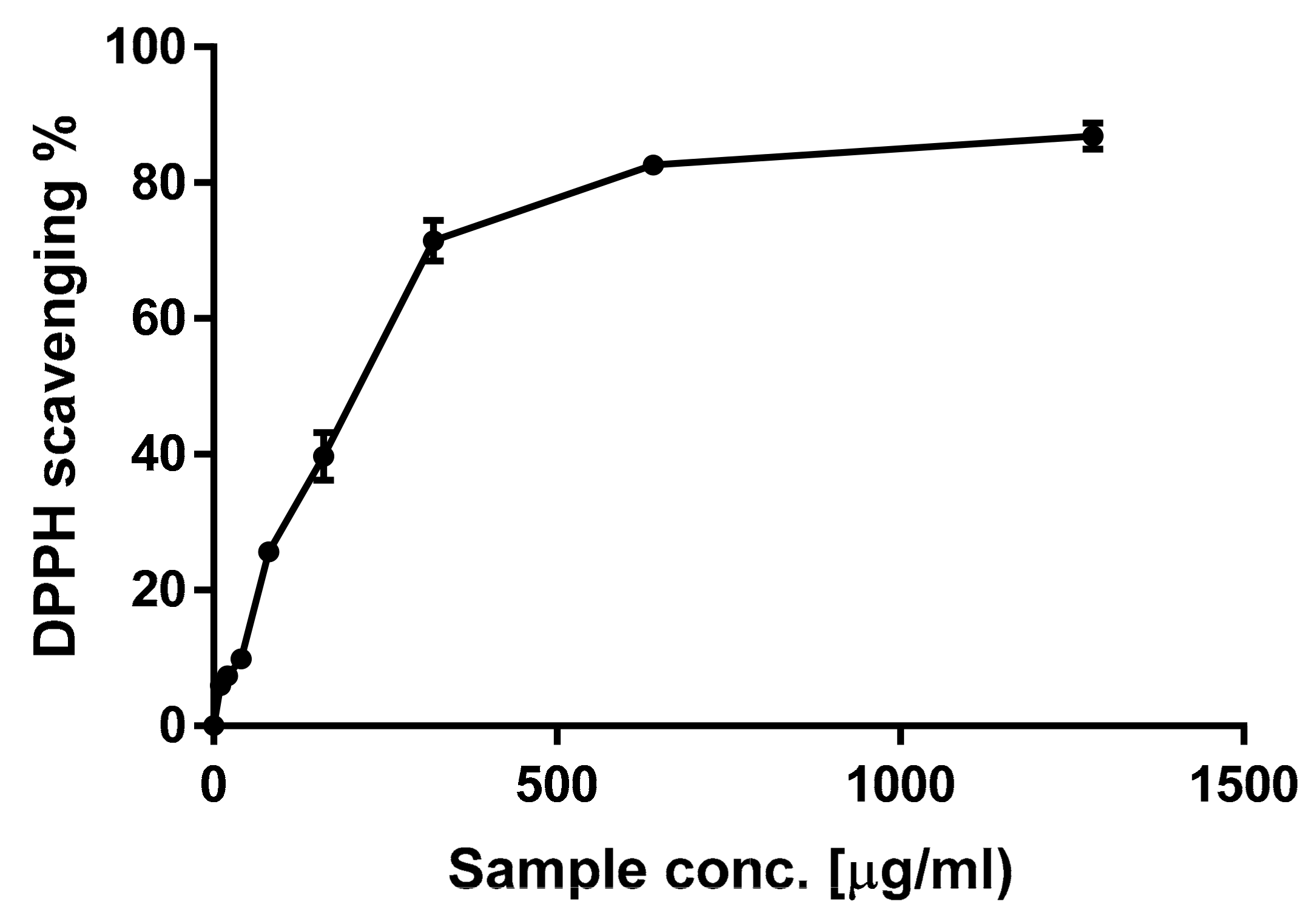
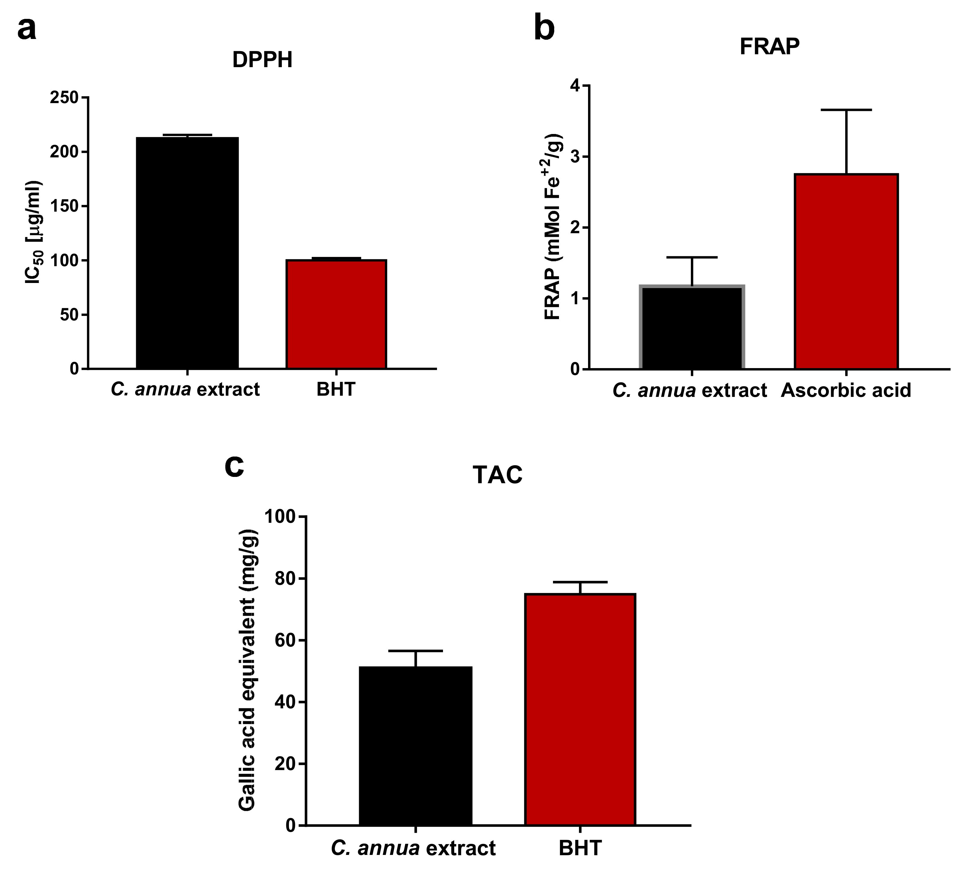
3.2. Evaluation of In Vitro Antioxidant Activity of C. annua Extract

Flavonoids and phenolic acids are characterized by being electron or hydrogen donors, reducing and metal chelating agents. These properties arise from different conjugations and varying numbers of hydroxyl groups in their structures [16]. Therefore, due to the complexity of natural phytoconstituents and different scavenging modes of ROS (reactive oxygen species), a group of assays were used simultaneously in order to judge the antioxidant activity of C. annua extract, [36]. In the present study, three indicative tests (DPPH, FRAP, TAC) were applied to analyze the antioxidant power of C. annua crude extract. Figure 2 demonstrates that the crude extract had definite scavenging activity on DPPH exhibiting a dose-dependent scavenging rate. As shown in Figure 3a, C. annua crude extract with IC50 values of 211.9 ± 3.7 µg/mL revealed notable activities in DPPH radical scavenging assay compared to the positive control (BHT IC50 = 100 ± 2.1 µg/mL.). Results of FRAP (Figure 3b) demonstrates that C. annua had promising reduction ability with 1.17 mMol Fe+2 /g in comparison to the positive control (Ascorbic acid with 2.75 ± 0.91 respectively). Figure 3c demonstrates the total antioxidant capacity of C. annua extract and BHT (standard synthetic antioxidant) assessed by phosphomolybdenum test. C. annua extract exhibited remarkable antioxidant potential (50.92 ± 5.64 mg GAE/g) in comparison with BHT (74.86 ± 3.92 mg GAE/g).

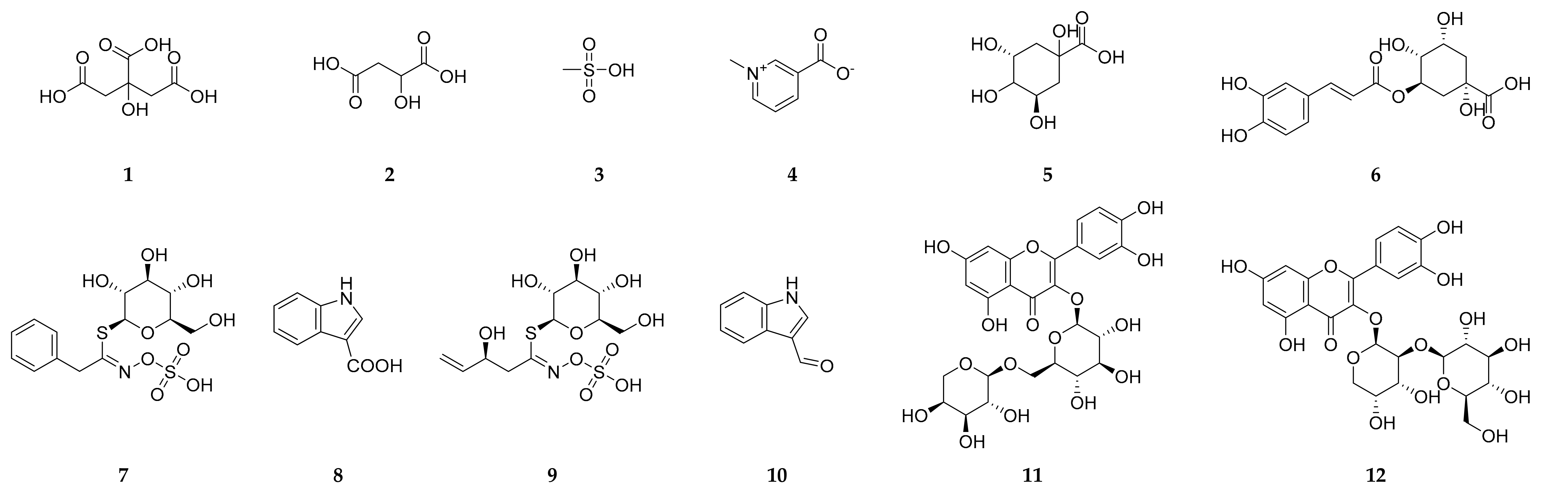
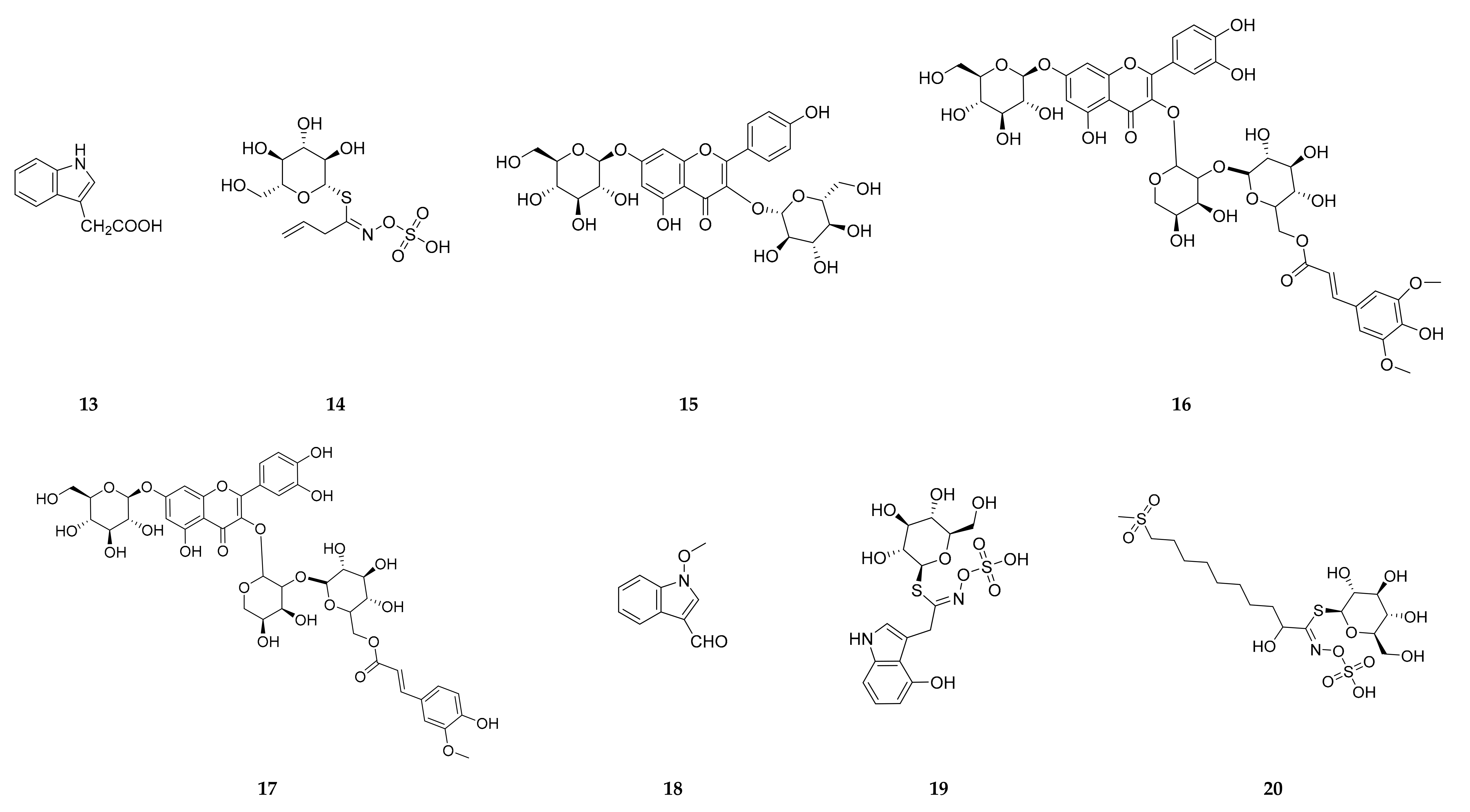
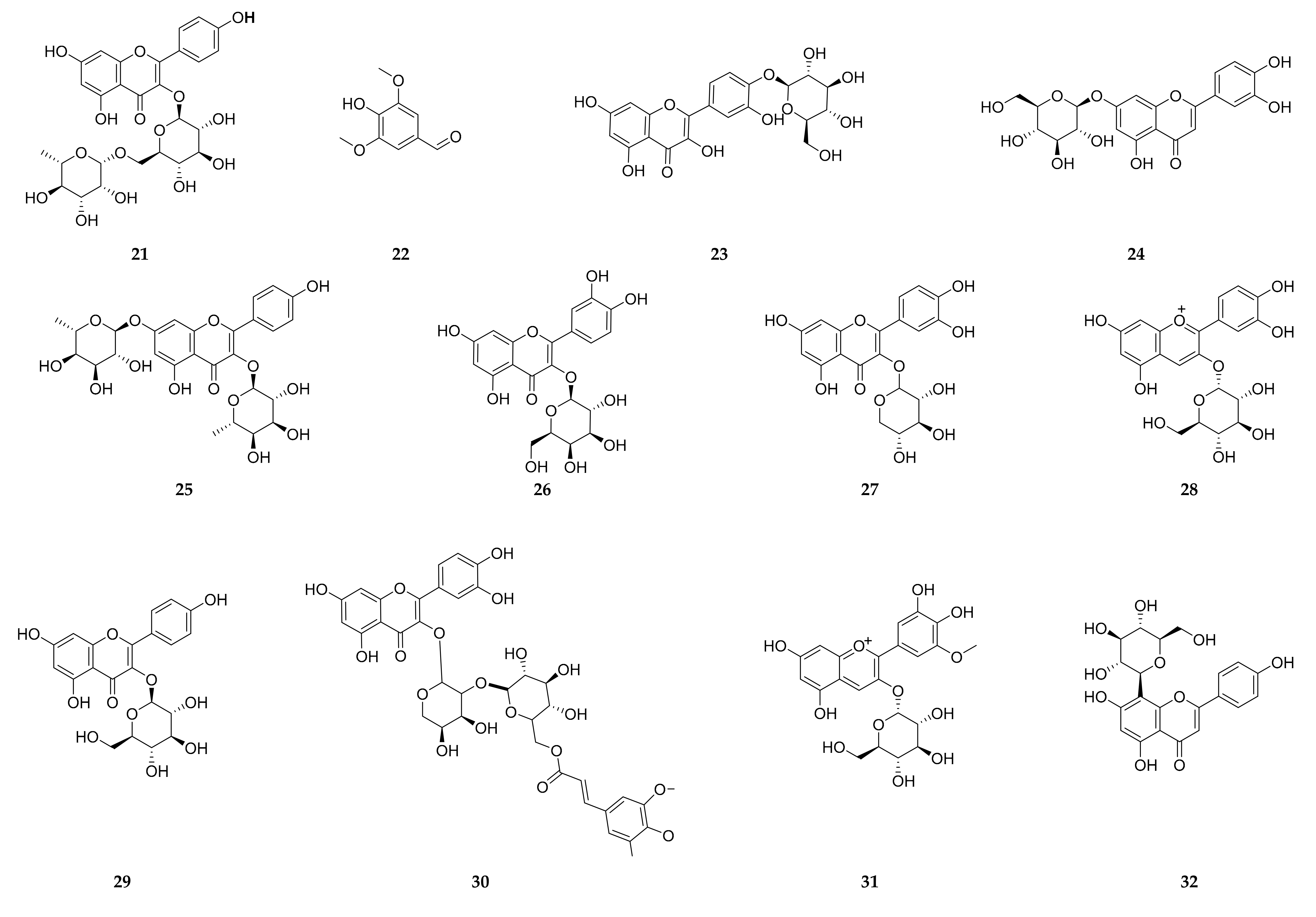
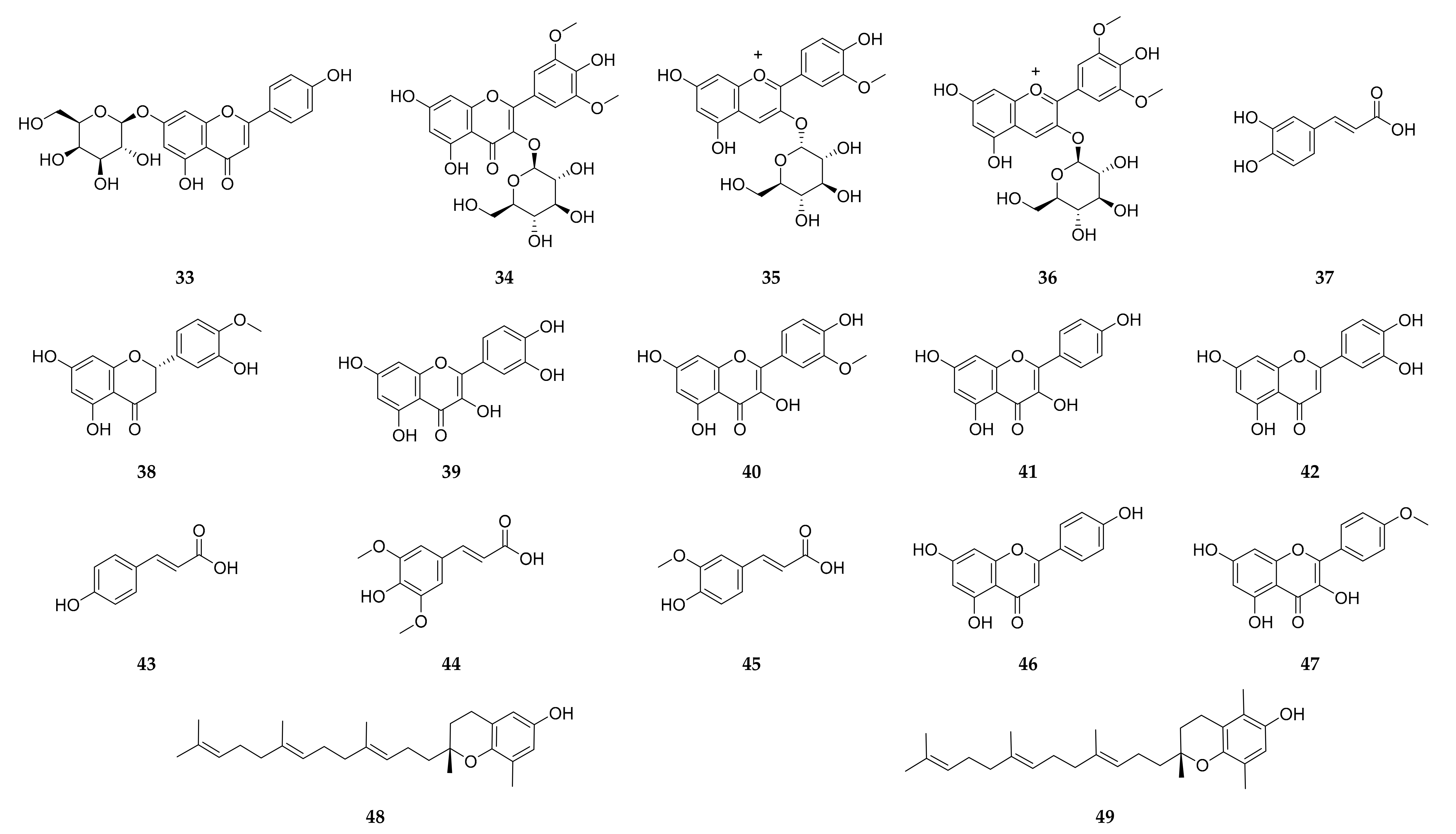
3.3. LC-ESI-TOF-MS/MS Analysis

Herein, Carrichtera annua was proven to be a rich source of phenolics and flavonoids (83.8 ± 5.3 mg GAE/g and 47.3 ± 17.9 mg RE/g respectively). It demonstrated promising antioxidant potential by exhibiting notable free radical scavenging activity (IC50 = 211.9 ± 3.7 µg/mL) and ferric reduction power (17 mMol Fe+2/g) as well as good antioxidant potential (50.92 ± 5.64 mg GAE/g). As a consequence, C. annua crude extract was investigated by LC-ESI-TOF-MS/MS (Agilant, Santa Clara, CA, USA) in order to fully understand the chemical diversity of its phytoconstituents including phenolics and other metabolites accountable for the estimated antioxidant activity of the plant. Data are represented in Table 2 and LC-ESI-TOF-MS/MS profile is shown in (Supplementary Materials Figures S1–S4). Tentative identification of the individual components was achieved by comparison of their chromatographic behavior, m/z values in the total ion chromatogram (TIC) and base peak chromatogram (BPC) profile as well as their fragmentation pattern with those described in the literature.
In particular, 49 hits were identified in C. annua (Table 2, Figure 4) belonging to different metabolic classes; mainly phenolics. Fifteen flavonol derivatives have been detected in C. annua extract among which quercetin-3-O-arabinoglucoside (peltatoside), quercetin 3-O-β-D-glucopyranosyl-(1–>2)-arabinopyranoside, quercetin 3-O-[(6 sinapoyl-β-glucopyranosyl)(1–>2)-β-arabinopyranoside]-7-O-β-glucopyranoside, quercetin 3-O-[(6-feruloyl-β-glucopyranosyl)-(1–>2)-β-arabinopyranoside]-7-O-β-glucopyranoside, quercetin 3-O-[(6-sinapoyl-β-glucopyranosyl)-(1–>2)-β arabinopyranoside, kaempferol-3 rutinoside and isorhamnetin have been reported previously in C. annua [17,19,20,21]. It is noteworthy to mention that, despite of quercetin-7-O-arabinosyl-3-O glucoside and quarecetin-3-O-glucoside [21] were isolated from C. annua, they were not detected in our extract, instead we recorded quercetin-4′-O-glucoside. In addition to flavonols, luteolin, and apigenin flavone aglycones and glycosides were recorded in the present study. Moreover, four anthocyanins (cyanidin-3-glucoside, petunidin-3-O-β-glucopyranoside, peonidine-3-O-glucoside and malvidin-3-galactoside) have been reported for the first time in C. annua. Ferulic, p coumaric, caffeic and sinapic acids were detected along with other organic acids.
Glucosinolates (GSs) are sulfur- and nitrogen-containing compounds widely distributed in Brassicaceae plants [37]. Five GSs (progoitrin, sinigrin, glucotropaeolin, 4-hydroxyglucobrassicin and 9-(methylsulfonyl) hydroxy nonyl glucosinolate) were detected in the current study. On the other hand, gluconapin, brassicanapin, erucin, glucoberteroin, glucoerucine and glucoraphanin which were reported in Spanish and Australian C. annua [38,39] were not detected.
Alkaloids are nitrogenous compounds synthesized from amino acids and have been reported in several Brassicaceae plants [13]. In the present investigation, four nitrogenous compounds particularly of indole type (1H-indole-3-carboxylic acid, 3-formyl indole, 2-(1H-indol-3-yl) acetic acid and 1-methoxy-1H-indole-3-carbaldehyde) which were previously reported in Isatis tinctoria; a plant belongs to family Brassicaceae [40] were also recorded in this study. No tropane alkaloids were detected in this study which coincide with Brock and coworker [41]. However, our findings went against them in the presence of calystegines since there were none of them were reported. Interestingly, trigonelline was recorded for the first time in a member of family Brassicaceae.
Tocotrienols and tocopherols are naturally occurring terpenes present in Brassicaceae plants and have diverse biological activities [13]. Our results revealed the presence of δ- tocotrienol and β-tocotrienol in C. annua.

3.4. Biology

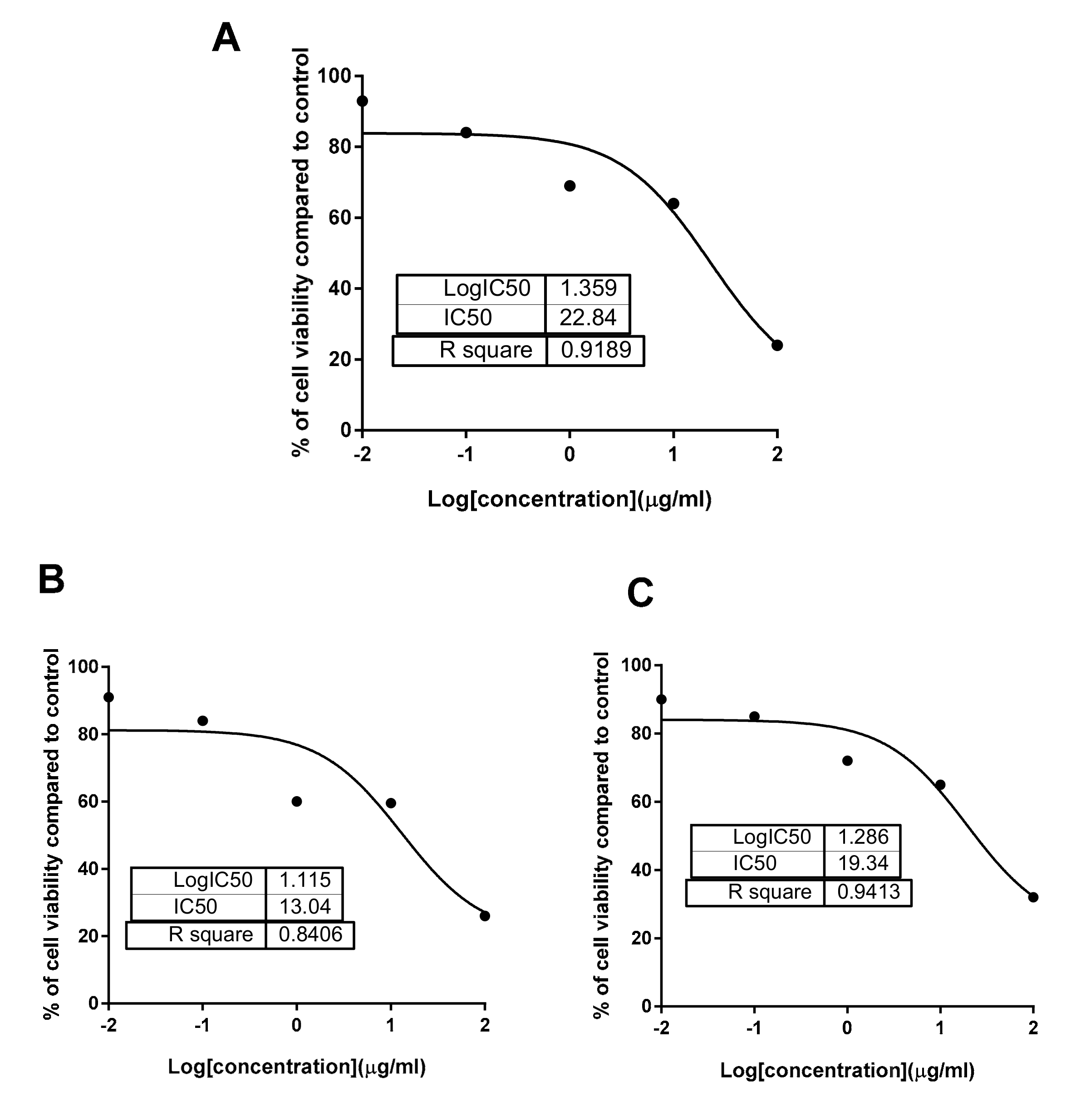
3.4.1. Cytotoxicity Using the MTT Assay

Evidence of the anticancer activities of family Brassicaceae plants against various types of malignancies have been acquired in numerous biological investigations [16,75,76,77,78,79]. These antitumor activities are mediated by different mechanisms such as antioxidant, cell cycle arrest, induction of apoptosis and prevention of angiogenesis and metastasis [80,81]. Candidate phytoconstituents responsible for these antineoplastic properties are glucosinolates and their hydrolytic products and phenolics (particularly flavonoids) as well [78]. Hence, the crude and phenolics extracts of C. annua were screened for their cytotoxic activities against panel of cancerous and non-cancerous breast and liver cell lines; MCF-7, MDA-MB-231, MCF-10A, HepG2 and THLE2, and to test their safety (selectivity) using the MTT assay (Table 3). C. annua crude extract was most cytotoxic against the MCF-7 cell line (IC50 = 22.8 µg/mL) with cell growth inhibition 25.4% at the highest concentrations 100 µg (Figure 5), while it was selective against the MDA-MB-231 with higher IC50 value of 46.2 µg/mL, and safe against normal breast and liver cells.
On the other hand, phenolics extract of C. annua was much more cytotoxic against the MCF-7 cells (IC50 = 13.04 µg/mL) than the crude extract, additionally, it showed more cytotoxic activity against the HepG2 cells (IC50 = 19.3 µg/mL) than the crude extract. On the other hand, it was not toxic against other cells, which elucidates the selectivity of its action. So, these results indicated the activity of phenolics extract against MCF-7 and HepG2 cells in a selective way for the other cells. Hence, phenolics extract was assumed of value to be investigated to determine its impact on induction of apoptosis in both MCF-7 and HepG2 cancer cells.

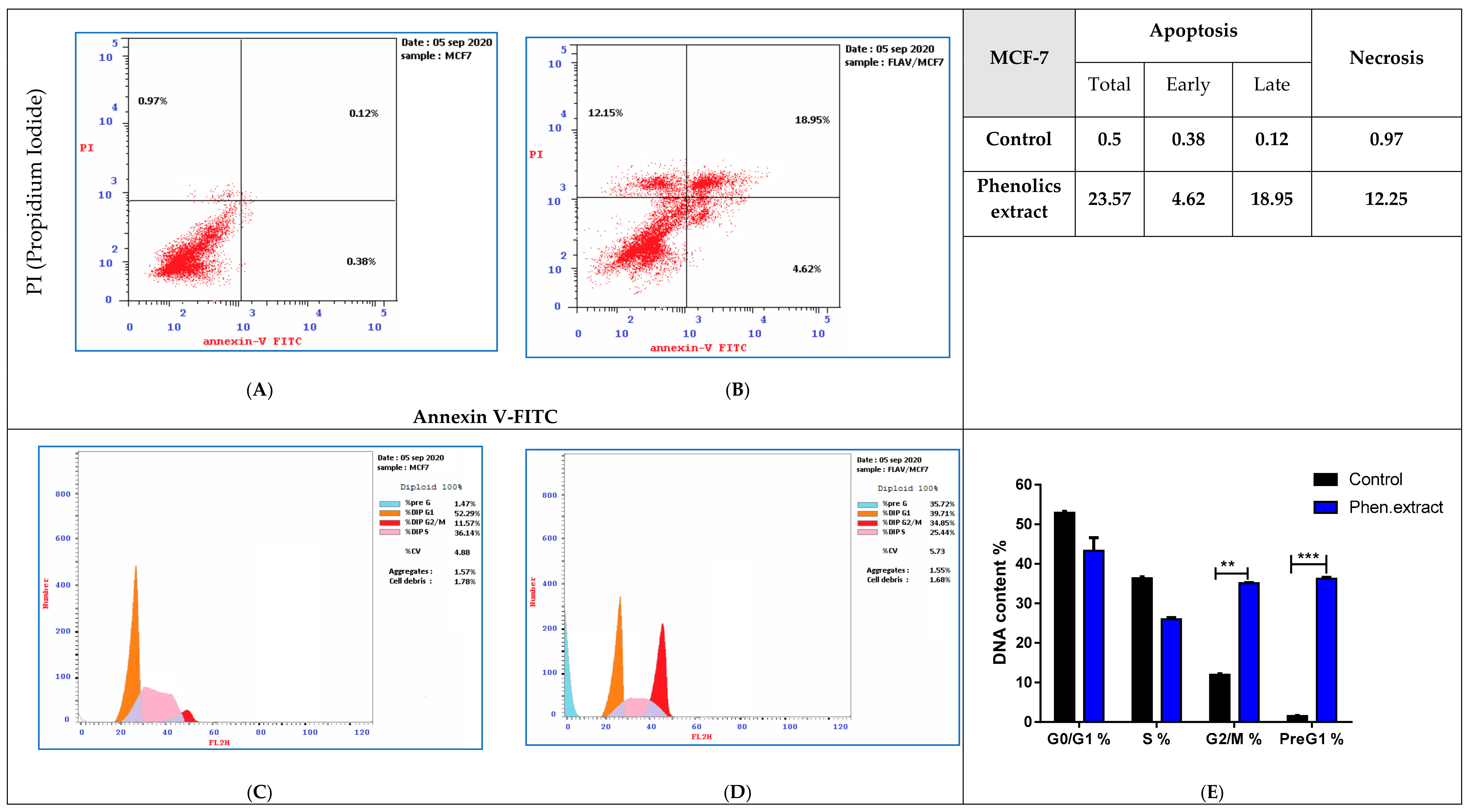
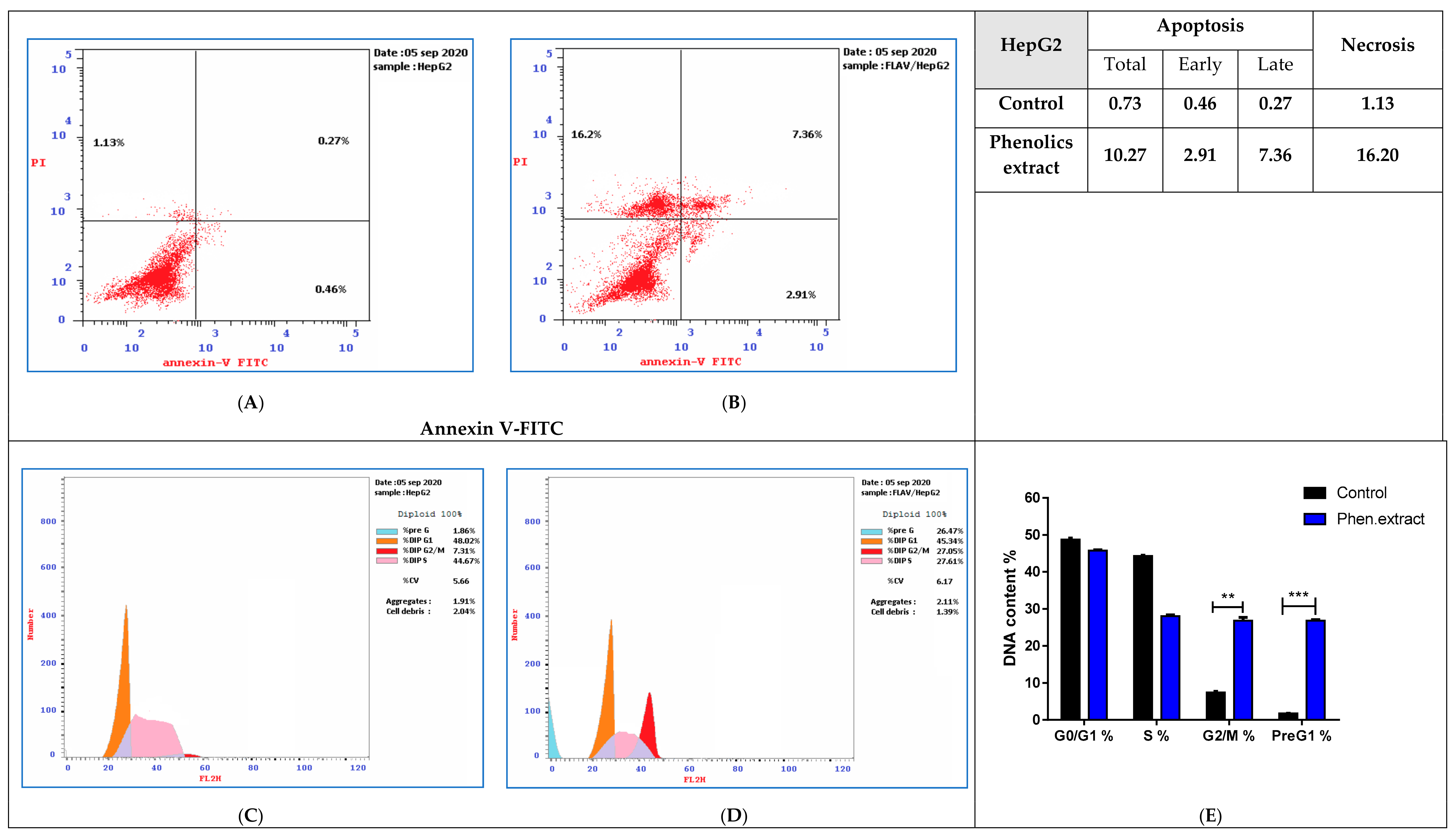
3.4.2. Annexin V/PI and Cell Cycle Analysis

Treatment of MCF-7 cancer cells with phenolics extract (IC50 = 13.04 μg/mL, 48 h) was performed. Investigations were done for its apoptosis-inducing activity using the cell cycle analysis with the cell population in different cell cycle phases. Investigation of the cell cycle is a decisive test that declares the cell accumulation percentage in each growth phase with cytotoxic substances after treatment. As shown in Figure 6 (upper panel), phenolics extract remarkably stimulated apoptotic breast cancer cell death with 47.14-fold (23.57% compared to 0.51% for the control). It induced early apoptosis by 12.15-fold (4.62% compared to 0.38% for control), and late apoptosis by 157.9-fold (18.95% compared to 0.12% for control). While it stimulated cell death via necrosis with 12.63-fold (12.25%, compared to 0.97% for the control). Moreover, MCF-7 cancer cells after phenolics extract treatment were subjected to DNA flow cytometry to analyze the cell cycle kinetics to determine the compound’s phase interference with the cell cycle. As seen in Figure 6C–E. It increased G2/M cell (34.85%, compared to 11.57% for control), and pre-G1 (35.72%, compared to 1.47% for the control) population, also it reduced cell number in the S (25.44% compared to 36.14% for control).
Similarly, HepG2 cancer cells were remedied with phenolics extract (IC50 = 19.34 μg/mL, 48 h). As seen in Figure 7 (upper panel), phenolics extract remarkably stimulated apoptotic breast cancer cell death with 14.07-fold (10.27% while it was 0.73% for the control). It induced early apoptosis by 6.32-fold (2.91% compared to 0.46% for control), and late apoptosis by 27.25-fold (7.36% compared to 0.27% for control). Moreover, HepG2 cancer cells after phenolics extract treatment were subjected to DNA flow cytometry, as seen in Figure 7 (lower panel). It enhanced G2/M cell (27.05%, compared to 7.3% for control), and pre-G1 (26.47%, compared to 1.86% for the control) population, also it reduced cell population in the S (27.61% compared to 44.67% for control).
Consequently, phenolics extract induced pre-G1 and G2/M-phase cell cycle arrest and blocked the progression of MCF-7 and HepG2 cancer cells that deteriorate the genetic metrical.

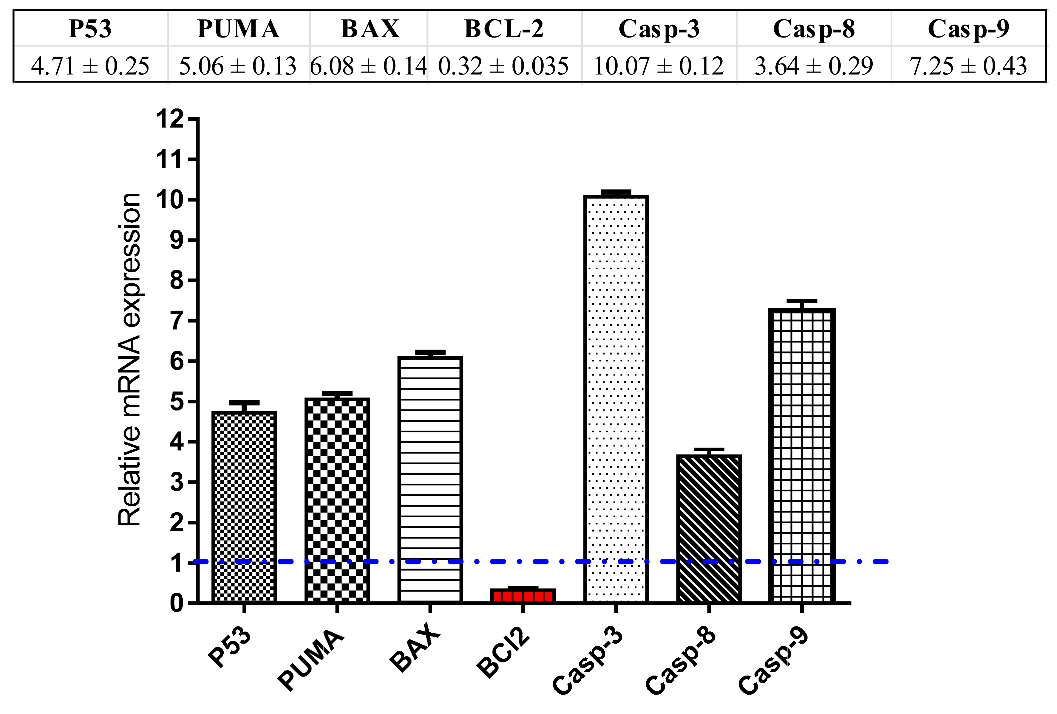
3.4.3. RT-PCR Analysis

For investigation the apoptotic pathway for the phenolics extract, 13.2 μg/mL of the sample was added to MCF-7 cells and left for 72 h to allow complete interaction, after RNA extraction, cDNA was produced. Then the expression of mRNA of Caspases 3, 8 9, pro-apoptotic (P53, BAX, PUMA) as well as anti-apoptotic genes (Bcl-2) in MCF-7 cells was traced by the RT-PCR analysis.
As demonstrated in Figure 8, the expression of P53 gene was noticeably elevated by the phenolics extract (≈4.7-fold) with concomitant activation of the PUMA and BAX levels which have been raised by ≈5.06-fold and 6.08-fold, respectively. Furthermore, flavonoid extract has remarkably raised the mRNA expression of caspases 3, 8, 9 genes by ≈10.7-fold, 3.64-fold and 7.25-fold, respectively. On the hand, it markedly suppressed the expression of Bcl-2 (the anti-apoptotic gene) by ≈0.32-fold. These findings are in harmony with the apoptotic mechanism suggested for anti-cancer activity.

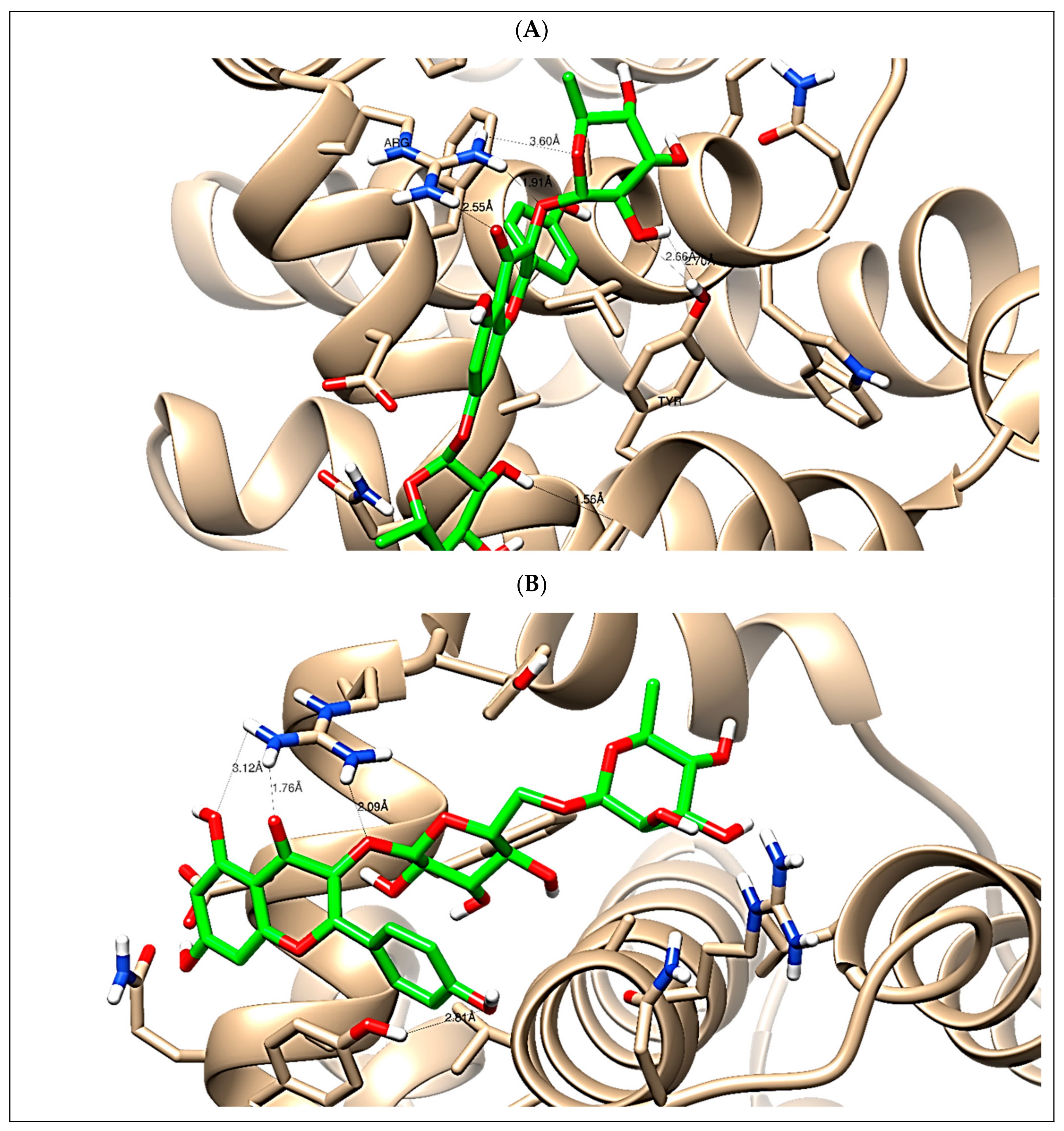
3.5. Simulated Molecular Docking Experiment

In this present study, C. annua phenolics extract exhibited antiproliferative activity against breast and liver carcinomas via apoptosis. Hence, the phenolics and flavonoids identified by the present LC-ESI-TOF-MS/MS analysis were selected for a simulated molecular docking investigation to gain insights into the possible molecular targets for the cytotoxic and apoptosis-inducing activities. The majority of the identified compounds revealed good binding interactions with binding energies (−9.24 to −27.28 Kcal/mol) inside the “B-cell lymphoma 2 (Bcl-2) (PDB ID: 4IEH) and their full interactions were summarized in Table 4. Other minor compounds didn’t show any binding activity towards the studied target. Accordingly, our docking experiment proposed their mechanism of action as Bcl-2 suppressors which is congruent with the flow cytometric and the RT-PCR analyses illustrating the apoptosis-inducing activity. As seen in Figure 9, Kaempferol-3,7-O-bis-α-L-rhamnoside and Kaempferol-3-rutinoside formed the maximum interactions with the interactive amino acids Arg 66 and Tyr 161 with binding energies of −23.67 and −18.28 (Kcal/mol), as they formed three hydrogen bonds with Arg 66 amino acid.
The current study has highlighted the noteworthy antioxidant and anticancer activities of C. annua. Therefore, this plant could be beneficial in cancer chemo preventive and chemotherapy. It represents a promising candidate to be used in combination with the existing chemotherapies. This combination tends to overcome drug resistance, increase the sensitivity to chemotherapy and counteract their dose dependent side effects especially those arise from the increased oxidative stress such as cardio and nephro toxicities. A future pharmacological study will be conducted to assess the in vivo efficacy and safety of C. annua as an antitumor herb individually and in combination with conventional chemotherapies to verify our assumption.

4. Conclusions

C. Annua herb can be considered as a promising chemo preventive and anticancer plant owing to its antioxidant and anti-proliferative effects that attributed to its unique chemical constituents. Herein, C. annua herb was studies for the first time for its chemical profiling and anticancer potential as well. Using LC-ESI-TOF-MS/MS analysis, 49 hits were identified mainly of polyphenolic type where flavonoid derivatives predominated and some of them were recorded in the plant for the first time. On the other hand, both of C. annua crude extract and its flavonoid fraction displayed significant and selective anticancer activity on HepG2 and MCF-7 cancer cells. However, the phenolic fraction was more active than the extract and it induced cell cycle arrest at pre-G1 and G2/M-phases by activation of pro apoptotic proteins and suppression of anti-apoptotic ones. The molecular docking studies indicated that most of the polyphenolics identified in the C. annua extract exhibited good binding interactions with binding energies (−9.24 to −27.28 Kcal/mol) inside the “B-cell lymphoma 2 (Bcl-2). Both of kaempferol-3,7-O-bis-α-L-rhamnoside and kaempferol-3-rutinoside were the most active inhibitors of Bcl-2. Therefore, C. annua herb seems to be a promising candidate to further advance anticancer research. In extrapolation, the intake of C. annua phenolics might be adventitious for alleviating breast and liver malignancies and tumoral proliferation in humans.

Supplementary Materials

The following are available online at https://www.mdpi.com/2076-3921/9/12/1286/s1; Figure S1. Total ion chromatogram (TIC) recorded in the negative mode for C. annua extract; Figure S2. Base peak chromatogram (BPC) recorded in negative ion mode C. annua extract; Figure S3. Total ion chromatogram (TIC) recorded in the positive mode for C. annua extract; Figure S4. Base peak chromatogram (BPC) recorded in the positive mode for C. annua extract.

Author Contributions

Conceptualization, J.M.B., S.A.A., E.E.E., S.S.E. and M.S.N.; methodology, E.E.E. and M.S.N., S.S.E. and H.A.A.; software, E.E.E., S.S.E. and M.S.N.; validation, E.E.E. and M.S.N.; formal analysis, E.E.E., A.O.N. and M.S.N.; investigation, E.E.E., M.S.N., S.S.E. and H.A.A.; resources, S.A.A., E.E.E., S.S.E. and M.S.N.; funding acquisition, S.S.E., A.O.N. and S.A.A.; data curation, E.E.E., S.S.E. and M.S.N.; writing—original draft preparation, E.E.E., H.A.A., S.S.E. and M.S.N.; writing—review and editing, H.A.A., E.E.E., S.S.E., S.A.A., J.M.B. and M.S.N.; supervision, J.M.B., E.E.E. and S.A.A. All authors have read and agreed to the published version of the manuscript.

Funding

This research was funded by the Deanship of Scientific Research (DSR) at King Abdulaziz University, Jeddah, Saudi Arabia under grant no. (FP-126-42).

Acknowledgments

The Deanship of Scientific Research (DSR) at King Abdulaziz University, Jeddah, Saudi Arabia has funded this project, under grant no. (FP-126-42). The authors, therefore, acknowledge with thanks DSR for technical and financial support.

Conflicts of Interest

The authors declare no conflict of interest.

References

  1. Losada-Echeberría, M.; Herranz-López, M.; Micol, V.; Barrajón-Catalán, E. Polyphenols as promising drugs against main breast cancer signatures. Antioxidants 2017, 6, 88. [Google Scholar] [CrossRef] [Green Version]
  2. Elhady, S.S.; Eltamany, E.E.; Shaaban, A.E.; Bagalagel, A.A.; Muhammad, Y.A.; El-Sayed, N.M.; Ayyad, S.-E.N.; Ahmed, A.A.M.; Elgawish, M.S.; Ahmed, S.A. Jaceidin flavonoid isolated from Chiliadenus montanus attenuates tumor progression in mice via VEGF inhibition: In Vivo and in silico studies. Plants 2020, 9, 1031. [Google Scholar] [CrossRef]
  3. Torić, J.; Brozovic, A.; Baus Lončar, M.; Jakobušić Brala, C.; Karković Marković, A.; Benčić, D.; Barbarić, M. Biological activity of phenolic compounds in extra virgin olive oils through their phenolic profile and their combination with anticancer drugs observed in human cervical carcinoma and colon adenocarcinoma cells. Antioxidants 2020, 9, 453. [Google Scholar] [CrossRef] [PubMed]
  4. World Health Organization. Available online: http://www.who.int/cancer/en/ (accessed on 7 November 2020).
  5. Efenberger-Szmechtyk, M.; Nowak, A.; Nowak, A. Cytotoxic and DNA-damaging effects of Aronia melanocarpa, Cornus mas, and Chaenomeles superba leaf extracts on the human colon adenocarcinoma cell line Caco-2. Antioxidants 2020, 9, 1030. [Google Scholar] [CrossRef] [PubMed]
  6. Hanahan, D.; Weinberg, R.A. The hallmarks of cancer. Cell 2000, 100, 57–70. [Google Scholar] [CrossRef] [Green Version]
  7. Anubhuti, S.; Ashok, S.; Prashant, Y.; Dhiraj, S. Isothiocyanates in Brassica: Potential anticancer agents. Asian Pac. J. Cancer Prev. 2016, 17, 4507–4510. [Google Scholar]
  8. Sasaki, K.; Takahashi, T. A flavonoid from Brassica rapa flower as the UV-absorbing nectar guide. Phytochemistry 2002, 61, 339–343. [Google Scholar] [CrossRef]
  9. Kaushik, N.; Agnihotri, A. GLC analysis of Indian rapeseed-mustard to study the variability of fatty acid composition. Biochem. Soc. Trans. 2000, 28, 581–583. [Google Scholar] [CrossRef]
  10. Cartea, M.E.; Francisco, M.; Soengas, P.; Velasco, P. Phenolic compounds in Brassica vegetables. Molecules 2010, 16, 251–280. [Google Scholar] [CrossRef]
  11. Jahangir, M.; Kim, H.K.; Choi, Y.H.; Verpoorte, R. Health-affecting compounds in Brassicaceae. Compr. Rev. Food Sci. Food Saf. 2009, 8, 31–43. [Google Scholar] [CrossRef]
  12. De Pascale, S.; Maggio, A.; Pernice, R.; Fogliano, V.; Barbieri, G. Sulphur fertilization may improve the nutritional value of Brassica rapa L. subsp. sylvestris. Eur. J. Agron. 2007, 26, 418–424. [Google Scholar] [CrossRef]
  13. Ramirez, D.; Abellán-Victorio, A.; Beretta, V.; Camargo, A.; Moreno, D.A. Functional ingredients from Brassicaceae species: Overview and perspectives. Int. J. Mol. Sci. 2020, 21, 1998. [Google Scholar] [CrossRef] [Green Version]
  14. Vallejo, F.; Gil-Izquierdo, A.; Pérez-Vicente, A.; García-Viguera, C. In Vitro gastrointestinal digestion study of broccoli inflorescence phenolic compounds, glucosinolates, and vitamin C. J. Agric. Food Chem. 2004, 52, 135–138. [Google Scholar] [CrossRef] [PubMed]
  15. Ateya, A.; Al-Gendy, A.; Kotob, S.; Hafez, A. Chemical constituents, antioxidant, antimicrobial and antiinflammatory activities of Erysimum corinthium boiss. (Brassicaceae). Int. J. Pharmacogn. Phytochem. Res. 2016, 8, 1601–1609. [Google Scholar]
  16. Miceli, N.; Cavò, E.; Ragusa, M.; Cacciola, F.; Mondello, L.; Dugo, L.; Acquaviva, R.; Malfa, G.A.; Marino, A.; D’Arrigo, M.; et al. Brassica incana Ten. (Brassicaceae): Phenolic constituents, antioxidant and cytotoxic properties of the leaf and flowering top extracts. Molecules 2020, 25, 1461. [Google Scholar] [CrossRef] [PubMed] [Green Version]
  17. Cuyckens, F.; Shahat, A.A.; Heuvel, H.V.D.; Abdel-Shafeek, K.A.; El-Messiry, M.M.; Elnasr, M.S.; Pieters, L.; Vlietinck, A.J.; Claeys, M. The Application of liquid chromatography-electrospray ionization mass spectrometry and collision-induced dissociation in the structural characterization of acylated flavonol O-Glycosides from the seeds of Carrichtera annua. Eur. J. Mass Spectrom. 2003, 9, 409–420. [Google Scholar] [CrossRef] [PubMed]
  18. Abdel-Shafeek, K.A.; El-Messiry, M.M.; Shahat, A.A.; Apers, S.; Pieters, L.; Seif-El Nasr, M.M. A new acylated flavonol triglycoside from Carrichtera annua. J. Nat. Prod. 2000, 63, 845–847. [Google Scholar] [CrossRef]
  19. Shahat, A.A.; Abdel-Shafeek, K.A.; Claeys, M.; Apers, S.; Pieters, L.; Vlietinck, A.J. A new flavonoid from Carrichtera annua. Nat. Prod. Sci. 2006, 12, 122–124. [Google Scholar]
  20. Shahat, A.A.; Abdelshafeek, K.; Husseiny, H.A. Isolation and identification of a new flavonoid glycoside from Carrichtera annua L. seeds. Pharmacogn. Res. 2011, 3, 151–154. [Google Scholar] [CrossRef] [Green Version]
  21. Abdelshafeek, K.; Elmissiry, M.M.; Hussiny, H.A.; Elnasr, M.M. The flavonoids and anticomplement activity of two cruciferous plants growing in Egypt. Int. J. Pharmacogn. Phytochem. Res. 2016, 8, 223–227. [Google Scholar]
  22. Saeed, N.; Khan, M.R.; Shabbir, M. Antioxidant activity, total phenolic and total flavonoid contents of whole plant extracts Torilis leptophylla L. BMC Complement. Altern. Med. 2012, 12, 221. [Google Scholar] [CrossRef] [PubMed] [Green Version]
  23. Fuochi, V.; Barbagallo, I.; Distefano, A.; Puglisi, F.; Palmeri, R.; Di Rosa, M.; Giallongo, C.; Longhitano, L.; Fontana, P.; Sferrazzo, G.; et al. Biological properties of Cakile maritima Scop. (Brassicaceae) extracts. Eur. Rev. Med. Pharmacol. Sci. 2019, 23, 2280–2292. [Google Scholar] [PubMed]
  24. Nsimba, R.Y.; Kikuzaki, H.; Konishi, Y. Antioxidant activity of various extracts and fractions of Chenopodium quinoa and Amaranthus spp. seeds. Food Chem. 2008, 106, 760–766. [Google Scholar] [CrossRef]
  25. Vijayalakshmi, A.; Kumar, P.R.; Priyadarsini, S.S.; Meenaxshi, C. In vitro antioxidant and anticancer activity of flavonoid fraction from the aerial parts of Cissus quadrangularis Linn. against human breast carcinoma cell lines. J. Chem. 2013, 2013, 1–9. [Google Scholar] [CrossRef] [Green Version]
  26. Eltamany, E.E.; Nafie, M.S.; Khodeer, D.M.; El-Tanahy, A.H.H.; Abdel-Kader, M.S.; Badr, J.M.; Abdelhameed, R.F.A. Rubia tinctorum root extracts: Chemical profile and management of type II diabetes mellitus. RSC Adv. 2020, 10, 24159–24168. [Google Scholar] [CrossRef]
  27. Freshney, R.I. Culture of tumor cells. In Culture of Animal Cells; John Wiley & Sons, Inc.: Hoboken, NJ, USA, 2010; ISBN 978-0-470-64936-7. [Google Scholar]
  28. Mosmann, T. Rapid colorimetric assay for cellular growth and survival: Application to proliferation and cytotoxicity assays. J. Immunol. Methods 1983, 65, 55–63. [Google Scholar] [CrossRef]
  29. Tantawy, E.S.; Amer, A.M.; Mohamed, E.K.; Alla, M.M.A.; Nafie, M.S. Synthesis, characterization of some pyrazine derivatives as anti-cancer agents: In vitro and in silico approaches. J. Mol. Struct. 2020, 1210, 128013. [Google Scholar] [CrossRef]
  30. Sarhan, A.A.M.; Boraei, A.T.A.; Barakat, A.; Nafie, M.S. Discovery of hydrazide-based pyridazino[4,5-b]indole scaffold as a new phosphoinositide 3-kinase (PI3K) inhibitor for breast cancer therapy. RSC Adv. 2020, 10, 19534–19541. [Google Scholar] [CrossRef]
  31. Nafie, M.S.; Arafa, K.; Sedky, N.K.; Alakhdar, A.A.; Arafa, R.K. Triaryl dicationic DNA minor-groove binders with antioxidant activity display cytotoxicity and induce apoptosis in breast cancer. Chem. Interact. 2020, 324, 109087. [Google Scholar] [CrossRef]
  32. Gad, E.M.; Nafie, M.S.; Eltamany, E.; Hammad, M.S.A.G.; Barakat, A.; Boraei, A.T.A. Discovery of new apoptosis-inducing agents for breast cancer based on ethyl 2-Amino-4,5,6,7-tetra hydrobenzo[b]thiophene-3-carboxylate: Synthesis, in vitro, and in vivo activity evaluation. Molecules 2020, 25, 2523. [Google Scholar] [CrossRef]
  33. Nafie, M.S.; Amer, A.M.; Mohamed, A.K.; Tantawy, E.S. Discovery of novel pyrazolo[3,4-b]pyridine scaffold-based derivatives as potential PIM-1 kinase inhibitors in breast cancer MCF-7 cells. Bioorg. Med. Chem. 2020, 28, 115828. [Google Scholar] [CrossRef] [PubMed]
  34. Nafie, M.S.; Tantawy, M.A.; Elmgeed, G.A. Screening of different drug design tools to predict the mode of action of steroidal derivatives as anti-cancer agents. Steroids 2019, 152, 108485. [Google Scholar] [CrossRef] [PubMed]
  35. Youssef, E.; El-Moneim, M.A.; Fathalla, W.; Nafie, M.S. Design, synthesis and antiproliferative activity of new amine, amino acid and dipeptide-coupled benzamides as potential sigma-1 receptor. J. Iran. Chem. Soc. 2020, 17, 2515–2532. [Google Scholar] [CrossRef]
  36. Fan, M.; Chen, G.-L.; Zhang, Y.-L.; Nahar, L.; Sarker, S.D.; Hu, G.; Guo, M.-Q. Antioxidant and anti-proliferative properties of Hagenia abyssinica roots and their potentially active components. Antioxidants 2020, 9, 143. [Google Scholar] [CrossRef] [Green Version]
  37. Lin, L.-Z.; Sun, J.; Chen, P.; Zhang, R.-W.; Fan, X.-E.; Li, L.-W.; Harnly, J.M. Profiling of glucosinolates and flavonoids in Rorippa indica (Linn.) Hiern. (Cruciferae) by UHPLC-PDA-ESI/HRMSn. J. Agric. Food Chem. 2014, 62, 6118–6129. [Google Scholar] [CrossRef]
  38. Daxenbichler, M.E.; Spencer, G.F.; Carlson, D.G.; Rose, G.B.; Brinker, A.M.; Powell, R.G. Glucosinolate composition of seeds from 297 species of wild plants. Phytochemistry 1991, 30, 2623–2638. [Google Scholar] [CrossRef]
  39. Camp, A.; Croxford, A.E.; Ford, C.S.; Baumann, U.; Clements, P.R.; Hiendleder, S.; Woolford, L.; Netzel, G.; Boardman, W.S.J.; Fletcher, M.T.; et al. Dual-locus DNA metabarcoding reveals southern hairy-nosed wombats (Lasiorhinus latifrons Owen) have a summer diet dominated by toxic invasive plants. PLoS ONE 2020, 15, e0229390. [Google Scholar] [CrossRef] [Green Version]
  40. Zhang, D.; Sun, Y.; Shi, Y.; Wu, X.; Jia, Q.; Chen, K.; Li, Y.; Wang, R. Four new indole alkaloids from the roots of Isatis tinctoria. Nat. Prod. Res. 2020, 1–7. [Google Scholar] [CrossRef]
  41. Brock, A.; Herzfeld, T.; Paschke, R.; Koch, M.; Dräger, B. Brassicaceae contain nortropane alkaloids. Phytochemistry 2006, 67, 2050–2057. [Google Scholar] [CrossRef]
  42. Sun, J.; Xiao, Z.; Lin, L.Z.; Lester, G.E.; Wang, Q.; Harnly, J.M.; Chen, P. Profiling polyphenols in five Brassica species microgreens by UHPLC-PDA-ESI/HRMSn. J. Agric. Food Chem. 2013, 61, 10960–10970. [Google Scholar] [CrossRef] [Green Version]
  43. Fernández-Fernández, R.; López-Martínez, J.C.; Romero-González, R.; Martínez-Vidal, J.L.; Flores, M.I.A.; Frenich, A.G. Simple LC–MS Determination of citric and malic acids in fruits and vegetables. Chromatographia 2010, 72, 55–62. [Google Scholar] [CrossRef]
  44. Jin, B.; Guo, K.; Zhang, T.; Li, T.; Ma, C. Simultaneous determination of 15 sulfonate ester impurities in phentolamine mesylate, amlodipine besylate, and tosufloxacin tosylate by LC-APCI-MS/MS. J. Anal. Methods Chem. 2019, 2019, 1–7. [Google Scholar] [CrossRef] [PubMed]
  45. Lang, R.; Yagar, E.F.; Eggers, R.; Hofmann, T. Quantitative Investigation of trigonelline, nicotinic acid, and nicotinamide in foods, urine, and plasma by means of LC-MS/MS and stable isotope dilution analysis. J. Agric. Food Chem. 2008, 56, 11114–11121. [Google Scholar] [CrossRef] [PubMed]
  46. Fan, Y.; Li, Y.; Wu, Y.; Li, L.; Wang, Y.; Li, Y.-B. Identification of the chemical constituents in Simiao Wan and rat plasma after oral administration by GC-MS and LC-MS. Evid. Based Complement. Altern. Med. 2017, 2017, 1–13. [Google Scholar] [CrossRef] [PubMed]
  47. Karar, M.G.E.; Kuhnert, N. UPLC-ESI-Q-TOF-MS/MS characterization of phenolics from Crataegus monogyna and Crataegus laevigata (Hawthorn) leaves, fruits and their herbal derived drops (Crataegutt Tropfen). J. Chem. Biol. Ther. 2016, 1, 102. [Google Scholar] [CrossRef] [Green Version]
  48. Clarke, D.B. Glucosinolates, structures and analysis in food. Anal. Methods 2010, 2, 310–325. [Google Scholar] [CrossRef]
  49. Revelou, P.; Kokotou, M.G.; Constantinou-Kokotou, V. Identification of auxin metabolites in Brassicaceae by ultra-performance liquid chromatography coupled with high-resolution mass spectrometry. Molecules 2019, 24, 2615. [Google Scholar] [CrossRef] [Green Version]
  50. Lee, K.-C.; Chan, W.; Liang, Z.; Liu, N.; Zhao, Z.; Lee, A.W.-M.; Cai, Z. Rapid screening method for intact glucosinolates in Chinese medicinal herbs by using liquid chromatography coupled with electrospray ionization ion trap mass spectrometry in negative ion mode. Rapid Commun. Mass Spectrom. 2008, 22, 2825–2834. [Google Scholar] [CrossRef]
  51. Tsugawa, H.; Nakabayashi, R.; Mori, T.; Yamada, Y.; Takahashi, M.; Rai, A.; Sugiyama, R.; Yamamoto, H.; Nakaya, T.; Yamazaki, M.; et al. A cheminformatics approach to characterize metabolomes in stable-isotope-labeled organisms. Nat. Methods 2019, 16, 295–298. [Google Scholar] [CrossRef]
  52. Engels, C.; Gräter, D.; Esquivel, P.; Jiménez, V.M.; Gänzle, M.G.; Schieber, A. Characterization of phenolic compounds in jocote (Spondias purpurea L.) peels by ultra high-performance liquid chromatography/electrospray ionization mass spectrometry. Food Res. Int. 2012, 46, 557–562. [Google Scholar] [CrossRef]
  53. Liang, X.; Lee, H.W.; Li, Z.; Lu, Y.; Zou, L.; Ong, C.N. Simultaneous quantification of 22 glucosinolates in 12 Brassicaceae vegetables by hydrophilic interaction chromatography–Tandem mass spectrometry. ACS Omega 2018, 3, 15546–15553. [Google Scholar] [CrossRef] [PubMed]
  54. Paganelli, C.J.; Siebert, D.A.; Vitali, L.; Micke, G.A.; Alberton, M.D. Quantitative analysis of phenolic compounds in crude extracts of Myrcia splendens leaves by HPLC-ESI-MS/MS. Rodriguésia 2020, 71, 00552019. [Google Scholar] [CrossRef]
  55. Bonaccorsi, P.; Caristi, C.; Gargiulli, C.; Leuzzi, U. Flavonol glucoside profile of southern Italian red onion (Allium cepa L.). J. Agric. Food Chem. 2005, 53, 2733–2740. [Google Scholar] [CrossRef] [PubMed]
  56. Song, H.-P.; Zhang, H.; Fu, Y.; Mo, H.-Y.; Zhang, M.; Chen, J.; Qi, L.-W. Screening for selective inhibitors of xanthine oxidase from Flos Chrysanthemum using ultrafiltration LC–MS combined with enzyme channel blocking. J. Chromatogr. B. 2014, 961, 56–61. [Google Scholar] [CrossRef]
  57. Negri, G.; de Santi, D.; Tabach, R. Chemical composition of hydroethanolic extracts from Siparuna guianensis, medicinal plant used as anxiolytics in Amazon region. Rev. Bras. Farm. 2012, 22, 1024–1034. [Google Scholar] [CrossRef] [Green Version]
  58. Olennikov, D.N.; Chirikova, N.K.; Kashchenko, N.I.; Nikolaev, V.M.; Kim, S.-W.; Vennos, C. Bioactive phenolics of the Genus Artemisia (Asteraceae): HPLC-DAD-ESI-TQ-MS/MS profile of the Siberian species and their inhibitory potential against α-amylase and α-glucosidase. Front. Pharmacol. 2018, 9, 756. [Google Scholar] [CrossRef]
  59. Sánchez-Ilárduya, M.; Sánchez-Fernández, C.; Viloria-Bernal, M.; López-Márquez, D.; Berrueta, L.A.; Gallo, B.; Vicente, F. Mass spectrometry fragmentation pattern of coloured flavanol-anthocyanin and anthocyanin-flavanol derivatives in aged red wines of Rioja. Aust. J. Grape Wine Res. 2012, 18, 203–214. [Google Scholar] [CrossRef]
  60. Lopes-Lutz, D.; Dettmann, J.; Nimalaratne, C.; Schieber, A. Characterization and quantification of polyphenols in Amazon grape (Pourouma cecropiifolia Martius). Molecules 2010, 15, 8543–8552. [Google Scholar] [CrossRef]
  61. Mazzoni, L.; Giampieri, F.; Álvarez-Suarez, J.M.; Gasparrini, M.; Mezzetti, B.; Forbes-Hernández, T.Y.; Battino, M. Isolation of strawberry anthocyanin-rich fractions and their mechanisms of action against murine breast cancer cell lines. Food Funct. 2019, 10, 7103–7120. [Google Scholar] [CrossRef]
  62. Jang, G.H.; Kim, H.W.; Lee, M.K.; Jeong, S.Y.; Bak, A.R.; Lee, D.J.; Kim, J.-B. Characterization and quantification of flavonoid glycosides in the Prunus genus by UPLC-DAD-QTOF/MS. Saudi J. Biol. Sci. 2018, 25, 1622–1631. [Google Scholar] [CrossRef]
  63. Shin, S.; Lee, S.-J.; Chung, J.; Bae, D.-W.; Kim, S.; Sung, N.-J. Comparison of anthocyanin content in seed coats of black soybean [Glycine max (L.) merr.] cultivars using liquid chromatography coupled to tandem mass spectrometry. Food Sci. Biotechnol. 2009, 18, 1470–1475. [Google Scholar]
  64. Chen, G.; Mutie, F.M.; Xu, Y.-B.; Saleri, F.D.; Hu, G.-W.; Guo, M. Antioxidant, anti-inflammatory activities and polyphenol profile of Rhamnus prinoides. Pharmaceuticals 2020, 13, 55. [Google Scholar] [CrossRef] [Green Version]
  65. Lee, S.-H.; Kim, H.-W.; Lee, M.-K.; Kim, Y.J.; Asamenew, G.; Cha, Y.-S.; Kim, J.-B. Phenolic profiling and quantitative determination of common sage (Salvia plebeia R. Br.) by UPLC-DAD-QTOF/MS. Eur. Food Res. Technol. 2018, 244, 1637–1646. [Google Scholar] [CrossRef] [Green Version]
  66. De Rosso, M.; Tonidandel, L.; Larcher, R.; Nicolini, G.; Vedova, A.D.; De Marchi, F.; Gardiman, M.; Giust, M.; Flamini, R. Identification of new flavonols in hybrid grapes by combined liquid chromatography–mass spectrometry approaches. Food Chem. 2014, 163, 244–251. [Google Scholar] [CrossRef] [PubMed]
  67. Wu, Z.-J.; Ma, X.-L.; Fang, D.-M.; Qi, H.-Y.; Ren, W.-J.; Zhang, G. Analysis of caffeic acid derivatives from Osmanthus yunnanensis using electrospray ionization quadrupole time-of-flight mass spectrometry. Eur. J. Mass Spectrom. 2009, 15, 415–429. [Google Scholar] [CrossRef]
  68. Sun, J.; Liang, F.; Bin, Y.; Li, P.; Duan, C.-Q. Screening non-colored phenolics in red wines using liquid chromatography/ultraviolet and mass spectrometry/mass spectrometry libraries. Molecules 2007, 12, 679–693. [Google Scholar] [CrossRef] [Green Version]
  69. Scigelova, M.; Hornshaw, M.; Giannakopulos, A.; Makarov, A. Fourier transform mass spectrometry. Mol. Cell. Proteom. 2011, 10, M111.009431. [Google Scholar] [CrossRef] [Green Version]
  70. Chen, Y.; Yu, H.; Wu, H.; Pan, Y.; Wang, K.; Jin, Y.; Zhang, C. Characterization and quantification by LC-MS/MS of the chemical components of the heating products of the flavonoids extract in Pollen Typhae for transformation rule exploration. Molecules 2015, 20, 18352–18366. [Google Scholar] [CrossRef] [Green Version]
  71. Falcão, S.I.; Vale, N.; Gomes, P.; Domingues, M.R.M.; Freire, C.; Cardoso, S.M.; Vilas-Boas, M. Phenolic profiling of Portuguese propolis by LC-MS spectrometry: Uncommon propolis rich in flavonoid glycosides. Phytochem. Anal. 2013, 24, 309–318. [Google Scholar] [CrossRef] [Green Version]
  72. Fang, N.; Yu, S.; Prior, R.L. LC/MS/MS characterization of phenolic constituents in dried plums. J. Agric. Food Chem. 2002, 50, 3579–3585. [Google Scholar] [CrossRef]
  73. Farias, L.D.S.; Mendez, A.S.L. LC/ESI-MS method applied to characterization of flavonoids glycosides in B. forficata subsp. pruinosa. Química Nova 2014, 37, 483–486. [Google Scholar] [CrossRef]
  74. Zhu, L.; Yang, S.; Li, G.; Zhang, X.; Yang, J.; Lai, X.; Yang, G. Simultaneous analysis of tocopherols, tocotrienols, phospholipids, γ-oryzanols and β-carotene in rice by ultra-high performance liquid chromatography coupled to a linear ion trap-orbitrap mass spectrometer. Anal. Methods 2016, 8, 5628–5637. [Google Scholar] [CrossRef]
  75. Pereira, L.P.; Silva, P.; Duarte, M.; Rodrigues, L.A.; Duarte, C.M.M.; Albuquerque, C.; Serra, A.T. Targeting colorectal cancer proliferation, stemness and metastatic potential using Brassicaceae extracts enriched in isothiocyanates: A 3D cell model-based study. Nutrients 2017, 9, 368. [Google Scholar] [CrossRef] [PubMed]
  76. Miceli, N.; Filocamo, A.; Ragusa, S.; Cacciola, F.; Dugo, P.; Mondello, L.; Celano, M.; Maggisano, V.; Taviano, M.F. Chemical characterization and biological activities of phenolic-rich fraction from cauline Leaves of Isatis tinctoria L. (Brassicaceae) growing in Sicily, Italy. Chem. Biodivers. 2017, 14, e1700073. [Google Scholar] [CrossRef] [PubMed]
  77. Al-Gendy, A.A.; El-Gindi, O.D.; Hafez, A.; Ateya, A. Glucosinolates, volatile constituents and biological activities of Erysimum corinthium Boiss. (Brassicaceae). Food Chem. 2010, 118, 519–524. [Google Scholar] [CrossRef]
  78. Avato, P.; Argentieri, M.P. Brassicaceae: A rich source of health improving phytochemicals. Phytochem. Rev. 2015, 14, 1019–1033. [Google Scholar] [CrossRef]
  79. Le, T.N.; Chiu, C.H.; Hsieh, P.C. Bioactive compounds and bioactivities of Brassica oleracea L. var. Italica sprouts and microgreens: An updated overview from a nutraceutical perspective. Plants 2020, 9, 946. [Google Scholar]
  80. Mitsiogianni, M.; Koutsidis, G.; Mavroudis, N.; Trafalis, D.T.; Botaitis, S.; Franco, R.; Zoumpourlis, V.; Amery, T.; Galanis, A.; Pappa, A.; et al. The role of isothiocyanates as cancer chemo-preventive, chemo-therapeutic and anti-melanoma agents. Antioxidants 2019, 8, 106. [Google Scholar] [CrossRef] [Green Version]
  81. Abotaleb, M.; Samuel, S.M.; Varghese, E.; Varghese, S.; Kubatka, P.; Líšková, A.; Büsselberg, D. Flavonoids in cancer and apoptosis. Cancers 2018, 11, 28. [Google Scholar] [CrossRef] [Green Version]
Figure 1. C. annua total phenolics contents (TPC) expressed as gallic acid equivalent per gram of dry weight GAE/g dw (83.8%) and total flavonoids contents (TFC) expressed as rutin equivalent per gram of dry weight RE/g dw (47.3%).
Antioxidants 09 01286 g001
Figure 2. The scavenging rate (%) of 2,2-diphenyl- 1-picrylhydrazyl (DPPH) by C. annua crude extract. All of the values in the figure are expressed as means (%) and SD of triplicated experiments.
Antioxidants 09 01286 g002
Figure 3. Antioxidant activity of C. annua crude extract (a) The IC50 value of 2,2-diphenyl-1-picrylhydrazyl (DPPH) radical scavenging assay, (b) ferric-ion reducing antioxidant power (FRAP) assay and (c) total antioxidant capacity (TAC) assay.
Antioxidants 09 01286 g003
Figure 4. Chemical structures of the identified compounds by LC-ESI-TOF-MS/MS.
Antioxidants 09 01286 g004aAntioxidants 09 01286 g004bAntioxidants 09 01286 g004cAntioxidants 09 01286 g004d
Figure 5. IC50 nonlinear regression curve fit of percentage of cell viability vs log [con. µg/mL], R square ≈1, using the GraphPad prism software. (A) Cytotoxicity of crude extract against MCF–7, (B) Cytotoxicity of phenolics extract against MCF–7, (C) Cytotoxicity of phenolics extract against HepG2.
Antioxidants 09 01286 g005
Figure 6. FITC/Annexin-V-FITC/PI differential apoptosis/necrosis (A) untreated control, (B) Phenolics extract (IC50 = 13.04 μg/mL, 48 h) and DNA content-flow cytometry aided cell cycle analyses (C) untreated control, (D) Flavonoid extract (IC50 = 13.04 μg/mL, 48 h), (E) bar chart representation) in MCF–7. ** p ≤ 0.05 and *** p ≤ 0.001 are significant different.
Antioxidants 09 01286 g006
Figure 7. FITC/Annexin-V-FITC/PI differential apoptosis/necrosis (A) untreated control, (B) Phenloics extract (IC50 = 19.34 μg/mL, 48 h) and DNA content-flow cytometry aided cell cycle analyses (C) untreated control, (D) Flavonoid extract (IC50 = 13.04 μg/mL, 48 h), (E) bar chart representation in HepG2 cells. ** p ≤ 0.05 and *** p ≤ 0.001 are significant different
Antioxidants 09 01286 g007
Figure 8. RT-PCR analysis of the apoptosis-related genes was performed after the MCF-7 cells were treated with phenolics extract (13.04 μg/mL) for 72 h.
Antioxidants 09 01286 g008
Figure 9. Visualized docked compounds: (A) Kaempferol-3,7-O-bis-α-L-rhamnoside (25) and (B) Kaempferol-3-rutinoside (21) inside the “B-cell lymphoma 2 (Bcl-2) (PDB ID: 4IEH) representing key interactions with bond length (Å) with interactive amino acids (Arg 66 and Tyr 161).
Antioxidants 09 01286 g009
Table 1. Primers used for real-time RT-PCR.
PrimerSequence
β-ActinFOR: 5’-GCACTCTTCCAGCCTTCCTTCC-3’
REV: 5’-GAGCCGCCGATCCACACG-3’
P53FOR: 5’-CTTTGAGGTGCGTGTTTGTG-3’
REV: 5’-GTGGTTTCTTCTTTGGCTGG-3’
Bcl-2FOR: 5’-GAGGATTGTGGCCTTCTTTG-3’
REV: 5’-ACAGTTCCACAAAGGCATCC-3’
PUMAFOR: 5’-GAGGAGGAACAGTGGGC-3’
REV: 5’-CTAATTGGGCTCCATCTCGG-3’
BAXFOR: 5’-TTTGCTTCAGGGTTTCATCC-3’
REV: 5’-CAGTTGAAGTTGCCGTCAGA-3’
Casp-3FOR: 5’-TGGCCCTGAAATACGAAGTC-3’
REV: 5’-GGCAGTAGTCGACTCTGAAG-3’
Casp-8FOR: 5’-AATGTTGGAGGAAAGCAAT-3’
REV: 5’-CATAGTCGTTGATTATCTTCAGC-3’
Casp-9FOR: 5’-CGAACTAACAGGCAAGCAGC-3’
REV: 5’-ACCTCACCAAATCCTCCAGAAC-3’
Table 2. Metabolites identified in C. annua crude extract using LC-ESI/TOF/MS/MS.
Compound NoRt (Min.)Proposed CompoundMolecular FormulaPrecursor TypeCalcd. m/z for PrecursorObs. m/z for PrecursorMS/MSRef.
11.05Citric acidC6H8O7[M−H]191.0192191.0193173, 111[42]
21.18Malic acidC4H6O5[M−H]133.0137133.0141155, 71[42,43]
31.24Methanesulfonic acidCH4O3S[M−H]94.980394.980780[44]
41.39TrigonillineC7H7NO2[M+H]+138.0550138.055194, 92[45]
51.44D-(-)-Quinic acidC7H12O6[M−H]191.0556191.0556173, 147, 85[46,47]
62.02Caffeoyl-quinic acid C16H18O9[M−H]353.0872353.0868191, 179, 135[42]
72.23GlucotropaeolinC14H19NO9S2[M−H]408.0423408.0268408, 162, 195, 246, 228[48]
82.981H-indole-3 carboxylic acidC9H7NO2[M−H]160.0399160.0399116, 142, 143[40,49]
93.22ProgoitrinC11H19NO10S2[M−H]388.0372388.0370388, 274, 259, 210, 192[50,51]
104.183-formylindoleC9H7NO[M+H]+146.0606146.0608118[40,49]
114.60 *Quercetin-3-O-arabinoglucosideC26H28O16[M−H]595.1299595.1303462, 433, 301, 300, 299[21,52]
124.68 *Quercetin 3-O-β-D-glucopyranosyl-(1→2)-arabinopyranosideC26H28O16[M−H]595.1299595.1306301, 311, 433[19,52]
134.752-(1H-indol-3-yl) acetic acidC10H9NO2[M+H]+176.0712176.0736176, 159, 158 130, 118[40,49]
145.10SinigrinC10H17NO9S2[M−H]358.0266358.0569358, 278, 275, 259, 241[50]
155.29Kaempferol 3, 7 di-glucosideC27H30O16[M−H]609.1456609.1460489, 447, 285[42]
165.69Quercetin3-O- [(6 sinapoyl-β-glucopyranosyl) (1→2)-β-arabinopyranoside]-7-O-β-glucopyranosideC43H48O25[M−H]963.2406963.2443801, 595, 463, 385[17,20,42]
175.70Quercetin3-O-[(6-feruloyl-β-glucopyranosyl) -(1→2)-β-arabinopyranoside]-7-O-β-glucopyranosideC42H46O24[M−H]933.2301933.2311771, 739, 301[17,18,42]
185.931-methoxy-1H-indole-3-carbaldehydeC10H9NO2[M+H] +176.0712176.0749161, 133, 117[40,49]
196.094-HydroxyglucobrassicinC16H20N2O10S2[M−H]463.0481463.1205463, 291, 275, 259, 241, 195[48,53]
206.469(methylsulfonyl)hydroxy nonyl glucosinolateC17H33NO12S3[M−H]538.1087538.8553259, 275, 291[37]
216.46Kaempferol-3 rutinosideC27H30O15[M−H]593.1506593.1503285, 447[19,42]
226.66SyringaldehydeC9H10O4[M−H]181.0501181.0511181, 151[54]
236.84Quercetin-4’-glucosideC21H20O12[M+H]+465.1033465.1028465, 303[55]
246.91Luteolin-7-O-glucosideC21H20O11[M−H]447.0927447.09366447, 285[56]
256.94Kaempferol-3,7-O-bis-α-L-rhamnosideC27H30O14[M−H]577.1557577.1576431, 285[57]
266.95Quercetin 3-O-galactosideC21H20O12[M+H]+465.1033465.1049465, 303[58]
276.97Quercetin-3-D-xylosideC20H18O11[M−H]433.0771433.0772301, 300, 271, 151[52]
286.99 *Cyanidin-3-glucosideC21H21O11[M]+449.1084449.1075449, 287[59,60,61]
297.00 *Kaempferol-3-O glucosideC21H20O1[M+H]+449.1084449.1085449, 287[58,62]
307.09Quercetin 3-O-[(6-sinapoyl-β-glucopyranosyl)-(1→2)-β arabinopyranosideC37H38O20[M−H]801.1878801.1855801, 595, 300[17]
317.23Petunidin-3-O-β-glucopyranosideC22H23O12[M]+479.1190479.116317, 302[63]
327.75VitexinC21H20O10[M−H]431.0978431.0989431, 311, 283,[64]
337.77CosmosiinC21H20O10[M+H]+433.1135433.1154433, 271[65]
347.79Syringetin-3-O-glucosideC23H24O13[M−H]507.1139507.1134507, 345, 179[66]
357.97Peonidine-3-O-glucosideC22H23O11[M]+463.1240463.1223463, 301, 286[59,60]
368.14Malvidin-3-galactosideC23H25O12[M]+493.1346493.1358493, 331[59,60]
378.31Caffeic acidC9H8O4[M−H]179.0344179.0343179, 151, 136, 133[67]
388.69HesperetinC16H14O6[M−H]301.0712301.0711301, 283, 271, 258[68]
398.72QuercetinC15H10O7[M+H]+303.0505303.0457303, 153[69]
409.13IsorhamnetinC16H12O7[M−H]315.0505315.0513315, 300, 151[20,70]
419.39LuteolinC15H10O6[M−H]285.0399285.0402285, 267, 257, 241, 223, 197,175[64,71]
429.40KaempferolC15H10O6[M−H]285.0399285.0392285, 257, 241, 223, 197, 151[64,71]
439.60P -coumaric acidC9H8O3[M−H]163.0395163.0389163, 119[72]
449.88Sinapic acidC11H12O5[M−H]223.0606223.0964223, 208, 179, 164[42]
4510.23Ferulic acidC10H10O4[M−H]193.0501193.0503193, 178, 149[42]
4610.68ApigeninC15H10O5[M−H]269.0450269.0458269, 241, 225, 181, 169[64]
4711.23KaempferideC16H12O6[M+H]+301.0712301.0712301, 286[73]
4824.77δ- tocotrienolC27H40O2[M+H]+397.3107397.3115397, 201, 187[74]
4926.89β- tocotrienolC28H42O2[M+H]+411.3263411.3271411, 205, 191, 151[74]
* Interchangeable values.
Table 3. IC50 values of both C. annua crude and phenolics extracts against panel of breast and liver cancer and normal cells using the MTT assay.
SampleWorking ConcentrationsIC50 (µg/mL) *
BreastLiver
MCF-7MDA-MB-231MCF-10AHepG2THLE2
C. annua crude extract20, 50, 100, 150, 200 (µg/mL)22.8 ± 1.0146.2 ± 1.4≥5032.3 ± 1.1ND
C. annua phenolics extract13.04 ± 0.87ND≥5019.3 ± 0.98≥50
* Values are expressed as mean ± SD of triplet trials, and calculated using GraphPad prism 7 software using nonlinear regression Dose-Inhibition curve ft.
Table 4. Summary of ligand-receptor interactions with binding energy (Kcal/mol) of the identified compounds to “B-cell lymphoma 2 (Bcl-2) protein, PDB = 4IEH).
CompoundBinding Energy
(Kcal/mol)
Ligand-Receptor Interactions with the Key Amino Acids
HB InteractionsLipophilic Interactions
Caffeoyl-quinic acid (6)−9.241 HB with Arg 66-
Quercetin-3-O-arabinoglucoside (11)−19.302 HB with Arg 66 and Tyr 161-
Quercetin 3-O-β-D-glucopyranosyl-(1→2)-arabinopyranoside (12)−21.202 HB with Arg 66 and Tyr 161Arene-Cation Arg 66
Kaempferol 3,7diglucoside (15)−18.172 HB with Arg 66 and 1 HB with Tyr 161Arene-Cation Arg 66
Quercetin 3-O-[(6-sinapoyl-β-glucopyranosyl) -(1→2)-β-arabinopyranoside]-7-O-β-glucopyranoside (16)−27.282 HB with Arg 66 and Tyr 161Arene-Cation Arg 66
Quercetin 3-O-[(6-feruloyl-β-glucopyranosyl)-(1→2)-β-arabinopyranoside]-7-O-β-glucopyranoside (17)−27.52 HB with Arg 66 and Tyr 161Arene-Cation with Arg 66
Kaempferol-3-rutinoside (21)−18.283 HB with Arg 66, 1 HB with Tyr 161-
Quercetin-4’-glucoside (23)−16.571 HB with Arg 66Arene-Cation Tyr 161
Luteolin-7-O-glucoside (24)−21.272 HB with Arg 66, and Tyr 161-
Kaempferol-3,7-O-bis-α-L-rhamnoside (25)−23.673 HB with Arg 66, 3 HB with Tyr 161-
Quercetin 3-O-galactoside (26)−18.172 HB with Arg 66 and Tyr 161-
Quercetin-3-D-xyloside (27)−20.511 HB with Tyr 161Arene-Cation Arg 66
Cyanidin-3-glucoside (28)−18.782 HB with Arg 66 and Tyr 161-
Kaempferol-3-O-glucoside (29)−16.782HB with Arg 661 arene-cation with Arg 66
Quercetin 3-O-[(6-sinapoyl-β-glucopyranosyl) -(1→2)-β arabinopyranoside (30)−15.292HB with Arg 661 arene-cation with Arg 66
Bolded numbers in parenthesis represent the number of the chemical structure (Figure 4).
Publisher’s Note: MDPI stays neutral with regard to jurisdictional claims in published maps and institutional affiliations.

