Antioxidant and Anticholinesterase Activities of Macrosphyra Longistyla (DC) Hiern Relevant in the Management of Alzheimer’s Disease


Abstract
:1. Introduction
2. Materials and Methods
2.1. Plant Material
2.2. Plant Extraction and Partitioning
2.3. Phytochemical Screening
2.4. Determination of the Total Phenol Content (TPC)
2.5. Determination of Total Flavonoid Contents (TFC)
2.6. Determination of Pro-Anthocyanidin Content (PAC)
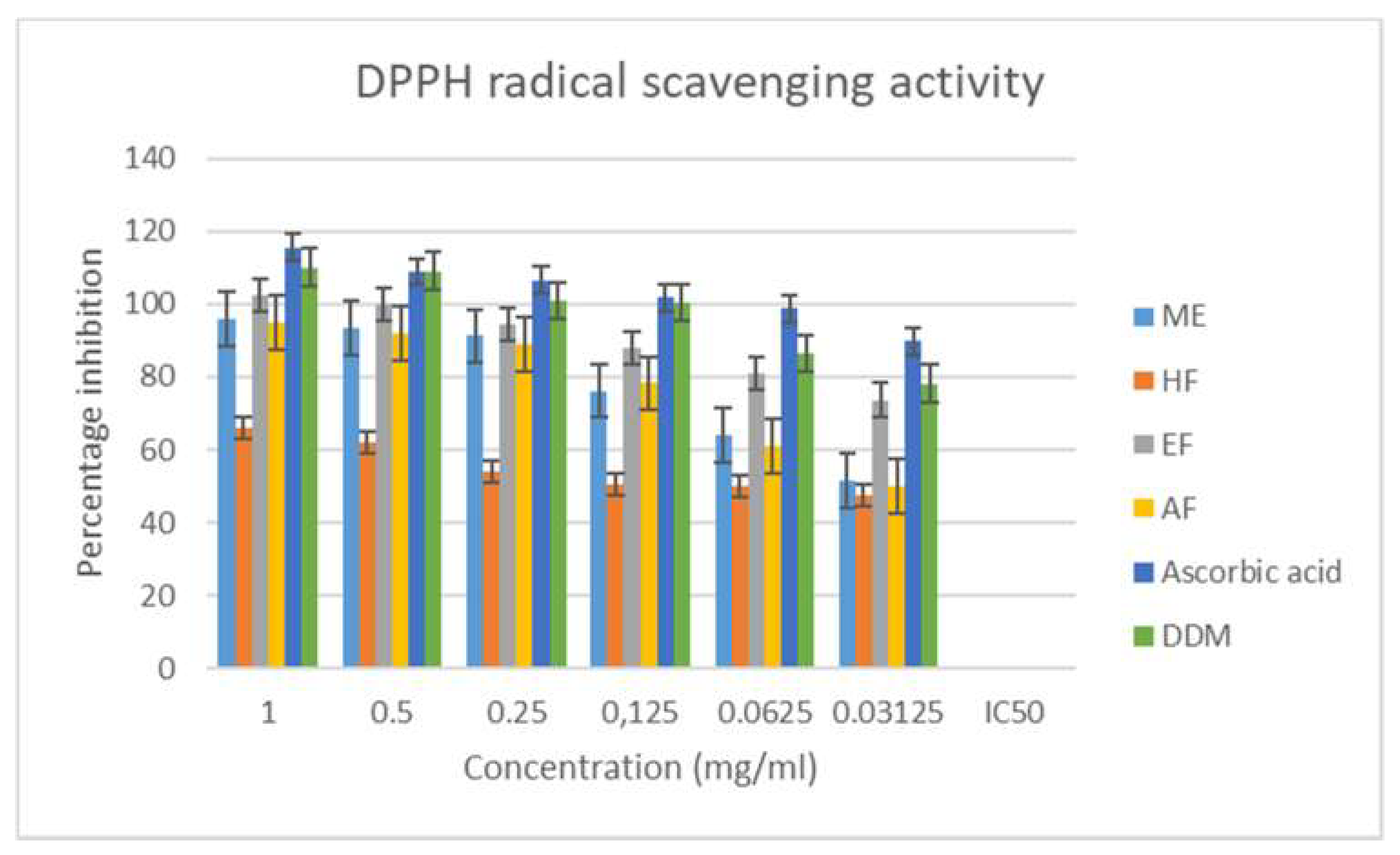
2.7. DPPH (2,2-Diphenyl-1-Picrylhydrazyl Hydrate) Radical Scavenging Assay
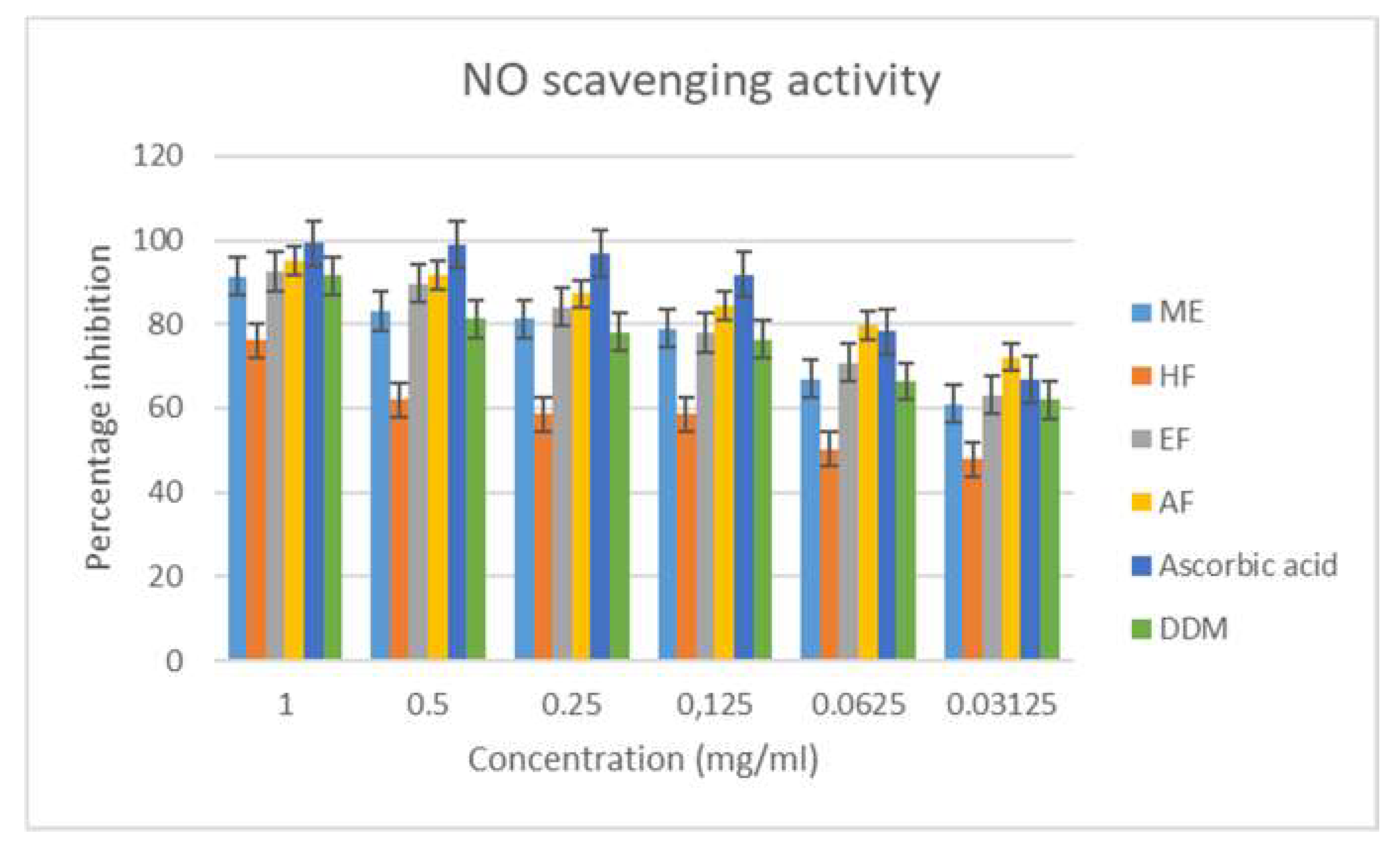
2.8. Nitric Oxide (NO) Scavenging Assay
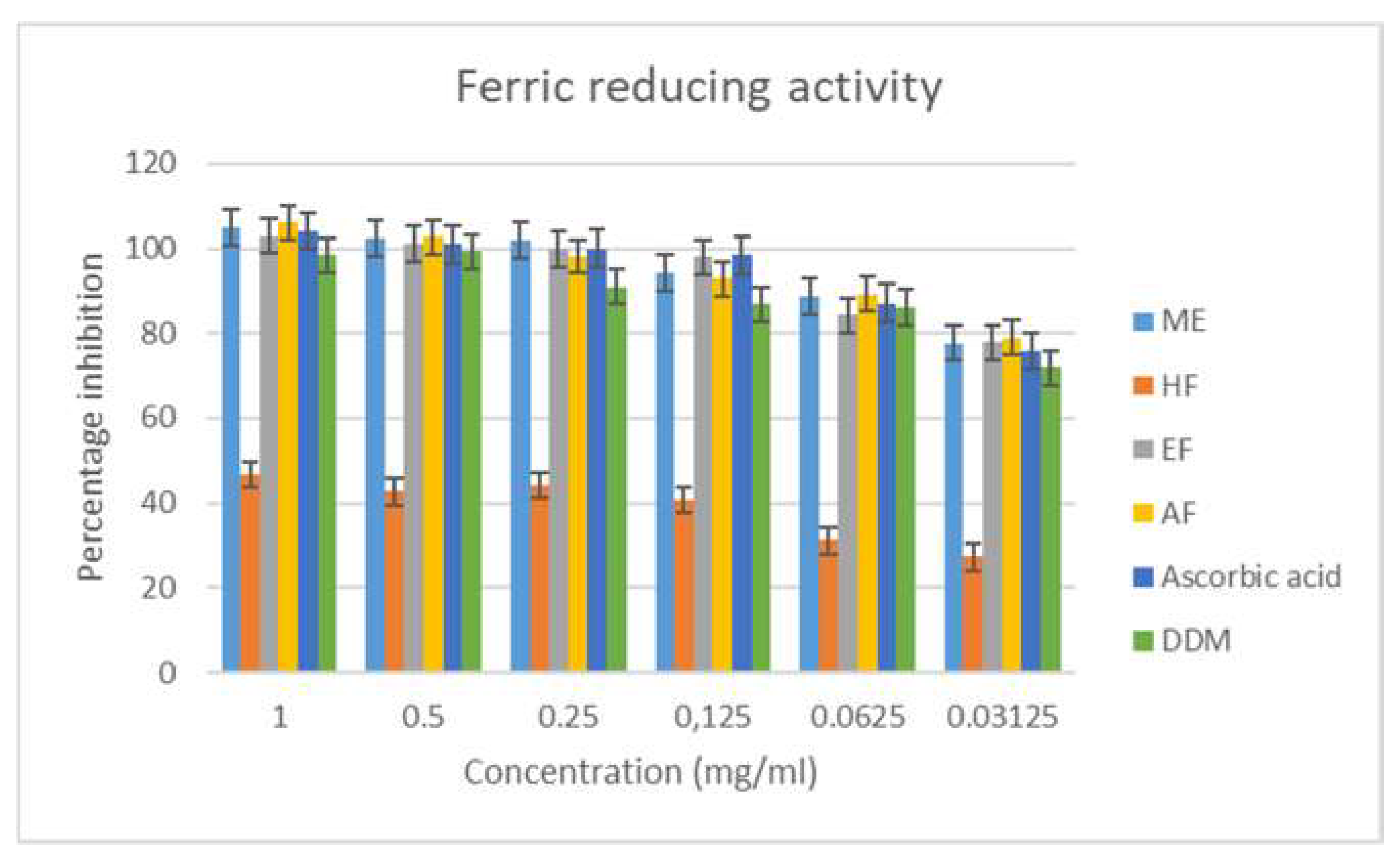
2.9. Ferric Reducing Antioxidant Assay
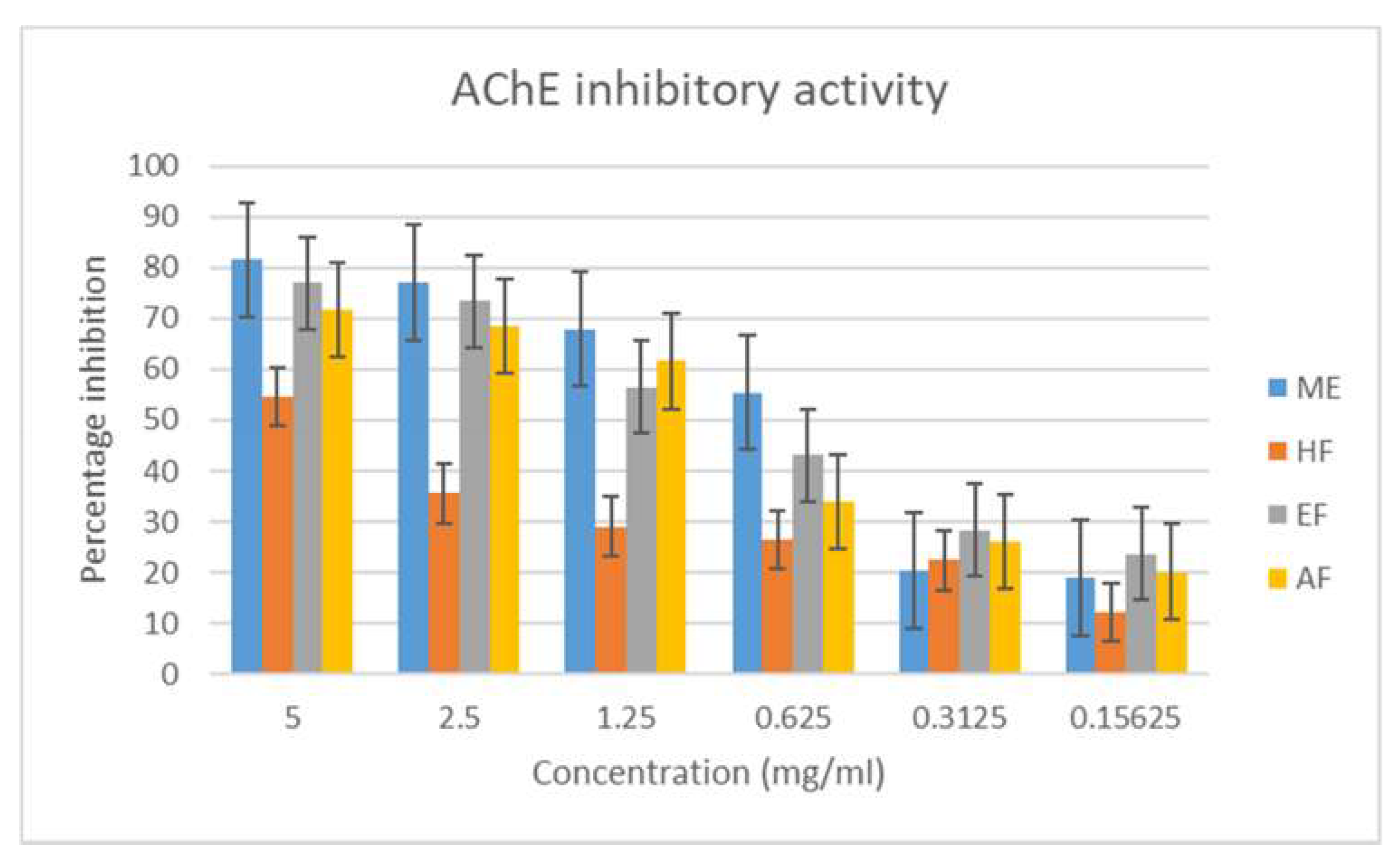
2.10. Cholinesterase Inhibitory Assay
2.11. Gas Chromatography Mass Spectrometry (GCMS) Analysis
2.12. Statistical Analysis
3. Results and Discussion
4. Conclusions
Author Contributions
Funding
Acknowledgments
Conflicts of Interest
References
- Amor, S.; Puentes, F.; Baker, D.; Van Der Valk, P. Inflammation in neurodegenerative diseases. Immunology 2010, 129, 154–169. [Google Scholar] [CrossRef]
- Chen, H.; Kwong, J.C.; Copes, R.; Tu, K.; Villeneuve, P.J.; Van Donkelaar, A.; Hystad, P.; Martin, R.V.; Murray, B.J.; Jessiman, B.; et al. Living near major roads and the incidence of dementia, Parkinson’s disease, and multiple sclerosis: A population-based cohort study. Lancet 2017, 389, 718–726. [Google Scholar] [CrossRef]
- Prince, M.; Guerchet, M.; Prina, M. The Epidemiology and Impact of Dementia: Current State and Future Trends; World Health Organization: Geneva, Switzerland, 2015. [Google Scholar]
- Solanki, I.; Parihar, P.; Mansuri, M.L.; Parihar, M.S. Flavonoid-Based Therapies in the Early Management of Neurodegenerative Diseases. Adv. Nutr. 2015, 6, 64–72. [Google Scholar] [CrossRef] [PubMed] [Green Version]
- Tan, C.-C.; Yu, J.-T.; Tan, M.-S.; Jiang, T.; Zhu, X.-C.; Tan, L. Autophagy in aging and neurodegenerative diseases: Implications for pathogenesis and therapy. Neurobiol. Aging 2014, 35, 941–957. [Google Scholar] [CrossRef] [PubMed]
- Johri, A.; Beal, M.F. Mitochondrial Dysfunction in Neurodegenerative Diseases. J. Pharmacol. Exp. Ther. 2012, 342, 619–630. [Google Scholar] [CrossRef] [PubMed] [Green Version]
- Takalo, M.; Salminen, A.; Soininen, H.; Hiltunen, M.; Haapasalo, A. Protein aggregation and degradation mechanisms in neurodegenerative diseases. Am. J. Neurodegener. Dis. 2013, 2, 1–14. [Google Scholar]
- Chen, W.-W.; Zhang, X.; Huang, W.-J. Role of neuroinflammation in neurodegenerative diseases (Review). Mol. Med. Rep. 2016, 13, 3391–3396. [Google Scholar] [CrossRef] [PubMed] [Green Version]
- Schliebs, R.; Arendt, T. The cholinergic system in aging and neuronal degeneration. Behav. Brain Res. 2011, 221, 555–563. [Google Scholar] [CrossRef] [PubMed]
- Butterfield, D.A.; Swomley, A.M.; Sultana, R. Amyloid β-Peptide (1–42)-Induced Oxidative Stress in Alzheimer Disease: Importance in Disease Pathogenesis and Progression. Antioxid. Redox Signal. 2013, 19, 823–835. [Google Scholar] [CrossRef]
- Kumar, A.; Singh, A. A review on Alzheimer’s disease pathophysiology and its management: An update. Pharmacol. Rep. 2015, 67, 195–203. [Google Scholar] [CrossRef]
- Keay, R.W. Randia and Gardenia in West Africa; Bulletin du Jardin botanique de l’Etat, Bruxelles/Bulletin van den Rijksplantentuin: Brussel, Belgium, 1958; pp. 15–72. [Google Scholar]
- Klotoé, J.; Dougnon, T.V.; Koudouvo, K.; Atègbo, J.-M.; Loko, F.; Akoègninou, A.; Aklikokou, K.; Dramane, K.; Gbeassor, M. Ethnopharmacological Survey on Antihemorrhagic Medicinal Plants in South of Benin. Eur. J. Med. Plants 2013, 3, 40–51. [Google Scholar] [CrossRef]
- Olabanji, S.; Adebajo, A.; Omobuwajo, O.; Ceccato, D.; Buoso, M.; Moschini, G. PIXE analysis of some Nigerian anti-diabetic medicinal plants (II). Nucl. Instrum. Methods Phys. Res. Sect. B Beam Interact. Mater. Atoms 2014, 318, 187–190. [Google Scholar] [CrossRef]
- Konkon, N.G.; Ouatara, D.; Kpan, W.B.; Kouakou, T.H. Medicinal plants used for treatment of diabetes by traditional practitioners in the markets of Abidjan district in Côte d’Ivoire. J. Med. Plants Stud. 2017, 5, 39–48. [Google Scholar]
- Soladoye, M.O.; Ikotun, T.; Chukwuma, E.C.; Ariwaodo, J.O.; Ibhanesebor, G.A.; Agbo-Adediran, O.A.; Owolabi, S.M. Our plants, our heritage: Preliminary survey of some medicinal plant species of Southwestern University Nigeria Campus, Ogun State, Nigeria. Annu. Biol. Res. 2013, 4, 27–34. [Google Scholar]
- Allabi, A.C.; Busia, K.; Ekanmian, V.; Bakiono, F. The use of medicinal plants in self-care in the Agonlin region of Benin. J. Ethnopharmacol. 2011, 133, 234–243. [Google Scholar] [CrossRef]
- Agbankpé, A.J.; Dougnon, T.V.; Bankolé, H.S.; Yèhouénou, B.; Yédomonhan, H.; Lègonou, M.; Dougnon, T.J. Etude ethnobotanique des légumes feuilles thérapeutiques utilisés dans le traitement des diarrhées au sud-Bénin (Afrique de l’Ouest). Int. J. Biol. Chem. Sci. 2014, 8, 1784–1795. [Google Scholar] [CrossRef]
- Achigan-Dako, E.G.; Pasquini, M.W.; Assogba Komlan, F.; N’danikou, S.; Yédomonhan, H.; Dansi, A.; Ambrose-Oji, B. Traditional Vegetables in Benin; Institut National des Recherches Agricoles du Bénin, Imprimeries du CENAP: Cotonou, Benin, 2010; p. 174. [Google Scholar]
- Atato, A.; Wala, K.; Batawila, K.; Lamien, N.; Akpagana, K. Edible wild fruit highly consumed during food shortage period in Togo: State of knowledge and conservation status. J. Life Sci. 2011, 5, 1046–1057. [Google Scholar]
- Chadare, F.J.; Madode, Y.E.; Fanou-Fogny, N.; Kindossi, J.M.; Ayosso, J.O.; Honfo, S.H.; Kayodé, A.P.; Linnemann, A.R.; Hounhouigan, D.J. Indigenous food ingredients for complementary food formulations to combat infant malnutrition in Benin: A review. J. Sci. Food Agric. 2018, 98, 439–455. [Google Scholar] [CrossRef]
- Sofowora, A. Screening Plants for Bioactive Agents. In Medicinal Plants and Traditional Medicinal in Africa, 2nd ed.; Spectrum Books Ltd.: Ibadan, Nigeria, 1993; pp. 134–156. [Google Scholar]
- Trease, G.E.; Evans, W.C. Pharmacognosy, 15th ed.; Saunders Publishers: London, UK, 2002; pp. 221–393. [Google Scholar]
- Singleton, V.L.; Rossi, J.A. Colorimetric of total phenolics with phosphomolybdic-phosphotungstic acid reagents. Am. J. Enol. Vinic. 1965, 16, 144–158. [Google Scholar]
- Köksal, E.; Gülçin, I. Antioxidant activity of cauliflower (Brassica oleracea L.). Turk. J. Agric. For. 2008, 32, 65–78. [Google Scholar]
- Broadhurst, R.B.; Jones, W.T. Analysis of condensed tannins using acidified vanillin. J. Sci. Food Agric. 1978, 29, 788–794. [Google Scholar] [CrossRef]
- Brand-Williams, W.; Cuvelier, M.; Berset, C. Use of a free radical method to evaluate antioxidant activity. LWT 1995, 28, 25–30. [Google Scholar] [CrossRef]
- Aiyegoro, O.A.; Okoh, A.O. Preliminary phytochemical screening and In vitro antioxidant activities of the aqueous extract of Helichrysum longifolium DC. BMC Complement. Altern. Med. 2010, 10, 21. [Google Scholar] [CrossRef] [PubMed]
- Oyaizu, M. Studies on products of browning reaction. Antioxidative activities of products of browning reaction prepared from glucosamine. Jpn. J. Nutr. Diet. 1986, 44, 307–315. [Google Scholar] [CrossRef]
- Ellman, G.L.; Courtney, K.; Andres, V.; Featherstone, R.M. A new and rapid colorimetric determination of acetylcholinesterase activity. Biochem. Pharmacol. 1961, 7, 88–95. [Google Scholar] [CrossRef]
- Quest Graph™. Linear, Logarithmic, Semi-Log Regression Calculator; AAT Bioquest, Inc.: Sunnyvale, CA, USA; Available online: https://www.aatbio.com/tools/linear-logarithmic-semi-log-regression-online-calculator (accessed on 20 May 2019).
- Shahidi, F.; Ambigaipalan, P. Phenolics and polyphenolics in foods, beverages and spices: Antioxidant activity and health effects—A review. J. Funct. Foods 2015, 18, 820–897. [Google Scholar] [CrossRef]
- Brewer, M. Natural Antioxidants: Sources, Compounds, Mechanisms of Action, and Potential Applications. Compr. Rev. Food Sci. Food Saf. 2011, 10, 221–247. [Google Scholar] [CrossRef]
- Han, R.-M.; Zhang, J.-P.; Skibsted, L.H. Reaction Dynamics of Flavonoids and Carotenoids as Antioxidants. Molecules 2012, 17, 2140–2160. [Google Scholar] [CrossRef] [Green Version]
- Wolfe, K.; Wu, X.; Liu, R.H. Antioxidant Activity of Apple Peels. J. Agric. Food Chem. 2003, 51, 609–614. [Google Scholar] [CrossRef]
- Koleckar, V.; Kubikova, K.; Řeháková, Z.; Kuca, K.; Jun, D.; Jahodář, L.; Opletal, L. Condensed and Hydrolysable Tannins as Antioxidants Influencing the Health. Mini-Rev. Med. Chem. 2008, 8, 436–447. [Google Scholar] [CrossRef]
- Kedare, S.B.; Singh, R.P. Genesis and development of DPPH method of antioxidant assay. J. Food Sci. Technol. 2011, 48, 412–422. [Google Scholar] [CrossRef] [PubMed] [Green Version]
- Pacher, P.; Beckman, J.S.; Liaudet, L. Nitric Oxide and Peroxynitrite in Health and Disease. Physiol. Rev. 2007, 87, 315–424. [Google Scholar] [CrossRef] [PubMed] [Green Version]
- Garrat, D.C. The Quantitative Analysis of Drugs; Chapman and Hall Ltd.: Tokyo, Japan, 1964; Volume 3, pp. 456–458. [Google Scholar]
- Al-Majedy, Y.K.; Al-Amiery, A.A.; Kadhum, A.A.H.; Mohamad, A.B. Antioxidant Activities of 4-Methylumbelliferone Derivatives. PLoS ONE 2016, 11, 0156625. [Google Scholar] [CrossRef] [PubMed]
- Benzie, I.F.; Strain, J. Ferric reducing/antioxidant power assay: Direct measure of total antioxidant activity of biological fluids and modified version for simultaneous measurement of total antioxidant power and ascorbic acid concentration. In Methods in Enzymology; Elsevier BV: Amsterdam, The Netherlands, 1999; Volume 299, pp. 15–27. [Google Scholar]
- Aliev, G.; Obrenovich, M.E.; Reddy, V.P.; Shenk, J.C.; Moreira, P.I.; Nunomura, A.; Zhu, X.; Smith, M.A.; Perry, G. Antioxidant Therapy in Alzheimer’s Disease: Theory and Practice. Mini-Rev. Med. Chem. 2008, 8, 1395–1406. [Google Scholar] [CrossRef] [PubMed]
- Schneider, L.S. Treatment of Alzheimer’s disease with cholinesterase inhibitors. Clin. Geriatr. Med. 2001, 17, 337–358. [Google Scholar] [CrossRef]
- Ranjan, N.; Kumari, M. Acetylcholinesterase inhibition by medicinal plants: A Review. Ann. Plant Sci. 2017, 6, 1640–1644. [Google Scholar] [CrossRef] [Green Version]
- Venneri, A.; McGeown, W.J.; Shanks, M.F. Empirical evidence of neuroprotection by dual cholinesterase inhibition in Alzheimer’s disease. NeuroReport 2005, 16, 107–110. [Google Scholar] [CrossRef]
- Khan, I.; Nisar, M.; Khan, N.; Saeed, M.; Nadeem, S.; Rehman, F.U.; Ali, F.; Karim, N.; Kaleem, W.A.; Qayum, M.; et al. Structural insights to investigate Conypododiol as a dual cholinesterase inhibitor from Asparagus adscendens. Fitoterapia 2010, 81, 1020–1025. [Google Scholar] [CrossRef]
- Mathew, M.; Subramanian, S. In Vitro Screening for Anti-Cholinesterase and Antioxidant Activity of Methanolic Extracts of Ayurvedic Medicinal Plants Used for Cognitive Disorders. PLoS ONE 2014, 9, e86804. [Google Scholar] [CrossRef]
- Owokotomo, I.; Ekundayo, O.; Abayomi, T.; Chukwuka, A. In-vitro anti-cholinesterase activity of essential oil from four tropical medicinal plants. Toxicol. Rep. 2015, 2, 850–857. [Google Scholar] [CrossRef] [Green Version]
- Samaradivakara, S.P.; Samarasekera, R.; Handunnetti, S.M.; Weerasena, O.J. Cholinesterase, protease inhibitory and antioxidant capacities of Sri Lankan medicinal plants. Ind. Crop. Prod. 2016, 83, 227–234. [Google Scholar] [CrossRef]
- Sheeja, M.D.; Beema, S.R.; Karutha, P.S.; Pandima, D.K. Cholinesterase inhibitory, anti-amyloidogenic and neuroprotective effect of the medicinal plant Grewia tiliaefolia—An in vitro and in silico study. Pharm. Biol. 2017, 55, 381–393. [Google Scholar] [CrossRef] [PubMed]
- Ovais, M.; Ayaz, M.; Khalil, A.T.; Shah, S.A.; Jan, M.S.; Raza, A.; Shahid, M.; Shinwari, Z.K. HPLC-DAD finger printing, antioxidant, cholinesterase, and α-glucosidase inhibitory potentials of a novel plant Olax nana. BMC Complement. Altern. Med. 2018, 18, 1. [Google Scholar] [CrossRef] [PubMed]
- Steele, M.; Stuchbury, G.; Münch, G. The molecular basis of the prevention of Alzheimer’s disease through healthy nutrition. Exp. Gerontol. 2007, 42, 28–36. [Google Scholar] [CrossRef] [PubMed]
- Mecocci, P.; Polidori, M.C. Antioxidant clinical trials in mild cognitive impairment and Alzheimer’s disease. Biochim. Biophys. Acta Mol. Basis Dis. 2012, 1822, 631–638. [Google Scholar] [CrossRef] [PubMed]
- Zhang, L.; Ravipati, A.S.; Koyyalamudi, S.R.; Jeong, S.C.; Reddy, N.; Smith, P.T.; Bartlett, J.; Shanmugam, K.; Münch, G.; Wu, M.J. Antioxidant and Anti-inflammatory Activities of Selected Medicinal Plants Containing Phenolic and Flavonoid Compounds. J. Agric. Food Chem. 2011, 59, 12361–12367. [Google Scholar] [CrossRef] [PubMed]
- Gülçin, İ.; Huyut, Z.; Elmastaş, M.; Aboul-Enein, H.Y. Radical scavenging and antioxidant activity of tannic acid. Arab. J. Chem. 2010, 3, 43–53. [Google Scholar] [CrossRef] [Green Version]
- Babbar, N.; Oberoi, H.S.; Uppal, D.S.; Patil, R.T. Total phenolic content and antioxidant capacity of extracts obtained from six important fruit residues. Food Res. Int. 2011, 44, 391–396. [Google Scholar] [CrossRef]
- Hussain, A.; Tian, M.-Y.; He, Y.-R.; Bland, J.M.; Gu, W.-X. Behavioral and electrophysiological responses of Coptotermes formosanus Shiraki towards entomopathogenic fungal volatiles. Biol. Control 2010, 55, 166–173. [Google Scholar] [CrossRef]
- Choi, H.-S. Analysis of the Terpenoids from Syneilesis palmata Essential Oil and the Variation of the Sesquiterpene Compounds by Harvest Year. Korean J. Food Nutr. 2013, 26, 287–294. [Google Scholar] [CrossRef]
- Palani, S.; Raja, S.; Kumar, R.P.; Selvaraj, R.; Kumar, B.S. Evaluation of phytoconstituents and anti-nephrotoxic and antioxidant activities of Monochoria vaginalis. Pak. J. Pharm. Sci. 2011, 24, 293–301. [Google Scholar] [PubMed]
- Ntalli, N.G.; Manconi, F.; Leonti, M.; Maxia, A.; Caboni, P. Aliphatic Ketones from Ruta chalepensis (Rutaceae) Induce Paralysis on Root Knot Nematodes. J. Agric. Food Chem. 2011, 59, 7098–7103. [Google Scholar] [CrossRef] [PubMed]
- Walia, M.; Mann, T.S.; Kumar, D.; Agnihotri, V.K.; Singh, B. Chemical Composition and In Vitro Cytotoxic Activity of Essential Oil of Leaves of Malus domestica Growing in Western Himalaya (India). Evid. Based Complement. Altern. Med. 2012, 2012, 649727. [Google Scholar] [CrossRef] [PubMed]
- Innocent, E.; Gikonyo, N.K.; Nkunya, M.H. Repellency property of long chain aliphatic methyl ketones against Anopheles gambiae s.s. Tanzan. J. Health Res. 2008, 10, 50–54. [Google Scholar]
- Manosroi, A.; Jantrawut, P.; Sainakham, M.; Manosroi, W.; Manosroi, J. Anticancer activities of the extract from Longkong (Lansium domesticum) young fruits. Pharm. Biol. 2012, 50, 1397–1407. [Google Scholar] [CrossRef] [PubMed]
- Hernández-Villegas, M.; Borges-Argáez, R.; Rodríguez-Vivas, R.; Torres-Acosta, J.; Méndez-González, M.; Cáceres-Farfán, M.; Torres-Acosta, J.F.D.J. In vivo anthelmintic activity of Phytolacca icosandra against Haemonchus contortus in goats. Veter Parasitol. 2012, 189, 284–290. [Google Scholar] [CrossRef] [PubMed]
- Hamza, F.; Zinjarde, S. Marine Biodiversity As a Resource for Bioactive Molecules As Inhibitors of Microbial Quorum Sensing Phenotypes. In Biotechnological Applications of Quorum Sensing Inhibitors; Springer Science and Business Media LLC: Berlin/Heidelberg, Germany, 2018; pp. 329–350. [Google Scholar]
- Carretero, M.; López-Pérez, J.L.; Abad, M.; Bermejo, P.; Tillet, S.; Israel, A.; Noguera, P.B. Preliminary study of the anti-inflammatory activity of hexane extract and fractions from Bursera simaruba (Linneo) Sarg. (Burseraceae) leaves. J. Ethnopharmacol. 2008, 116, 11–15. [Google Scholar] [CrossRef]
- Silici, S.; Kutluca, S. Chemical composition and antibacterial activity of propolis collected by three different races of honeybees in the same region. J. Ethnopharmacol. 2005, 99, 69–73. [Google Scholar] [CrossRef]
- Conley, A.J.; Kabara, J.J. Antimicrobial Action of Esters of Polyhydric Alcohols. Antimicrob. Agents Chemother. 1973, 4, 501–506. [Google Scholar] [CrossRef] [Green Version]
- Santos, C.C.; Salvadori, M.S.; Mota, V.G.; Costa, L.M.; de Almeida, A.A.; de Oliveira, G.A.; Costa, J.P.; de Sousa, D.P.; de Freitas, R.M.; de Almeida, R.N. Antinociceptive and antioxidant activities of phytol in vivo and in vitro models. Neurosci. J. 2013, 2013, 949452. [Google Scholar] [CrossRef]
- Elufioye, T.O.; Obuotor, E.M.; Agbedahunsi, J.M.; Adesanya, S.A. Cholinesterase inhibitory activity and structure elucidation of a new phytol derivative and a new cinnamic acid ester from Pycnanthus angolensis. Rev. Bras. Farm. 2016, 26, 433–437. [Google Scholar] [CrossRef] [Green Version]
- Ghalib, R.M.; Hashim, R.; Sulaiman, O.; Mehdi, S.H.; Anis, Z.; Rahman, S.Z.; Ahamed, B.K.; Abdul Majid, A.M. Phytochemical analysis, cytotoxic activity and constituents–activity relationships of the leaves of Cinnamomum iners (Reinw. ex Blume-Lauraceae). Nat. Prod. Res. 2012, 26, 2155–2158. [Google Scholar] [PubMed]
- Gecibesler, I.H.; Yaglıoglu, A.S.; Gul, F.; Temirturk, M.; Demirtas, I. Phytochemicals of Chrysophthalmum montanum (DC.) Boiss. Roots and their antiproliferative activities Against HeLa and C6 Cell Lines. Proc. Natl. Acad. Sci. India Sect. B Biol. Sci. 2019, 89, 145–154. [Google Scholar] [CrossRef]
- El-Din, S.M.M.; Mohyeldin, M.M. Component Analysis and Antifungal Activity of the Compounds Extracted from Four Brown Seaweeds with Different Solvents at Different Seasons. J. Ocean Univ. China 2018, 17, 1178–1188. [Google Scholar] [CrossRef]
- Conforti, F.; Menichini, F.; Loizzo, M.R.; Statti, A.G.; Rapisarda, A.; Menichini, F.; Houghton, P.J. Antioxidant, α-amylase inhibitory and brine-shrimp toxicity studies on Centaurea centaurium L. methanolic root extract. Nat. Prod. Res. 2008, 22, 1457–1466. [Google Scholar] [CrossRef]
- Vakharia, P.P.; Silverberg, J.I. New therapies for atopic dermatitis: Additional treatment classes. J. Am. Acad. Dermatol. 2018, 78, S76–S83. [Google Scholar] [CrossRef]
- Jíchová, Š.; Kopkan, L.; Husková, Z.; Doleželová, Š.; Neckář, J.; Kujal, P.; Vernerová, Z.; Kramer, H.J.; Sadowski, J.; Kompanowska-Jezierska, E.; et al. Epoxyeicosatrienoic acid analog attenuates the development of malignant hypertension, but does not reverse it once established: A study in Cyp1a1-Ren-2 transgenic rats. J. Hypertens. 2016, 34, 2008–2025. [Google Scholar] [CrossRef]
- Sales, D.L.; Oliveira, O.P.; Cabral, M.E.; Dias, D.Q.; Kerntopf, M.R.; Coutinho, H.D.; Costa, J.G.; Freitas, F.R.; Ferreira, F.S.; Alves, R.R.; et al. Chemical identification and evaluation of the antimicrobial activity of fixed oil extracted from Rhinella jimi. Pharm. Biol. 2015, 53, 98–103. [Google Scholar] [CrossRef]
- Nagarjunakonda, S.; Amalakanti, S.; Dhishana, S.R.; Ramaiah, M.; Rajanala, L. GC-MS Analysis of Indrakeeladri Native Medicine used in the Treatment of Stroke. Pharmacogn. J. 2017, 9, 102–106. [Google Scholar] [CrossRef]
- Sandoval-Montemayor, N.E.; García, A.; Elizondo-Treviño, E.; Garza-González, E.; Alvarez, L.; Camacho-Corona, M.D.R. Chemical Composition of Hexane Extract of Citrus aurantifolia and Anti-Mycobacterium tuberculosis Activity of Some of Its Constituents. Molecules 2012, 17, 11173–11184. [Google Scholar] [CrossRef]
- Mormile, R.; Vittori, G.; De Michele, M.; Squarcia, U.; Quaini, F. Linoleic acid and colorectal cancer cell growth suppression: Is the deregulation of mitochondrial surviving the key factor? Int. J. Colorectal Dis. 2012, 27, 1383–1384. [Google Scholar] [CrossRef] [PubMed]
- Kim, Y.-S.; Lee, S.-J.; Hwang, J.-W.; Kim, E.-K.; Kim, S.-E.; Kim, E.-H.; Moon, S.-H.; Jeon, B.-T.; Park, P.-J. In vitro protective effects of Thymus quinquecostatus Celak extracts on t-BHP-induced cell damage through antioxidant activity. Food Chem. Toxicol. 2012, 50, 4191–4198. [Google Scholar] [CrossRef] [PubMed]
- Gok, M.; Zeybek, N.D.; Bodur, E. Butyrylcholinesterase expression is regulated by fatty acids in HepG2 cells. Chem. Interact. 2016, 259, 276–281. [Google Scholar] [CrossRef] [PubMed]
- Agboke, A.A.; Attama, A.A. Bioactive components and antibacterial activities of n-hexane extract of Moringa oleifera root bark on clinical isolates of methicilin resistant Staphylococcus aureus. Int. J. Curr. Res. Chem. Pharm. Sci. 2016, 3, 1–9. [Google Scholar]
- Avis, T.J.; Boulanger, R.R.; Belanger, R.R. Synthesis and Biological Characterization of (Z)-9-Heptadecenoic and (Z)-6-Methyl-9-Heptadecenoic Acids: Fatty Acids with Antibiotic Activity Produced by Pseudozyma flocculosa. J. Chem. Ecol. 2000, 26, 987–1000. [Google Scholar] [CrossRef]
- Wang, J.; Liu, H.; Gao, H.; Zhao, J.; Zhou, L.; Han, J.; Yu, Z.; Yang, F. Antimicrobial and antioxidant activities of the flower essential oil of Halimodendron halodendron. Nat. Prod. Commun. 2011, 6. [Google Scholar] [CrossRef]


| Tests | Observations | Inferences |
|---|---|---|
| 1. Alkaloids | ||
| a. Dragendorff | Deep yellow color | Alkaloid absent |
| b. Wagner test | Orange color | Alkaloid absent |
| 2. Anthraquinones | ||
| a. Borntrager’s test | Milky color | Anthraquinone absent |
| 3. Flavonoids | Yellow coloration | Flavonoids present |
| 4. Phenols | Dark coloration | Phenols present |
| 5. Tannins | Blue black coloration | Tannin present |
| 6. Saponin | Frothing which disappear after sometime | Saponin present |
| 7. Terpenoid | Dark green coloration | Terpenoids present |
| Assays | ME | HF | EF | AF |
|---|---|---|---|---|
| % Yield | 6.18 | 4.70 | 7.11 | 40.00 |
| Total phenolics (mg GAE/g) | 18.30 ± 0.04 | 7.56 ± 0.12 | 16.06 ± 0.13 | 9.02 ± 0.02 |
| Total flavonoids (mg GAE/g) | 16. 07 ± 0.14 | 5.02 ± 0.01 | 10.49 ± 0.014 | 11.62 ± 0.01 |
| Total tannins (mg GAE/g) | 24. 44 ± 0.32 | 2.99 ± 0.06 | 9.12 ± 0.17 | 26.11 ± 0.02 |
| Assays | IC50 | |||||
|---|---|---|---|---|---|---|
| ME | HF | EF | AF | Ascorbic Acid | DDM | |
| DPPH scavenging | 0.090 | 0.363 | 0.079 | 0.089 | 0.006 | 0.050 |
| NO scavenging | 0.008 | 5.678 | 0.056 | 0.010 | 0.072 | 0.063 |
| Ferric reducing | 0.051 | 0.087 | 0.078 | 0.009 | 0.053 | 0.003 |
| Assays | IC50 | |||||
|---|---|---|---|---|---|---|
| ME | HF | EF | AF | Eserin | Donepezil | |
| AChE | 0.556 | 25.871 | 0.914 | 0.846 | 0.002 | 0.001 |
| BuChE | 5.541 | 11.957 | 23.338 | ND | 0.002 | 0.001 |
| Assays | r2 Values | ||
|---|---|---|---|
| Total Phenolics | Total Flavonoids | Proautocyanidin | |
| DPPH scavenging | 0.439 | 0.695 | 0.515 |
| NO scavenging | 0.430 | 0.724 | 0.558 |
| Ferric reducing | 0.012 | 0.276 | 0.801 |
| AChE inhibition | 0.439 | 0.730 | 0.557 |
| BuChE inhibition | 0.00154 | 0.131 | 0.325 |
| S/N | Name of Identified Compounds | Retention Time (min) | % Abundance | Molecular Formula | Class of Compound | Reported Biological Effect | References |
|---|---|---|---|---|---|---|---|
| 1 | 2,6,8-trimethyl-decane | 27.084 | 0.084 | C13H28 | Alkane | Antifungal | [57] |
| 2 | 2-methyl-hexadecanal | 30.833 | 9.285 | C17H34O | Aldehyde | Antifungal | [58] |
| 3 | Z,Z,Z-1,4,6,9-nonadecatetraene | 34.147 | 0.349 | C19H32 | Alkene | Antioxidant | [59] |
| 4 | 2-dodecanone | 34.831 | 0.357 | C12H24O | Aliphatic ketones | Nematocidal | [60] |
| 5 | 2-pentadecanone | 35.591 | 1.630 | C15H30O | Ketone | Cytotoxic and repellant | [61,62] |
| 6 | 17-octadecanal | 39.211 | 0.119 | C18H36O | Long-chain aldehyde | NR | NR |
| 7 | Hexadecanoic acid | 39.564 | 0.152 | C16H32O2 | Saturated fatty acid | Anticancer and anthelmintic | [63,64] |
| 8 | 2-methyl-dodecanoic acid | 43.071 | 0.283 | C11H22O2 | Fatty acid | Antimicrobial | [65] |
| 9 | Neophytadiene | 43.482 | 0.109 | C20H38 | Sesquiterpene | Anti-inflammatory | [66] |
| 10 | 2-nonadecanone | 43.611 | 1.942 | C19H38O | Alkanone | Antimicrobial | [67] |
| 11 | Decanoic acid methyl ester | 46.414 | 24.303 | C11H22O2 | Fatty acid ester | Antimicrobial | [68] |
| 12 | Phytol | 47.053 | 0.202 | C20H40O | Diterpene alcohol | Antinociceptive, antioxidant, and anticholinesterase | [69,70] |
| 13 | Eicosanoic acid ethyl ester | 48.585 | 5.265 | C22H44O2 | Fatty acid | Anticancer | [71] |
| 14 | Pentadecanoic acid | 48.592 | 9.300 | C15H30O2 | Saturated fatty acid | Anthelmintic | [64] |
| 15 | tetradecanoic acid-12-methyl-methyl ester | 49.580 | 0.048 | C16H32O2 | Fatty acid | Anticancer and antifungal | [72,73] |
| 16 | 11,14-eicosadienoic acid methyl ester | 51.562 | 16.788 | C21H38O2 | Fatty acid | Antioxidant and anti-amylase | [74] |
| 17 | 8,11,14-ecosatrienoic acid | 51.712 | 2.299 | C20H34O2 | Omega fatty acid | Atopic dermatitis and malignant hypertension | [75,76] |
| 18 | Z-methyl-hexadec-11-enoate | 51.963 | 4.204 | C17H32O2 | Fatty acid methyl ester | Antimicrobial | [77] |
| 19 | Dodecanoic acid-10-methyl-methyl ester | 52.643 | 1.957 | C14H28O2 | Fatty acid methyl ester | Anticoagulant | [78] |
| 20 | Linoelaidic acid | 53.559 | 10.444 | C18H32O2 | Omega-6 trans fatty acid | Anticholinesterase, anti-mycobacterium, anticancer, and antioxidant | [79,80,81,82] |
| 21 | Z,E-3,13-octadecadien-1-ol | 53.763 | 2.707 | C18H34O | Fatty alcohol | Antimicrobial | [83] |
| 22 | (Z)-methyl-Heptadec-9-enoate | 53.953 | 0.179 | C18H34O2 | Fatty acid | Antibiotic | [84] |
| 23 | Hexadecanoic acid-2-methyl-methyl ester | 54.615 | 1.139 | C18H36O2 | Fatty acid methyl esters | Antimicrobial and antioxidant | [85] |
© 2019 by the authors. Licensee MDPI, Basel, Switzerland. This article is an open access article distributed under the terms and conditions of the Creative Commons Attribution (CC BY) license (http://creativecommons.org/licenses/by/4.0/).
Share and Cite
Elufioye, T.O.; Chinaka, C.G.; Oyedeji, A.O. Antioxidant and Anticholinesterase Activities of Macrosphyra Longistyla (DC) Hiern Relevant in the Management of Alzheimer’s Disease. Antioxidants 2019, 8, 400. https://doi.org/10.3390/antiox8090400
Elufioye TO, Chinaka CG, Oyedeji AO. Antioxidant and Anticholinesterase Activities of Macrosphyra Longistyla (DC) Hiern Relevant in the Management of Alzheimer’s Disease. Antioxidants. 2019; 8(9):400. https://doi.org/10.3390/antiox8090400
Chicago/Turabian StyleElufioye, Taiwo O., Chidimma G. Chinaka, and Adebola O. Oyedeji. 2019. "Antioxidant and Anticholinesterase Activities of Macrosphyra Longistyla (DC) Hiern Relevant in the Management of Alzheimer’s Disease" Antioxidants 8, no. 9: 400. https://doi.org/10.3390/antiox8090400