Article

Interdependencies of Gene Expression and Function between Two Redox Enzymes and REG Family Proteins in Murine Pancreatic Islets and Human Pancreatic Cells

1
Department of Animal Science, Cornell University, Ithaca, NY 14853, USA
2
College of Light Industry and Food Technology, Zhongkai University of Agriculture and Engineering, Guangzhou 510225, China
*
Author to whom correspondence should be addressed.
Antioxidants 2023, 12(4), 849; https://doi.org/10.3390/antiox12040849
Submission received: 19 January 2023 / Revised: 10 March 2023 / Accepted: 30 March 2023 / Published: 1 April 2023
(This article belongs to the Special Issue Trace Elements Metabolism and Oxidative Stress)

Abstract

:
Our laboratory previously revealed that regenerating islets-derived protein 2 (REG2) was diminished in pancreatic islets of glutathione peroxidase-1-overexpressing mice (Gpx1-OE). It remained unknown if there is an inverse relationship between the expression and function of all Reg family genes and antioxidant enzymes in the pancreatic islets or human pancreatic cells. This research was to determine how altering the Gpx1 and superoxide dismutase-1 (Sod1) genes alone or together (dKO) affected the expression of all seven murine Reg genes in murine pancreatic islets. In Experiment 1, Gpx1-/-, Gpx1-OE, their wild-type (WT), Sod1-/-, dKO, and their WT (male, 8-wk old, n = 4–6) were fed a Se-adequate diet and their islets were collected to assay the mRNA levels of Reg family genes. In Experiment 2, islets from the six groups of mice were treated with phosphate-buffered saline (PBS), REG2, or REG2 mutant protein (1 µg/mL), and/or GPX mimic (ebselen, 50 µM) and SOD mimic (copper [II] diisopropyl salicylate, CuDIPS, 10 µM) for 48 h before the proliferation assay using bromodeoxyuridine (BrdU). In Experiment 3, human pancreatic cells (PANC1) were treated with REG2 (1 µg/mL) and assayed for REG gene expression, GPX1 and SOD1 activities, viability, and responses to Ca2+. Compared with the WT, knockouts of Gpx1 and/or Sod1 up-regulated (p < 0.05) the mRNA levels of most of the murine Reg genes in islets whereas the Gpx1 overexpression down-regulated (p < 0.05) Reg mRNA levels. REG2, but not the REG2 mutant, inhibited islet proliferation in Gpx1 or Sod1-altered mice. Such inhibition was abolished by co-incubation the Gpx1-/- islets with ebselen and the Sod1-/- islets with CuDIPS. Treating PANC1 cells with murine REG2 protein induced expression of its human orthologue REG1B and three other REG genes, but decreased SOD1 and GPX1 activities and cell viability. In conclusion, our results revealed an interdependence of REG family gene expression and/or function on intracellular GPX1 and SOD1 activities in murine islets and human pancreatic cells.

1. Introduction

Reactive oxygen species (ROS) are involved in pathogeneses of chronic diseases including diabetes and cancer [1,2,3,4]. Se-dependent glutathione peroxidase 1 (GPX1) [5,6] and Cu-Zn superoxide dismutase (SOD1) [7] represent two major scavengers of intracellular ROS. Our laboratory has applied knockouts of GPX1 (Gpx1-/-) [8], SOD1 (Sod1-/-), [9] and both (dKO) [10,11] mice and GPX1-overexpressing (Gpx1-OE) mice [12,13] to study the metabolic roles and molecular mechanisms of these redox enzymes in the development of insulin resistance and diabetes. Paradoxically, overexpression of Gpx1 in mice induced a type 2 diabetes-like phenotype, featuring augmented glucose-stimulated insulin secretion (GSIS) and chronic hyperinsulinemia [12,13]. Most strikingly, we found a diminished expression of regenerating islet-derived 2 (REG2) mRNA and protein in the Gpx1-OE islets [14]. A similar inverse relationship between GPX1 and REG2 expression was shown in high-fat diet-induced diabetic mice [15]. Subsequently, we revealed that the down-regulation of Reg2 expression in the GPX1-overproducing pancreatic islets was mediated by a transcriptional inhibition of the gene through two ROS responsive transcription factors: activator protein-1 (AP-1) and albumin D box-binding protein (DBP) [14]. Because ROS-generating compounds diquat, streptozotocin, and hydrogen peroxide counteracted GPX1 overproduction on pancreatic islet REG2 protein and/or secretion, while ROS-scavenging compounds ebselen and N-acetylcysteine duplicated these effects, we proposed that ROS and GPX1 are counter-acting regulators of Reg2 expression [14].
REG2 is one of the seven murine REG family proteins which include REG1, REG2, REG3α, REG3β, REG3γ, REG3δ, and REG4, and their respective genes are localized on chromosome 3 or 6 [16,17]. Because these REG proteins are abundantly produced in the pancreas and can be substantially induced by pancreatectomy [18], they are considered to be important for pancreatic regeneration [19,20]. They have also been shown to function as acute phase reactants with antiapoptotic and growth-promoting effects in islet β-cells, hepatocytes, and neuronal and epithelial cells [21,22,23] as well as in different tissues [24,25,26]. Because the excessive production of GPX1 activity in Gpx1-OE mice suppressed Reg2 expression in the pancreatic islets [14], it becomes interesting to determine if the inverse relationship between Reg2 gene expression and GPX1 activity could extend to all murine Reg family genes and alterations of another ROS-scavenging enzyme SOD1. Specifically, it is meaningful to find out if knockout of Gpx1 induces responses of these Reg genes opposite to those in the Gpx1-OE mice and if knockouts of Gpx1 and Sod1 alone and together exert similar effects on Reg family gene expression. Functionally, it is important to find out if effects of exogenous REG2 on cell proliferation are modulated by altering Gpx1 and/or Sod1 expression as well as by the GPX mimic ebselen and SOD mimic CuDIPS.
In the present study, we isolated islets from the pancreases of Gpx1-/-, Sod1-/-, Gpx1-/-Sod1-/- (dKO), and Gpx1-OE mice and their respective wild-type (WT) controls. We determined effects of altering these antioxidant enzyme genes on: (1) mRNA abundances of the seven murine Reg family genes in pancreatic islets and (2) cell proliferation responses of cultured islets to treatments with exogenous, recombinant murine REG2 protein. To explore the biomedical potential of recombinant REG proteins, we treated a human pancreatic cell line (PANC1) with murine REG2 protein and evaluated its effects on REG gene expression, cell viability, intracellular GPX and SOD1 activities, and its interactions with Ca2+.

2. Materials and Methods

2.1. Animals, Tissue Sample Collection, and Islet Isolation

Our animal protocols were approved by the Institutional Animal Care and Use Committee of Cornell University. Male mice (8 weeks of age) were housed in boxes at 20–26 °C and 40–60% humidity under a 12 h light/dark cycle. They had free access to food containing adequate levels of all required nutrients (0.3 mg Se/kg) and water. Gpx1-/-, Sod1-/-, and their WT littermate mice were generated from C57BL/6 mice [27,28]. The dKO mice were generated in our laboratory by crossing the Gpx1-/- and Sod1-/- knockouts [9]. The Gpx1-OE mice were derived from a B6C3 (C57B1 × C3H) hybrid line [8]. The genotypes of the mice were confirmed by PCR using tail genomic DNA as the template. After overnight food deprivation, the experimental mice (n = 4–6 per genotype) were euthanized by CO2 and pancreatic islets were isolated using a standard procedure with minor modifications [12].

2.2. In Vitro Islets Treatment and Proliferation Assay

The pancreatic islets (400 per sample, n = 6 mice per genotype) from all the six genotypes were cultured in RPMI 1640 (Gibco, Grand Island, NY, USA) containing 10% fetal bovine serum (FBS) and 1% antibiotic solution, and incubated in a humidified atmosphere of 5% CO2 at 37°C. The pancreatic islets were incubated with murine REG2 protein (2 μg/mL), murine REG2 mutant (2 μg/mL, Reg2 Mut), copper diisopropylsalicylate (CuDIPS, 10 μM) (Sigma-Aldrich, St. Louis, MO, USA), or ebselen (50 µM) (Sigma-Aldrich) for 48 h before the proliferation assay. The murine REG2 and REG2 Mut proteins were expressed in Pichia pastoris [29]. Briefly, the NOD mouse Reg2 cDNA (NM 009043, G22 to A173) [24] was inserted into the Pichia pastoris expression vector pPICZ (Invitrogen, Grand Island, NY, USA). The mutant contained substitutions of three amino acid residues in the C-type lectin binding domain [30] with alanine (G142A, K157A, and D158A) and was created by site-directed mutagenesis of the Reg2/pPICZ gamma C construct through touchdown PCR (Integrated DNA Technologies, Coralville, IA, USA). Isolated Reg2 plasmids were transformed into the yeast P. pastoris GS200 using electroporation (BTX, Holliston, MA, USA), and cultured in BMMY media for 5 d. The secreted REG2 proteins were isolated and purified by ammonium sulfate precipitation and ion-exchange chromatography, and verified by SDS-PAGE and immunoblotting.
The proliferation of pancreatic islets was determined using 10 μM bromodeoxyuridine (BrdU, Sigma-Aldrich), following the manufacturer’s instructions. After fixation with 70% ethanol, the cells were incubated with 1.5 M HCl for 30 min and washed with PBS. Thereafter, the islets were incubated with the BrdU antibody (Invitrogen) at room temperature for 1 h in the dark, and then washed with PBS. The islets were stained with the anti-mouse secondary antibody (Invitrogen) at room temperature in the dark for 1 h. After the cells were washed with PBS, nuclear counterstaining was performed using 4′,6-diamidino-2-phenylindole (DAPI, Invitrogen). Immunofluorescence images were acquired with a Zeiss LSM 710 confocal microscope (Carl Zeiss, Aalen, Germany) and evaluated using the Image J software version 1.53f 25 (https://imagej.net/ij/index.html; accessed on 2 November 2019).

2.3. Cell Culture and Viability Assay

Human pancreatic cancer cells, PANC1, were maintained in DMEM (Gibco, Grand Island, NY, USA) containing 10% FBS and 1% antibiotic solution. The cells were incubated in a humidified atmosphere of 5% CO2 at 37 °C. The incubation media was replaced every 48 h. Cell viability was assessed as previously described. Briefly, cells in growth media were plated in 24-well plates at 5 × 10⁴ cells/well and incubated at 37 °C in 5% CO2 for 24 h. Then, the medium was replaced by the growth medium and incubated for another 20 h. The number of viable cells was counted using a hemocytometer based on the trypan blue exclusion method [31].

2.4. Quantitative Real-Time PCR and Enzyme Activity Assays

Total RNA was isolated from freshly prepared islets (n = 6 mice/group, 150 islets per sample) and PANC1 cells using TRIzol reagent (Invitrogen) according to the manufacturer’s instructions. Reverse transcription of RNA and real-time PCR were carried out using one step SYBR Green Master Mix regents and an ABI 7700 (Applied Biosystems, Foster City, CA, USA). The normalized expression levels of the genes were calculated by the 2−ΔΔCt method, using β-actin, a ubiquitous cytoskeletal protein, as a control gene. The primers used are listed in Table S1. SOD1 activity was measured using a water-soluble formazan dye kit (Dojindo Molecular Technologies, Gaithersburg, MD, USA) according to the manufacturer’s instructions. GPX activity was determined using the coupled assay of NADPH oxidation using H2O2 as the substrate [32].

2.5. Statistical Analysis

Data were presented as mean ± standard deviation (SD). The effects of genotypes (Gpx1-/- or Gpx1-OE vs. WT and Sod1-/- or DKO vs. WT) on the mRNA levels of each Reg family gene (Figure 1) were analyzed using one-way analysis of variance (ANOVA) for individual genes. Duncan’s multiple comparison post-test was used for the mean comparisons. Data from the rest of study were analyzed using Student’s t test to compare the various treatment effects with the respective controls. The statistical significance was set at p < 0.05. All statistical analyses were performed using SPSS 22.0 (SPSS Inc., Chicago, IL, USA).

3. Results

3.1. Effects of Knockouts of Gpx1 and/or Sod1 on Expression of Reg Family Genes in Mouse Islets

Compared with WT islets, Gpx1-/- islets had greater (p < 0.05) mRNA levels of all seven Reg genes except for Reg1 (Figure 1A). The difference between these two genotypes in Reg4 was approximately 20-fold (p < 0.05). In contrast, Gpx1-OE islets showed nearly diminished or substantially decreased (p < 0.05) mRNA levels of all Reg genes except for Reg1 and Reg4 compared with the WT islets. Intriguingly, the Reg4 mRNA level of Gpx1-OE islets was approximately 5-fold higher (p < 0.05) than that of the WT islets.
As shown in Figure 1B, the mRNA levels of four Reg genes, but not Reg2, Reg3α, or Reg3γ, showed graded increases (p < 0.05) from the WT to Sod1-/- to dKO islets. In contrast, Sod1-/- islets had greater (p < 0.05) increases in Reg2 mRNA levels than dKO islets, and only the dKO islets had greater (p < 0.05) mRNA levels of Reg3α and Reg3γ compared to the WT islets.

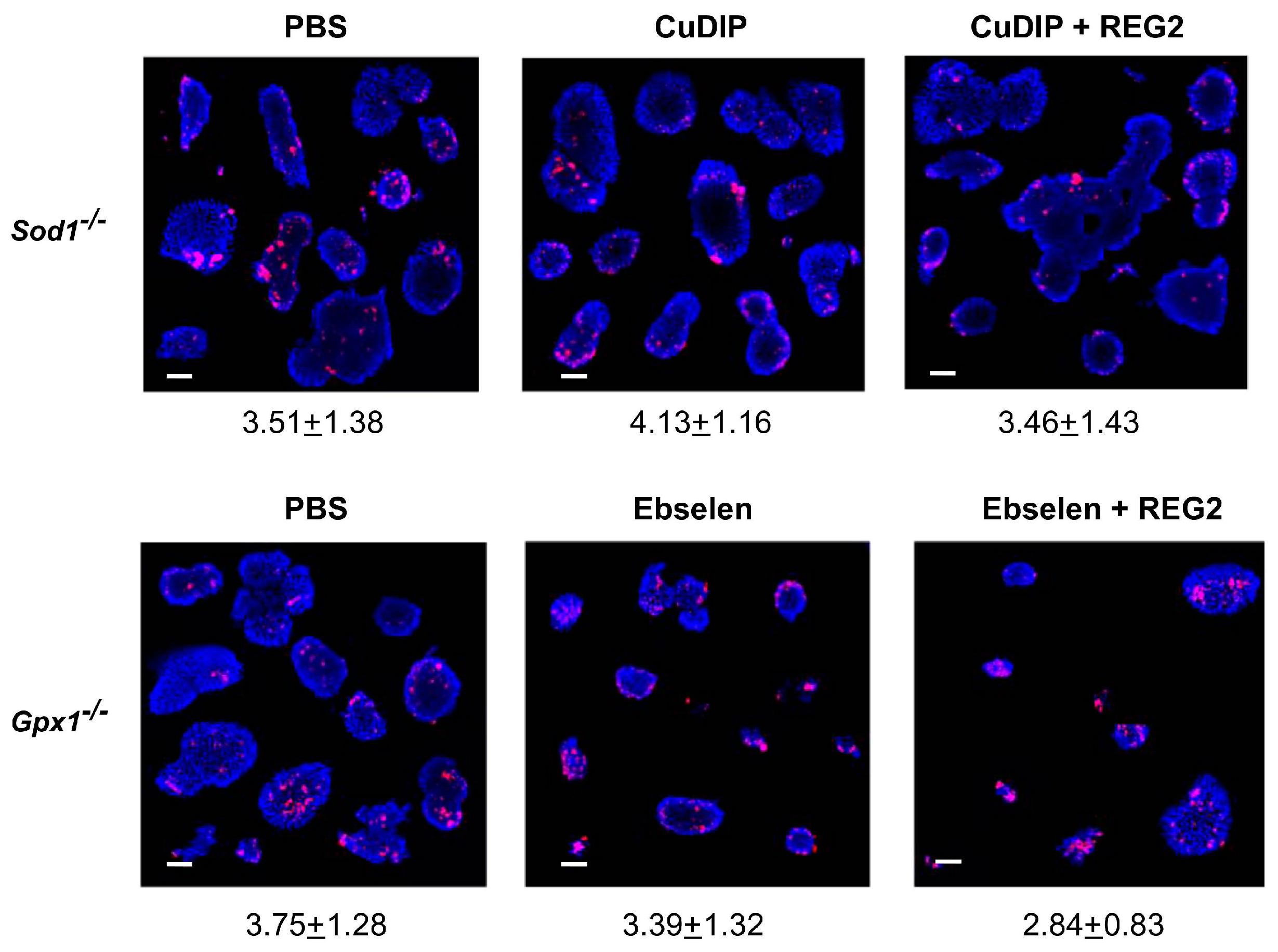
3.2. Impacts of GPX1 and SOD1 Status on Inhibitions of Islet Proliferation by Exogenous REG2 Protein

Compared with the respective PBS treatments (Figure 2), the incubation with REG2 protein led to substantial reductions of BrdU incorporation into islets of the four genetically altered groups of mice. Specifically, the staining intensity was decreased (p < 0.01) by 81% in Gpx1-/- islets, 75% in Gpx1-OE islets, 62% in Sod1-/- islets, and 88% in dKO islets. However, the incubation with the same amount of REG2 mutant (REG2 Mut) protein produced no significant changes in BrdU incorporation or staining intensity in islets of any of these four genotypes compared with the PBS treatments. The two groups of WT islets showed no response to the treatments with either REG2 or REG2 Mut compared with the PBS treatments.
Meanwhile, incubating the Gpx1-/- islets with ebselen and the Sod1-/- islets with CuDIPS (Figure 3) did not produce apparent BrdU staining changes over their respective controls treated with PBS. Strikingly, the inhibition of BrdU incorporation by REG2 disappeared in the Gpx1-/- islets co-incubated with ebselen or in the Sod1-/- islets co-incubated with CuDIPS.

3.3. Effects of Murine REG2 Protein on REG Gene Expression and Antioxidant Enzyme Activities in PANC1 Cells

To determine if murine REG2 exerted a cross-species effect on the expression of human REG genes, we treated PANC1 cells with recombinant murine REG2 for 5 or 20 h. The treatment elevated (p < 0.05 or 0.01) mRNA levels of REG1A, REG1B, REG3A, and REG3G compared with the controls. In contrast, the treatments decreased (p < 0.05) or diminished the expression of REG4 (Figure 4A). Incubating PANC1 cells with the recombinant murine REG2 for 20 h decreased GPX (p < 0.01) and SOD1 (p < 0.05) activities by 20–30% compared with the control (Figure 4B).

3.4. Effects of Murine REG2 Protein on Cell Viability and Its Dependence on Free Calcium in PANC1 Cells

To explore putative metabolic roles of murine REG2 in human cell growth and survival, we tested the effects of the recombinant murine REG2 protein on the viability of PANC1 cells (Figure 4C). Indeed, the cell viability was decreased (p < 0.05) by the recombinant murine REG2 treatment, suggesting a cross-species effectiveness. Because we revealed a potential of REG2 as a novel regulator of calcium channel activity in islets [29], we tested if adding extra free calcium to the media could affect the decrease in viability of PANC1 cells caused by the REG2 treatment (Figure 4D). In fact, adding 1.8 or 3.6 nM of free Ca2+ into the media attenuated the REG2-mediated decrease in cell viability.

4. Discussion

Our present study revealed a largely inverse relationship between murine Reg family gene expression and intracellular ROS-scavenging GPX1 and SOD1 activities in the pancreatic islets, with several exceptions. While the Gpx1-/- and Sod1-/- islets had higher mRNA levels of most of the Reg genes than their respective WT islets, the dKO islets showed further elevations in or higher mRNA levels of six genes compared to the Sod1-/- islets. In contrast, the Gpx1-OE islets had lower or diminished expression of five genes, the exceptions being Reg1 and Reg4, compared with the WT islets. This extends our earlier finding on the down-regulation of Reg2 expression in the pancreatic islets of Gpx1-OE mice [14] to a rather common negative interaction in gene expression between the two major antioxidant enzymes GPX1 and SOD1 and Reg family genes. Alternatively, our previously illustrated counteracting regulations of Reg2 by ROS and GPX1 [14] might be viewed as an interdependence of intracellular ROS-scavenging enzyme activity and Reg family gene expression [14]. Our findings may qualify antioxidant enzymes and their counterpart ROS as strong regulators of Reg family gene expression that is up-regulated by pancreatectomy [18] and pro- and anti-inflammatory cytokines [33,34,35]. It is well known that pancreatic islets have a low baseline level of antioxidant enzyme expression [36]. This feature allows islets to possess the high ROS sensitivity required for regulating insulin secretion [3]. Our results suggest that altering ROS production may influence islet functions through affecting the expression of Reg genes. It is puzzling that up-regulated Reg expression in the Gpx1-/-, Sod1-/-, and dKO islets was associated with decreased islet β-cell mass and insulin secretion [11], while down-regulated Reg expression in Gpx1-OE islets was accompanied with islet hyperplasia, hyperinsulinemia, and augmented glucose-stimulated insulin secretion [12,13].
Despite the demonstrated main trends, there were several intriguing exceptions for the relationship between antioxidant enzymes and Reg family gene expression in murine pancreatic islets. Although we showed that down-regulation of Reg2 expression in the GPX1-overproducing pancreatic islets was mediated by a transcriptional inhibition of the gene through two ROS-responsive transcription factors AP-1 and DBP [14], we could not find a uniform presence of such domains in the proximate promoters of the rest of the Reg family genes. Thus, intracellular GPX1 and SOD1 likely regulate their transcription through other transcriptional factors or mechanisms. Compared with other Reg genes, Reg1 expression was not altered whereas Reg4 expression was much more substantially (~20 fold) elevated by the knockout of Gpx1. Meanwhile, the mRNA levels of islet Reg3α and Reg3γ were not affected by knockout of Sod1 which induced greater levels of Reg2 expression than the double knockouts. Seemingly, the regulation of Reg family gene expression by GPX1 and SOD1 alterations involves additional mechanisms associated with specific ROS species instead of just total levels, individual responsive elements in the Reg gene promoters, and their interactions. Most notably, islet Reg4 expression was induced by both knockout and overproduction of GPX1. These unilateral responses to both increases and decreases in intracellular antioxidant enzyme activities were distinctly different from those of the other six Reg genes. Whereas a detailed mechanism for this difference remains to be revealed, Reg4 is the most phylogenetically different gene within the Reg family. In mice, the other six Reg genes were mapped to a 75 kb region of chromosome 6C while Reg4 is located on chromosome 3 [37]. In humans, four of the five REG genes are located in a 95 kb region of chromosome 2p12 [38], but REG4 is on chromosome 1 [39].
Another interesting finding of our study is the inhibition of proliferation by exogenous REG2 protein in the Gpx1-/- and Sod1-/-, but not WT, islets. While REG2 Mut had no such effect on islet proliferation, GPX1 and SOD1 mimics obviated the REG2-mediated inhibition. This raises a serious question against the potential of REG2 as a crucial growth factor for pancreatic islet proliferation [40]. In fact, Reg2-/- mice maintained normal β-cell mass until later stages of life or the onset of obesity induced by a high-fat diet [25]. As mentioned above, we have shown that the diminished Reg2 expression in the pancreatic islets of Gpx1-OE mice was associated with elevated β-cell mass [12]. In contrast, pancreatic β-cell mass was decreased in the Gpx1-/- and Sod1-/- mice [12] that expressed greater levels of Reg genes than the WT mice. Reciprocally, a pancreatic acinar overexpression of Reg2 did not confer any protection against pancreatitis or diabetes [41]. Seemingly, we should not simply view REG2 as a “default” growth factor for intact or unstressed islet β-cells or other types of cells. Instead, it may function as an acute stress reactant in regulating the growth or survival of cells with altered or compromised metabolism. Thus, our study suggests a link between the function and the outcome of REG2 to the intracellular redox status or antioxidant activity context. This may help explain why exogenous REG2 inhibited the islet proliferation of Gpx1-/- and Sod1-/- mice but not the WT mice, why REG2 inhibited islet proliferation in both Gpx1-/- and Gpx1-OE mice, and why restoring normal redox status with GPX1 and SOD1 mimics precluded such inhibition. In fact, REG2 was shown to inhibit nuclear entry of apoptosis-inducing factor by promoting the nuclear presence of Scythe and inducing heat shock protein 70 (HSP70) in mouse insulinoma cells [42]. In addition, Gpx1 overexpression was associated with a thermo-sensitive phenotype via a direct participation of peroxides in the induction of cytoprotective protein HSP70 [43]. A possible extended linking of GPX1-HSP70-Scythe-REG2 may depict a dependence of cell apoptosis/division on its redox state [44]. In addition, REG3β is considered unlikely to be an islet growth factor but a putative protector that prevents streptozotocin-induced damage by inducing expression of specific genes [45]. Expression of human REG1B, an orthologue of mouse REG2, is altered under conditions of inflammatory diseases [33,34,35], cancer [46], and childhood stunting [33].
Our third interesting finding is that exogenous murine REG2 protein decreased the viability of human pancreatic cancer cells (PANC1), and the inhibition disappeared after the addition of extra free Ca2+ ions into the media. This action was consistent with its inhibition of the proliferation of Gpx1-/- and Sod1-/- islets, along with a down-regulation of both GPX and SOD activities in the cells. These results suggest a cross-species effectiveness of REG2 with an anti-human cancer potential and an involvement of calcium channels in the mechanism [33,46]. The lack of inhibition of islet proliferation by REG2 Mut (with a destruction of the C-type lectin binding domain) supports this notion. Interestingly, murine REG2 enhanced the expression of REG1A, REG1B, REG3A, and REG3G, but decreased or diminished the expression of REG4 in PANC1 cells. Different types of cancer are associated with increased level of REG family proteins [47]. REG1A, REG1B, and REG3A were reported to be increased in cancer ductal fluid [48], and REG1A was linked to pancreatic cancer [47]. However, the metabolic functions of REG3β remain controversial. Gironello reported that REG3β deletion in mice drastically impaired pancreatic tumor growth, correlating with decreased angiogenesis and increased apoptosis of tumor cells [48]. Liu and colleagues reported that REG3β negatively regulated cytokine-induced activation of STAT-3 in colon epithelial cells thus reducing carcinogenesis [49]. However, in HIP/PAP (REG3α) knockout mice, PAP showed an antiapoptotic and anti-inflammatory effect during cerulean-induced acute pancreatitis [50], while antisense knockdown of PAP expression exacerbated the severity of pancreatitis, suggesting a protective function against pancreatitis [51].

5. Conclusions

Altering GPX1 and SOD1 activities, either by single or double knockout of the two major ROS-scavenging enzymes, enhanced the expression of most murine Reg family genes in the pancreatic islets. These increases were contrary to the down-regulation induced by the overproduction of GPX1 activity in the islets. However, there were variations with individual Reg genes and antioxidant enzymes. Exogenous murine REG2 protein inhibited the proliferation of Gpx1-/- and Sod1-/- islets and decreased cell viability of human pancreatic cancer cells (PANC1). Meanwhile, these actions of REG2 were obviated by the additions of GPX and SOD mimics or free Ca2+ ions, and thus there was no simple cascade to explain the underlying mechanism. Although additional research is still needed to completely unveil the complexity of this matter, our current findings reveal an interdependence of gene expression and function between two redox enzymes and the REG family proteins in murine pancreatic islets and human pancreatic cancer cells.

Supplementary Materials

The following are available online at https://www.mdpi.com/article/10.3390/antiox12040849/s1, Table S1: Primers used for Q-PCR analysis.

Author Contributions

X.G.L. conceived and designed the study and edited the manuscript; H.W. performed the study and analyzed the results; Z.Z. prepared the Reg2 and Reg2 mutant proteins for the study; M.Z.V., H.W. and X.G.L. wrote the manuscript. All authors have read and agreed to the published version of the manuscript.

Funding

This research received no external funding.

Institutional Review Board Statement

This study was approved by the Institutional Animal Care and Use Committee of Cornell University (Protocol # 2007-0006, approved on 05.20/2014).

Informed Consent Statement

Not applicable.

Data Availability Statement

All data are contained within the article.

Conflicts of Interest

The authors declare no conflict of interest.

References

  1. Dröge, W. Free radicals in the physiological control of cell function. Physiol. Rev. 2002, 82, 47–95. [Google Scholar] [CrossRef] [PubMed] [Green Version]
  2. Valko, M.; Leibfritz, D.; Moncol, J.; Cronin, M.T.; Mazur, M.; Telser, J. Free radicals and antioxidants in normal physiological functions and human disease. Int. J. Biochem. Cell Biol. 2007, 39, 44–84. [Google Scholar] [CrossRef] [PubMed]
  3. Pi, J.; Bai, Y.; Zhang, Q.; Wong, V.; Floering, L.M.; Daniel, K.; Reece, J.M.; Deeney, J.T.; Andersen, M.E.; Corkey, B.E.; et al. Reactive oxygen species as a signal in glucose-stimulated insulin secretion. Diabetes 2007, 56, 1783–1791. [Google Scholar] [CrossRef] [Green Version]
  4. Houstis, N.; Rosen, E.D.; Lander, E.S. Reactive oxygen species have a causal role in multiple forms of insulin resistance. Nature 2006, 440, 944–948. [Google Scholar] [CrossRef] [PubMed]
  5. Mills, G.C. Hemoglobin catabolism. I. Glutathione peroxidase, an erythrocyte enzyme which protects hemoglobin from oxidative breakdown. J. Biol. Chem. 1957, 229, 189–197. [Google Scholar] [CrossRef]
  6. Flohe, L.; Gunzler, W.; Schock, H. Glutathione peroxidase: A selenoenzyme. FEBS Lett. 1973, 32, 132–134. [Google Scholar] [CrossRef] [Green Version]
  7. Fridovich, I. Superoxide radical and superoxide dismutases. Annu. Rev. Biochem. 1995, 64, 97–112. [Google Scholar] [CrossRef]
  8. Cheng, W.H.; Ho, Y.S.; Ross, D.A.; Han, Y.; Combs, G.F., Jr.; Lei, X.G. Overexpression of cellular glutathione peroxidase does not affect expression of plasma glutathione peroxidase or phospholipid hydroperoxide glutathione peroxidase in mice offered diets adequate or deficient in selenium. J. Nutr. 1997, 127, 675–680. [Google Scholar] [CrossRef] [Green Version]
  9. Lei, X.G.; Zhu, J.H.; McClung, J.P.; Aregullin, M.; Roneker, C.A. Mice deficient in Cu,Zn-superoxide dismutase are resistant to acetarninophen toxicity. Biochem. J. 2006, 399, 455–461. [Google Scholar] [CrossRef] [PubMed] [Green Version]
  10. Lei, X.G.; Vatamaniuk, M.Z. Two tales of antioxidant enzymes on β cells and diabetes. Antioxid. Redox Signal. 2011, 14, 489–503. [Google Scholar] [CrossRef] [PubMed] [Green Version]
  11. Wang, X.D.; Vatamaniuk, M.Z.; Roneker, C.A.; Pepper, M.P.; Hu, L.G.; Simmons, R.A.; Lei, X.G. Knockouts of SOD1 and GPX1 exert different impacts on murine islet function and pancreatic integrity. Antioxid. Redox Signal. 2011, 14, 391–401. [Google Scholar] [CrossRef] [PubMed] [Green Version]
  12. Wang, X.D.; Vatamaniuk, M.Z.; Wang, S.K.; Roneker, C.A.; Simmons, R.A.; Lei, X.G. Molecular mechanisms for hyperinsulinaemia induced by overproduction of selenium-dependent glutathione peroxidase-1 in mice. Diabetologia 2008, 51, 1515–1524. [Google Scholar] [CrossRef] [Green Version]
  13. McClung, J.P.; Roneker, C.A.; Mu, W.; Lisk, D.J.; Langlais, P.; Liu, F.; Lei, X.G. Development of insulin resistance and obesity in mice overexpressing cellular glutathione peroxidase. Proc. Natl. Acad. Sci. USA 2004, 101, 8852–8857. [Google Scholar] [CrossRef] [PubMed] [Green Version]
  14. Yun, J.W.; Zhao, Z.; Yan, X.; Vatamaniuk, M.Z.; Lei, X.G. Glutathione peroxidase-1 inhibits transcription of regenerating islet-derived protein-2 in pancreatic islets. Free Radic. Biol. Med. 2019, 134, 385–393. [Google Scholar] [CrossRef] [PubMed]
  15. Qiu, L.; List, E.O.; Kopchick, J.J. Differentially expressed proteins in the pancreas of diet-induced diabetic mice. Mol. Cell Proteom. 2005, 4, 1311–1318. [Google Scholar] [CrossRef] [Green Version]
  16. Liu, J.L.; Cui, W. Which gene, Reg2 or Reg3beta, was targeted that affected liver regeneration? Hepatology 2007, 45, 1584–1585. [Google Scholar] [CrossRef]
  17. Narushima, Y.; Unno, M.; Nakagawara, K.; Mori, M.; Miyashita, H.; Suzuki, Y.; Noguchi, N.; Takasawa, S.; Kumagai, T.; Yonekura, H.; et al. Structure, chromosomal localization and expression of mouse genes encoding type III Reg, RegIII alpha, RegIII beta, RegIII gamma. Gene 1997, 185, 159–168. [Google Scholar] [CrossRef]
  18. Terazono, K.; Yamamoto, H.; Takasawa, S.; Shiga, K.; Yonemura, Y.; Tochino, Y.; Okamoto, H. A novel gene activated in regenerating islets. J. Biol. Chem. 1988, 263, 2111–2114. [Google Scholar] [CrossRef]
  19. Huszarik, K.; Wright, B.; Keller, C.; Nikoopour, E.; Krougly, O.; Lee-Chan, E.; Qin, H.Y.; Cameron, M.J.; Gurr, W.K.; Hill, D.J.; et al. Adjuvant immunotherapy increases beta cell regenerative factor Reg2 in the pancreas of diabetic mice. J. Immunol. 2010, 185, 5120–5129. [Google Scholar] [CrossRef] [Green Version]
  20. Lieu, H.T.; Simon, M.T.; Nguyen-Khoa, T.; Kebede, M.; Cortes, A.; Tebar, L.; Smith, A.J.; Bayne, R.; Hunt, S.P.; Bréchot, C.; et al. Reg2 inactivation increases sensitivity to Fas hepatotoxicity and delays liver regeneration post-hepatectomy in mice. Hepatology 2006, 44, 1452–1464. [Google Scholar] [CrossRef]
  21. Baeza, N.J.; Moriscot, C.I.; Renaud, W.P.; Okamoto, H.; Figarella, C.G.; Vialettes, B.H. Pancreatic regenerating gene overexpression in the nonobese diabetic mouse during active diabetogenesis. Diabetes 1996, 45, 67–70. [Google Scholar] [CrossRef]
  22. Cavard, C.; Terris, B.; Grimber, G.; Christa, L.; Audard, V.; Radenen-Bussiere, B.; Simon, M.T.; Renard, C.A.; Buendia, M.A.; Perret, C. Overexpression of regenerating islet-derived 1 alpha and 3 alpha genes in human primary liver tumors with beta-catenin mutations. Oncogene 2006, 25, 599–608. [Google Scholar] [CrossRef] [Green Version]
  23. Harris, A.C.; Ferrara, J.L.; Braun, T.M.; Holler, E.; Teshima, T.; Levine, J.E.; Choi, S.W.; Landfried, K.; Akashi, K.; Vander Lugt, M.; et al. Plasma biomarkers of lower gastrointestinal and liver acute GVHD. Blood 2012, 119, 2960–2963. [Google Scholar] [CrossRef] [PubMed] [Green Version]
  24. Gurr, W.; Shaw, M.; Li, Y.; Sherwin, R. RegII is a beta-cell protein and autoantigen in diabetes of NOD mice. Diabetes 2007, 56, 34–40. [Google Scholar] [CrossRef] [PubMed] [Green Version]
  25. Li, Q.; Li, B.; Miao, X.; Ramgattie, C.; Gao, Z.H.; Liu, J.L. Reg2 expression is required for pancreatic islet compensation in response to aging and high-fat diet-induced obesity. Endocrinology 2017, 158, 1634–1644. [Google Scholar] [CrossRef] [PubMed] [Green Version]
  26. Duplan, L.; Michel, B.; Boucraut, J.; Barthellémy, S.; Desplat-Jego, S.; Marin, V.; Gambarelli, D.; Bernard, D.; Berthézène, P.; Alescio-Lautier, B.; et al. Lithostathine and pancreatitis-associated protein are involved in the very early stages of Alzheimer’s disease. Neurobiol. Aging 2001, 22, 79–88. [Google Scholar] [CrossRef] [PubMed]
  27. Ho, Y.S.; Gargano, M.; Cao, J.; Bronson, R.T.; Heimler, I.; Hutz, R.J. Reduced fertility in female mice lacking copper-zinc superoxide dismutase. J. Biol. Chem. 1998, 273, 7765–7769. [Google Scholar] [CrossRef] [Green Version]
  28. Ho, Y.S.; Magnenat, J.L.; Bronson, R.T.; Cao, J.; Gargano, M.; Sugawara, M.; Funk, C.D. Mice deficient in cellular glutathione peroxidase develop normally and show no increased sensitivity to hyperoxia. J. Biol. Chem. 1997, 272, 16644–16651. [Google Scholar] [CrossRef] [PubMed] [Green Version]
  29. Yan, X.; Zhao, Z.; Weaver, J.; Sun, T.; Yun, J.W.; Roneker, C.A.; Hu, F.; Doliba, N.M.; McCormick, C.C.W.; Vatamaniuk, M.Z.; et al. Role and mechanism of REG2 depletion in insulin secretion augmented by glutathione peroxidase-1 overproduction. Redox Biol. 2022, 56, 102457. [Google Scholar] [CrossRef] [PubMed]
  30. Walker, J.R.; Nagar, B.; Young, N.M.; Hirama, T.; Rini, J.M. X-ray crystal structure of a galactose-specific C-type lectin possessing a novel decameric quaternary structure. Biochemistry 2004, 43, 3783–3792. [Google Scholar] [CrossRef]
  31. Sun, L.H.; Li, J.G.; Zhao, H.; Shi, J.; Huang, J.Q.; Wang, K.N.; Xia, X.J.; Li, L.; Lei, X.G. Porcine serum can be biofortified with selenium to inhibit proliferation of three types of human cancer cells. J. Nutr. 2013, 143, 1115–1122. [Google Scholar] [CrossRef] [PubMed] [Green Version]
  32. Lawrence, R.A.; Sunde, R.A.; Schwartz, G.L.; Hoekstra, W.G. Glutathione peroxidase activity in rat lens and other tissues in relation to dietary selenium intake. Exp. Eye Res. 1974, 18, 563–569. [Google Scholar] [CrossRef] [PubMed]
  33. Peterson, K.M.; Buss, J.; Easley, R.; Yang, Z.; Korpe, P.S.; Niu, F.; Ma, J.Z.; Olortegui, M.P.; Haque, R.; Kosek, M.N.; et al. REG1B as a predictor of childhood stunting in Bangladesh and Peru. Am. J. Clin. Nutr. 2013, 97, 1129–1133. [Google Scholar] [CrossRef] [Green Version]
  34. Yamauchi, A.; Itaya-Hironaka, A.; Sakuramoto-Tsuchida, S.; Takeda, M.; Yoshimoto, K.; Miyaoka, T.; Fujimura, T.; Tsujinaka, H.; Tsuchida, C.; Ota, H.; et al. Synergistic activations of REGIα and REGIβ promoters by IL-6 and glucocorticoids through JAK/STAT pathway in human pancreatic β cells. J. Diabetes Res. 2015, 2015, 173058. [Google Scholar] [CrossRef] [PubMed] [Green Version]
  35. van Beelen Granlund, A.; Østvik, A.E.; Brenna, Ø.; Torp, S.H.; Gustafsson, B.I.; Sandvik, A.K. REG gene expression in inflamed and healthy colon mucosa explored by in situ hybridisation. Cell Tissue Res. 2013, 352, 639–646. [Google Scholar] [CrossRef] [Green Version]
  36. Lenzen, S.; Drinkgern, J.; Tiedge, M. Low antioxidant enzyme gene expression in pancreatic islets compared with various other mouse tissues. Free Radic. Biol. Med. 1996, 20, 463–466. [Google Scholar] [CrossRef]
  37. Abe, M.; Nata, K.; Akiyama, T.; Shervani, N.J.; Kobayashi, S.; Tomioka-Kumagai, T.; Ito, S.; Takasawa, S.; Okamoto, H. Identification of a novel Reg family gene, Reg IIIδ, and mapping of all three types of Reg family gene in a 75 kilobase mouse genomic region. Gene 2000, 246, 111–122. [Google Scholar] [CrossRef]
  38. Miyashita, H.; Nakagawara, K.; Mori, M.; Narushima, Y.; Noguchi, N.; Moriizumi, S.; Takasawa, S.; Yonekura, H.; Takeuchi, T.; Okamoto, H. Human REG family genes are tandemly ordered in a 95-kilobase region of chromosome 2p12. FEBS Lett. 1995, 377, 429–433. [Google Scholar]
  39. Hartupee, J.C.; Zhang, H.; Bonaldo, M.F.; Soares, M.B.; Dieckgraefe, B.K. Isolation and characterization of a cDNA encoding a novel member of the human regenerating protein family: Reg IV. Biochim. Et Biophys. Acta (BBA)-Gene Struct. Expr. 2001, 1518, 287–293. [Google Scholar] [CrossRef]
  40. De León, D.D.; Farzad, C.; Crutchlow, M.F.; Brestelli, J.; Tobias, J.; Kaestner, K.H.; Stoffers, D.A. Identification of transcriptional targets during pancreatic growth after partial pancreatectomy and exendin-4 treatment. Physiol. Genom. 2006, 24, 133–143. [Google Scholar] [CrossRef]
  41. Li, B.; Wang, X.; Liu, J.L. Pancreatic acinar-specific overexpression of Reg2 gene offered no protection against either experimental diabetes or pancreatitis in mice. Am. J. Physiol. Gastrointest. Liver Physiol. 2010, 299, G413–G421. [Google Scholar] [CrossRef] [Green Version]
  42. Liu, L.; Chowdhury, S.; Uppal, S.; Fang, X.; Liu, J.L.; Srikant, C.B. mReg2 inhibits nuclear entry of apoptosis-inducing factor in mouse insulinoma cells. Growth Factors 2015, 33, 1–7. [Google Scholar] [CrossRef] [PubMed]
  43. Mirochnitchenko, O.; Palnitkar, U.; Philbert, M.; Inouye, M. Thermosensitive phenotype of transgenic mice overproducing human glutathione peroxidases. Proc. Natl. Acad. Sci. USA 1995, 92, 8120–8124. [Google Scholar] [CrossRef] [PubMed] [Green Version]
  44. Alenzi, F.Q. Links between apoptosis, proliferation and the cell cycle. Br. J. Biomed. Sci. 2004, 61, 99–102. [Google Scholar] [CrossRef]
  45. Xiong, X.; Wang, X.; Li, B.; Chowdhury, S.; Lu, Y.; Srikant, C.B.; Ning, G.; Liu, J.L. Pancreatic islet-specific overexpression of Reg3beta protein induced the expression of pro-islet genes and protected the mice against streptozotocin-induced diabetes mellitus. Am. J. Physiol. Endocrinol. Metab. 2011, 300, E669–E680. [Google Scholar] [CrossRef] [Green Version]
  46. Liu, Z.; Zhang, Y.; Xie, J.; Li, C.; Wang, X.; Shen, J.; Zhang, Y.; Wang, S.; Cheng, N. Regenerating gene 1B silencing inhibits colon cancer cell HCT116 proliferation and invasion. Int. J. Biol. Markers 2015, 30, 217–225. [Google Scholar] [CrossRef]
  47. Zhou, L.; Zhang, R.; Wang, L.; Shen, S.; Okamoto, H.; Sugawara, A.; Xia, L.; Wang, X.; Noguchi, N.; Yoshikawa, T.; et al. Upregulation of REG Ialpha accelerates tumor progression in pancreatic cancer with diabetes. Int. J. Cancer 2010, 127, 1795–1803. [Google Scholar] [CrossRef] [PubMed]
  48. Gironella, M.; Calvo, C.; Fernández, A.; Closa, D.; Iovanna, J.L.; Rosello-Catafau, J.; Folch-Puy, E. Reg3beta deficiency impairs pancreatic tumor growth by skewing macrophage polarization. Cancer Res. 2013, 73, 5682–5694. [Google Scholar] [CrossRef] [PubMed] [Green Version]
  49. Liu, X.; Wei, W.; Li, X.; Shen, P.; Ju, D.; Wang, Z.; Zhang, R.; Yang, F.; Chen, C.; Cao, K. BMI1 and MEL18 promote colitis-associated cancer in mice via REG3B and STAT3. Gastroenterology 2017, 153, 1607–1620. [Google Scholar] [CrossRef] [Green Version]
  50. Gironella, M.; Folch-Puy, E.; LeGoffic, A.; Garcia, S.; Christa, L.; Smith, A.; Tebar, L.; Hunt, S.P.; Bayne, R.; Smith, A.J. Experimental acute pancreatitis in PAP/HIP knock-out mice. Gut 2007, 56, 1091–1097. [Google Scholar] [CrossRef] [Green Version]
  51. Zhang, H.; Kandil, E.; Lin, Y.Y.; Levi, G.; Zenilman, M.E. Targeted inhibition of gene expression of pancreatitis-associated proteins exacerbates the severity of acute pancreatitis in rats. Scand. J. Gastroenterol. 2004, 39, 870–881. [Google Scholar] [CrossRef] [PubMed] [Green Version]
Figure 1. Relative abundance of Reg mRNA in pancreatic islets of wild-type (WT), Gpx1-/-, and Gpx1-OE mice (A), and WT, Sod1-/-, and Gpx1-/-Sod1-/- (dKO) mice (B). Gene expression was quantified by Q-PCR and normalized with β-actin expression. Values were expressed as relative expression levels to respective WT and presented as mean ± SD. Data were analyzed using one-way analysis of variance (ANOVA) for individual genes. Means of the same gene without sharing a common letter differ at p < 0.05; n = 4–6.
Antioxidants 12 00849 g001
Figure 2. BrdU (red) immunostaining of pancreatic islets (blue) isolated from WT, Gpx1-/-, Gpx1-OE, Sod1-/-, and dKO mice. Islets were incubated with REG2 and REG2 Mut for 48 h before the proliferation assay. Numbers under the image panel represent relative BrdU fluorescent intensity, and means with * differ at p < 0.01 compared with the PBS control. Data were analyzed using Student’s t test within each genotype. Representative images are displayed for each condition; n = 4. Scale bar = 50 μm.
Antioxidants 12 00849 g002
Figure 3. Effects of co-incubation with GPX mimic ebselen or SOD mimic CuDIP on the REG2-mediated inhibition of islet proliferation measured by the BrdU (red) immunostaining in the Gpx1-/- and Sod1-/- islets. Numbers under the image panel represent relative BrdU fluorescent intensity. Data were analyzed using Student’s t test within each genotype. Images are representative of replicated experiments: n = 3–4. Scale bar = 50 μm.
Antioxidants 12 00849 g003
Figure 4. Effects of treating human PANC1 (cancer) cells with murine REG2 protein on mRNA levels of 5 human REG genes [(A), after 5 and 20 h of treatment], GPX1 and SOD activities [(B), after 20 h of treatment], cell viability [(C), after 20 h of treatment], and cell viability at three doses of additional calcium ions [(D), after 20 h of treatment]. Columns with * or ** differ significantly at p < 0.05 or p < 0.01 from control, respectively. Data were analyzed using Student’s t test within each REG gene (A) or calcium concentration (D).
Antioxidants 12 00849 g004
Disclaimer/Publisher’s Note: The statements, opinions and data contained in all publications are solely those of the individual author(s) and contributor(s) and not of MDPI and/or the editor(s). MDPI and/or the editor(s) disclaim responsibility for any injury to people or property resulting from any ideas, methods, instructions or products referred to in the content.

