Article

Interaction and Redox Chemistry between Iron, Dopamine, and Alpha-Synuclein C-Terminal Peptides

1
Department of Chemistry, University of Pavia, Via Taramelli 12, 27100 Pavia, Italy
2
IUSS School for Advanced Studies of Pavia, Palazzo del Broletto, Piazza della Vittoria 15, 27100 Pavia, Italy
*
Authors to whom correspondence should be addressed.
Antioxidants 2023, 12(4), 791; https://doi.org/10.3390/antiox12040791
Submission received: 25 February 2023 / Revised: 17 March 2023 / Accepted: 20 March 2023 / Published: 24 March 2023
(This article belongs to the Special Issue Iron Metabolism, Oxidative Stress and Cellular Dysfunction)

Abstract

:
α-Synuclein (αS), dopamine (DA), and iron have a crucial role in the etiology of Parkinson’s disease. The present study aims to investigate the interplay between these factors by analyzing the DA/iron interaction and how it is affected by the presence of the C-terminal fragment of αS (Ac-αS119–132) that represents the iron-binding domain. At high DA:Fe molar ratios, the formation of the [FeIII(DA)2] complex prevents the interaction with αS peptides, whereas, at lower DA:Fe molar ratios, the peptide is able to compete with one of the two coordinated DA molecules. This interaction is also confirmed by HPLC-MS analysis of the post-translational modifications of the peptide, where oxidized αS is observed through an inner-sphere mechanism. Moreover, the presence of phosphate groups in Ser129 (Ac-αSpS119–132) and both Ser129 and Tyr125 (Ac-αSpYpS119–132) increases the affinity for iron(III) and decreases the DA oxidation rate, suggesting that this post-translational modification may assume a crucial role for the αS aggregation process. Finally, αS interaction with cellular membranes is another key aspect for αS physiology. Our data show that the presence of a membrane-like environment induced an enhanced peptide effect over both the DA oxidation and the [FeIII(DA)2] complex formation and decomposition.

1. Introduction

The interplay between iron, dopamine (DA), and α-synuclein (αS) seems to be a key feature of degenerating neurons in Parkinson’s disease (PD) [1]. In fact, from one side, it is well known that iron is able to exacerbate DA toxicity by promoting its oxidation to neurotoxic species [2], while, on the other hand, αS is able to bind to iron in both oxidation states [3]. The presence of an intracellular pool of labile, chelatable iron has long been recognized [4], and recent progress toward its characterization and distribution has been made in bacterial cells [5]. More iron can be released by damaged neurons, particularly those of the substantia nigra (SN), which contain the iron-rich neuromelanin pigment [6]. The dopaminergic neurons of SN are, in fact, particularly vulnerable to oxidative stress, mainly due to their direct involvement in DA metabolism which, if not controlled, can lead to the production of reactive quinones and ROS [7]. Furthermore, the produced quinones are able to react with proteins, like αS, thus preventing the protein from exerting its physiological role and ultimately leading to a vicious cycle in which the increased cytosolic DA exacerbates the oxidative stress [8]. Iron, being a redox-active transition metal, can promote DA oxidation (Scheme 1) by overcoming the spin restriction nature of dioxygen reduction. In addition, the produced superoxide and hydrogen peroxide can be converted into hydroxyl radical through Fenton- and Haber–Weiss-type reactions. Alongside the quinones and ROS production, iron–DA interaction can produce 6-hydroxydopamine, a neurotoxin used to simulate PD in animal models, although to a modest extent [2].
The interaction between iron and DA is strongly dependent on the DA:Fe molar ratio; at low molar ratio the interaction is pro-oxidant, leading to the production of ROS and Fenton chemistry, while at high molar ratio, where the coordinatively saturated [FeIII(DA)3]3– species is formed, the interaction is protective, since the strong DA complexation hinders the reaction between iron and hydrogen peroxide. Despite this, the internal electron transfer is still possible, leading to the same toxic DA quinones [9]. It is worth noting that DA is able to mobilize iron from amorphous aggregates of iron hydroxide through a reductive mechanism (Scheme 1). These latter aspects of Fe–DA redox chemistry are of paramount importance in promoting toxicity in the long term, also considering the boundary conditions observed in PD, like decreased ferritin levels, impaired iron homeostasis, and decreased DA levels as well [10].
Regarding the effects of αS-iron interaction, in addition to the ability of the protein to bind to the metal ion, the protein contains an iron-responsive element in its 5’-untranslated region, like ferritin; thus, the protein translation appears to be regulated by iron levels. In depleted iron conditions, protein synthesis is inhibited, while, in the case of iron overload, the protein is synthesized [3]. Nevertheless, metal binding makes the protein more prone to aggregation by stabilizing particular local conformations, reducing electrostatic repulsion, and even acting as a cross-linker [11]. Remarkably, iron overload could start a vicious toxic cycle in which the excess iron level promotes protein translation and, ultimately, the related toxicity.
The iron-binding site in αS is located in the C-terminus of the protein encompassing the 119DPDNEA124 motif, in which D119 acts as the main anchoring residue. The coordination sphere is poorly characterized, but it seems that D121, E123, and S129 participate in iron binding (Figure 1) [12,13]. Furthermore, precise binding location also relies on the phosphorylation state of the protein where, upon such modification, a shift in the coordination sphere is likely to occur [12,13]. Phosphorylation also enhances the iron-binding properties between 10 and 50-fold when compared to the wild type [13,14]. In spite of the appreciable protein affinity towards iron [15], in aerated solutions at circumneutral pH and in the presence of DA, iron hydrolysis and [FeIII(DA)2] complex formation overcomes iron binding to αS, as can be deducted from the log K values reported in Scheme 2 [9,16].
The reactivity of DA and iron ions has been extensively studied by Waite et al. in various conditions, and complex kinetic modeling was used to deduce rate constants for the various reactions involved [9,10]. The effect of iron binding by αS on this chemistry, however, is not known, and we aimed to investigate the reactivity of the Fe–DA system using the αS C-terminal peptide Ac-119DPDNEAYEMPSEEG132-NH2 (Ac-αS119–132), containing the iron-binding site and the phosphorylated mutant Ac-119DPDNEAYEMP(pS)EEG132-NH2 (Ac-αSpS119–132). The choice of this particular mutant was made based on the finding that phosphorylated αS at S129 is the major form present in Lewy bodies [19], and that phosphorylation increases the binding affinity of the protein for both iron(II) and iron(III) [14,20].
Furthermore, the phosphorylation of Y125 is indicated to be involved in the pathological mechanism of αS aggregation [21,22]; however, to date, the role of this post-translational modification has not been analyzed at the molecular level. For this reason, we decided to study the interaction between iron and αS also with the peptide bearing the phosphate group at both S129 and Y125 (Ac-119DPDNEA(pY)EMP(pS)EEG132-NH2 (Ac-αSpYpS119–132).
Another important issue to consider is that, whereas αS is unstructured in solution, its interaction with lipid membrane modulates the conformation by inducing an α-helical structure of the N-terminal region [23]. This conformational change delays the oligomerization of the protein [24] and modulates the interaction with metal ions, as recently observed for copper-αS complex formation [25,26]. For this reason, the analysis of the interaction between iron and αS should also consider the presence of a membrane-like environment.

2. Materials and Methods

2.1. Materials

Protected amino acids, rink amide resin, and benzotriazole-1-yl-oxy-tris-pyrrolidino-phosphonium hexafluorophosphate (PyBOP) were purchased from Novabiochem, while all other reagents were purchased from Merck at the highest purity available and used as received. All glassware was acid-washed with 6 M HCl for 1 week and rinsed thoroughly with ultrapure water. All aqueous solutions were prepared in ultrapure 18 MΩ Milli-Q water. Stock solutions of FeII were prepared by dissolving FeSO4·7H2O in 10 mM H2SO4. Stock solutions of Cu(II) were made by dissolving CuSO4·5H2O in water. Stock solutions of dopamine were prepared in HCl 10 mM. Stock solutions of Ac-αS119–132, Ac-αSpS119–132, and Ac-αSpYpS119–132 were prepared by dissolving the corresponding peptide in H2O; the concentrations were assessed by UV-Vis spectroscopy, measuring the absorbance of Y125 (λmax = 275 nm, ε275 = 1420 M−1 cm−1) or pY125 (λmax = 266 nm, ε266 = 500 M−1 cm−1) [20]. Stock solutions of horseradish peroxidase (HRP) were prepared by dissolving the enzyme in H2O, and the concentration was assessed by UV-Vis spectroscopy, measuring the absorbance of the Soret band of the protein (λmax = 402 nm, ε402 = 103,000 M−1 cm−1). The buffers used were adjusted to proper pH using NaOH, H3PO4, HCl, or HNO3, depending on the buffer type.

2.2. Instrumentation

2.2.1. RP-HPLC

The chromatographic separations were operated with a Shimadzu Prominence instrument equipped with a DGU-20A3R degassing unit, two LC-20AD pumps, an SPD-M20A photodiode array (190–800 nm wavelength range), and a CTO-20A column oven. The columns used were a Phenomenex Jupiter 4U Proteo 90A, 250 × 10 mm as semi-preparative and a Phenomenex Jupiter 4U Proteo 90A, 250 × 4.6 mm or a Supelco Ascentis Express 2.7 µm C18, 100 × 4.6 mm as analytical. The solvents used were CH3CN and H2O both with 0.1% TFA for gradient elution or 98% HCOOH pH 2 plus 2% CH3CN for isocratic elution.

2.2.2. Mass Spectrometry

The MS and MS/MS spectra were recorded with a Thermo Finnigan LCQ ADV MAX equipped with an ionic-trap and an ESI ion source. The spectra were analyzed with Xcalibur 2.0.7 SP1 software.

2.2.3. HPLC-MS

The HPLC-MS analysis was performed on a Thermo Finnigan HPLC autosampler Surveyor IC pump coupled with a Thermo Finnigan LCQ ADV MAX. Fragmentation was achieved by collision-induced dissociation (CID), with an isolation width of 2 Th (m/z); the activation amplitude was around 35% of the ejection radio frequency amplitude of the instrument. The column used was a Phenomenex Jupiter 4U Proteo 90A, 150 × 2 mm, flow rate 0.2 mL/min. The solvents used were H2O plus 0.1% HCOOH (A) and CH3CN plus 0.1% HCOOH (B). Separations were performed with a semi-linear gradient (0–5 min 2% B, 5–50 min 2–33% B, 50–55 min 33–100% B, 55–73 min 100% B, 73–78 100–2% B, and 78–90 min 2% B). Data were analyzed with XcaliburTM 2.0.7 SP1 software.

2.2.4. UV-Vis Spectroscopy

The UV-Vis spectra were acquired with an Agilent 8453 spectrophotometer, equipped with a 1024 photodiode array detector (190–1100 nm wavelength range) and a magnetically stirred, thermostated sample holder. All spectra were recorded in a quartz cell with an optical path of 1 cm.

2.3. General Methods

2.3.1. Peptide Synthesis

The Ac-αS119–132 (Ac-119DPDNEAYEMPSEEG132-NH2, M.W. 1623.6 g/mol) was synthesized by standard 9-fluorenylmethoxycarbonyl (Fmoc) solid-phase peptide synthesis (SPPS) in DMF, using a 0.33 scale. In order to obtain the amidated C-terminus of the peptide, MBHA rink amide resin (100–200 mesh) was employed as solid support. Resin deprotection was achieved by treating the solid phase with 2 × 10 mL of 20% (v/v) piperidine in DMF for 3 and 7 mins. After this, the first amino acid was condensed by adding 2 equivalents (vs reactive sites) 1-hydroxybenzotriazole (HOBt), PyBOP and N,N-diisopropylethylamine (Dipea). Coupling time was set to 45 min and, after this, the resin was washed with 10 mL of DMF and the coupling procedure was repeated. Unreacted N-terminal groups were acetylated with 2 × 10 mL 4.7% acetic anhydride and 4% pyridine in DMF for 15 and 7 mins, respectively. Coupling yield was evaluated spectrophotometrically reading the absorbance of the dibenzofulvene–piperidine adduct (λmax = 301 nm, ε301 = 7800 M−1 cm−1) generated upon the deprotection process. In order to have acetylated N-terminus of the peptide, after the last coupling, the peptide was first deprotected and then acetylated. Resin was dried by washing with 3 × 10 mL of DMF, DCM, 2-propanol, and Et2O. Cleavage of the peptide from the resin and deprotection of the side chains were achieved at once by treating the resin with 25 mL 95/2.5/2.5 (v/v) of TFA/H2O/triisopropylsilane, under stirring in the dark at r.t. for 3 h. Then, the resin was filtered out and the peptide was precipitated with 200 mL of cold Et2O, washed with Et2O, and filtered. The crude peptide was solubilized in H2O and purified by RP-HPLC (flow rate 5 mL/min, loop 2 mL, oven 25 °C, semi-preparative column) using a semi-linear gradient of 0.1% TFA in H2O (A) and 0.1% of TFA in CH3CN (B) (0–8 min 2% B, 8–40 min 2–55% B, and 40–50 min 55–100% B, retention time 16.5 min). Elution was monitored reading the absorbance at 200 and 280 nm. The purified peptide was lyophilized and stored at −20 °C. The product was characterized by ESI-MS (direct injection, MeOH, negative-ion mode, capillary temperature 200 °C): m/z 1622 ([M-H]), 811 ([M-2H]2−). The purity was determined by HPLC-MS (>95%).
Ac-αSpS119–132 (Ac-119DPDNEAYEMP(pS)EEG132-NH2, M.W. 1703.6 g/mol) was synthesized and purified following the same protocol; phosphorylated amino acid was incorporated as monobenzyl-protected Fmoc-Ser(PO(OBzl)OH). RP-HPLC: retention time 17.4 min; ESI-MS: m/z 1702 ([M-H]), 851 ([M-2H]2−); purity > 95%.
Ac-αSpYpS119–132 (Ac-119DPDNEA(pY)EMP(pS)EEG132-NH2, M. W. 1783.6 g/mol) was synthesized and purified following the same protocol, but the elution was monitored reading the absorbance at 200 and 266 nm. Phosphorylated amino acids were incorporated as monobenzyl-protected Fmoc-Tyr(PO(OBzl)OH)OH and Fmoc-Ser(PO(OBzl)OH). RP-HPLC: retention time 18.8 min; ESI-MS: m/z 1782 ([M-H]), 891 ([M-2H]2−); purity > 95%.

2.3.2. Effect of αS-Peptides on FeIII-DA Interaction

The effect of peptides on FeIII-DA complex formation was studied at 37 °C in 10 mM Mops buffer pH 7.0 with 100 mM NaCl as the ionic strength buffer. The reactions were monitored by UV-Vis spectroscopy, following the absorption of the [FeIII(DA)2] complex (λmax = 580 nm, ε580 = 3312 M−1 cm−1) for 150 s with 0.5 s shutter cycle. The kinetic traces were obtained by subtracting the absorbance at 900 nm to the absorbance of the [FeIII(DA)2] complex at 580 nm and plotting the results against time. The DA:Fe molar ratio was kept constant at 2:1, with 200 µM DA and 100 µM FeII, while the selected peptide concentration was changed from 0 to 200 µM, with FeII being the last reagent added. The experiments with 20 mM SDS, as the membrane model, were carried out with 200 µM DA, 100 µM FeII, and 200 µM of the selected peptide. The experiments were also repeated with a higher DA:Fe molar ratio of 20:1, changing the FeII concentration to 10 µM.

2.3.3. Kinetic Studies on the Oxidation of DA Promoted by FeIII-Peptide Complexes

UV-Vis spectroscopy: The catalytic oxidation of DA promoted by FeIII-peptide complexes was studied at 37 °C in 10 mM Mops buffer pH 7.0 with 100 mM NaCl as the ionic strength buffer. The reactions were monitored by UV-Vis spectroscopy, following the absorption of DA (λmax = 281 nm) for 24 h, recording spectra at 0, 1.5, 3, 6, and 24 h. The background contribution was removed with a three-point-dropline correction. Briefly: two reference wavelengths, which define the slope, were selected on either side of the analytical wavelength and used to extrapolate the background contribution at the analytical wavelength (i.e., 281 nm). The absorbance readings were plotted against time obtaining the kinetic traces. For DA autoxidation, in a plastic tube covered with tinfoil, DA was added to a final volume of 10 mL of buffer (DA final concentration 200 µM). An aliquot of 2 mL was then taken and used to record a UV-Vis spectrum (t = 0 h), and then unified with the bulk solution. The tube was covered with a holed sheet of tinfoil and left in a thermostatic bath at 37 °C. After 1.5 h, a new aliquot was taken, centrifuged (14,000 rpm, 4 min), analyzed, and unified again with the bulk solution. The latter procedure was repeated at the selected times. For evaluating the effect of Fe, after the recording of the first spectrum, FeII was added to the DA solution (final concentration 10 µM) and an aliquot of 2 mL was newly taken and used to record a second UV-Vis spectrum (t = 0 h), and then reunified with the bulk solution. Last, the effect of FeIII-peptide complexes was evaluated by adding 20 µM of selected peptide before adding the metal ion. The whole protocol was repeated adding 20 mM of sodium dodecyl sulfate (SDS) to the buffer solution in order to mimic the effect of the membrane [27].
RP-HPLC: The catalytic oxidation of DA promoted by FeIII-peptide complexes was studied at 37 °C in 10 mM Mops buffer pH 7.0 with 100 mM NaCl as the ionic strength buffer. The reaction was followed through DA quantification by isocratic RP-HPLC at 0, 1.5, 3, 6, and 24 h. Chromatographic separations were carried out by adapting the protocol reported by Sun et al. [28]. A solution made of 98% H2O, 0.1% HCOOH, and 2% CH3CN was used as the mobile phase, while the C18 Supelco Ascentis Express 100 × 4.6 mm column was used as the solid phase. In these conditions, DA retention time was 5.8 min. A 0–250 µM calibration curve was also prepared. For DA autoxidation, in a plastic tube covered with tinfoil, DA was added to a final volume of 10 mL of buffer (DA final concentration 200 µM). A 20 µL aliquot of this solution was withdrawn and injected into the HPLC system. The tube was covered with a holed sheet of tinfoil and left in a thermostatic bath at 37 °C. The procedure was repeated at the selected times. The effects of Fe and FeIII-peptide complexes were evaluated by adding 10 µM of metal ion and 20 µM of selected peptide, with iron as the last reagent. The procedure was also repeated with a lower DA:Fe ratio of 2:1 by adding 100 µM of the metal ion and 200 µM of the selected peptide.
HPLC-MS: The samples analyzed with RP-HPLC were also subjected to HPLC-MS analysis in order to identify peptide modifications. Samples at 0, 6, and 24 h were diluted with buffer to a final peptide concentration of 20 µM and analyzed in negative-ion mode.

2.3.4. Kinetic Studies on DA Oxidation Promoted by CuII-Peptide Complexes

The catalytic oxidation of DA (3 mM) promoted by Cu(II)-peptide complexes was studied at 37 °C in 25 mM Hepes buffer pH 7.4, following the absorbance of aminochrome at λmax = 475 nm, according to the procedure reported previously [29]. The reaction rates were evaluated with 25 µM CuII and increasing amounts of Ac-αS119–132 (0–2 eq vs. copper(II)), with copper added as the last reagent.

2.3.5. Identification of αS-Peptides Modification by HPLC-MS

Chemical modifications of Ac-αS119–132 promoted by FeIII-DA complexes were studied at 37 °C in 10 mM Mops buffer pH 7.0 with 100 mM NaCl as the ionic strength buffer. The reaction was followed every 90 min by HPLC-MS for 6 h, in negative-ion mode. The Ac-αS119–132 oxidation by buffer, FeIII and DA were firstly considered. In a dark vial, 20 µM of Ac-αS119–132 were added to the buffer, loaded into the HPLC sample holder, and thermostated to 37 °C. Fractions of 100 µL were automatically injected into the HPLC systems every 90 min and analyzed. The effect of DA was taken into account by adding 200 µM of DA into the vial. Last, the effect of FeIII-DA complexes was evaluated by adding 10 µM of FeII as the last reagent. The whole protocol was repeated with a lower DA:Fe ratio of 2:1 by adding 20 µM of DA and 10 µM of FeII. The effect of Ac-αS119–132 concentration was evaluated by preparing two samples: one with 40 µM peptide, 10 µM FeII, and 200 µM DA, and another sample with a lower DA:Fe ratio of 2:1 with 20 µM of DA. The samples were incubated at 37 °C in a thermostated bath for 6 h. Later, the samples were diluted to a peptide final concentration of 20 µM and analyzed. The whole protocol was also repeated for Ac-αSpS119–132.

2.3.6. Ac-αS119–132 Enzymatic Oxidation

The enzymatic oxidation of Ac-αS119–132 was carried out in 10 mM Mops buffer pH 7.0 with 100 mM NaCl as the ionic strength buffer, at 37 °C, using HRP. In a dark vial were added 20 µM of Ac-αS119–132, 0.2 µM of HRP, and 1.5 mM of hydrogen peroxide, as the last reagent, and the mixture was left at 37 °C for 2 h. Then, 100 µL of the sample were taken and analyzed through HPLC-MS in negative-ion mode.

3. Results and Discussion

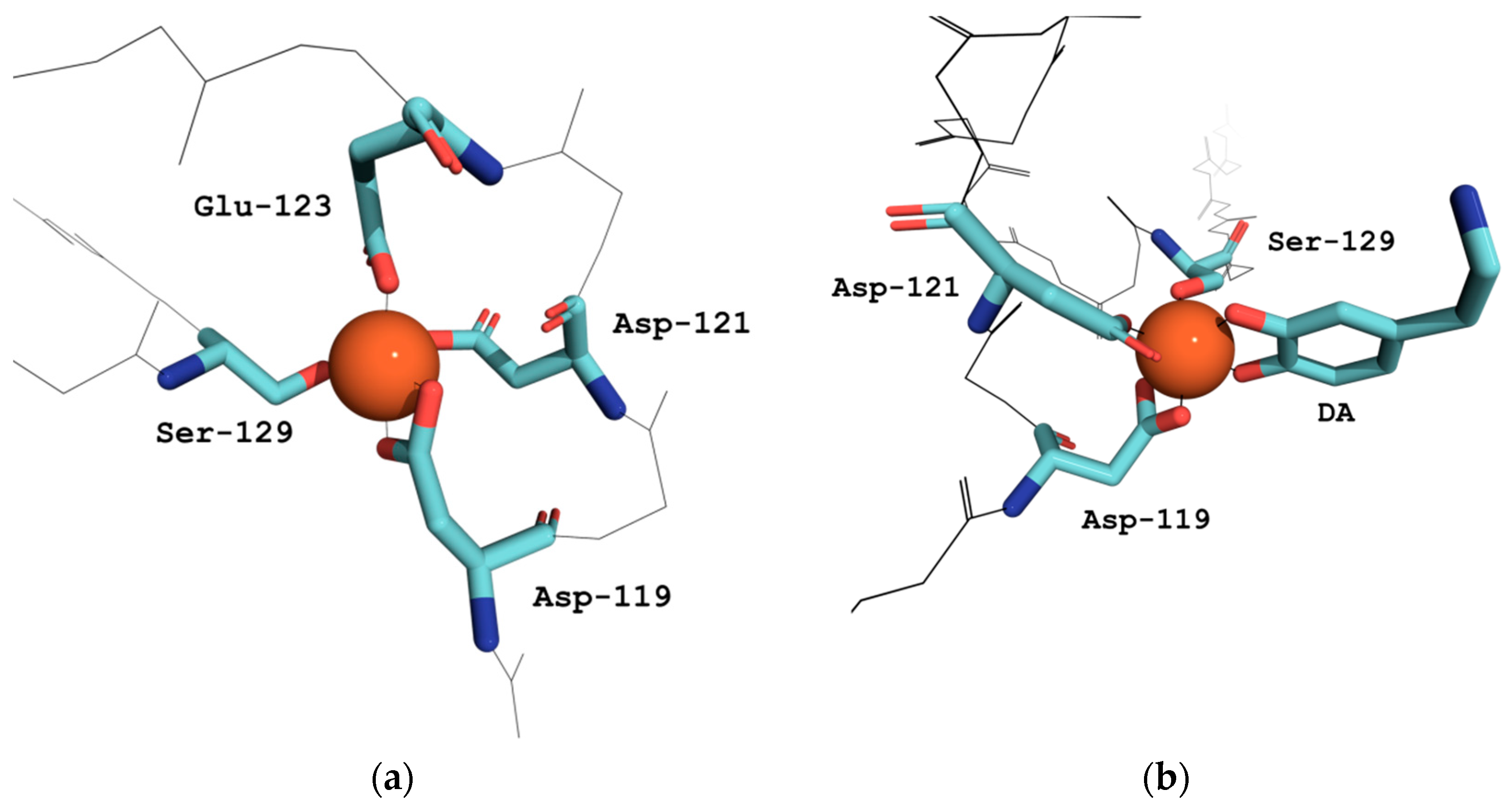
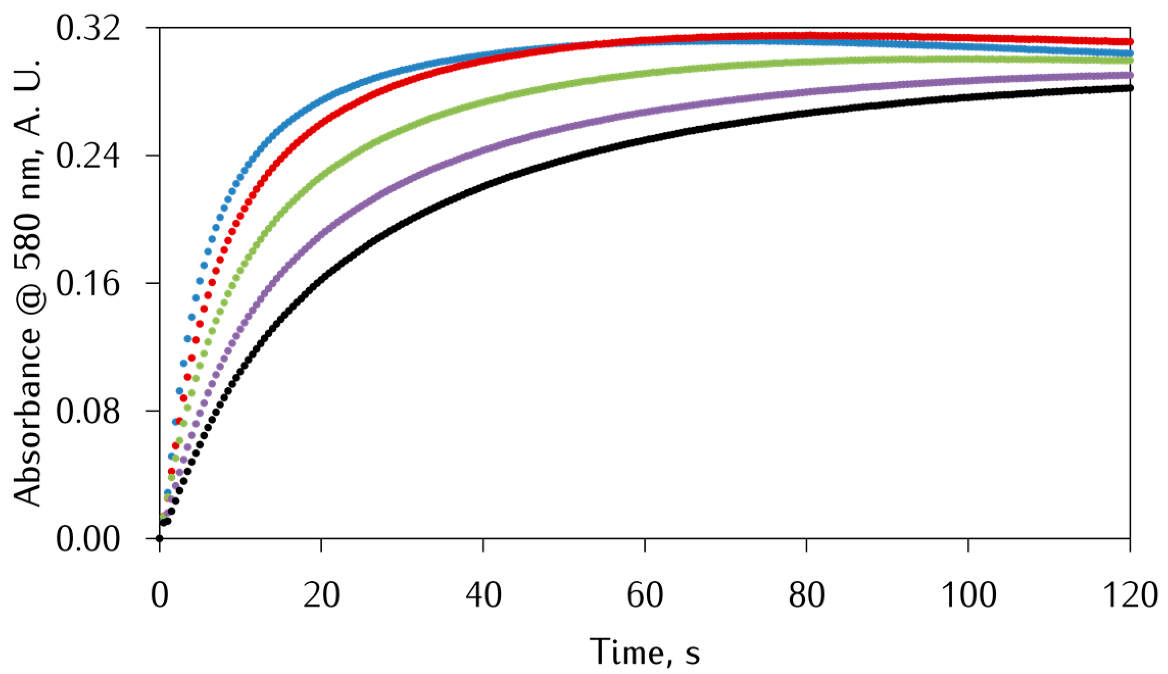
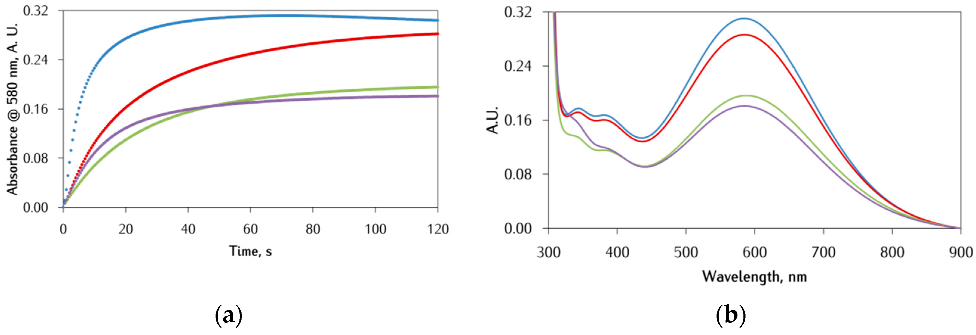
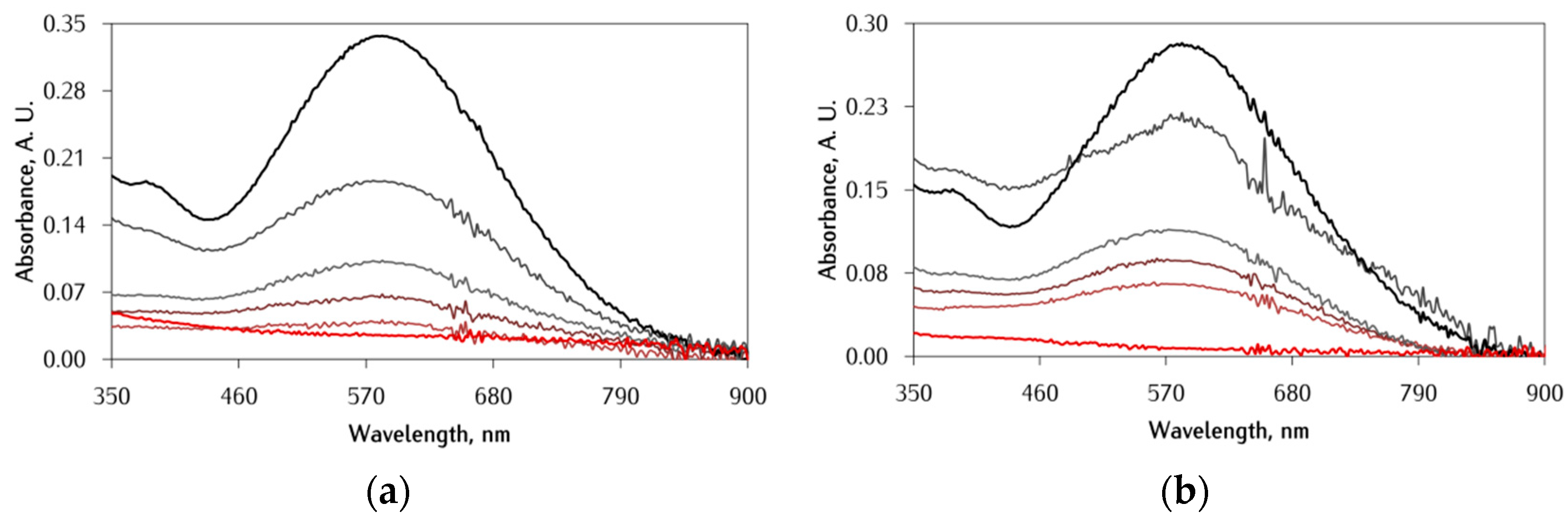
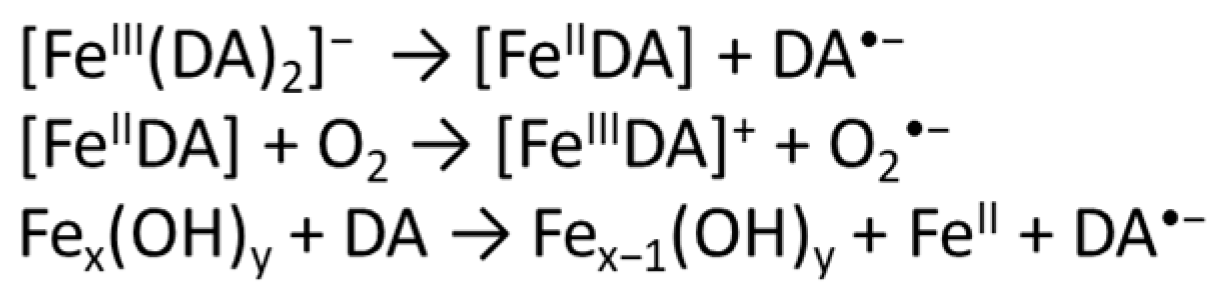
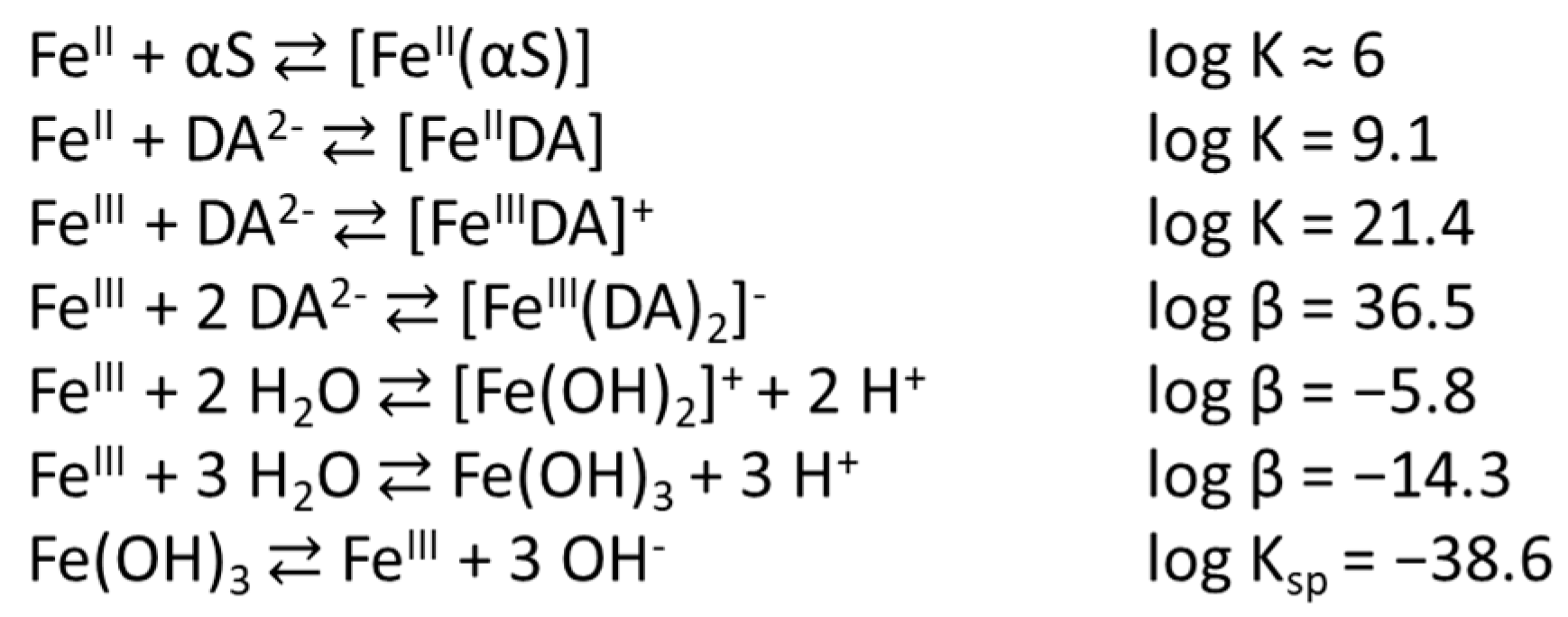
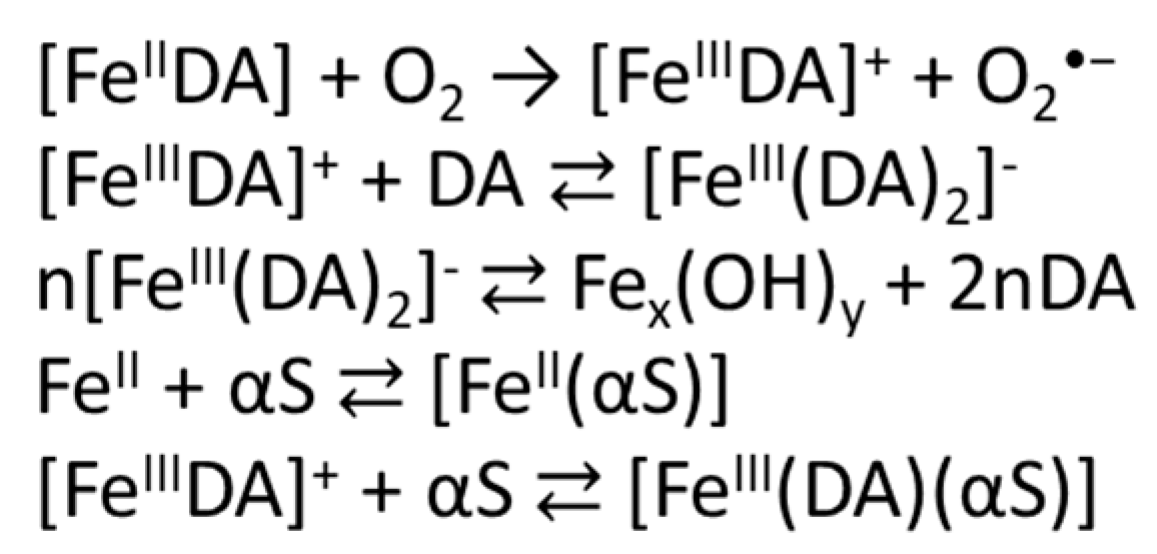
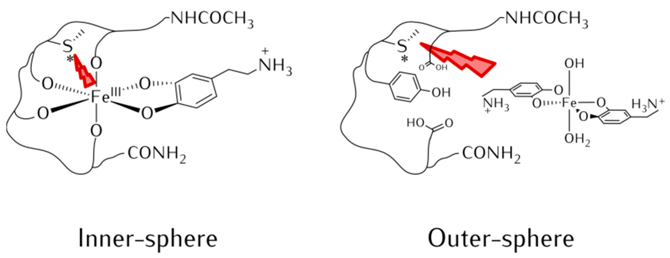
3.1. Effect of αS C-Terminal Peptides on FeIII-DA Interaction

The effect of αS model peptides on the interaction between iron and DA was investigated in 10 mM Mops buffer pH 7.0 by varying both the DA:Fe ratio and the peptide concentration. The selected pH was slightly acidic compared to the physiological pH due to the tissue acidosis found in PD brains [7,10]. The combined use of iron and DA gives rise to some problems: (i) the well-known propensity of DA to form insoluble DA-quinone oligomers upon oxidation; (ii) the strong competition of FeIII hydrolysis at pH above 4 in aerated solutions [30]; and (iii) the formation of the bis-catecholato-FeIII complex [FeIII(DA)2], which has a strong LMCT band in the visible region (λmax = 580 nm, ε580 = 3300 M−1 cm−1) [9,31] thus preventing observation of the UV-Vis band of aminochrome (λmax = 475 nm), the observable product of DA oxidation [32]. Therefore, the experiments with Fe–DA-αS model peptides were monitored through the formation and decay of the [FeIII(DA)2] species at 580 nm.
First, the effect of Ac-αS119–132 on the formation and decay of [FeIII(DA)2] was evaluated, at a DA:Fe ratio of 2:1, and the kinetic traces of absorbance against time are reported in Figure 2. The obtained kinetic traces display a biphasic behavior due to the complex mechanism described below. As reported by Sun et al. [9], upon mixing FeII and DA, the initial FeII-DA complex is readily oxidized by dioxygen resulting in superoxide radical anion and FeIII-DA; this is then converted in the [FeIII(DA)2] complex which is responsible for the strong absorption in the visible region (Scheme 3). The growth rate of the 580 nm band depends on the DA:Fe ratio but, at the lowest ratio (i.e., 2:1), FeIII hydrolysis is competitive [16] and, thus, the [FeIII(DA)2] complex decomposes causing the decrease of the complex concentration and, consequently, of the kinetic traces slope. When the experiment was carried out in the presence of Ac-αS119–132, a decrease in both the final concentration and formation rate of the [FeIII(DA)2] complex was observed. It is worth noting that these effects are greater as the peptide concentration increases. This effect could be explained considering the establishment of a competition between the peptide and the DA towards iron(II) or iron(III) binding, hence reducing the amount of the [FeIII(DA)2] complex. Nevertheless, peptide binding seems to also affect the second phase of the reaction, decreasing the rate of iron(III) hydrolysis. Further information can be extracted from the UV-Vis spectra (Figure 3) recorded during the kinetic study, in which a red-shift of ca. 15 nm of the LMCT band suggests that the Ac-αS119–132 reaction with the [FeIII(DA)2] complex is likely to occur, forming a ternary complex [Fe(DA)(Ac-αS119–132)] (Figure 1) showing the characteristic band of the FeIII-monocatecholato complex [FeIIIDA]+. This last assumption is in good agreement with the results reported by Jiang et al., which claim that the formation of the ternary complex [FeIII(DA)(ATP)] is followed by a marked red shift of the LMCT band [33].
The phosphorylated peptides Ac-αSpS119–132 and Ac-αSpYpS119–132 have a stronger impact on the formation and degradation rate of the [FeIII(DA)2] complex as, in both cases, the rates decrease more significantly than with the non-phosphorylated analogue (Figure 3). Furthermore, the shift towards lower energies of the LMCT band is slightly larger compared to the one caused by the non-phosphorylated analogue (Figure 3), also suggesting the formation of the ternary complex. This finding is in line with the gain in the stability constant of the peptides for FeIII binding, offered by the phosphorylation of S129 and Y125 [13,14,20]. Compared to the kinetic trace of Ac-αSpS119–132, it is worth noting that the kinetic trace of Ac-αSpYpS119–132 appears to be different: the initial rate of the [FeIII(DA)2] complex formation is higher, but the final amount of complex is lower. We hypothesize that this phenomenon is caused by an initial competition between the two phosphorylated sites due to difficult conformational changes needed for FeIII coordination coupled with the negative repulsion offered by two phosphate groups, with the pY125 being the strongest ligand [14], thus resulting in a lower final concentration of the [FeIII(DA)2] complex.
Since the interaction of αS peptides with lipid membrane modulates their conformation, analysis of the interaction between iron and αS is incomplete if it remains confined in the aqueous milieu. For this reason, the anionic surfactant SDS is used in this investigation because the negative surface charge and high affinity to hydrophobic domains of the micelles formed above the critical concentration (≈8 mM in water at 25 °C) [34,35] represent a useful and simple model of biological membranes. The addition of SDS (Figure 4) to the buffer enhances the effect of αS peptides over [FeIII(DA)2] formation and consumption, making the slope of the first phase of the kinetics much less steep. A possible explanation could be that, at the investigated pH (i.e., 7.0), the DA aromatic hydroxyl group is protonated and, thus, the aromatic ring can be inserted into the SDS micelles [27] causing a decrease in the effective DA concentration and making DA less competitive towards FeIII binding compared to the αS peptides.
Similar conclusions were drawn after performing the experiments at the 20:1 DA:Fe molar ratio (Figure S1). The main difference highlighted upon increasing the DA concentration is the diminution of the peptide effect. This aspect was expected since the large DA excess and its high affinity for FeIII binding hampers the interaction of αS-peptides with the [FeIII(DA)2] complex.

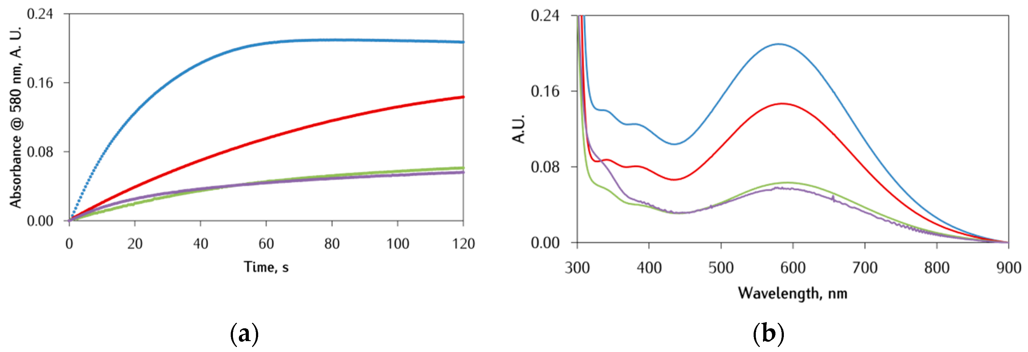
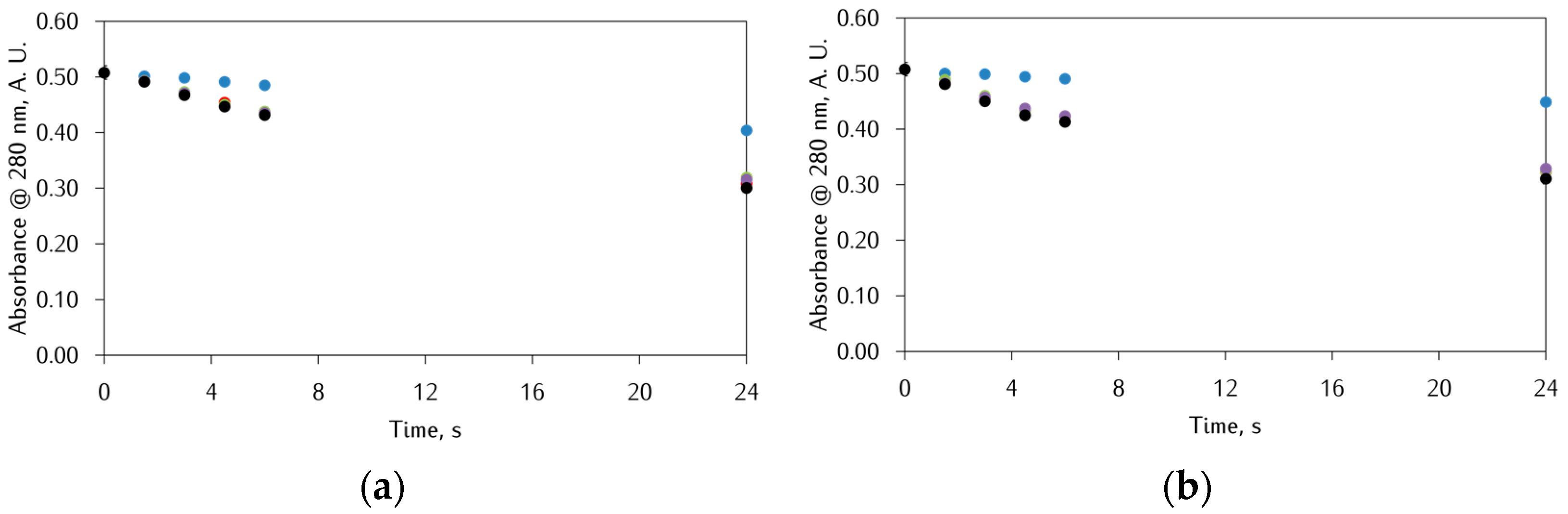
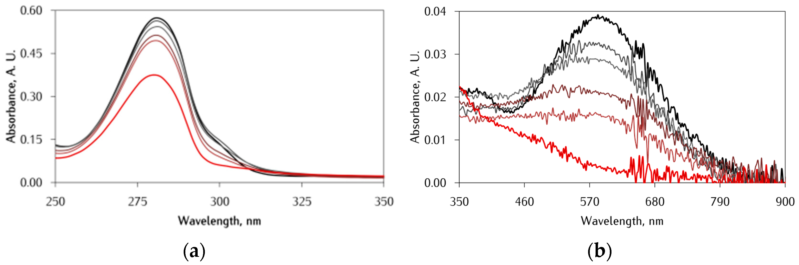
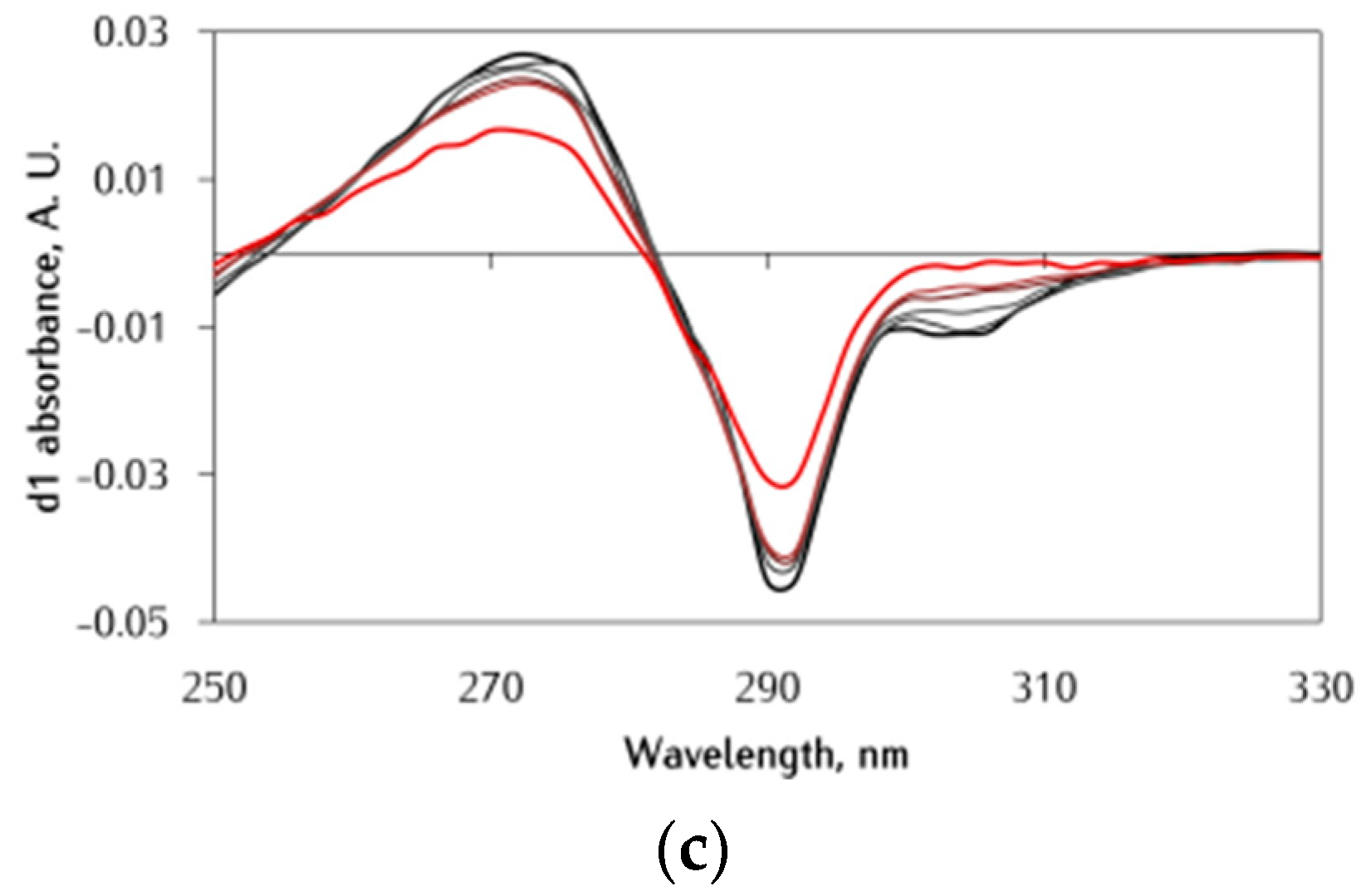
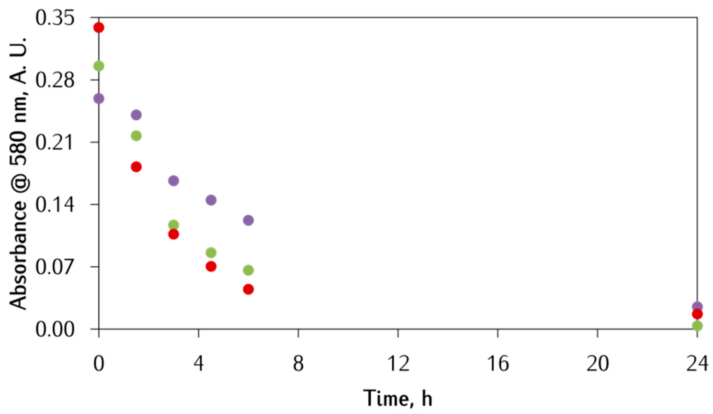
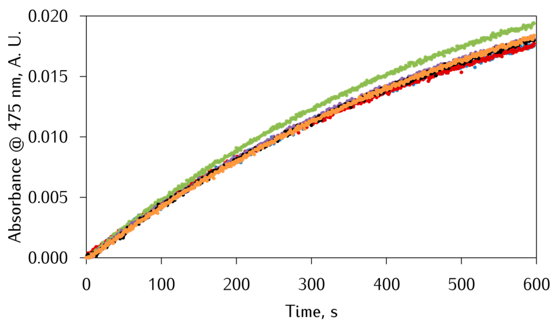
3.2. Kinetic Studies on the Oxidation of Catechol Substrates Promoted by FeIII-Peptide Complexes

The catalytic oxidation of DA promoted by Fe-αS peptides was studied in Mops buffer at pH 7.0, at the two different DA:Fe ratios of 2:1 and 20:1, adding 2 equiv. of Ac-αS119–132, Ac-αSpS119–132, or Ac-αSpYpS119–132. In order to overcome the difficulties related to monitoring FeIII-DA chemistry, different techniques were applied—i.e., RP-HPLC, UV-Vis spectroscopy, and HPLC-MS/MS. Due to the intense [FeIII(DA)2] LMCT band, it is not possible to follow DA oxidation through the development of the absorption band of aminochrome at 475 nm, which accumulates in the reaction initial stages. Furthermore, the DA oxidation products can polymerize and form insoluble aggregates with a general increase of baseline absorption over the whole wavelength range of the spectrum due to light scattering. For these reasons, DA oxidation was monitored following a decrease of the absorbance of DA (λmax = 280 nm) over time, removing background contribution by a three-point-dropline correction.
Starting from the experiments at the higher DA:Fe molar ratio of 20:1, the kinetic traces of absorbance against time are reported in Figure 5, which refers to DA oxidation by FeIII-αS-peptides. The results clearly show that the catalytic oxidation of DA by iron is not significantly promoted by the addition of either Ac-αS119–132, Ac-αSpS119–132, or Ac-αSpYpS119–132, since the kinetic traces obtained with the peptides are almost identical to the control experiment without peptides, in line with what is expected. In fact, as seen for the Fe–DA coordination experiments, a large excess of DA makes the interaction of αS-peptides with the metal center difficult. Furthermore, comparing the UV-Vis spectra obtained with and without αS-peptides (Figure 6), the following similarities are observed: (i) the DA absorption band (λmax = 280 nm) and the [FeIII(DA)2] band (λmax = 580 nm) show a similar decrease; (ii) the [FeIII(DA)2] complex band shows the same shape, and at 6 h the available FeIII is still bound to the DA; (iii) the DA band has a shoulder at around 305 nm, better highlighted in the first derivative spectra (panel C), which is due to the DA fraction bound to FeIII [36]; and (iv) the band intensity decrease is similarly followed by a progressive background increase in both cases, due to the melanic pigment particle formation [37].
Further, DA oxidation was also followed by RP-HPLC (Figure S2). As in the UV-Vis experiments, the chromatographic data show no difference in DA oxidation rates between the control experiment and the experiments in the presence of αS.
The addition of SDS (Figure 5–panel B) to the buffer caused a slight increase in global DA oxidation rate in all samples. We hypothesize that this effect is driven by the possible interaction between SDS and DA oligomers. In the UV-Vis spectra in the presence of SDS (Figure S3), an increase of the background absorption in the first 3 h is observed, followed by a decrease from 3 to 6 h due to the formation of melanic aggregates. These aggregates may stick to the SDS micelles [27], thus accumulating in the solution before precipitating as melanin particles. Consequently, the melanic pigments do not bind iron which can, in turn, catalyze DA oxidation.
Considering the experiments at the lower DA:Fe molar ratio of 2:1, a different situation is observed. Herein, DA oxidation cannot be reliably monitored following the absorbance of DA (λmax = 280 nm) over time, because this band is altered by coordination to FeIII and overlapped with the peptide tyrosine absorption band (Figure S4). Therefore, the LMCT band of the [FeIII(DA)2] complex at 580 nm was monitored instead. In Figure 7, the kinetic traces of absorbance against time are reported for DA oxidation by FeIII-Ac-αS119–132. The starting points of the kinetic traces are different among the samples due to the impact of the peptides on the [FeIII(DA)2] complex, already discussed in the previous section. Nevertheless, the results clearly indicate that addition of Ac-αS119–132 and particularly Ac-αSpS119–132 causes a decrease in the DA oxidation rate, as suggested by the lower slope of the traces, probably due to the [Fe(DA)(Ac-αS119–132)] ternary complex formation (Figure 1) which stabilizes the FeIII oxidation state, or by hampering DA binding and thus redox cycling.
In the UV-Vis spectra with and without Ac-αS119–132, reported in Figure 8, the main difference appears to be the higher absorption in the 350–500 nm portion of the 1.5 h spectra. Even if we do not know the nature of this spectral feature, it may be related to peptide-promoted FeIII aggregation or to a form of melanic intermediate. Both are in line with the reduced oxidation rate of DA.
Also in this case, DA oxidation was followed by RP-HPLC (Figure S5). In agreement with UV-Vis spectral changes, the chromatographic data show a reduction in DA oxidation rate upon the addition of αS-peptides, but no significant difference was found between Ac-αS119–132 and Ac-αSpS119–132.

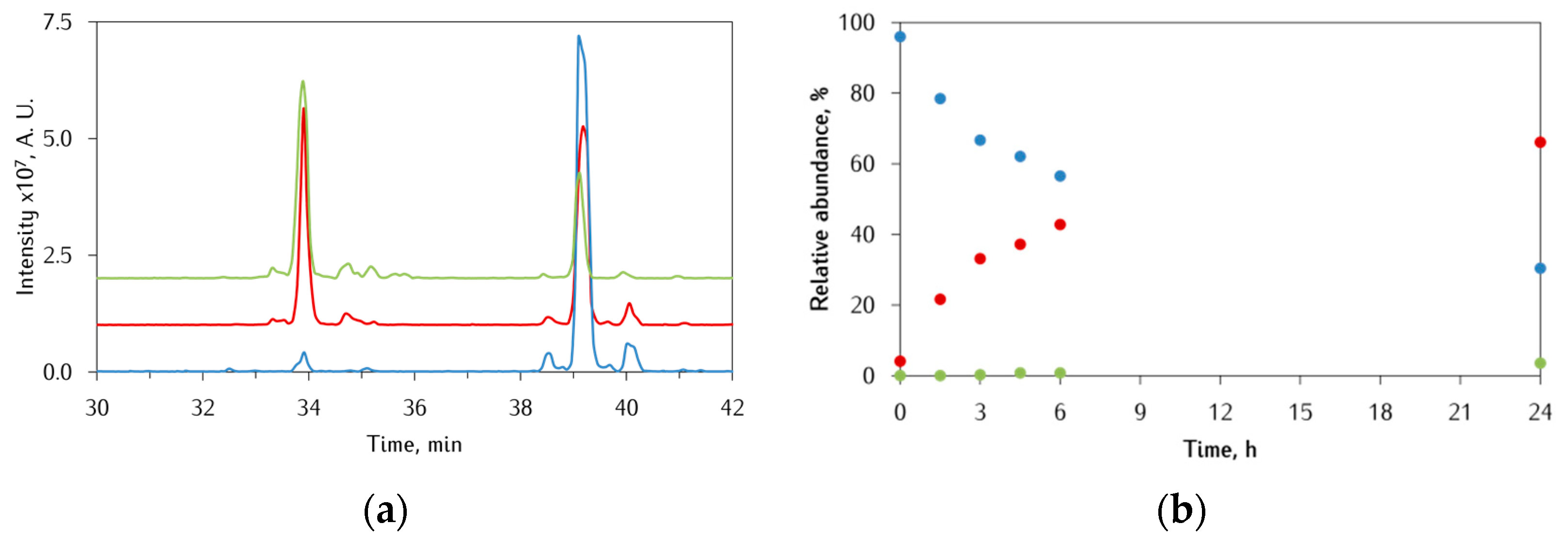
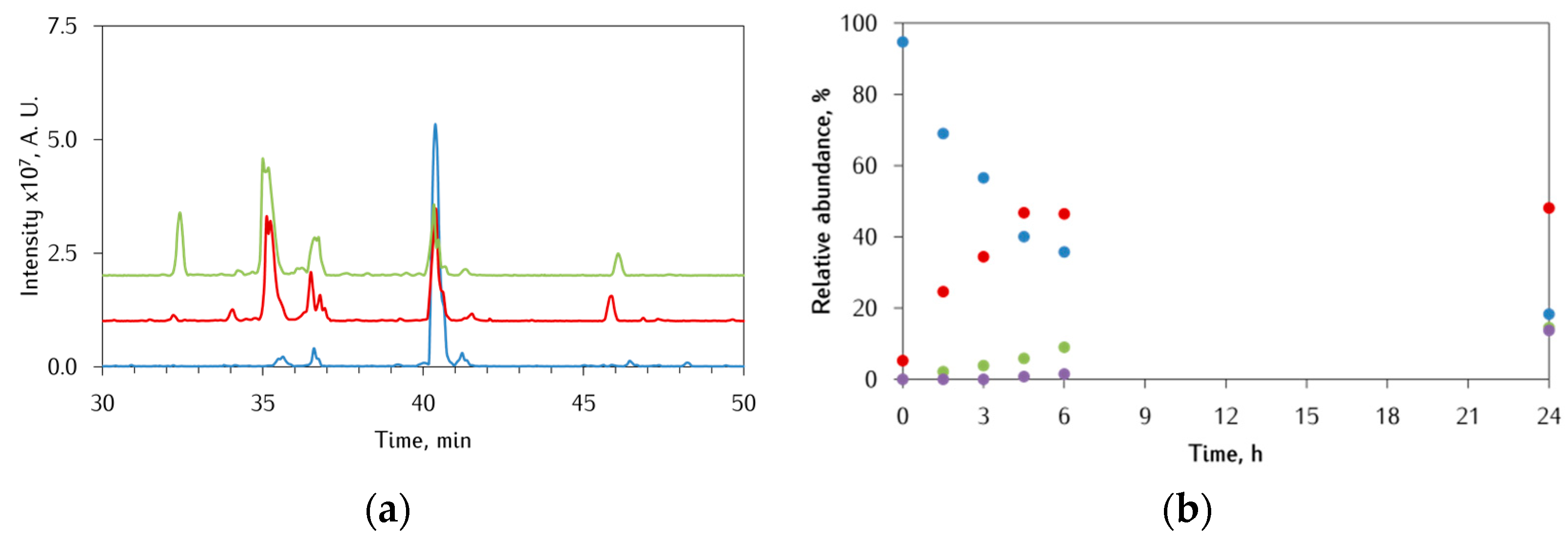
3.3. Identification of αS-Peptide Modifications by HPLC-MS

It is well known that upon metal-catalyzed oxidation of catechols such as DA, ROS species are generated [7,38,39]. Such harmful species include O2•−, H2O2, and OH, which ultimately lead to protein damage. Based on the results obtained so far, one important target of this damage are the αS-peptides themselves due to their ability to influence both the [FeIII(DA)2] complex formation and degradation. Furthermore, studies on the peptide modification could also shed light on the type of interaction and on the mechanism of reaction. The modifications undergone by Ac-αS119–132 and Ac-αSpS119–132 concomitantly with DA oxidation were identified by performing a series of HPLC-MS/MS analyses. The experiments were run in 10 mM Mops buffer pH 7.0, by varying both the DA:Fe ratio and the peptide concentration. Typical oxidative modifications usually consist of an O-atom insertion (+16 Da) in oxidation-sensitive residues such as tyrosine and methionine, which are converted into 3-hydroxytyrosine and methionine sulfoxide, respectively [40]. Unfortunately, the mass spectra recorded in negative-ion mode fail to be fed into algorithms like turboSEQUEST and, hence, the MS/MS spectra had to be analyzed by inspection.

3.3.1. Ac-αS119–132 Peptide

When the Ac-αS119–132 peptide was used at the lower DA:Fe ratio of 2:1, the elution profiles of Ac-αS119–132 together with the modifications detected by the HPLC-MS/MS analysis were obtained as shown in Figure 9. After 6 h of incubation, the elution profile is dominated by two main species: (i) the unmodified Ac-αS119–132 peptide (tr 39 min, 56%) and (ii) the single oxidized derivative (tr 33.8 min, 43%). After 24 h, the overall amount of oxidized peptide increased (66%) and a new species was detected at tr 35.2 min (3.5%), which appears to be the double oxidized peptide. Together with the above species, other minor modifications were detected, mainly related to peptide truncation. However, due to the low intensities of such signals, it was not possible to assess the exact nature of the corresponding species. Unfortunately, none of the peptide-DA and DA-quinone oligomeric species could be detected, probably due to the lack of strong nucleophilic residues in the peptide. Considering the peptide primary sequence, the most likely sites of oxidation are Y125 and M127. However, from the analysis of the ion fragmentation series found in the MS/MS spectra, it was not possible to assess the exact oxidation site. To overcome this problem, the peptide was selectively oxidized by hydrogen peroxide and horseradish peroxidase, which can differentiate the two amino acids by promoting the methionine sulfoxidation and tyrosine dimerization. The elution profile obtained showed an intense peak at tr 33.6 min with the same MS/MS fragmentation patterns of the mono oxidized Ac-αS119–132; hence. we can easily conclude that the oxidation occurs at M127 since HRP does not catalyze tyrosine hydroxylation but its dimerization. Based on this finding, the second oxidation of the peptide could occur either at the oxidized M127 (to form sulfone) or at the Y125. Furthermore, control experiments were carried out by incubating the peptide alone and in the presence of only DA or only FeII. The data obtained showed no appreciable oxidation of M127 when the peptide was incubated alone in the buffer or with only DA. On the other hand, in the case of only FeII, the single oxidized peptide was only detected after 90 min, albeit being less than 10%.
When the reaction was carried out at the higher DA:Fe molar ratio of 20:1, the elution profiles of Ac-αS119–132 and the peptide modifications detected by HPLC-MS shown in Figure S6 were obtained. Also in this case, as seen above for the DA oxidation, the increase in the DA:Fe ratio is followed by a decrease of the observed modification, being 15% at 6 h and 28% at 24 h.
To further investigate both the type of Fe-peptide interaction and the peptide modification mechanism, the experiments were replicated by doubling the peptide concentration (40 µM) for the studied case. Considering the possible scenarios, two representative cases were considered: (i) the peptide oxidation occurs through an outer-sphere (OS) mechanism, where the peptide is not directly bound to the iron site, or (ii) the oxidation occurs through an inner-sphere (IS) mechanism and, therefore, it undergoes oxidation when bound to iron (Scheme 4). As a rule of thumb, in the former case, by doubling the peptide concentration, we expect to observe the same percentage of modification, while, in the latter case, a lower (a halving in the case of strong binding) of the percentage is expected. However, it is worth noting that the two mechanisms could be operative at the same time, with IS generally more efficient. The data obtained (Table 1) suggest an overlap between the two mechanisms depending on the DA:Fe ratio. In fact, at the lower DA:Fe ratio, upon doubling the peptide concentration, a 25% decrease of oxidized peptide was observed, thus suggesting an initial IS mechanism with the excess peptide being oxidized through an OS mechanism, which accounts for the observed decrease of the percentage. At the higher DA:Fe ratio, an initial OS mechanism is likely to occur as the percentage of oxidized peptide is lower, even though we cannot exclude that the mechanism could be IS-type but with a low fraction of the peptide coordinated. By doubling the peptide concentration, a doubling in the modification percentage is observed, suggesting that the higher Ac-αS119–132 concentration leads to an increase of the coordinated fraction which will be oxidized faster through the IS mechanism.
We can, therefore, conclude that αS-peptide oxidation is likely to occur through an IS-type mechanism, in which the coordinated peptide is oxidized, while the OS mechanism could contribute only to a modest extent. These findings are in line with what was found in the previous sections, where a strong interaction between Ac-αS119–132 and [FeIII(DA)2] can only occur at a low DA:Fe ratio.

3.3.2. Ac-αSpS119–132 Peptide

Considering the oxidation undergone by the phosphorylated peptide Ac-αSpS119–132, Figure 10 shows the elution profiles together with the modifications detected by the HPLC-MS/MS analysis, at the lower DA:Fe ratio of 2:1. As discussed in the previous sections, phosphorylation of S129 has a strong impact on the ability of Ac-αSpS119–132 to compete with DA for the interaction with the [FeIII(DA)2] complex. In fact, the unmodified peptide (tr 40 min) found in solution is 35% at 6 h of incubation, with a further decrease to less than 20% after 24 h. Contrary to the previous case, the oxidative modifications appear to be dominant species, especially at 24 h. Among them, it is worth noting the presence of the double oxidized peptide (tr 36.4 min, 15%) and several truncated species. The exact nature of such species is currently undefined, but we hypothesize that the species eluted at tr 32 min (14%) corresponds to the truncation of the mono oxidized peptide, with sequence Ac-119DPDNEAYEM*127, where M127 is oxidized to sulfoxide. This latter peptide fragment suggests that the presence of the phosphate group on S129 causes a shift of the metal coordinating region towards the peptide C-terminus.
At the higher DA:Fe ratio of 20:1, a decrease in the oxidation rate of Ac-αSpS119–132 was observed (Figure S7), even though it was less pronounced compared with that of Ac-αS119–132, again in agreement with the higher affinity towards FeIII contributed by the phosphoserine pS129. Interestingly, after 24 h of incubation, two new peaks appeared at tr 38.6 and 31.8 min, which share the same MS/MS pattern of the Ac-αS119–132 peptide and the corresponding mono oxidized product, respectively; hence, dephosphorylation of pS129 is likely to occur.
Upon doubling the concentration of Ac-αSpS119–132 peptide to 40 µM, the data obtained (Table 2) suggest that the oxidation mechanism still depends on the DA:Fe ratio but appear to be less ambiguous. In fact, at the lower ratio of 2:1, after doubling the peptide concentration, a halving of the per cent of oxidized peptide was observed, indicating an IS mechanism. At the higher ratio of 20:1, an OS mechanism occurs instead since the percentage of oxidized peptide remained almost unchanged with only a small decrease probably related to the competitive peptide truncation. Similar conclusions can be drawn for Ac-αSpS119–132 when compared to Ac-αS119–132. Again, serine phosphorylation has a strong impact on the peptide effect over Fe–DA chemistry since, at both DA:Fe ratios, a greater percentage of peptide modification and truncation was found, in line with a stronger αS-peptide-Fe interaction.

3.4. Comparison with Copper

Besides iron, it is well known that copper is able to promote DA oxidation in the presence of neuronal peptides [41]. It is, thus, useful to compare the behavior of the two metal ions bound to the same αS peptide. Following our previous studies on the ability of CuII-Aβ16 complexes to promote catechol oxidation [29], we aimed to investigate whether αS, and in particular the Ac-αS119–132 peptide, can catalyze this reaction. In fact, the protein region contains a binding site for divalent metal ions in the 119DPDNEA124 motif [12].
The catalytic oxidation of DA by Cu(II)-Ac-αS119–132 complex was studied in Hepes buffer at pH 7.4 adding increasing amounts of Ac-αS119–132. The kinetic traces of absorbance against time are reported in Figure 11, where the readings at 475 nm refer to the formation of aminochrome [29]. As can be observed from Figure 11, catalytic oxidation of DA by CuII is not promoted by the addition of Ac-αS119–132, since kinetic traces obtained with the peptide are almost identical. These findings are even clearer by plotting the slope of the kinetic traces against equivalents of peptide added, as reported in Figure S8. Again, since the slope is quite constant over the peptide addition, we can conclude that Ac-αS119–132 is neither able to catalyze nor suppress DA oxidation.

4. Conclusions

The interaction between C-terminal fragments of αS with biologically relevant metal ions and DA was investigated. The data suggest that Ac-αS119–132 and particularly Ac-αSpS119–132 and Ac-αSpYpS119–132 are able to interact with the [FeIII(DA)2] complex, likely forming a ternary complex—[Fe(DA)(αS)]. The interplay between the three species is dominated by two factors: (i) the DA:Fe molar ratio and (ii) the phosphorylation of the peptide. At high DA:Fe molar ratios, DA coordination is too strong and hampers the peptide coordination, while, at lower DA:Fe molar ratios, the peptide is able to compete with one of the two coordinated DA molecules. This kind of interaction is also confirmed by the analysis of the post-translational modifications (PTMs) of the peptide, where an inner-sphere mechanism was observed only at the DA:Fe ratio of 2:1. Nevertheless, peptide phosphorylation enhances affinity for FeIII, causing a decrease in DA oxidation rate and increase in peptide oxidation. Furthermore, phosphorylation at S129 causes the shift of the coordination sphere towards the C-terminus of the peptide, as a consistent fraction of fragmented peptide was observed.
Studies conducted in a membrane-like environment showed an enhanced peptide effect over both the DA oxidation and the [FeIII(DA)2] complex formation and degradation by lowering the effective DA concentration and thus shifting the DA:Fe molar ratio towards lower values.
In the case of copper(II), we demonstrated that the Ac-αS119–132 peptide does not influence the DA oxidation promoted by the free ion. This behavior contrasts with what was observed with other neuronal peptides like Aβ and prion fragments [41], and is due to the lack of free N-terminal amine and histidine in the C-terminal αS, which are the residues that typically offer a strong CuII coordination sphere and stabilize the CuI redox state necessary to cycle during the catalytic event.
Future perspectives will address the full characterization of the bis-phosphorylated αS fragment at both Y125 and S129. Despite that, the preliminary data indicate that the second phosphorylation (i.e., on Y125) does not enhance interaction with [FeIII(DA)2], if compared with the Ac-αSpS119–132 peptide, and rather a competition between the two residues occurs. A further aspect to be taken into consideration will be the use of more sophisticated membrane models than the micelles formed by SDS; for example, with large or small unilamellar vesicles (LUV and SUV, respectively). Since the membrane alters the effective DA concentration, other DA:Fe molar ratios than those used in the present work need to be investigated.

Supplementary Materials

Additional kinetic traces (Figures S1, S2, S4, S5 and S8), additional UV-vis spectra (Figure S3), additional HPLC-MS elution profiles, and percentage modifications (Figures S6 and S7) tables with kinetic constants (Table S1) are available online and can be downloaded at: https://www.mdpi.com/article/10.3390/antiox12040791/s1.

Author Contributions

Conceptualization, F.S., S.D., S.N., E.M. and L.C.; methodology, F.S.; formal analysis, F.S., S.D., S.N. and E.M.; writing—original draft preparation, F.S., E.M. and L.C.; writing—review and editing, F.S., S.D., S.N., E.M. and L.C. All authors have read and agreed to the published version of the manuscript.

Funding

Italian Ministry of Education, University, and Research (MIUR)-Research Projects of National Interest (PRIN) 2015 prot. 2015T778JW.

Institutional Review Board Statement

Not applicable.

Informed Consent Statement

Not applicable.

Data Availability Statement

Data supporting the reported results can be obtained from the corresponding author.

Acknowledgments

The authors acknowledge support from the Ministero dell’Università e della Ricerca (MUR) and the University of Pavia through the program “Dipartimenti di Eccellenza 2023–2027”.

Conflicts of Interest

The authors declare no conflict of interest.

References

  1. Wise, R.M.; Wagener, A.; Fietzek, U.M.; Klopstock, T.; Mosharov, E.V.; Zucca, F.A.; Sulzer, D.; Zecca, L.; Burbulla, L.F. Interactions of dopamine, iron, and alpha-synuclein linked to dopaminergic neuron vulnerability in Parkinson’s disease and Neurodegeneration with Brain Iron Accumulation disorders. Neurobiol Dis. 2022, 175, 105920. [Google Scholar] [CrossRef]
  2. Hare, D.J.; Double, K.L. Iron and dopamine: A toxic couple. Brain 2016, 139, 1026–1035. [Google Scholar] [CrossRef] [PubMed] [Green Version]
  3. Lingor, P.; Carboni, E.; Koch, J.C. Alpha-synuclein and iron: Two keys unlocking Parkinson’s disease. J. Neural Transm. 2017, 124, 973–981. [Google Scholar] [CrossRef]
  4. Rothman, R.J.; Serroni, A.; Farber, J.L. Cellular pool of transient ferric iron, chelatable by deferoxamine and distinct from ferritin, that is involved in oxidative cell injury. Mol. Pharm. 1992, 42, 703–710. [Google Scholar]
  5. Brawley, H.N.; Kreinbrink, A.C.; Hierholzer, J.D.; Vali, S.W.; Lindahl, P.A. Labile Iron Pool of Isolated Escherichia coli Cytosol Likely Includes Fe-ATP and Fe-Citrate but not Fe-Glutathione or Aqueous Fe. J. Am. Chem. Soc. 2023, 145, 2104–2117. [Google Scholar] [CrossRef]
  6. Zucca, F.A.; Segura-Aguilar, J.; Ferrari, E.; Muñoz, P.; Paris, I.; Sulzer, D.; Sarna, T.; Casella, L.; Zecca, L. Interactions of iron, dopamine and neuromelanin pathways in brain aging and Parkinson’s disease. Prog. Neurobiol. 2017, 155, 96–119. [Google Scholar] [CrossRef]
  7. Monzani, E.; Nicolis, S.; Dell’Acqua, S.; Capucciati, A.; Bacchella, C.; Zucca, F.A.; Mosharov, E.V.; Sulzer, D.; Zecca, L.; Casella, L. Dopamine, Oxidative Stress and Protein-Quinone Modifications in Parkinson’s and Other Neurodegenerative Diseases. Angew. Chem. Int. Ed. Engl. 2019, 58, 6512–6527. [Google Scholar] [CrossRef] [PubMed]
  8. Bisaglia, M.; Mammi, S.; Bubacco, L. Kinetic and structural analysis of the early oxidation products of dopamine: Analysis of the interactions with alpha-synuclein. J. Biol. Chem. 2007, 282, 15597–15605. [Google Scholar] [CrossRef] [Green Version]
  9. Sun, Y.; Pham, A.N.; Waite, T.D. Elucidation of the interplay between Fe(II), Fe(III), and dopamine with relevance to iron solubilization and reactive oxygen species generation by catecholamines. J. Neurochem. 2016, 137, 955–968. [Google Scholar] [CrossRef]
  10. Sun, Y.; Pham, A.N.; Hare, D.J.; Waite, T.D. Kinetic modeling of pH-dependent oxidation of dopamine by iron and its relevance to Parkinson’s disease. Front. Neurosci. 2018, 12, 859. [Google Scholar] [CrossRef] [PubMed]
  11. Viles, J.H. Metal ions and amyloid fiber formation in neurodegenerative diseases. Copper, zinc and iron in Alzheimer’s, Parkinson’s and prion diseases. Coord. Chem. Rev. 2012, 256, 2271–2284. [Google Scholar] [CrossRef]
  12. Binolfi, A.; Rasia, R.M.; Bertoncini, C.W.; Ceolin, M.; Zweckstetter, M.; Griesinger, C.; Jovin, T.M.; Fernández, C.O. Interaction of alpha-synuclein with divalent metal ions reveals key differences: A link between structure, binding specificity and fibrillation enhancement. J. Am. Chem. Soc. 2006, 128, 9893–9901. [Google Scholar] [CrossRef] [PubMed]
  13. Lu, Y.; Prudent, M.; Fauvet, B.; Lashuel, H.A.; Girault, H.H. Phosphorylation of α-Synuclein at Y125 and S129 Alters Its Metal Binding Properties: Implications for Understanding the Role of α-Synuclein in the Pathogenesis of Parkinson’s Disease and Related Disorders. ACS Chem. Neurosci. 2011, 2, 667–675. [Google Scholar] [CrossRef] [PubMed] [Green Version]
  14. Liu, L.L.; Franz, K.J. Phosphorylation of an α-synuclein peptide fragment enhances metal binding. J. Am. Chem. Soc. 2005, 127, 9662–9663. [Google Scholar] [CrossRef]
  15. Rao, K.S.J. Thermodynamics imprinting reveals differential binding of metals to α-synuclein: Relevance to parkinson’s disease. Biochem. Biophys. Res. Commun. 2007, 359, 115–120. [Google Scholar] [CrossRef]
  16. Stefánsson, A. Iron (III) hydrolysis and solubility at 25 degrees C. Environ. Sci. Technol. 2007, 41, 6117–6123. [Google Scholar] [CrossRef] [PubMed]
  17. Hanwell, M.D.; Curtis, D.E.; Lonie, D.C.; Vandermeersch, T.; Zurek, E.; Hutchison, G.R. Avogadro: An advanced semantic chemical editor, visualization, and analysis platform. J. Cheminform. 2012, 4, 17. [Google Scholar] [CrossRef] [Green Version]
  18. Schrödinger, L.; DeLano, W. PyMOL. 2020. Available online: http://www.pymol.org/pymol (accessed on 1 October 2022).
  19. Anderson, J.P.; Walker, D.E.; Goldstein, J.M.; de Laat, R.; Banducci, K.; Caccavello, R.J.; Barbour, R.; Huang, J.; Kling, K.; Lee, M.; et al. Phosphorylation of Ser-129 is the dominant pathological modification of alpha-synuclein in familial and sporadic Lewy body disease. J. Biol. Chem. 2006, 281, 29739–29752. [Google Scholar] [CrossRef] [Green Version]
  20. Liu, L.L.; Franz, K.J. Phosphorylation-dependent metal binding by α-synuclein peptide fragments. J. Biol. Inorg. Chem. 2007, 12, 234–247. [Google Scholar] [CrossRef]
  21. Xu, Y.; Deng, Y.; Qing, H. The phosphorylation of α-synuclein: Development and implication for the mechanism and therapy of the Parkinson’s disease. J. Neurochem. 2015, 135, 4–18. [Google Scholar] [CrossRef]
  22. Chen, L.; Periquet, M.; Wang, X.; Negro, A.; McLean, P.J.; Hyman, B.T.; Feany, M.B. Tyrosine and serine phosphorylation of alpha-synuclein have opposing effects on neurotoxicity and soluble oligomer formation. J. Clin. Investig. 2009, 119, 3257–3265. [Google Scholar] [CrossRef] [PubMed] [Green Version]
  23. Davidson, W.S.; Jonas, A.; Clayton, D.F.; George, J.M. Stabilization of alpha-synuclein secondary structure upon binding to synthetic membranes. J. Biol. Chem. 1998, 273, 9443–9449. [Google Scholar] [CrossRef] [PubMed] [Green Version]
  24. Kurochka, A.S.; Yushchenko, D.A.; Bouř, P.; Shvadchak, V.V. Influence of Lipid Membranes on α-Synuclein Aggregation. ACS Chem. Neurosci. 2021, 12, 825–830. [Google Scholar] [CrossRef] [PubMed]
  25. Bacchella, C.; Camponeschi, F.; Kolkowska, P.; Kola, A.; Tessari, I.; Baratto, M.C.; Bisaglia, M.; Monzani, E.; Bubacco, L.; Mangani, S.; et al. Copper Binding and Redox Activity of α-Synuclein in Membrane-Like Environment. Biomolecules 2023, 13, 287. [Google Scholar] [CrossRef] [PubMed]
  26. Wang, H.; Mörman, C.; Sternke-Hoffmann, R.; Huang, C.Y.; Prota, A.; Ma, P.; Luo, J. Cu(2+) ions modulate the interaction between α-synuclein and lipid membranes. J. Inorg. Biochem. 2022, 236, 111945. [Google Scholar] [CrossRef] [PubMed]
  27. Bacchella, C.; Nicolis, S.; Dell’Acqua, S.; Rizzarelli, E.; Monzani, E.; Casella, L. Membrane Binding Strongly Affecting the Dopamine Reactivity Induced by Copper Prion and Copper/Amyloid-beta (Abeta) Peptides. A Ternary Copper/Abeta/Prion Peptide Complex Stabilized and Solubilized in Sodium Dodecyl Sulfate Micelles. Inorg. Chem. 2020, 59, 900–912. [Google Scholar] [CrossRef]
  28. Sun, Y.; Pham, A.N.; Hider, R.C.; Zheng, H.; Waite, T.D. Effectiveness of the Iron Chelator CN128 in Mitigating the Formation of Dopamine Oxidation Products Associated with the Progression of Parkinson’s Disease. ACS Chem. Neurosci. 2020, 11, 3646–3657. [Google Scholar] [CrossRef]
  29. Pirota, V.; Dell’Acqua, S.; Monzani, E.; Nicolis, S.; Casella, L. Copper-Aβ Peptides and Oxidation of Catecholic Substrates: Reactivity and Endogenous Peptide Damage. Chem.—A Eur. J. 2016, 22, 16964–16973. [Google Scholar] [CrossRef]
  30. Morgan, B.; Lahav, O. The effect of pH on the kinetics of spontaneous Fe(II) oxidation by O2 in aqueous solution—Basic principles and a simple heuristic description. Chemosphere 2007, 68, 2080–2084. [Google Scholar] [CrossRef]
  31. Sever, M.J.; Wilker, J.J. Visible absorption spectra of metal–catecholate and metal–tironate complexes. J. Chem. Society. Dalton Trans. 2004, 4, 1061–1072. [Google Scholar] [CrossRef]
  32. Herlinger, E.; Jameson, R.F.; Linert, W. Spontaneous autoxidation of dopamine. J. Chem. Soc. Perkin Trans. 1995, 2, 259–263. [Google Scholar] [CrossRef]
  33. Jiang, D.; Shi, S.; Zhang, L.; Liu, L.; Ding, B.; Zhao, B.; Yagnik, G.; Zhou, F. Inhibition of the Fe(III)-catalyzed dopamine oxidation by ATP and its relevance to oxidative stress in Parkinson’s disease. ACS Chem. Neurosci. 2013, 4, 1305–1313. [Google Scholar] [CrossRef] [PubMed] [Green Version]
  34. Sachin, K.M.; Karpe, S.A.; Singh, M.; Bhattarai, A. Self-assembly of sodium dodecylsulfate and dodecyltrimethylammonium bromide mixed surfactants with dyes in aqueous mixtures. R. Soc. Open Sci. 2019, 6, 181979. [Google Scholar] [CrossRef] [PubMed] [Green Version]
  35. Niraula, T.P.; Shah, S.K.; Chatterjee, S.K.; Bhattarai, A. Effect of methanol on the surface tension and viscosity of sodiumdodecyl sulfate (SDS) in aqueous medium at 298.15–323.15 K. Karbala Int. J. Mod. Sci. 2018, 4, 26–34. [Google Scholar] [CrossRef]
  36. Barreto, W.J.; Barreto, S.R.G.; Ponzoni, S.; Kawano, Y.; Di Mauro, E.; Magosso, H.A.; Silva, W.P. Preparation and characterization of a stable semiquinone-iron complex. Mon. Chem. 2005, 136, 701–712. [Google Scholar] [CrossRef]
  37. Young, A.R. Chromophores in human skin. Phys. Med. Biol. 1997, 42, 789–802. [Google Scholar] [CrossRef]
  38. Greenough, M.A.; Camakaris, J.; Bush, A.I. Metal dyshomeostasis and oxidative stress in Alzheimer’s disease. Neurochem. Int. 2013, 62, 540–555. [Google Scholar] [CrossRef] [PubMed]
  39. Valko, M.; Morris, H.; Cronin, M.T. Metals, toxicity and oxidative stress. Curr. Med. Chem. 2005, 12, 1161–1208. [Google Scholar] [CrossRef] [PubMed] [Green Version]
  40. Verrastro, I.; Pasha, S.; Jensen, K.T.; Pitt, A.R.; Spickett, C.M. Mass spectrometry-based methods for identifying oxidized proteins in disease: Advances and challenges. Biomolecules 2015, 5, 378–411. [Google Scholar] [CrossRef] [PubMed] [Green Version]
  41. Bacchella, C.; Dell’Acqua, S.; Nicolis, S.; Monzani, E.; Casella, L. The reactivity of copper complexes with neuronal peptides promoted by catecholamines and its impact on neurodegeneration. Coord. Chem. Rev. 2022, 471, 214756. [Google Scholar] [CrossRef]
Scheme 1. Iron-mediated DA oxidation occurs mainly by the [FeIII(DA)2] complex, through internal electron transfer from bound DA to FeIII, yielding FeII and DA semi-quinone (DA•−). The [FeII-DA] complex is then readily oxidized by O2, producing superoxide anion [9]. Alternatively, amorphous aggregates of iron hydroxide promote DA oxidation with concomitant FeII release. Please note that when DA is complexed to iron(III), as in [FeIIIDA]+, it is assumed that it binds in its dianionic form (DA•−).
Antioxidants 12 00791 sch001
Figure 1. (a) Schematic representation of the putative iron coordination sphere of αS [12,13]. (b) Schematic representation of one of the possible [Fe(DA)(Ac-αS119–132)] ternary complexes, where FeIII is bound to D119, D121, and S129. Both sketches were drawn using the Avogadro and PyMol softwares [17,18]; the peptide’s backbone was drawn as poly-Gly.
Antioxidants 12 00791 g001
Scheme 2. Reactions and related constants for FeII-DA and FeIII-DA binding, FeIII hydrolysis, and Fe(OH)3 solubility. All reported constants refer to 25 °C and infinite dilution [9,15,16].
Antioxidants 12 00791 sch002
Figure 2. Kinetic traces of DA-FeII (2:1) interaction as a function of Ac-αS119–132 concentration (λmax = 580 nm) in 10 mM Mops buffer pH 7.0, 37 °C, FeII 0.1 mM, DA 0.2 mM (blue), and increasing amounts of Ac-αS119–132 (0.5 equiv.: red, 1 equiv.: green, 1.5 equiv.: purple, 2 equiv.: black).
Antioxidants 12 00791 g002
Scheme 3. Proposed mechanism for [FeIII(DA)2] complex formation and degradation [9]. αS peptides compete with DA towards iron binding with either FeII or [FeIIIDA]+.
Antioxidants 12 00791 sch003
Figure 3. (a) Kinetic traces (λmax = 580 nm) and (b) UV-Vis spectra of DA-FeII (2:1) interaction as a function of peptide concentration in 10 mM Mops buffer pH 7.0, 37 °C, FeII 0.1 mM, DA 0.2 mM (blue), and 2 equiv. of peptide (Ac-αS119–132: red, Ac-αSpS119–132: green, Ac-αSpYpS119–132: purple).
Antioxidants 12 00791 g003
Figure 4. (a) Kinetic traces (λmax = 580 nm) and (b) UV-Vis spectra of DA-FeII (2:1) interaction as a function of peptide concentration in 10 mM Mops buffer and 20 mM SDS, pH 7.0, 37 °C, FeII 0.1 mM, DA 0.2 mM (blue), and 2 equiv. of peptide (Ac-αS119–132: red, Ac-αSpS119–132: green, Ac-αSpYpS119–132: purple).
Antioxidants 12 00791 g004
Figure 5. (a) Kinetic traces of 200 µM DA in Mops buffer pH 7.0 and 37 °C (blue) in the presence of 10 µM of FeII (red) and 2 equiv. of peptide (Ac-αS119–132: green, Ac-αSpS119–13: purple, Ac-αSpYpS119–132: black). (b) Kinetic traces of 200 µM DA in Mops buffer pH 7.0, SDS 20 mM, 37 °C (blue) in the presence of 10 µM of FeII (red) and 2 equiv. of peptide (Ac-αS119–132: green, Ac-αSpS119–13: black).
Antioxidants 12 00791 g005
Figure 6. UV-Vis spectra of DA oxidation kinetics by FeIII-Ac-αS119–132 complex (DA:Fe 20:1) in 10 mM Mops buffer pH 7.0 and 37 °C; progression of time is rendered as color hue transition from black (0 h) towards red (24 h). Spectra were recorded at 0, 1.5, 3.0, 4.5, 6, and 24 h. (a) 250–350 nm region magnification of UV-Vis spectra. (b) 350–900 nm region magnification of UV-Vis spectra. (c) UV-Vis spectra first derivative.
Antioxidants 12 00791 g006aAntioxidants 12 00791 g006b
Figure 7. Kinetic traces of DA (200 µM) oxidation in Mops buffer pH 7.0 and 37 °C in the presence of 100 µM of FeII (red) and 200 µM (2 equiv.) of peptide (Ac-αS119–132: green, Ac-αSpS119–132: purple).
Antioxidants 12 00791 g007
Figure 8. (a) UV-Vis spectra of DA oxidation at various times by FeII (DA:Fe 2:1) in 10 mM Mops buffer pH 7.0 and 37 °C; progression of time is rendered as color hue transition from black (0 h) towards red (24 h). Spectra were recorded at 0, 1.5, 3.0, 4.5, 6, and 24 h. (b) UV-Vis spectra of DA oxidation progress by FeIII-Ac-αS119–132 complex (DA:Fe:peptide 2:1:1) in 10 mM Mops buffer pH 7.0 and 37 °C, progression of time is rendered as color hue transition from black (0 h) towards red (24 h). Spectra were recorded at 0, 1.5, 3.0, 4.5, 6, and 24 h.
Antioxidants 12 00791 g008
Figure 9. (a) HPLC-MS elution profiles of Ac-αS119–132 (20 µM) in Mops buffer pH 7.0 at 37 °C, upon reaction with FeII (10 µM) and DA (20 µM) at the beginning (blue), and after 6 h (red) and 24 h of incubation (green). (b) Per cent modification detected by the HPLC-MS analysis: Ac-αS119–132 (tr 39 min, blue), Ac-αS119–132 +16 (tr 33.8 min, red), Ac-αS119–132 +32 (tr 35.2 min, green).
Antioxidants 12 00791 g009
Scheme 4. The possible oxidation mechanisms of Ac-αS119–132 upon FeIII-DA promoted oxidation: left, inner-sphere, where the bound peptide is oxidized; right: outer-sphere, where the unbound peptide is oxidized.
Antioxidants 12 00791 sch004
Figure 10. (a) HPLC-MS elution profiles of Ac-αSpS119–132 (20 µM) in Mops buffer pH 7.0 at 37 °C, in the presence of FeII (10 µM) and DA (20 µM) at the beginning (blue), and after 6 h (red) and 24 h of incubation (green). (b) Per cent modification detected by HPLC-MS analysis: Ac-αSpS119–132 (tr 40 min, blue), Ac-αSpS119–132 +16 (tr 34.6 min, red), Ac-αSpS119–132 +32 (tr 36.4 min, green), Ac-119DPDNEAYEM*127 fragment (tr 32 min, purple).
Antioxidants 12 00791 g010
Figure 11. Kinetic traces of 3 mM DA in Hepes buffer pH 7.4 and 37 °C (blue) in the presence of 25 μM of Cu(II) (red) and increasing amounts of Ac-αS119–132 (0.5 eq: green, 1 eq: purple, 1.5 eq: black, 2 eq: orange). The DA autoxidation (without copper or peptide) overlaps with the lower, purple, trace.
Antioxidants 12 00791 g011
Table 1. Effect of Ac-αS119–132 concentration on the extent of peptide oxidation.
DA:Fe:Peptide (Equiv.)Incubation Time (h)Ac-αS119–132 +16 (%)
2:1:2643
2:1:4633
20:1:2615
20:1:4628
Table 2. Effect of Ac-αSpS119–132 concentration over oxidation mechanism.
DA:Fe:Peptide (Mol Ratio)Incubation Time (h)Ac-αSpS119–132 +16 (%)
2:1:2656
2:1:4629
20:1:2654
20:1:4641
Disclaimer/Publisher’s Note: The statements, opinions and data contained in all publications are solely those of the individual author(s) and contributor(s) and not of MDPI and/or the editor(s). MDPI and/or the editor(s) disclaim responsibility for any injury to people or property resulting from any ideas, methods, instructions or products referred to in the content.

