Article

Characterization of Antioxidant and α-Glucosidase Inhibitory Compounds of Cratoxylum formosum ssp. pruniflorum and Optimization of Extraction Condition

1
College of Pharmacy, Chungbuk National University, Cheongju 28160, Republic of Korea
2
Department of Medicinal Chemistry Technology, Institute of Marine Biochemistry, Vietnam Academy of Science & Technology (VAST), Hanoi 10000, Vietnam
3
Graduate University of Science and Technology, VAST, Hanoi 10000, Vietnam
4
Hatinh Pharmaceutical Company (HADIPHAR), Ha Tinh 45000, Vietnam
5
College of Pharmacy, Korea University, Sejong 47236, Republic of Korea
*
Author to whom correspondence should be addressed.
Antioxidants 2023, 12(2), 511; https://doi.org/10.3390/antiox12020511
Submission received: 27 January 2023 / Revised: 13 February 2023 / Accepted: 15 February 2023 / Published: 17 February 2023

Abstract

:
Cratoxylum formosum ssp. pruniflorum (Kurz.) Gogel (Guttiferae), called kuding tea, is widely distributed in Southeast Asia. In this study, the constituents and biological activity of C. formosum ssp. pruniflorum were investigated. Extract of its leaves, roots and stems showed antioxidant and α-glucosidase inhibitory activity. Interestingly, comparison of the metabolite profiles of leaves, roots and stems of C. formosum ssp. pruniflorum by LC-MS analysis showed a great difference between the roots and leaves, whereas the roots and stems were quite similar. Purification of the roots and leaves of C. formosum ssp. pruniflorum through various chromatographic techniques resulted in the isolation of 25 compounds. The structures of isolated compounds were elucidated on the basis of spectroscopic analysis as 18 xanthones, 5 flavonoids, a benzophenone and a phenolic compound. Among them, a xanthone (16) and a benzophenone (19) were first reported from nature. Evaluation of biological activity revealed that xanthones had a potent α-glucosidase inhibitory activity, while flavonoids were responsible for the antioxidant activity. To maximize the biological activity, yield and total phenolic content of C. formosum ssp. pruniflorum, extraction conditions such as extraction solvent, time and temperature were optimized using response surface methodology with Box–Behnken Design (BBD). Regression analysis showed a good fit of the experimental data, and the optimal condition was obtained as MeOH concentration in EtOAc, 88.1%; extraction time, 6.02 h; and extraction temperature 60.0 °C. α-Glucosidase inhibitory activity, yield and total phenolic content under the optimal condition were found to be 72.2% inhibition, 10.3% and 163.9 mg GAE/g extract, respectively. These results provide useful information about C. formosum ssp. pruniflorum as functional foods for oxidative stress–related metabolic diseases.

1. Introduction

Diabetes is one of the most common metabolic diseases worldwide. According to the International Diabetes Federation, 463 million adults had diabetes worldwide as of 2019, with these numbers increasing to 578 million by 2030 and 700 million by 2045. In diabetes, the increased blood glucose level leads to release of glucose into the urine. Diabetes is caused by a malfunction of carbohydrate metabolism due to insufficient or abnormal insulin function. Sustained hyperglycemia progresses to various diabetes complications such as cardiovascular diseases, nephropathy, neuropathy and retinopathy [1,2,3].
Oxidative stress is caused by the excessive production of reactive oxygen species (ROS), which results from an increase in free radical production and/or a decrease in endogenous antioxidant defenses. Persistent oxidative stress by excessive production of ROS eventually leads to diverse severe diseases such as cancer, inflammation and metabolic diseases. The ROS production increases in diabetes, which exacerbates the inflammatory response and causes complications of diabetes [4,5,6].
Research to develop therapeutic agents for diabetes is being actively conducted in various ways [7,8]. Suppression of the increase in blood sugar is the primary therapeutic target for diabetes. Ingested carbohydrates are broken down into single monosaccharides to be absorbed, and α-glucosidase plays an important role in this process. Therefore, α-glucosidase inhibitors are suggested to retard the absorption of carbohydrates in the small intestine and further reduce postprandial glucose. Several α-glucosidase inhibitors such as acarbose and voglibose are used for the treatment of carbohydrate-mediated diseases [9,10]. Antioxidant action is also used as a therapeutic strategy to suppress the onset and complications of diabetes [11,12].
Natural products contain many substances with various activities, so they are important materials for disease treatment. Natural products with α-glucosidase inhibitory activity have been considered important targets for the treatment and prevention of diabetes by controlling blood glucose [13,14]. The antioxidant effect of natural products is widely known and has long been used for the prevention and treatment of diseases. Among various types of compounds, polyphenols are rich in plants and considered beneficial for oxidative stress and metabolic diseases [15,16].
Cratoxylum formosum ssp. pruniflorum (Kurz.) Gogel (Guttiferae) is widely distributed in Southeast Asia. It is also called kuding tea and has been used routinely in traditional foods and remedies for the treatment of metabolic diseases, inflammation, fever, coughs and diarrhea [17,18]. Investigations have revealed xanthones, flavonoid and terpenes as constituents [19,20,21,22]. Anti-cancer potentials of this plant have been suggested by many researchers [23,24,25,26]. In addition, various biological activities, including neuroprotective, anti-inflammatory, and antibacterial effects, were also reported [27,28,29].
In this study, the constituents and biological activity of the leaves, roots and stems of C. formosum ssp. pruniflorum were investigated. The metabolic profiles and biological activities of the leaves, roots and stems of C. formosum ssp. pruniflorum were compared by LC-MS/MS analysis. The constituents were purified and characterized. In addition, its antioxidant and α-glucosidase inhibitory activity were evaluated. For efficient use, the optimal extraction condition that maximizes efficacy and yield was also established using response surface analysis.

2. Materials and Methods

2.1. Plant Material

The leaves, roots and stems of C. formosum ssp. Pruniflorum,were collected from trees of 3–4 m height at Huongkhe District at Hatinh Province (GPS: 18°24′30.3″ N 105°25′54.1″ E, 34 m), Vietnam, by Ha Tinh Pharmaceutical Company (HADIPHAR) in August 2019. After identification by Prof. Tran The Bach at Institute of Ecology and Biological Resources—Vietnam Academy of Science and Technology, voucher specimens (CBNU2019-CFL, CFR and CFS for leaves, roots and stems, respectively) were deposited in a specimen room of the herbarium of the College of Pharmacy Chungbuk National University.

2.2. General Experimental Procedure

A Bruker DRX 400 or 500 MHz spectrometer (Bruker-Biospin, Karlsruhe, Germany) was used for the analysis of NMR signals using methanol-d4 as a solvent. The UV and IR spectra were obtained using Jasco UV-550 (JASCO, Tokyo, Japan) and Perkin–Elmer model LE599 (Perkin–Elmer, Waltham, MA, USA) spectrometers, respectively. ESIMS and HRESI-TOF-MS data were obtained with LCQ Fleet and maXis 4G mass spectrometers (Bruker Daltonics, Bremen, Germany), respectively. Semi-preparative HPLC (Waters, Milford, MA, USA) was performed using a Waters 515 HPLC pump with a 996-photodiode array detector, and Waters Empower software using a Gemini-NX ODS-column (150 × 10.0 mm and 150 × 21.2 mm). Column chromatography procedures were performed using silica gel (200–400 mesh, Fisher Scientific, Waltham, MA, USA) and Sephadex LH-20 (25–100 µm, Pharmacia Fine Chemical Industries Co., Uppsala, Sweden). Thin-layer chromatography (TLC) was performed using aluminum plates precoated with Kieselgel 60 F254 (0.25 mm, Merck, Darmstadt, Germany).

2.3. Analysis of Chemical Profile Using LC-MS/MS

For the LC-HRMS/MS study, an Orbitrap Exploris 120 mass spectrometer was linked to a Vanquish UHPLC and diode array detector. The extracts of the leaves, roots and stems of C. formosum ssp. pruniflorum (0.5 mg/mL) were analyzed by YMC-Triart C18 column (100 × 2.1 mm, 1.9 μm), using a gradient system (H2O with 0.1% formic acid—CH3CN with 0.1% formic acid, 90:10 to 0:100) with a flow rate of 0.3 mL/min. The column oven was preheated to 30 °C, and the injection volume of samples was set at 5 μL. Orbitrap mass analyzer resolution was set at 60,000 for the whole MS scan and 15,000 for the data-dependent MSn scan, and mass detection was performed in the m/z range of 200–2000. Spray voltage of 3.5 kV, vaporizer temperature of 275 °C, ion transfer tube temperature of 320 °C, sheath gas flow rate of 6.4 L/min, aux gas flow rate of 12 L/min, and sweep gas flow rate of 2.2 L/min were the ion source characteristics for HESI. Ion collisions in the Orbitrap detector occurred at a normalized higher-energy collision dissociation (HCD) energy of 30%. The four most intense ions’ MS2 spectra were acquired using MS/MS fragmentation with the data-dependent MSn mode, and a dynamic exclusion filter was used to prevent further fragmentation of the ions within 2.5 s after getting the MS2 spectrum.

2.4. Measurement of Antioxidant and α-Glucosidase Activity

The inhibitory effect on α-glucosidase was measured using α-glucosidase from Saccharomyces cerevisiae (EC 3.2.1.20) [30]. A test sample was mixed with 80 μL enzyme buffer and 10 μL α-glucosidase and incubated for 15 min at 37 °C. Then, after the addition of 10 μL p-nitrophenyl α-D-glucopyranoside solution for enzyme reaction, the amount of p-nitrophenol that was cleaved by the enzyme was determined by measuring the absorbance at 405 nm in a 96-well microplate reader. Acarbose was used as a positive control. The antioxidant activity was evaluated by measuring the DPPH radical scavenging activity using ascorbic acid as a positive control [30].

2.5. Quantitation of Phenolic and Flavonoid Contents

The leaves, roots and stems of C. formosum ssp. pruniflorum were extracted respectively with 80% MeOH. The total amounts of phenolic and flavonoid contents of each extract were quantitated using Folin–Ciocalteu assay and aluminum chloride colorimetirc assay, respectively [31,32].

2.6. Extraction and Isolation

For the purification of compounds, the dried powder of C. formosum ssp. pruniflorum root (87.0 g) was extracted with 80% MeOH (1 L × 2) at room temperature. The MeOH extract (4.8 g) was suspended in H2O and partitioned successively with CH2Cl2, EtOAc and n-BuOH.
The CH2Cl2 fraction (CPRC, 1.3 g) was chromatographed on Sephadex LH-20 eluted with a mixture of n-hexane-CH2Cl2-MeOH (5:5:1) to obtain 17 subfractions (CPRC1-C17). Subfraction CPRC4 was subjected to semi-preparative HPLC eluted with acetonitrile-H2O (80:20) to yield compounds 3, 6 and 13. Semi-preparative HPLC (acetonitrile-H2O, 80:20) of CPRC5 and CPRC17 gave compounds 10 and 14, and compounds 4, 7, 8 and 15, respectively. Compounds 9 and 16 were isolated from CPRC11 and compounds 1 and 12 from CPRC8, respectively, by semi-preparative HPLC eluted with acetonitrile-H2O (55:45). Compounds 2, 5 and 11 were purified from CPRC9, CPRC15 and CPRC7, respectively, by semi-preparative HPLC (acetonitrile-H2O, 40:60).
The dried powder of C. formosum ssp. pruniflorum leaves (73.1 g) was extracted with 80% MeOH (1 L × 2) at room temperature. The MeOH extract (13.9 g) was suspended in H2O and partitioned successively with CH2Cl2, EtOAc and n-BuOH.
The EtOAc fraction (CPLE, 1.6 g) was chromatographed on Sephadex LH-20 eluted with a mixture of CH2Cl2-MeOH (9:1) to obtain eight subfractions (CPLE1-E8). Compound 18 was isolated from CPRE6 by recrystallization. CPLE3 was subjected to Sephadex LH-20 chromatography eluted with CH2Cl2-MeOH (9:1) to yield seven subfractions (CPLE3A-G). Compounds 17 and 24 were purified from CPLE3E and compound 19 from CPLE3G, respectively, by semi-preparative HPLC eluted with acetonitrile-H2O (18:82). Semi-preparative HPLC (acetonitrile-H2O, 18:82) of CPLE4 and CPLE5 using acetonitrile-H2O (15:85) as eluent gives compound 20 and compounds 21, 22, 23 and 25, respectively.

2.6.1. Pruniflonone A (16)

Brown amorphous powder; α D 25 + 3.5 (c 0.01, MeOH); FT-IR νmax 3680, 1558 cm−1; 1H-NMR (methanol-d4, 400 MHz) δH 7.49 (1H, d, J = 2.9 Hz, H-8), 7.38 (1H, d, J = 9.0 Hz, H-5), 7.24 (1H, dd, J = 2.9, 9.0 Hz, H-7), 5.54 (1H, brs, H-6″), 5.53 (1H, m, H-5″), 5.27 (1H, t, J = 7.1 Hz, H-2″), 5.20 (1H, t, J = 7.1 Hz, H-2′), 3.56 (2H, d, J = 7.1 Hz, H-1″), 3.39 (2H, d, J = 7.1 Hz, H-1′), 2.67 (2H, m, H-4″), 1.89 (3H, s, CH3-10″), 1.80 (3H, s, CH3-4′), 1.67 (3H, s, CH3-5′), 1.18 (6H, s, CH3-8″, 9″); 13C-NMR (methanol-d4, 100 MHz) δC 180.8 (C-9), 157.9 (C-2), 153.8 (C-5a), 153.1 (C-3), 149.8 (C-6), 147.9 (C-4a), 139.1 (C-6″), 133.8 (C-3″), 131.2 (C-3′), 124.5 (C-5″), 123.8 (C-7), 122.9 (C-2″), 121.9 (C-2′), 120.5 (C-8a), 120.5 (C-9a), 118.4 (C-5), 110.2 (C-1), 107.9 (C-8), 105.9 (C-4), 69.7 (C-7″), 42.0 (C-4″), 28.5 (C-8″, C-9″), 24.6 (C-5′), 21.3 (C-1″), 21.0 (C-1′), 16.6 (C-4′), 15.2 (C-10″); HRESI-TOF-MS m/z 463.2124 [M-H] (calcd. 463.2126) (Figures S1–S5).

2.6.2. Pruniflonone B (19)

Brown syrup; α D 25 − 45.5 (c 0.01, MeOH); FT-IR νmax 3709, 1056 cm−1; 1H-NMR (methanol-d4, 400 MHz) δH 7.69 (1H, m, H-2′), 7.52 (1H, m, H-4′), 6.21 (1H, d, J = 1.9 Hz, H-2), 6.07 (1H, d, J = 1.9 Hz, H-6), 7.41 (1H, m, H-3′), 7.41 (1H, m, H-5′), 7.69 (1H, m, H-6′), 4.81 (1H, d, J = 7.7 Hz, H-1″) and 13C-NMR (methanol-d4, 100 MHz), δC 109.1 (C-1), 95.8 (C-2), 159.7 (C-3), 164.0 (C-4), 162.0 (C-5), 98.1 (C-6)], 141.6 (C-1′), 130.1 (C-2′), 129.0 (C-3′), 133.2 (C-4′), 129.0 (C-5′), 130.1 (C-6′)], 199.5 (C-7), 101.9 (C-1″), 73.3 (C-2″), 76.0 (C-3″), 70.6 (C-4″), 74.3 (C-5″), 63.5 (C-6″)]; HRESI-TOF-MS m/z 391.1034 [M-H] (calcd. 391.1035) (Figures S6–S10).

2.7. Response Surface Methodology

A Box–Behnken design (BBD) with three variables such as extraction solvent (X1), extraction time (X2) and extraction temperature (X3) was chosen, with the three variables serving as independent variables, and α-glucosidase inhibitory effects together with yield and total phenolic content were determined as the dependent responses. Regression analysis was performed according to the experimental data; the mathematical model can be explained by the following equation: Y is the response, β0 is the constant coefficient, βi are the linear coefficients, βii are the quadratic coefficients and βij are the interaction coefficients. The statistical significance of the coefficients in the regression equation was checked by analysis of variance (ANOVA). The fitness of the polynomial model equation to the responses was evaluated with the coefficients of R2.

3. Results and Discussion

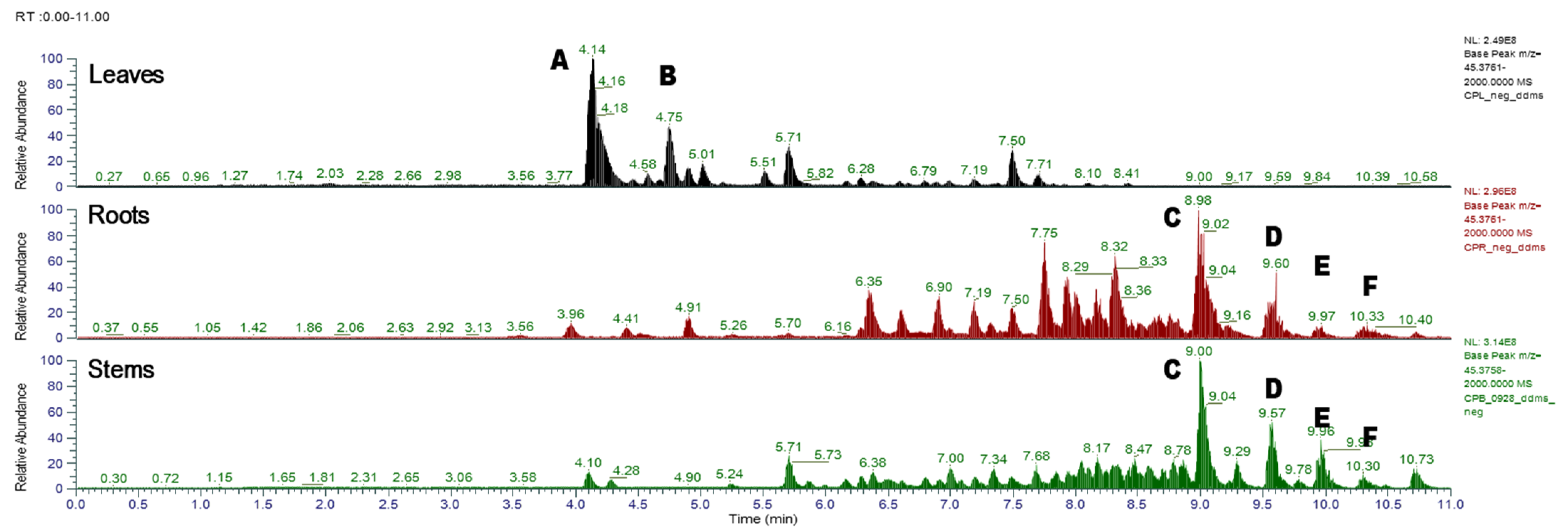
3.1. Comparison of Different Parts of C. formosum ssp. pruniflorum

Plant components are synthesized through plant-specific biosynthetic pathways, so there are similarities throughout the plant. However, if you subdivide it a little more, it shows some differences in constituents for each part of the plant, which leads to a difference in efficacy [32,33,34]. Therefore, we first compared the antioxidant and anti-diabetic efficacy of the parts of this plant, such as leaves, stems and roots. Since xanthone and flavonoid components have been known as major components of this plant, the contents in each part of the plant were also compared.
As shown in Table 1, all the parts of this plant, including the leaves, roots, and stems, showed antioxidant and α-glucosidase inhibitory effects. However, there were differences in the efficacy. The antioxidant effect was observed most strongly in the leaves, and the roots and stems also showed the efficacy. However, the roots showed the most excellent α-glucosidase inhibitory efficacy with an IC50 value of 2.0 μg/mL, followed by the leaves with an IC50 value of 3.9 μg/mL, but relatively weak efficacy in the case of the stem. As a result of comparing the contents of components, both flavonoid and phenol contents were highest in leaves. In particular, in the case of leaves, the content of flavonoid was relatively high, whereas phenolic compounds were observed to be high in roots and stems.
We further compared the chemical profiles of each part of C. formosum ssp. pruniflorum. As shown in Figure 1, the MS/MS chromatogram of leaves of C. formosum ssp. pruniflorum was quite different from that of roots and stems. Peak analysis by LC-MS/MS showed that mangiferin and quercetin-3-O-glucopyranoside were the major constituents of leaves, whereas α-mangostin, 7-geranyloxy-1,3-dihydroxyxanthone, and cochinchinone A were the major constituents of roots and stems (Table 2). The chemical patterns of the roots and stems were quite similar, but the components of the roots were more diverse than those of the stems and showed a higher content. Therefore, roots and leaves were selected for further purification of compounds.

3.2. Isolation and Characterization of the Constituents of C. formosum ssp. pruniflorum

Using various chromatography methods, 16 (1–16) and 9 (17–25) compounds were isolated from the roots and leaves of this plant, respectively. The structures of the isolated compounds were identified using spectroscopic methods as 2 new compounds (16 and 19) together with 23 known compounds.

3.2.1. Structure Elucidation of New Compounds

Compound 16 was isolated as a light brown amorphous powder. The molecular formula of 16 was determined as C28H31O6 from the HRESIMS (m/z 463.2124 [M-H], calcd. 463.2126) and the 13C NMR data. The characteristic UV absorption at 233, 266 and 317 nm suggested compound 16 as a xanthone skeleton [35]. The presence of the xanthone skeleton was confirmed from the 12 aromatic signals at [δC 110.2 (C-1), 157.9 (C-2), 153.1 (C-3), 105.9 (C-4), 147.9 (C-4a), 118.4 (C-5), 153.8 (C-5a), 149.8 (C-6), 123.8 (C-7), 107.9 (C-8), 120.5 (C-8a), 120.5 (C-9a)] together with a carbonyl carbon at δC 180.8 (C-9) in the 13C NMR spectrum and the signals for a 1,3,4-trisubstituted benzene ring at [δH 7.38 (1H, d, J = 9.0 Hz, H-5), 7.24 (1H, dd, J = 2.9, 9.0 Hz, H-7), 7.49 (1H, d, J = 2.9 Hz, H-8)] in the 1H NMR spectrum, which was also supported by the HSQC spectrum. The presence of a prenyl group was deduced by the signals at [δH 3.39 (2H, d, J = 7.1 Hz, H-1′), 5.20 (1H, t, J = 7.1 Hz, H-2′), 1.80 (3H, s, CH3-4′), 1.67 (3H, s, CH3-5′); δC 21.0 (C-1′), 121.9 (C-2′), 131.2 (C-3′), 16.6 (C-4′), 24.6 (C-5′)]. Additionally, the signals at [δH 3.56 (2H, d, J = 7.1 Hz, H-1″), 5.27 (1H, t, J = 7.1 Hz, H-2″), 2.67 (2H, m, H-4″), 5.53 (1H, m, H-5″), 5.54 (1H, brs, H-6″), 1.18 (6H, s, CH3-8″, 9″), 1.89 (3H, s, CH3-10″); δC 21.3 (C-1″), 122.9 (C-2″), 133.8 (C-3″), 42.0 (C-4″), 124.5 (C-5″), 139.1 (C-6″), 69.7 (C-7″), 28.5 (C-8″, C-9″), 15.2 (C-10″)] suggested the presence of a geranyl group. The presence of hydroxy group in the geranyl group was suggested by the oxymethine carbon at δC 69.7 (C-7″) and two methyl signals at δH 1.18 (6H, s, CH3-8″, CH3-9″), which was confirmed by the HMBC correlations from CH3-8″, 9″ to C-7″. Therefore, compound 16 was suggested to be a xanthone derivative with prenyl and hydroxygeranyl moieties. The positions of the prenyl and hydroxygeranyl moieties were deduced to C-1 and C-4, respectively, by the correlation from H-1′ to C-1 and from H-1″ to C-4 in the HMBC spectrum. On the basis of the obtained data, compound 16 was determined as shown and named pruniflonone A.
Compound 19 was purified as brown syrup with the molecular of C19H20O9 by HRESI-TOF-MS analysis (m/z 391.1034, calcd. for C19H19O9, 391.1035) and 13C NMR data. The 1H and 13C NMR spectra revealed the signals of a tetrasubstituted aromatic ring at [δH 6.21 (1H, d, J = 1.9 Hz, H-2), 6.07 (1H, d, J = 1.9 Hz, H-6); δC 109.1 (C-1), 95.8 (C-2), 159.7 (C-3), 164.0 (C-4), 162.0 (C-5), 98.1 (C-6)], a monosubstituted aromatic ring at [δH 7.69 (2H, m, H-2′, 6′), 7.41 (1H, m, H-3′, 5′), 7.52 (1H, m, H-4′); δC 141.6 (C-1′), 130.1 (C-2′, 6′), 129.0 (C-3′, 5′), 133.2 (C-4′)] and a carbonyl carbon at δC 199.5 (C-7). The presence of a glucosyl moiety was also confirmed by an anomeric proton at δH 4.81 (1H, d, J = 7.7 Hz, H-1″) together with the glucosyl carbon signals at [δC 101.9 (C-1″), 73.3 (C-2″), 76.0 (C-3″), 70.6 (C-4″), 74.3 (C-5″), 63.5 (C-6″)]. The HMBC correlations from H-2/6 and H-2′/6′ to C-7 (C=O) suggested the presence of a benzophenone skeleton. The position of the glucose was determined to be located at C-3 on the basis of HMBC correlation from the anomeric proton (H-1″) to C-3. Based on these data, compound 19 was determined as shown in Figure 2 and named pruniflonone B.

3.2.2. Identification of Known Compounds

The known compounds were identified as 17 xanthones—cochinchinone F (1), 1,3,7-cratosumatranone D (2), isocudraniaxanthone B (3), viellardixanthone B (4), diisoprenylxanthone (5), γ-mangostin (6), α-mangostin (7), β-mangostin (8), garcinone C (9), garcinone D (10), 11-hydroxy-1-garciniacowones E (11), isomangostin (12), garcinone B (13), trihydroxy-2,4-7-geranyloxy-1,3-dihydroxyxanthone (14), cochinchinone A (15), caloxanthone E (17) and mangiferin (18); a phenolic compound—protocatechuic acid (20); and 5 flavonoids—epicatechin (21), quercetin-3-O-glucopyranoside (22), isorhamnetin-3-O-glucoside (23), gujaverin (24) and quercetin-3-O-α-L-rhamnoside (25) via analysis of their physical data and comparison with values in the literature [36,37,38,39,40,41,42,43,44,45,46,47,48,49,50,51,52,53,54,55,56].

3.3. Evaluation of Antioxidant and α-Glucosidase Inhibitory Activity

The biological activity of the isolated compounds were evaluated by measuring the DPPH radical scavenging and α-glucosidase inhibitory activity. As described above, compounds isolated from C. formosum ssp. pruniflorum in this study are aromatic compounds and can be subdivided according to the compound skeleton as follows: xanthones (1–18), a benzophenone (19), a simple phenolic (20) and flavonoids (21–25). These isolated compounds showed good antioxidant and α-glucosidase inhibitory activity but differential efficacy depending on the structures (Figure 3).
Xanthones are more effective in the inhibition of α-glucosidase activity, whereas flavonoids are effective in antioxidant activity. Xanthones inhibited α-glucosidase activity with IC50 values of <50 μM. However, the addition of a hydroxyl group to prenyl or geranyl groups reduced the efficacy, as observed in 1 and 9. The addition of a sugar moiety also showed negative effects on α-glucosidase inhibition. In the case of antioxidant activity, xanthones 3, 6, 9, 17 and 18 showed more than 50% DPPH radical scavenging activity at 50 μM. Considering the structure, dihydroxy groups are important for the antioxidant activity of xanthones. In the case of flavonoids, flavonoids except compound 23 showed good antioxidant activity. Similar to xanthones, flavonoids with dihydroxy groups exerted antioxidant activity. Related to the α-glucosidase inhibitory activity of flavonoids of C. formosum ssp. pruniflorum, compound 21 without any sugar moieties showed good inhibition. However, benzophenone (19) exerted a weak effect on both antioxidant and α-glucosidase inhibition.
As described in Table 1, the extract of C. formosum ssp. pruniflorum exhibited α-glucosidase inhibitory and antioxidant activity. It contains xanthones, flavonoids and benzophenone, and most of them showed α-glucosidase inhibitory and/or antioxidant activity (Figure 3). In the case of the newly reported compounds in this study, compound 19 showed antioxidant efficacy, but compound 16, unfortunately, had weak efficacy. Conclusively, although the efficacies of compounds were quite different in each compound, xanthones and flavonoids were suggested to contribute to the antioxidant and α-glucosidase inhibitory potentials of C. formosum ssp. pruniflorum.
Differences were also observed depending on plant parts. For the α-glucosidase inhibition, the root extract showed the best activity, whereas the leaf extract showed the strongest antioxidant activity. Investigation of the constituents showed that the roots contained xanthones as major components and the leaves had flavonoids, which were consistent with the HRESI-MS/MS chromatogram (Figure 1). Measurement of biological activities of isolated compounds suggested that xanthone had α-glucosidase inhibitory potential, whereas flavonoids were more effective in antioxidant activity, which supported differential efficacy of the extract for each part.
Taken together, these results suggested the components and efficacy of C. formosum ssp. pruniflorum, which are differential depending on each part and can be used for the development of a marker component of each part.

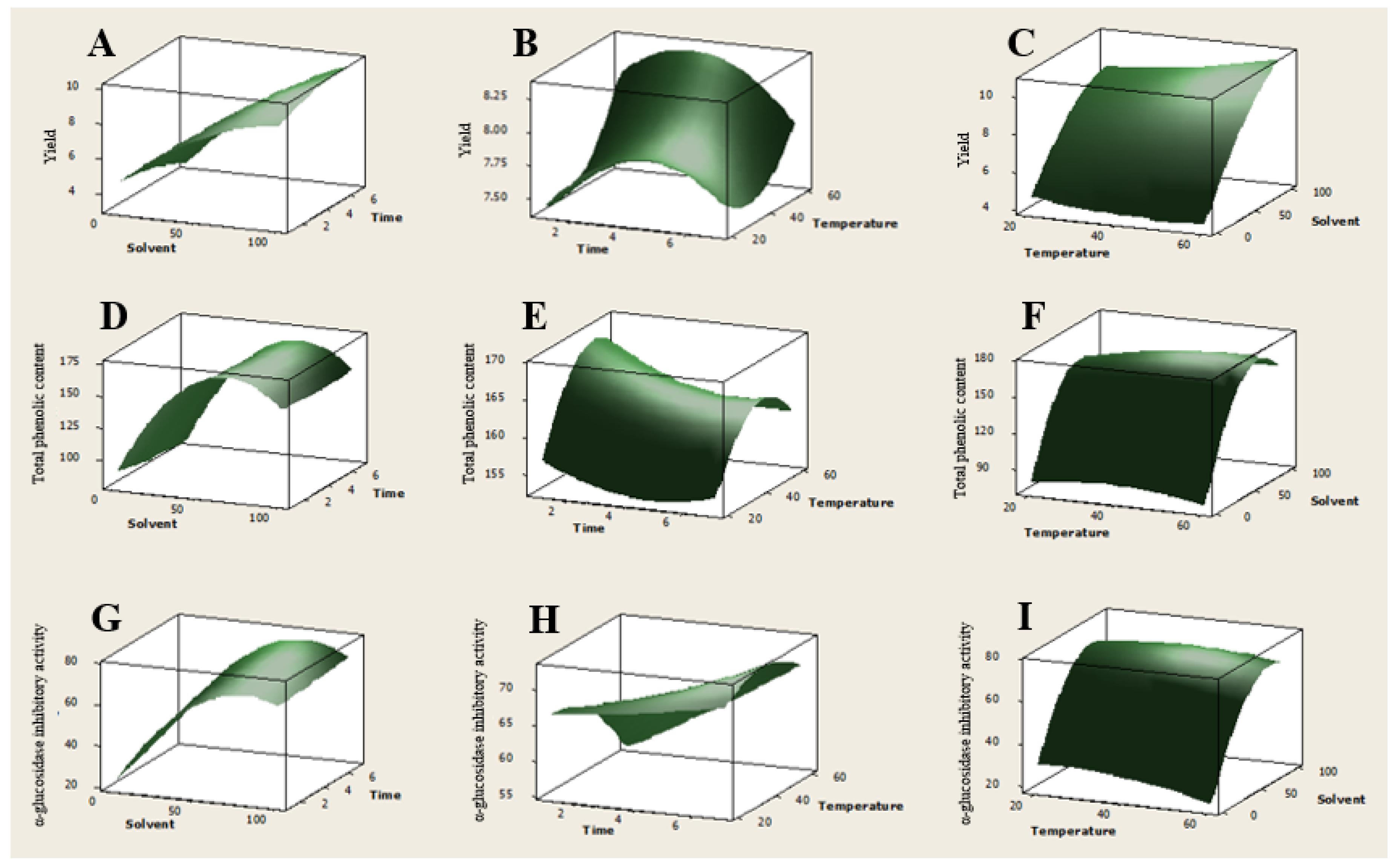
3.4. Optimization of Extraction Conditions Using Response Surface Metholodogy

The roots of C. formosum ssp. pruniflorum showed strong α-glucosidase inhibitory effects, and xanthones were assigned as active compounds. The content of active constituents in extract is highly affected by extraction conditions such as extraction solvent, extraction time and extraction temperature, which resulted in the difference in their biological activity [57,58]. Therefore, we further optimized the extraction conditions for maximum α-glucosidase inhibitory effects. Response surface methodology (RSM) is a statistical tool that takes several factors into account simultaneously using rationally designed experiments. The optimal condition can be derived effectively, especially in the case of several variables [59,60]. Therefore, RSM using a Box–Behnken design (BBD) was chosen for the optimization of extraction conditions of C. formosum ssp. pruniflorum for maximum efficiency.
Three variables such as extraction solvent (X1), extraction time (X2) and extraction temperature (X3) were chosen as independent variables, and the range of each variable was determined in the preliminary study. α-Glucosidase inhibitory effects together with yield and total phenolic content were determined as the dependent responses. The variables were coded at three levels (−1, 0 and 1), and the complete design consisted of 15 experimental points including three replications of the center points whose variables were all coded as zero (Table 3). Multiple regression analysis of the experiment data yielded the following second-order polynomial regression equation:
α-Glucosidase inhibition = 67.83 + 20.47X1 + 4.39X2 − 2.81X3 − 19.25X12 + 0.43X22 − 2.80X32 − 2.40X1X2 + 2.43X1X3 + 2.61X2X3.
Yield = 7.83 + 2.77X1 − 0.02X2 + 0.23X3 − 0.87X12 − 0.33X22 + 0.26X32 + 0.54X1X2 + 0.45X1X3 − 0.10X2X3.
Total phenolic content = 163.28 + 33.00X1 − 2.15X2 + 3.88X3 − 46.44X12 + 2.38X22 − 6.11X32 + 1.50X1X2 + 6.38X1X3 − 0.95X2X3.
The values of the coefficient determination (R2) and the adjusted coefficient determination (adj. R2) of the predicted model in this response suggested that the regression equation can explain the observed value to a high degree. Insignificant p-values of lack of fit (>0.05) for three responses also indicated the adaptability of this analysis (Table 4).
Among extraction variables, the linear term (X1) of MeOH concentration showed the most significant effect on all three responses. Relationships between the two variables in each response were also shown in a three-dimensional response surface (Figure 4). Consistent with multiple regression analysis, extraction solvent showed the strongest effect on yield, phenolic content and α-glucosidase inhibition (Figure 4A,D,G). Yield was increased with increasing MeOH concentration, but phenolic content and α-glucosidase inhibition were decreased with a continuing increase in MeOH concentration. On fixed temperature at 40 °C, yield was also affected by extraction time (Figure 4B), whereas total phenolic content was affected by extraction temperature when extracted with the mixture of MeOH-EtOAc (1:1) (Figure 4E). However, compared with extraction solvent, α-glucosidase inhibition showed slight changes as extraction time and temperature changed.
Based on these results, the extraction condition for maximum yield, α-glucosidase inhibitory effects and total phenolic content was optimized. The extract prepared using the optimized extraction condition was found to exert 73.9% α-glucosidase inhibitory effects at 1 μg/mL with a yield of 10.9% and a total phenolic content of 163.9 mg GAE/g extract (Table 5). The total phenolic content in the extract prepared using 15 different extraction conditions showed good correlation with α-glucosidase inhibitory effects, which is consistent with our present study about α-glucosidase inhibitory xanthones.
Collectively, the extraction yield and efficacy of C. formosum ssp. pruniflorum vary depending on the extraction conditions, and an extract with excellent efficacy can be efficiently secured through optimization of the extraction conditions. In addition, consistent with the efficacy of the isolated components, which was demonstrated in this study, the phenolic compounds were important for the efficacy of this plant and can be used as reference components for future product development.

4. Conclusions

Comparison of the roots, stems and leaves of C. formosum ssp. pruniflorum showed differences in the chemical profiles and biological activity. An investigation of C. formosum ssp. pruniflorum led to the isolation of 25 phenolic compounds, including 2 new compounds. The structures of the isolated compounds were determined to be xanthones, benzophenone, flavonoids and phenol. Two new compounds were defined as pruniflonone A (16) and pruniflonone B (19). The isolated compounds showed good antioxidant and α-glucosidase inhibitory activity with differences in activity depending on the structures. Optimization of extraction conditions was also studied using RSM for maximum efficacy. In conclusion, the C. formosum ssp. pruniflorum with antioxidant and α-glucosidase inhibitory activity might be beneficial for glucose-related diseases.

Supplementary Materials

The following are available online at https://www.mdpi.com/article/10.3390/antiox12020511/s1, Figures S1–S10: 1H, 13C, HSQC, HMBC and HRESI-MS spectrums of new compounds 16 and 19.

Author Contributions

Conceptualization, H.A. and M.K.L.; methodology, H.A., L.N.T., L.Q.K., S.H.R., S.L., S.W.Y., H.H.L. and A.T.; software, H.A. and K.Y.L.; validation, H.A., B.Y.H. and M.K.L.; formal analysis, H.A., B.Y.H. and M.K.L.; investigation, H.A., S.H.R., S.L., S.W.Y., H.H.L., A.T. and M.K.L.; writing—original draft preparation, H.A. and M.K.L.; writing—review and editing, H.A. and M.K.L.; supervision, M.K.L.; project administration, M.K.L.; funding acquisition, M.K.L. All authors have read and agreed to the published version of the manuscript.

Funding

This work was supported by the National Research Foundation of Korea (NRF) grant funded by the Korea Government (MSIT, 2022R1A2C1008081) and Medical Research Center Program (2017R1A5A2015541).

Institutional Review Board Statement

Not applicable.

Informed Consent Statement

Not applicable.

Data Availability Statement

The data are contained within the article or Supplementary Material.

Acknowledgments

The authors wish to thank the Korea Basic Science Institute for the NMR spectroscopic measurements.

Conflicts of Interest

The authors declare no conflict of interest.

References

  1. Brownlee, M. The pathobiology of diabetic complications a unifying mechanism. Diabetes 2005, 54, 1615–1625. [Google Scholar] [CrossRef] [PubMed] [Green Version]
  2. Yao, D.; Brownlee, M. Hyperglycemia-induced reactive oxygen species increase expression of the receptor for advanced glycation end products (RAGE) and RAGE ligands. Diabetes 2010, 59, 249–255. [Google Scholar] [CrossRef] [PubMed] [Green Version]
  3. Yaribeygi, H.; Sathyapalan, T.; Atkin, S.L.; Sahebkar, A. Molecular mechanisms linking oxidative stress and diabetes mellitus. Oxid. Med. Cell Longev. 2020, 2020, 8609213. [Google Scholar] [CrossRef] [Green Version]
  4. Reuter, S.; Gupta, S.C.; Chaturvedi, M.M.; Aggarwal, B.B. Oxidative stress, inflammation and cancer: How are they linked? Free Radic. Biol. Med. 2010, 49, 1603–1616. [Google Scholar] [CrossRef] [Green Version]
  5. Rendra, E.; Riabov, V.; Mossel, D.M.; Sevastyanova, T.; Harmsen, M.C.; Kzhyshkowska, J. Reactive oxygen species (ROS) in macrophage activation and function in diabetes. Immunobiology 2018, 224, 242–253. [Google Scholar] [CrossRef]
  6. Maritim, A.C.; Sanders, R.A.; Watkins, J.B. Diabetes, oxidative stress, and antioxidants: A review. J. Biochem. Mol. Toxicol. 2003, 17, 24–38. [Google Scholar] [CrossRef]
  7. Governa, P.; Baini, G.; Borgonetti, V.; Cettolin, G.; Giachetti, D.; Magnano, A.R.; Miraldi, E.; Biagi, M. Phytotherapy in the management of diabetes: A review. Molecules 2018, 23, 105. [Google Scholar] [CrossRef] [PubMed] [Green Version]
  8. Jo, Y.H.; Lee, S.; Yeon, S.W.; Turk, A.; Lee, J.H.; Hong, S.M.; Han, Y.K.; Lee, K.Y.; Hwang, B.Y.; Kim, S.Y.; et al. Anti-diabetic potential of Masclura tricuspidata leaves: Prenylated isoflavonoids with α-glucosidase inhibitory and anti-glycation activity. Bioorg. Chem. 2021, 114, 105098. [Google Scholar] [CrossRef]
  9. Ghani, U. Re-exploring promising α-glucosidase inhibitors for potential development into oral anti-diabetic drugs: Finding needle in the haystack. Eur. J. Med. Chem. 2015, 103, 133–162. [Google Scholar] [CrossRef]
  10. Joshi, S.R.; Standl, E.; Tong, N.; Shah, P.; Kalra, S.; Rathod, R. Therapeutic potential of α-glucosidase inhibitors in type 2 diabetes mellitus: An evidence-based review. Expert Opin. Pharmacother. 2015, 16, 1959–1981. [Google Scholar] [CrossRef]
  11. Tabatabaei-Malazy, O.; Peimani, M.; Mohseni, S.; Nikfar, S.; Abdollahi, M.; Larijani, B. Therapeutic effects of dietary antioxidative supplements on the management of type 2 diabetes and its complications; umbrella review of observational/trials meta-analysis studies. J. Diabetes Metab. Disord. 2022, 21, 1833–1859. [Google Scholar] [CrossRef]
  12. Zhang, P.; Li, T.; Wu, X.; Nice, E.C.; Huang, C.; Zhang, Y. Oxidative stress and diabetes: Antioxidative strategies. Front. Med. 2020, 14, 583–600. [Google Scholar] [CrossRef] [PubMed]
  13. Ryu, H.W.; Cho, J.K.; Curtis-Long, M.J.; Joo, Y.H.; Kim, Y.S.; Jung, S.I.; Kim, Y.S.; Lee, B.W.; Park, K.H. α-Glucosidase inhibition and antihyperglycemic activity of prenylated xanthones from Garcinia mangostana. Phytochemistry 2011, 72, 2148–2154. [Google Scholar] [CrossRef] [PubMed]
  14. Hedrington, M.S.; Davis, S.N. Considerations when using alpha-glucosidase inhibitors in the treatment of type 2 diabetes. Expert Opin. Pharmacother. 2019, 20, 2229–2235. [Google Scholar] [CrossRef] [PubMed]
  15. Rakha, A.; Umar, N.; Rabail, R.; Butt, M.S.; Kieliszek, M.; Hassoun, A.; Aadil, R.M. Anti-inflammatory and anti-allergic potential of dietary flavonoids: A review. Biomed. Pharmacother. 2022, 156, 113945. [Google Scholar] [CrossRef]
  16. Umeno, A.; Horie, M.; Murotomi, K.; Nakajima, Y.; Yoshida, Y. Antioxidative and antidiabetic effects of natural polyphenols and isoflavones. Molecules 2016, 30, 708. [Google Scholar] [CrossRef] [Green Version]
  17. Na, Z. Chemical constituents of volatile oil from leaf of Cratoxylum formosum subsp. pruniflorum in Xishuangbanna of Yunna Province. J. Plant Resour. Environ. 2007, 16, 75–77. [Google Scholar]
  18. Srithi, K.; Balslev, H.; Wangpakapattanawong, P.; Srisanga, P.; Trisonthi, C. Medicinal plant knowledge and its erosion among the Mien (Yao) in northern Thailand. J. Ethnopharmacol. 2009, 123, 335–342. [Google Scholar] [CrossRef]
  19. Boonnak, N.; Karalai, C.; Chantrapromma, S.; Ponglimanont, C.; Kanjana-Opas, A.; Chantrapromma, K.; Kato, S. Chromene and prenylated xanthones from the roots of Cratoxylum formosum ssp. pruniflorum. Chem. Pharm. Bull. 2010, 58, 386–389. [Google Scholar] [CrossRef] [Green Version]
  20. Duan, Y.H.; Dai, Y.; Wang, G.H.; Zhang, X.; Chen, H.F.; Chen, J.B.; Yao, X.S.; Zhang, X.K. Bioactive xanthones from the stems of Cratoxylum formosum ssp. pruniflorum. J. Nat. Prod. 2010, 73, 1283–1287. [Google Scholar] [CrossRef] [PubMed]
  21. Xiong, J.; Liu, X.H.; Bui, V.B.; Hong, Z.L.; Wang, L.J.; Zhao, Y.; Fan, H.; Yang, G.X.; Hu, J.F. Phenolic constituents from the leaves of Cratoxylum formosum ssp. pruniflorum. Fitoterapia 2014, 94, 114–119. [Google Scholar] [CrossRef]
  22. Duan, Y.H.; Dai, Y.; Wang, G.H.; Chen, H.F.; Gao, H.; Chen, J.B.; Yao, X.S.; Zhang, X.K. Xanthone and benzophenone glycosides from the stems of Cratoxylum formosum ssp. pruniflorum. Chem. Pharm. Bull. 2011, 59, 231–234. [Google Scholar] [CrossRef] [PubMed] [Green Version]
  23. Pocasap, P.; Weerapreeyakul, N.; Wongpoomchai, R. Chemopreventive effect of Cratoxylum formosum (Jack) ssp. pruniflorum on initial stage hepatocarcinogenesis in rats. Molecules 2021, 26, 4235. [Google Scholar] [CrossRef] [PubMed]
  24. Kaewpiboon, C.; Boonnak, N.; Kaowinn, S.; Yawut, N.; Chung, Y.H. Formoxanthone C inhibits malignant tumor phenotypes of human A549 multidrug resistant-cancer cells through signal transducer and activator of transcription 1-histone deacetylase 4 signaling. J. Cancer Prev. 2022, 27, 112–121. [Google Scholar] [CrossRef]
  25. Senggunprai, L.; Thammaniwit, W.; Kukongviriyapan, V.; Prawan, A.; Kaewseejan, N.; Siriamornpun, S. Cratoxylum formosum extracts inhibit growth and metastasis of cholangiocarcinoma cells by modulating the NF-κB and STAT3 pathways. Nutr. Cancer 2016, 68, 328–341. [Google Scholar] [CrossRef]
  26. Kaewpiboon, C.; Boonnak, N.; Kaowinn, S.; Chung, Y.H. Formoxanthone C, isolated from Cratoxylum formosum ssp. pruniflorum, reverses anticancer drug resistance by inducing both apoptosis and autophagy in human A549 lung cancer cells. Bioorg. Med. Chem. Lett. 2018, 28, 820–825. [Google Scholar] [CrossRef] [PubMed]
  27. Keowkase, R.; Weerapreeyaku, N. Cratoxylum formosum extract protects against amyloid-beta toxicity in a Caenorhabditis elegans model of Alzheimerʼs disease. Planta Med. 2016, 82, 516–523. [Google Scholar] [CrossRef] [PubMed] [Green Version]
  28. Raksat, A.; Laphookhieo, S.; Cheenpracha, S.; Ritthiwigrom, T.; Maneerat, W. Antibacterial compounds from the roots of Cratoxylum formosum spp. Pruniflorum. Nat. Prod. Commun. 2014, 9, 1487–1489. [Google Scholar] [CrossRef] [Green Version]
  29. Boonsri, S.; Karalai, C.; Ponglimanont, C.; Kanjana-opas, A.; Chantrapromma, K. Antibacterial and cytotoxic xanthones from the roots of Cratoxylum formosum. Phytochemistry 2006, 67, 723–727. [Google Scholar] [CrossRef]
  30. Ahn, J.H.; Ryu, S.H.; Lee, S.; Yeon, S.W.; Turk, A.; Han, Y.K.; Lee, K.Y.; Hwang, B.Y.; Lee, M.K. Aromatic constituents from the leaves of Actinidia arguta with antioxidant and α-glucosidase inhibitory activity. Antioxidants 2021, 10, 1896. [Google Scholar] [CrossRef]
  31. Kim, S.B.; Jo, Y.H.; Liu, Q.; Ahn, J.H.; Hong, I.P.; Han, S.M.; Hwang, B.Y.; Lee, M.K. Optimization of extraction condition of bee pollen using response surface methodology: Correlation between anti-melanogenesis, antioxidant activity, and phenolic content. Molecules 2015, 20, 9656. [Google Scholar] [CrossRef] [Green Version]
  32. Jang, J.Y.; Shin, H.; Lim, J.W.; Ahn, J.H.; Jo, Y.H.; Lee, K.Y.; Hwang, B.Y.; Jung, S.J.; Kang, S.Y.; Lee, M.K. Comparison of antibacterial activity and phenolic constituents of bark, lignum, leaves and fruit of Rhus verniciflua. PLoS ONE 2018, 13, e0200257. [Google Scholar] [CrossRef] [PubMed]
  33. Jo, Y.H.; Kim, S.B.; Liu, Q.; Do, S.G.; Hwang, B.Y.; Lee, M.K. Comparison of pancreatic lipase inhibitory isoflavonoids from unripe and ripe fruits of Cudrania tricuspidata. PLoS ONE 2017, 12, e0172069. [Google Scholar] [CrossRef] [PubMed] [Green Version]
  34. Liu, J.; Mu, X.; Liang, J.; Zhang, J.; Qiang, T.; Li, H.; Li, B.; Liu, H.; Zhang, B. Metabolic profiling on the analysis of different parts of Schisandra chinensis based on UPLC-QTOF-MS with comparative bioactivity assays. Front. Plant Sci. 2022, 13, 970535. [Google Scholar] [CrossRef] [PubMed]
  35. Jo, Y.H.; Shin, B.; Liu, Q.; Lee, K.Y.; Oh, D.-C.; Hwang, B.Y.; Lee, M.K. Antiproliferative prenylated xanthones and benzophenones from the roots of Cudrania tricuspidata in HSC-T6 cells. J. Nat. Prod. 2014, 77, 2361–2366. [Google Scholar] [CrossRef] [PubMed]
  36. Ding, H.Y.; Lin, H.C.; Teng, C.M.; Wu, Y.C. Phytochemical and pharmacological studies on Chinese Paeonia species. J. Chin. Chem. Soc. 2000, 47, 381–388. [Google Scholar] [CrossRef]
  37. Park, C.H.; Kim, K.H.; Lee, I.K.; Lee, S.Y.; Choi, S.U.; Lee, J.H.; Lee, K.R. Phenolic constituents of Acorus gramineus. Arch. Pharm. Res. 2011, 34, 1289–1296. [Google Scholar] [CrossRef]
  38. Li, H.Z.; Song, H.J.; Li, H.M.; Pan, Y.Y.; Li, R.T. Characterization of phenolic compounds from Rhododendron alutaceum. Arch. Pharm. Res. 2012, 35, 1887–1893. [Google Scholar] [CrossRef] [PubMed]
  39. Lee, Y.G.; Cho, J.Y.; Kim, C.M.; Lee, S.H.; Kim, W.S.; Jeon, T.I.; Park, K.H.; Moon, J.H. Coumaroyl quinic acid derivatives and flavonoids from immature pear (Pyrus pyrifolia Nakai) fruit. Food Sci. Biotechnol. 2013, 22, 803–810. [Google Scholar] [CrossRef]
  40. Prachayasittikul, S.; Suphapong, S.; Worachartcheewan, A.; Lawung, R.; Ruchirawat, S.; Prachayasittikul, V. Bioacitive metabolites from Spilanthes acmella Murr. Molecules 2009, 14, 850. [Google Scholar] [CrossRef] [Green Version]
  41. Bohlmann, F.; Chen, Z.L.; Schuster, A. Aromatic esters from Solidago decurrens. Phytochemstry 1981, 20, 2601–2602. [Google Scholar] [CrossRef]
  42. Itoh, T.; Ninomiya, M.; Yasuda, M.; Koshikawa, K.; Deyashiki, Y.; Nozawa, Y.; Akao, Y.; Koketsu, M. Inhibitory effects of flavonoids isolated from Fragaria ananassa Duch on IgE-mediated degranulation in rat basophilic leukemia RBL-2H3. Bioorg. Med. Chem. 2009, 17, 5374–5379. [Google Scholar] [CrossRef]
  43. Han, J.T.; Bang, M.H.; Chun, O.K.; Kim, D.O.; Lee, C.Y.; Baek, N.I. Flavonol glycosides from the aerial parts of Aceriphyllum rossii and their antioxidant activities. Arch. Pharm. Res. 2004, 27, 390–395. [Google Scholar] [CrossRef]
  44. Jeon, S.H.; Chun, W.J.; Choi, Y.J.; Kwon, Y.S. Cytotoxic constituents from the bark of Salix hulteni. Arch. Pharm. Res. 2008, 31, 978–982. [Google Scholar] [CrossRef]
  45. Lim, E.K.; Ashford, D.A.; Hou, B.K.; Jackson, R.G.; Bowles, D.J. Arabidopsis glycosyltransferases as biocatalysts in fermentation for regioselective synthesis of diverse quercetin glucosides. Biotechnol. Bioeng. 2004, 87, 623–631. [Google Scholar] [CrossRef] [PubMed]
  46. Fico, G.; Rodondi, G.; Flamini, G.; Passarella, D.; Tome, F. Comparative phytochemical and morphological analyses of three Italian Primula species. Phytochemistry 2007, 68, 1683–1691. [Google Scholar] [CrossRef]
  47. He, Z.; Lian, W.; Liu, J.; Zheng, R.; Xu, H.; Du, G.; Liu, A. Isolation, structural characterization and neuraminidase inhibitory activities of polyphenolic constituents from Flos caryophylli. Phytochem. Lett. 2017, 19, 160–167. [Google Scholar] [CrossRef]
  48. Zheng, Z.P.; Cheng, K.W.; Chao, J.; Wu, J.; Wang, M. Tyrosinase inhibitors from paper mulberry (Broussonetia papyrifera). Food Chem. 2008, 106, 529–535. [Google Scholar] [CrossRef]
  49. Calzada, F.; Cedillo-Rivera, R.; Mata, R. Antiprotozoal activity of the constituents of Conyza filaginoides. J. Nat. Prod. 2001, 64, 671–673. [Google Scholar] [CrossRef] [PubMed]
  50. Wan, C.P.; Yuan, T.; Cirello, A.L.; Seeram, N.P. Antioxidant and α-glucosidase inhibitory phenolics isolated from highbush blueberry flowers. Food Chem. 2012, 135, 1929–1937. [Google Scholar] [CrossRef]
  51. Pizzolatti, M.G.; Venson, A.F.; Junior, A.S.; Smania, E.F.A.; Braz-Filho, R. Two epimeric flavalignans from Trichilia catigua (Meliaceae) with antimicrobial activity. J. Biosci. 2002, 57, 483–488. [Google Scholar] [CrossRef]
  52. Kim, T.H.; Ito, H.; Hayashi, K.; Hasegawa, T.; Machiguchi, T.; Yoshida, T. Aromatic constituents from the Heartwood of Santalum album L. Chem. Pharm. Bull. 2005, 53, 641–644. [Google Scholar] [CrossRef] [PubMed] [Green Version]
  53. Fang, J.M.; Lee, C.K.; Cheng, Y.S. Lignans from leaves of Juniperus chinensis. Phytochemistry 1992, 31, 3659–3661. [Google Scholar]
  54. Kang, W.; Wang, J. In vitro antioxidant properties and in vivo lowering blood lipid of Forsythia suspense leaves. Med. Chem. Res. 2010, 19, 617–628. [Google Scholar] [CrossRef]
  55. Moon, S.S.; Rahman, A.A.; Kim, J.Y.; Kee, S.H. Hanultarin, a cytotoxic lignin as an inhibitor of actin cytoskeleton polymerization from the seeds of Trichosanthes kirilowii. Bioorg. Med. Chem. 2008, 16, 7264–7269. [Google Scholar] [CrossRef] [PubMed]
  56. Kong, W.X.; Yuan, Z. New lignin glycosides from Glehnia littralis. Chin. Chem. Lett. 2008, 19, 1459–1461. [Google Scholar] [CrossRef]
  57. Bezerra, M.A.; Santelli, R.E.; Oliveira, E.P.; Villar, L.S.; Escaleira, L.A. Response surface methodology (RSM) as a tool for optimization in analytical chemistry. Talanta 2008, 76, 965–977. [Google Scholar] [CrossRef]
  58. Ferreira, S.L.C.; Bruns, R.E.; Ferreira, H.S.; Matos, G.D.; David, J.M.; Brandao, G.C.; da Silva, E.G.P.; Portugal, L.A.; Reis, P.S.; Souza, A.S. Box-Behnken design an alternative for the optimization of analytical methods. Anal. Chim. Acta 2007, 597, 179–186. [Google Scholar] [CrossRef]
  59. Hu, X.; Xu, F.; Li, J.; Li, J.; Mo, C.; Zhao, M.; Wang, L. Ultrasonic-assisted extraction of polysaccharides from coix seeds: Optimization, purification, and in vitro digestibility. Food Chem. 2022, 374, 131636. [Google Scholar] [CrossRef]
  60. Pinto, D.; Vieira, E.F.; Peixoto, A.F.; Freire, C.; Freitas, V.; Costa, P.; Delerue-Matos, C.; Rodrigues, F. Optimizing the extraction of phenolic antioxidants from chestnut shells by subcritical water extraction using response surface methodology. Food Chem. 2021, 334, 127521. [Google Scholar] [CrossRef]
Figure 1. HRESI-MS/MS chromatograms of leaves, roots and stems of C. formosum ssp. pruniflorum.
Antioxidants 12 00511 g001
Figure 2. Chemical structures of compounds 125 from C. formosum ssp. pruniflorum.
Antioxidants 12 00511 g002
Figure 3. Antioxidant and α-glucosidase inhibitory activity of compounds 125 from C. formosum ssp. pruniflorum.
Antioxidants 12 00511 g003
Figure 4. Response surface plots. Effect of extraction variables in yield (AC), total phenolic content (DF) and α-glucosidase inhibitory activity (GI). Three variables are extraction solvent (X1), extraction time (X2) and extraction temperature (X3).
Antioxidants 12 00511 g004
Table 1. Total phenolic and flavonoid content of leaves, roots and stems of C. formosum ssp. pruniflorum.
Antioxidant Activity
(IC50, μg/mL)
α-Glucosidase Inhibition
(IC50, μg/mL)
Total Phenolic Content
(mg GAE/g Extracts)
Total Flavonoid Content
(mg CE/g Extracts)
Leaves14.93.9132.3101.6
Roots17.02.094.949.8
Stems47.823.085.528.7
Table 2. Peak profiling by LC-MS/MS from leaves, roots and stems of C. formosum ssp. pruniflorum.
Peak No.Compound IdentificationtR (min)m/zMolecular Formular
[M-H]
UV
max, nm)
Compd No in This Study
ObservedCalculated
Amangiferin4.14421.0769421.0776C19H18O11204, 256, 316, 36418
Bquercetin-3-O-glucopyranoside4.75463.0877463.0882C21H20O12204, 256, 35622
Cγ-mangostin9.00395.1507395.1500C23H23O6208, 268, 316, 36412
Dα-mangostin9.57409.1664409.1657C24H26O6240, 3168
E7-geranyloxy-1,3-dihydroxyxanthone9.96379.1541379.1551C23H24O5224, 236, 260, 308, 3681
Fcochinchinone A10.31447.2177447.2177C28H32O5220, 240, 268, 316, 4085
Table 3. A Box–Behnken Design for independent variables and their responses.
RunActual Variables (Coded Variables)Observed Values
Extraction Solvent
(X1, %)
Extraction
Time
(X2, h)
Extraction Temperature (X3, °C)α-Glucosidase Inhibition
(% of Control)
Yield
(%)
Total Phenolic Content
(mg GAE/g Extract)
1100 (1)7 (1)40 (0)67.910.30154.2
20 (−1)4 (0)20 (−1)29.24.7680.2
3100 (1)4 (0)60 (1)67.210.58153.9
450 (0)1 (−1)20 (−1)64.37.73159.4
550 (0)4 (0)40 (0)70.77.70165.4
650 (0)1 (−1)60 (1)53.58.41166.1
70 (−1)7 (1)40 (0)34.63.5082.1
850 (0)4 (0)40 (0)68.57.74165.1
9100 (1)1 (−1)40 (0)68.38.68153.3
1050 (0)7 (1)20 (−1)72.27.31154.9
11100 (1)4 (0)20 (−1)68.19.21130.5
120 (−1)1 (−1)40 (0)25.34.0487.3
1350 (0)7 (1)60 (1)71,87.59157.8
1450 (0)4 (0)40 (0)64.38.01159.3
150 (−1)4 (0)60 (1)18.64.3178.2
Table 4. ANOVA for response surface regression equation.
ResponsesCategorySum of
Square
Degree of FreedomMean SquareF Valuep Value
YieldRegression67.324897.480545.57<0.001
Linear61.7689320.5896125.44<0.001
Square3.517731.17267.140.029
Interaction2.038230.67944.140.08
Residual error0.820750.1641
Lack-of-fit0.743930.2486.450.137
Pure error0.076920.0384
Total68.145514
R2 = 0.988, adjusted R2 = 0.966
Total phenolicRegression17,170.6191907.85139.74<0.001
Linear8868.8632956.29216.53<0.001
Square8126.4132708.8198.4<0.001
Interaction178.34358.454.280.076
Residual error68.27513.65
Lack-of-fit42.03315.011.290.464
Pure error23.24211.62
Total17,238.914
R2 = 0.996, adjusted R2 = 0.989
α-GlucosidaseRegression5033.59559.2837.13<0.001
inhibitionLinear9570.4331190.1479.02<0.001
Square1389.173463.0630.740.001
Interaction73.9324.631.640.294
Residual error75.31515.06
Lack-of-fit53.81317.941.670.396
Pure error21.5210.75
Total5108.8114
R2 = 0.985, adjusted R2 = 0.959
Table 5. Predicted and observed values of yield, total phenolic content and α-glucosidase inhibitory activity under optimized condition.
Optimized Extraction ConditionResponses
Extraction Solvent
(% MeOH in EtOAc)
Extraction Time
(h)
Extraction Temperature
(°C)
α-Glucosidase Inhibitory Activity a
(% of Control)
Yield
(%)
Total Phenolic Content
(mg GAE/g extract)
88.16.0260.0PredictedObserved PredictedObservedPredictedObserved
72.273.910.410.9163.9163.9
a α-Glucosidase inhibitory activity (%) was measured at 1 μg/mL.
Disclaimer/Publisher’s Note: The statements, opinions and data contained in all publications are solely those of the individual author(s) and contributor(s) and not of MDPI and/or the editor(s). MDPI and/or the editor(s) disclaim responsibility for any injury to people or property resulting from any ideas, methods, instructions or products referred to in the content.

