Article

Anti-Inflammatory and Antioxidant Properties of β-Sitosterol in Copper Sulfate-Induced Inflammation in Zebrafish (Danio rerio)

1
Key Laboratory of Exploration and Utilization of Aquatic Genetic Resources, Ministry of Education, Shanghai Ocean University, Shanghai 201306, China
2
Yangtze River Fisheries Research Institute, Chinese Academy of Fishery Sciences, Wuhan 430223, China
3
National Pathogen Collection Center for Aquatic Animals, Shanghai Ocean University, Shanghai 201306, China
*
Authors to whom correspondence should be addressed.
These authors contributed equally to this work.
Antioxidants 2023, 12(2), 391; https://doi.org/10.3390/antiox12020391
Submission received: 17 December 2022 / Revised: 31 January 2023 / Accepted: 1 February 2023 / Published: 6 February 2023
(This article belongs to the Topic Antioxidant Activity of Natural Products)

Abstract

:
β-Sitosterol, which is used extensively in pharmaceuticals, nutraceuticals, and cosmetics, has high nutritional value along with immunomodulatory and anti-inflammatory properties. In this study, we investigated the antioxidant and anti-inflammatory effects of β-sitosterol in zebrafish and explored the associated molecular mechanisms. In an in vivo antioxidant experiment, zebrafish (Danio rerio) larvae were treated with different concentrations of β-sitosterol and then exposed to a nonlethal concentration of CuSO4 to induce oxidative stress. Treatment with β-sitosterol at 70 or 100 μg/mL significantly reduced CuSO4-induced oxidative stress in the zebrafish, demonstrating the strong antioxidant activity of β-sitosterol. Treatment with β-sitosterol protected zebrafish larvae against oxidative damage from CuSO4 by upregulating the expressions of sod and gpx4b. In a zebrafish model of inflammation, pretreatment with β-sitosterol before CuSO4 exposure inhibited neutrophil recruitment and damage to lateral line neuromasts, indicating a potent anti-inflammatory effect derived from reductions in the expressions of il-8 and myd88. The results demonstrate the antioxidative and anti-inflammatory activities of β-sitosterol and suggest that β-sitosterol may be useful for the treatment of various inflammatory diseases.

1. Introduction

Inflammation is the root cause of many chronic diseases, such as obesity, hypertension, atherosclerosis, cancer, and Alzheimer’s disease [1]. In the presence of inflammatory signals, neutrophils first infiltrate into the site of infection, injury, or inflammation. Neutrophils are the major source of reactive oxygen species (ROS) during inflammation [2]. Therefore, oxidative stress develops and further exacerbates inflammation [3]. Conversely, when oxidative stress occurs, the overproduction of ROS and nitrogen species (NOS) will drive the inflammatory response by inducing the expression of proinflammatory genes [4]. Thus, the inflammatory process can exacerbate oxidative stress and vice versa. Traditional anti-inflammatory agents can cause severe side effects, such as hemorrhagic gastritis, upper gastrointestinal hemorrhage, gastrointestinal toxicity (diarrhea/colitis), and hypertension [5,6], limiting the use of these drugs [7]. While oxidative stress and inflammation are interdependent, the treatment of primary abnormalities alone is not always successful [3]. Thus, it is crucial to find effective anti-inflammatory drugs that simultaneously inhibit ROS production and mediate inflammation; this is considered a promising strategy for preventing and treating diseases associated with chronic inflammation. Oxidative and inflammatory responses can be easily induced and visualized in zebrafish, especially those in the early developmental stages [8]. Copper is an essential micronutrient that can also be toxic to many types of cells at certain levels [9]. Excessive inorganic copper in the environment can disrupt the copper balance in zebrafish [10], leading to elevated serum copper levels and triggering an oxidative stress responses. This ultimately results in an oxidative stress damage-mediated inflammatory response [11]. In addition, the zebrafish (Danio rerio) fish embryo toxicity (FET) assay is a commonly used methodology for determining the toxicity of environmental pollutants [12].
Phytosterol is a natural active substance that has been hailed as “the key of life” [13]. The most common and abundant phytosterols, including β-sitosterol, exhibit important physiological functions [14]. In animal models, β-sitosterol was found to alleviate dextran sulfate sodium-induced colitis in mice; β-sitosterol not only enhanced the body weight and improved the colonic morphology in the mice, it also decreased the mRNA expressions of proinflammatory cytokines in the colon in a dose-dependent manner [15]. In vivo animal studies showed that β-sitosterol has strong antigenotoxic effects, while in vitro tests revealed a dose-dependent antioxidant effect of β-sitosterol [16]. However, few studies have evaluated the effects of β-sitosterol in fish, and little is known about the mechanism of action of β-sitosterol in fish. In this study, we used CuSO4 to induce an inflammatory response through an oxidative stress reaction in zebrafish and investigated the antioxidant and anti-inflammatory effects of β-sitosterol. We also investigated the molecular mechanisms of β-sitosterol. This study provides a scientific basis for the application of β-sitosterol in traditional medicine and aquatic feed additives.

2. Materials and Methods

2.1. Fish and Experimental Conditions

The wild-type AB zebrafish (Danio rerio) and transgenic zebrafish line Tg (lyz: DsRED2) were purchased from China Zebrafish Resource Center (CZRC, Wuhan, China). Tg (lyz: DsRed2) was used for the neutrophil imaging experiments. The zebrafish were raised and maintained according to the guidelines available at http://zfin.org/ (accessed on 1 June 2022). The zebrafish were fed three times each day. The day before mating, males and females were placed in breeding tanks. Mating and spawning occurred within 1 h after turning on the lights in the morning. The embryos were picked under a microscope, and the selected fertilized eggs were placed in E3 medium at 28 °C. The media were replaced after 24 h, and all experiments were carried out using zebrafish larvae at 3 days postfertilization (dpf). All animal experiments were approved by the Animal Experimental Ethical Inspection of Laboratory Animal Centre, Yangtze River Fisheries Research Institute, Chinese Academy of Fishery Sciences (ID number: YFI 2022-zhouyong-07).

2.2. Main Reagents

β-Sitosterol was purchased from Shanghai Yuanye Bio-Technology Co., Ltd. (Shanghai, China). 2,2-Diphenyl-1-picrylhydrazyl (DPPH), quercetin, 2′,7′-dichlorodihydrofluorescein diacetate (H2DCFDA), pyrocatechol violet (PV), copper sulfate pentahydrate (CuSO4·5H2O), and methyl cellulose were purchased from Merck Co., Ltd. (Shanghai, China). Dimethylformamide (DMSO) was purchased from Sangon Biotech Co., Ltd. (Shanghai, China). The E3 medium was purchased from Nanjing EzeRinka Biotechnology Co., Ltd. (Nanjing, China). The components of E3 were NaCl (29.4 g/100 mL), KCl (1.27 g/100 mL), CaCl2·2H2O (4.85 g/100 mL), and MgSO4·7H2O (8.13 g/100 mL) [17].

2.3. Experimental Design

2.3.1. Preparation of Test Samples

Dimethyl sulfoxide (DMSO) was used as a solvent. β-Sitosterol stock solution (200 µg/mL) was prepared in DMSO. Working solutions were prepared by diluting the stock solution with E3 medium. The experimental concentrations of β-sitosterol were 0, 5, 20, 50, 70, and 100 µg/mL. The stock solution of H2DCFDA (20 µg/mL) was prepared in DMSO and frozen at −20 °C. The working solutions were prepared on the day of the experiment.

2.3.2. Chelating Ability of β-Sitosterol for Cu2+

The chelating ability of β-sitosterol for Cu2+ was evaluated according to the method described by Santos et al. [18] using PV as the chromogen agent. A 30 µL solution of β-sitosterol at different concentrations (5, 20, 50, 75, and 100 µg/mL) or water (control) were mixed with 200 µL of sodium acetate buffer (50 mmol/L). Then, 30 μL of 100 mg/L CuSO4 ·5H2O solution was added to each group for 2 min. After 2 min, 2 mmol/L 8.5 μL catechol violet solution was added to initiate the reaction. All reaction mixtures were shaken at 25 °C for 10 min, and then the absorbance was measured at 632 nm using an UV-Vis spectrophotometer (Palo Alto, CA, USA). The Cu2+ chelating ability of β-sitosterol was calculated as follows: Cu2+ chelating ability of β-sitosterol (%) = (Abs control − Abs sample) × 100/Abs control.

2.3.3. Antioxidant Effect of β-Sitosterol In Vitro

The antioxidant activity was measured in vitro based on the DPPH radical scavenging, following Cheng-zhong et al. [19]. A 20 µL solution of β-sitosterol at different concentrations (5, 20, 50, 75, and 100 µg/mL) was added to a 180 µL methanol solution of 0.1 mM DPPH. Methanol was used as a blank, and quercetin was used as a positive control. The reaction solution was shaken at room temperature for 30 min in dark. Then the absorbance was measured at 517 nm using a UV-Vis spectrophotometer. The percent inhibition of DPPH radical scavenging capacity was calculated as follows: scavenging activity (%) = (Abs control − Abs sample) × 100/(Abs control).

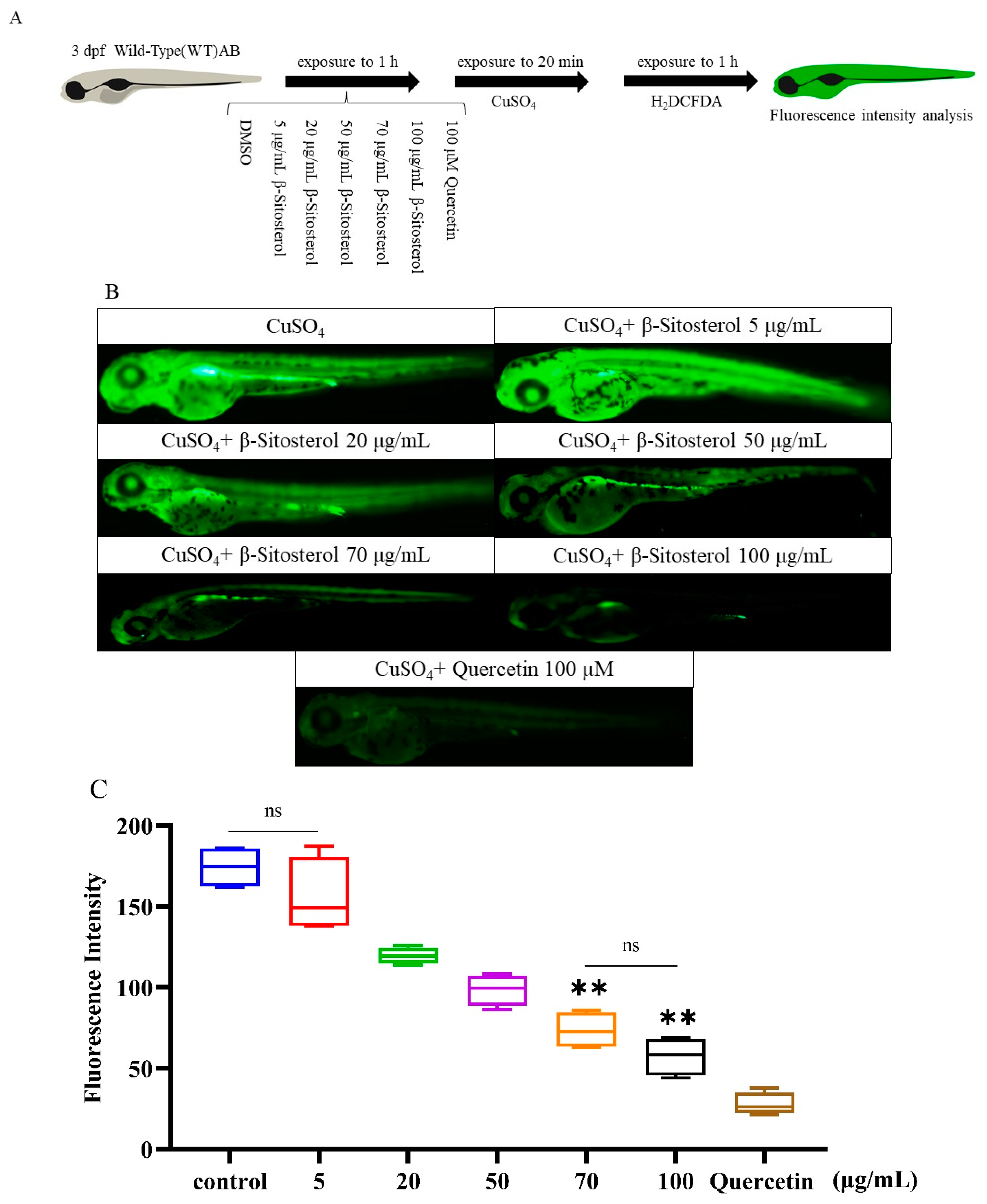
2.3.4. In Vivo Antioxidant Test

The production of ROS was detected by a H2DCFDA assay [20]. Zebrafish embryos (3 dpf) were transferred to a six-well plate (10 larvae per well) containing 940 μL E3 medium. The zebrafish larvae were treated with β-sitosterol (0, 5, 20, 50, 70, or 100 μg/mL) or 100 μM quercetin (positive control) for 1 h. The larvae were then treated with 10 μM CuSO4 for 20 min to induce oxidative stress [20], rinsed with fresh E3 medium, incubated with H2DCFDA for 1 h in the dark at 28 °C, and again rinsed in fresh E3 medium. The zebrafish larvae were anesthetized with 2% MS-222, immobilized in 3% methylcellulose, and observed under a fluorescence microscope (Olympus, Tokyo, Japan) and photographed. The average fluorescence intensity was quantified using Image J 1.48 software. The larvae treated with CuSO4 alone served as the negative control, while the larvae treated with quercetin were the positive control.

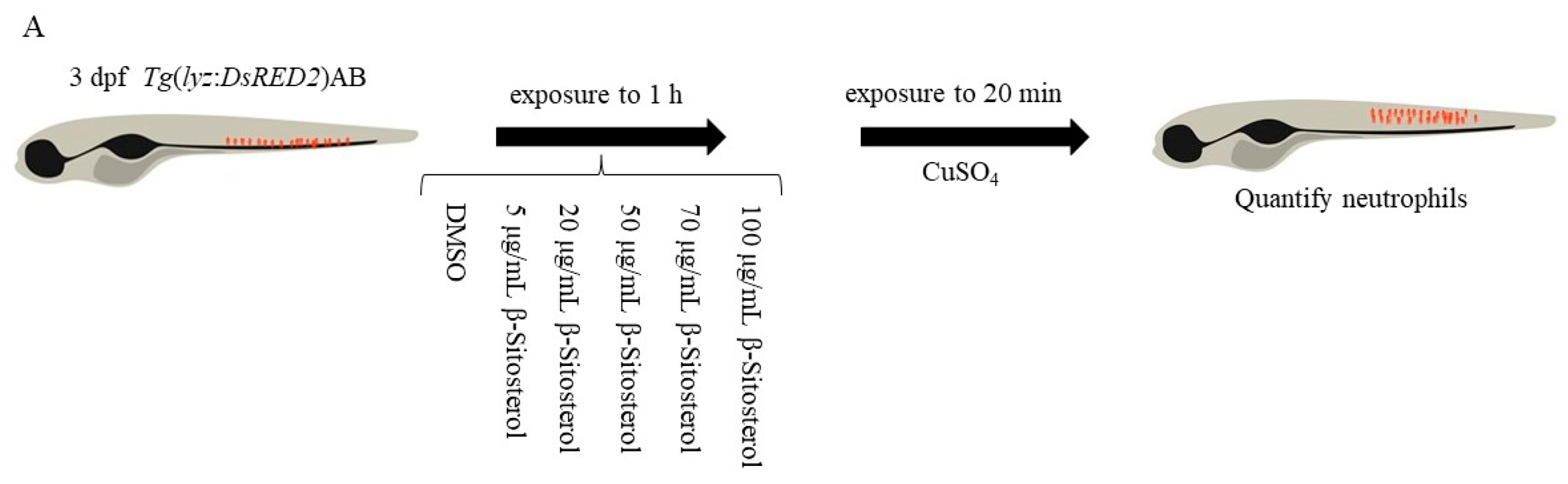
2.3.5. Neutrophil Migration Assay

The neutrophil migration assay was performed according to Reference [21] using the Tg (lyz: DsRED2) zebrafish larvae (3 dpf) as the study subjects. The zebrafish larvae were treated with β-sitosterol (0, 5, 20, 50, 70, or 100 μg/mL) for 1 h followed by 20 µM CuSO4 for 1 h [20], with 10 larvae in each group. The presence of neutrophils was monitored at the lateral line to evaluate the degree of inflammation. The neutrophil migration was quantified by calculating the number of labeled cells detected in the lateral line region. The larvae treated with DMSO alone served as the blank control, while the larvae treated with CuSO4 alone were the negative control.

2.3.6. Fluorescence Assay Quantification Method

For the in vivo antioxidant test, zebrafish fluorescence images were collected using the same magnification and exposure time. The individual zebrafish larvae fluorescence intensity was quantified using the Image J program. For the neutrophil migration assay, the number of neutrophils in the neuromast was quantified using the Image J program.

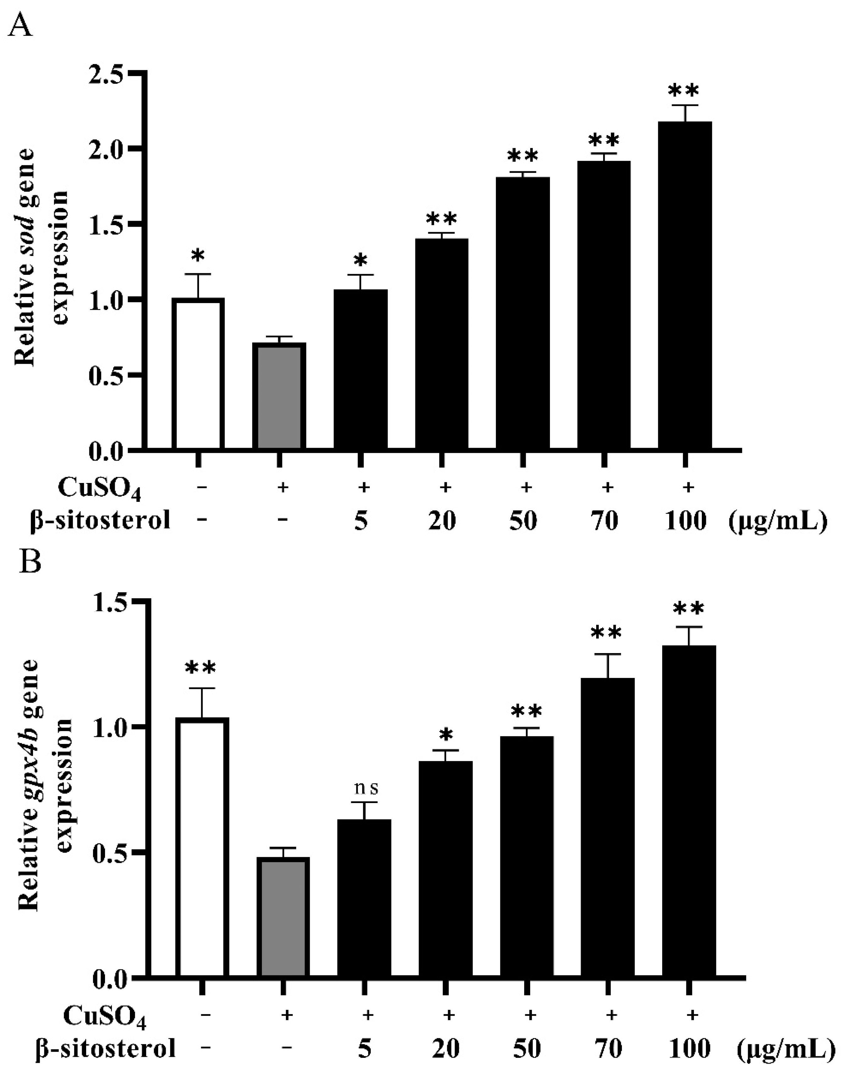
2.3.7. Quantitative Real-Time Polymerase Chain Reaction (RT-qPCR)

The transcriptome and RT-qPCR analysis methods referred to Reference [20]. The zebrafish larvae (3 dpf) were transplanted into a six-well plate containing 940 μL E3 medium (30 larvae per well). The 30 hatched larvae in each group were treated with different doses of β-sitosterol (0, 5, 20, 50, 70, or 100 μg/mL) for 1 h and then exposed to 10 μM CuSO4 (a concentration that induces oxidative stress and inflammation [11]) for 24 h. The larvae were collected after 24 h and stored at −80 °C for RNA extraction. Total RNA was extracted from the larvae using TRIzol reagent (Invitrogen, Carlsbad, CA, USA) [22]. The concentration (A260) and purity (A260/A280 and A260/A230) of the RNAs were measured using a NanoDrop 1000 instrument (Thermo Fisher Scientific). The RNA integrity was confirmed by electrophoresis in a 1.5% agarose denaturing gel and reverse-transcribed into cDNA using a cDNA reverse transcription kit (Trans Gen Biotech, Wuhan, China). The cycling conditions for RT-qPCR were 95 °C for 10 min followed by 40 cycles of 95 °C for 30 s and 60 °C for 30 s. The results were obtained using the 2−ΔΔct method [23]. The specific primers used are listed in Table 1.

2.3.8. Data Presentation and Statistical Analyses

The normally distributed data are expressed as the mean ± standard deviation. The differences between treatments were determined by t-test or one-way analysis of variance (ANOVA), as well as the least significant difference test. A probability level of 5% (p < 0.05) was considered significant. When the normality test failed, the Kruskal–Wallis nonparametric one-way ANOVA was used, and the differences between groups were determined by the Mann–Whitney test. The nonparametric data are presented using Tukey’s boxplot, where the bottom and top of the box are the minimum and maximum values, respectively.

3. Results

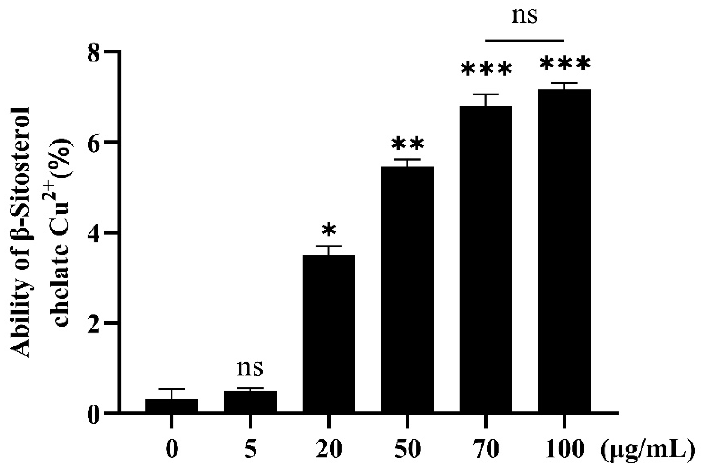
3.1. Chelating Ability of β-Sitosterol for Cu2+

In this experiment, CuSO4 solution was used as the Cu+ source. The chelating ability of β-sitosterol for Cu2+ increased with an increasing β-sitosterol concentration (Figure 1). The ability of β-sitosterol to complex with Cu2+ increased with an increasing β-sitosterol concentration. In terms of the chelation ability, the groups treated with 5 and 0 μg/mL β-sitosterol did not show a significant difference, while the remaining experimental groups were significantly different from the 0 μg/mL group. At the highest concentration of β-sitosterol (100 μg/mL), the chelating ability of β-sitosterol for Cu2+ was only 7%. No significant difference in the chelation ability was found between the groups treated with β-sitosterol at 70 and 100 μg/mL (Figure 1). Thus, β-sitosterol had a weak chelating activity for Cu2+, indicating that metal chelation was not important in the subsequent experiments.

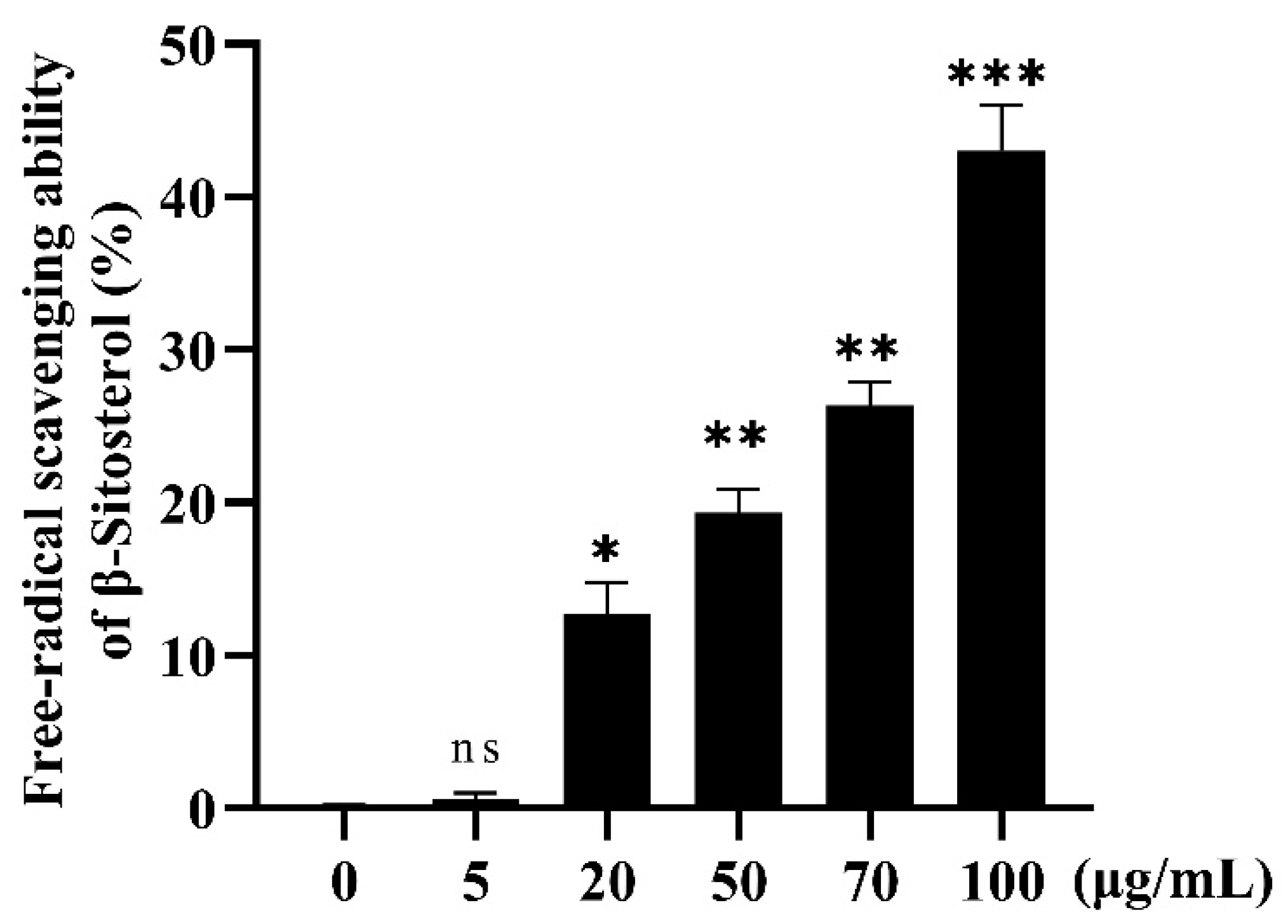
3.2. Antioxidant Effect of β-Sitosterol

3.2.1. In Vitro Antioxidant Effect of β-Sitosterol

The free radical scavenging ability of β-sitosterol was determined by the DPPH assay. The scavenging rate of the free radicals increased with an increasing β-sitosterol concentration in the experimental concentration range (Figure 2). With the exception of the group treated with 5 μg/mL β-sitosterol, the β-sitosterol-treated groups showed a significantly different free radical scavenging ability compared with the control group (0 μg/mL β-sitosterol). A the β-sitosterol concentration of 100 μg/mL, the scavenging rate was 43%, indicating that β-sitosterol had a strong ability to scavenge DPPH (Figure 2).

3.2.2. In Vivo Antioxidant Effect of β-Sitosterol

The ROS produced in zebrafish were detected using the fluorescent probe H2DCFDA, and quercetin was used as a positive control. Quercetin (100 μM) effectively inhibited the formation of ROS (Figure 3B), and the fluorescence intensity in the positive control was significantly reduced compared to that in the CuSO4 group (Figure 3C). Similar to quercetin, β-sitosterol at concentrations of 50, 70, and 100 μg/mL significantly reduced the ROS fluorescence intensity compared to the control. Treatment with β-sitosterol at 5 μg/mL did not have a significant effect on the fluorescence intensity compared to the control group (Figure 3C), indicating that this low concentration of β-sitosterol did not obviously inhibit ROS production.

3.2.3. Effect of β-Sitosterol on the Expressions of the Antioxidant Genes sod and gpx4b

We used RT-qPCR to analyze the mRNA expression levels of two antioxidant genes, sod and gpx4b, in the different treatment groups. Compared with the control group, the expressions of sod and gpx4b were significantly reduced in the presence of CuSO4 (Figure 4). However, after 24 h of β-sitosterol treatment, the expressions of sod and gpx4b were significantly upregulated compared with the CuSO4 group, with significant upregulation observed in the 50, 70, and 100 μg/mL β-sitosterol treatment groups for sod (Figure 4A) and in the 70 and 100 μg/mL β-sitosterol treatment groups for gpx4b (Figure 4B).

3.3. Anti-Inflammatory Effect of β-Sitosterol in Zebrafish

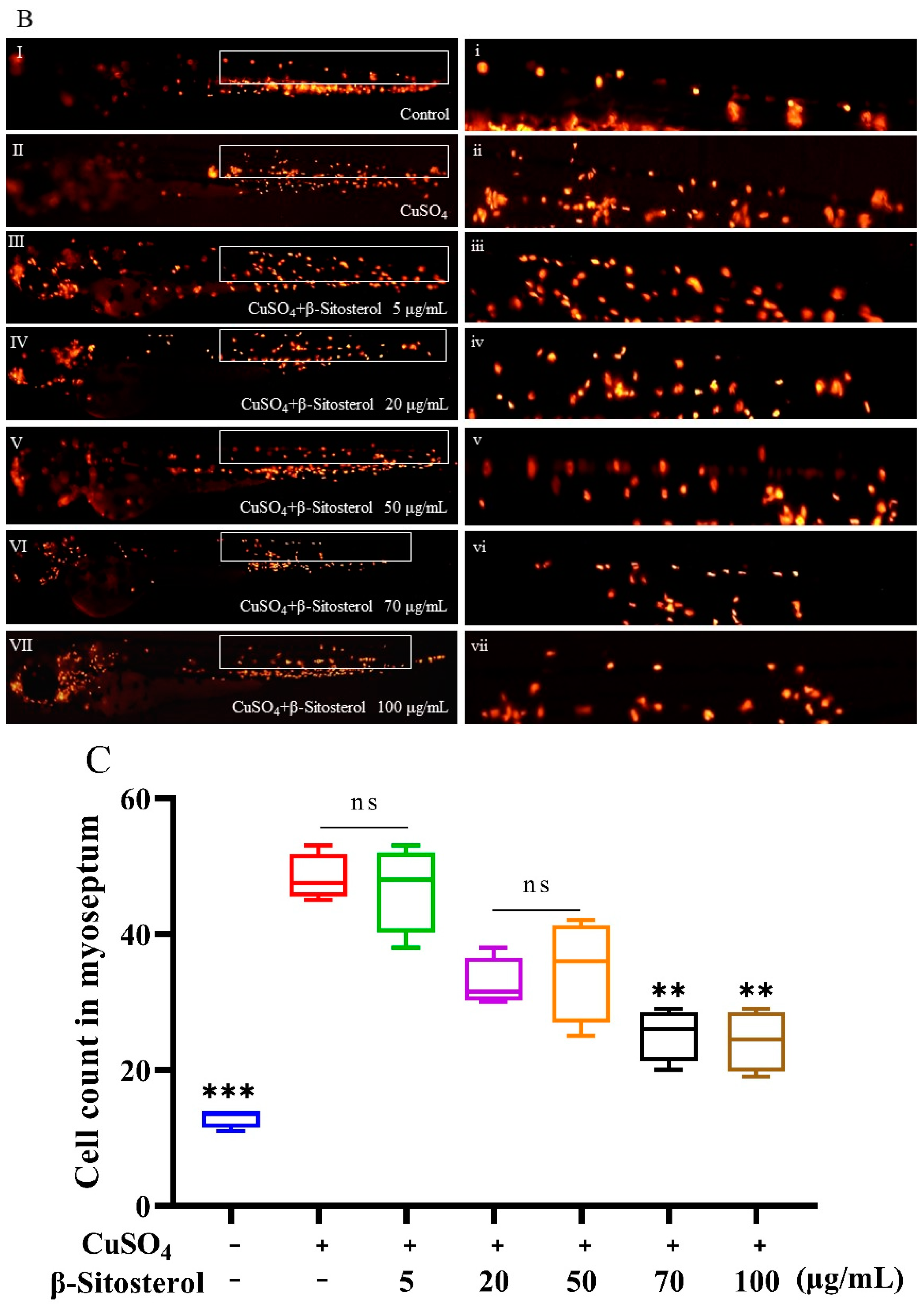
3.3.1. Effect of β-Sitosterol on Neutrophil Migration

The effect of β-sitosterol on the CuSO4-induced inflammatory response was evaluated using the transgenic Tg (lyz: DsRED2) zebrafish model. In the control group, most neutrophils were located in the tail hematopoietic tissues of the ventral stem and tail (Figure 5B). In the group treated with CuSO4 alone, the neutrophils migrated to the horizontal midline, as indicated by the white square box in Figure 5B(i), and formed clusters near the lateral line nerve hypertrophy. As shown in Figure 5B(i–vii), which respectively correspond to Figure 5B(I–VII), as the concentration of β-sitosterol increased, the number of neutrophils that migrated to the lateral line after the CuSO4 treatment gradually decreased. The neutrophils in the white boxes in Figure 5B(i–vii) were quantified using Image J software. The number of neutrophils was significantly different between the group treated with CuSO4 alone and the control group (Figure 5C). However, there was no significant difference with the group treated with 5 μg/mL β-sitosterol (Figure 5C). No significant difference in neutrophil migration was found between the groups treated with 20 and 50 μg/mL β-sitosterol (Figure 5C). Compared with the group treated with CuSO4 alone, the groups pretreated with β-sitosterol at 70 and 100 μg/mL showed significant differences in neutrophil migration (Figure 5C). Thus, the effect on neutrophil migration became stronger as the concentrations of β-sitosterol increased, suggesting that β-sitosterol significantly inhibited neutrophil migration to the lateral line.

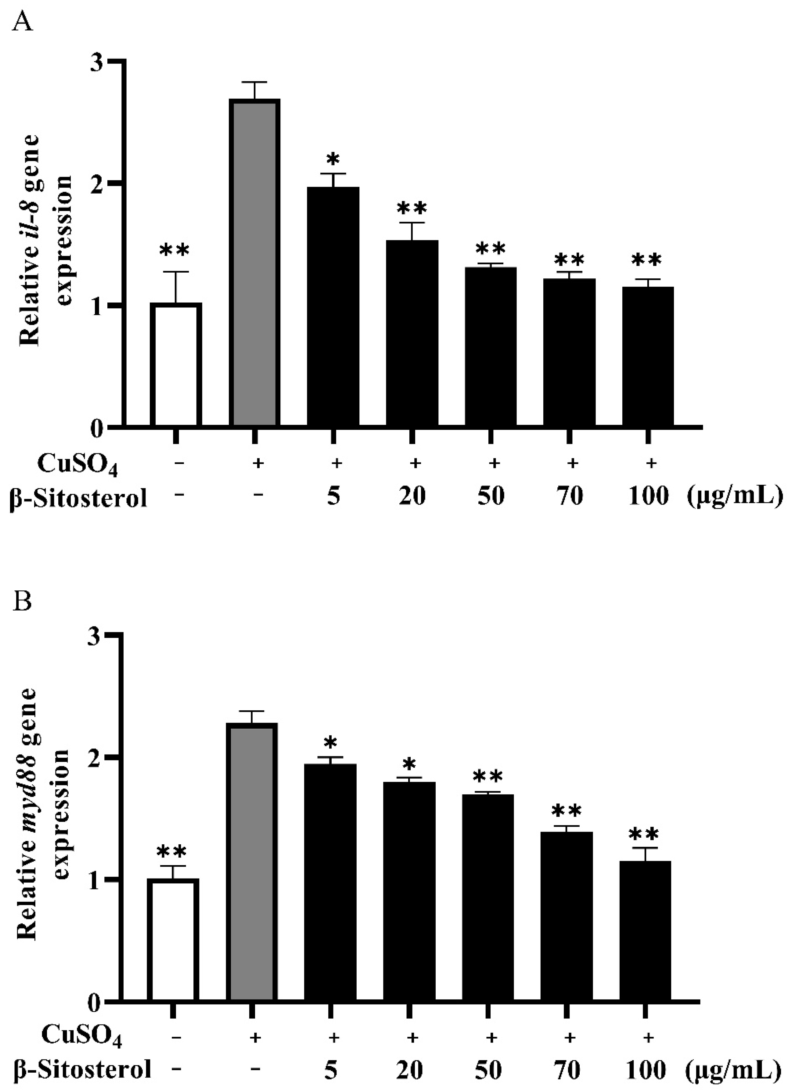
3.3.2. Effects of β-Sitosterol on the Expressions of il-8 and myd88

The expressions of the il-8 gene and myd88 gene in zebrafish stimulated by CuSO4 alone significantly increased compared with the blank control (Figure 6A,B). After 24 h of treatment with different doses of β-sitosterol, the expressions of il-8 and myd88 decreased. The expression of the il-8 gene decreased significantly after treatment with 50, 70, and 100 μg/mL β-sitosterol, with significant decreases in the myd88 expression in the 70 and 100 μg/mL β-sitosterol treatment groups.

4. Discussion

Cu2+ has been shown to cause oxidative stress [24,25]. Oxidative stress can occur through two mechanisms: (1) Cu2+ can directly damage DNA and cells and (2) Cu2+ can increase ROS release by activating phagocytic cells and increasing tissue damage [26]. Changes in ROS may induce the activation of activator protein-1 (AP-1) and nuclear factor-kappa B (NF-κB), both of which upregulate proinflammatory cytokines and chemokines that cause inflammation. Inflammation is the result of damages in zebrafish after Cu2+ stimulation. Oxidative stress and inflammation are related to the toxic mechanism of Cu2+. Therefore, we used a zebrafish CuSO4 inflammation model to study the antioxidant and anti-inflammatory effects of β-sitosterol. Our results show that CuSO4 stimulation in zebrafish resulted in an inflammatory response, while pretreatment with β-sitosterol reduced this inflammatory response and enhanced the anti-inflammatory and antioxidant capacity of zebrafish.

4.1. In Vivo Antioxidant Effect of β-Sitosterol

The antioxidant activity of β-sitosterol was verified in vitro by a DPPH assay, which is commonly used to evaluate the free radical scavenging activity of antioxidants due to the fact of its rapidity, reliability, and good repeatability [19,27]. The DPPH assay has been applied extensively to detect the antioxidant activity of pure antioxidant compounds and various plant extracts [28,29]. In this study, the free radical scavenging rate of 100 μg/mL β-sitosterol exceeded 40% (Figure 2), demonstrating the antioxidant effect of β-sitosterol. Although in vitro experiments are simple, fast, and inexpensive, they cannot completely replace in vivo animal testing [28]. CuSO4 can induce oxidative stress by inducing ROS [30]. Zebrafish are widely used as a model for studying anti-inflammatory research [20]. In this study, a zebrafish CuSO4 inflammatory model was used to confirm the antioxidant effect of β-sitosterol in vivo. Oxyflufen exposure has been shown to induce oxidative stress and inflammation in zebrafish by promoting ROS production, leading to acute kidney injury [31]. CuSO4 also stimulates ROS production and inhibits the expressions of antioxidant genes [32]. Therefore, we investigated the effects of β-sitosterol on ROS production and antioxidant gene expression. To exclude the effect of complexation between β-sitosterol and Cu2+, we first verified the complexation ability of β-sitosterol for Cu2+. The complexation rate between 100 μg/mL β-sitosterol and Cu2+ was only 7% (Figure 1), indicating that the complexation did not affect the subsequent experimental results. Subsequently, we demonstrated that CuSO4 immersion stimulated oxidative stress in zebrafish, while pretreatment with β-sitosterol produced an antioxidant effect. We then evaluated the ROS generation in zebrafish using H2DCFDA, a membrane-permeable fluorescent dye [26,33,34]. β-Sitosterol at concentrations of 50, 70, and 100 μg/mL significantly reduced the production of ROS in zebrafish (Figure 3B,C). Thus, β-sitosterol has a significant antioxidant effect and can reduce ROS production in zebrafish to protect against oxidative stress.
β-Sitosterol has been shown to demonstrate antioxidant activity without causing acute toxicity, subchronic toxicity, or abnormalities in the synthesis of antioxidant enzymes [35,36]. β-Sitosterol also increases the activities of superoxide dismutase (SOD) and glutathione peroxidase (GPX) while decreasing catalase activity [37]. Both SOD and GPX are antioxidant enzymes [38]. SOD is a primary substance that scavenges ROS [39]. GPX plays a crucial role against oxidative stress, which can turn toxic substances into innocuous products by scavenging their free radicals [40]. Thus, SOD and GPX can reflect the body’s ability to resist oxidative damage [41]. The extract of Clerodendrum Cyrtophyllum Turcz could increase the level of sod mRNA in zebrafish [20]. In addition, the extract of Monascus purpureus could increase the level of sod and gpx mRNA in LNCaP cells [42]. This research used the expression of the antioxidant genes sod and gpx to verify the antioxidant activity of the extract. Therefore, to further study the mechanism of the antioxidant effect, we measured the effects of β-sitosterol on the expressions of two zebrafish genes (superoxide dismutase sod and glutathione peroxidase gpx4b) involved in the antioxidant system. In this study, after 24 h of Cu2+ exposure, the expressions of sod and gpx4b were significantly lower compared with the control group. However, pretreatment with β-sitosterol significantly increased the expressions of the sod and gpx4b genes compared with group treated with CuSO4 alone. Pretreatment with 20, 50, 70, and 100 μg/mL β-sitosterol had the most obvious effects on sod expression (Figure 4A; p < 0.01), while pretreatment with 50, 70, and 100 μg/mL β-sitosterol had the most obvious effects on gpx4b expression (Figure 4B; p < 0.01). The effects of pretreatment with β-sitosterol at 70 and 100 μg/mL were particularly significant (Figure 4A,B). The effect of β-sitosterol on sod and gpx4b indicates that β-sitosterol has certain antioxidant effect in zebrafish after oxidative stress.

4.2. Anti-Inflammatory Effect of β-Sitosterol in Zebrafish

β-Sitosterol can inhibit NF-κB and extracellular signal-regulated kinases (ERK)/p38 mitogen-activated protein kinases (p38 MAPK) signal transduction to reduce Escherichia coli lipopolysaccharide-induced mouse microglial inflammatory response [43]. β-Sitosterol can also reduce the mRNA expressions of inflammatory factors, such as IL-6 and tumor necrosis factor-α (TNF-α) [43]. β-Sitosterol significantly reduced weight loss and colon length and alleviated the microscopic manifestations of dextran sulfate sodium-induced colitis in mice [15]. In J774A1 macrophages induced by lipopolysaccharide, β-sitosterol increased the activity of anti-inflammatory factor IL-10, decreased the activities of chemokines and pro-inflammatory factors, enhanced the activity of tyrosine phosphatase, and inhibited the migration of NF-κB [14]. In zebrafish larvae, exposure to CuSO4 can damage lateral neuromast hair cells and induce neutrophil migration to inflammatory foci. The temporary treatment of zebrafish larvae with CuSO4 selectively triggered the death of hair cells in the lateral line system and led to the rapid accumulation of neutrophils at the damaged site, resulting in a strong acute inflammatory response [44,45]. In this study, pretreatment with β-sitosterol before CuSO4 exposure significantly reduced the recruitment of neutrophils in the damaged parts of the zebrafish, and the number of neutrophils in the damaged parts gradually decreased with an increasing β-sitosterol concentration. These results confirm the good anti-inflammatory effect of β-sitosterol in zebrafish. While pretreatment with 5 μg/mL β-sitosterol did not significantly affect the number of neutrophils compared to the group treated with CuSO4 alone, the anti-inflammatory effects of the other concentrations of β-sitosterol, particularly 70 and 100 μg/mL (Figure 5B,C), were relatively significant. The gene myd88 encodes a cytoplasmic adaptor protein that plays a critical role in innate and adaptive immune responses. This gene is also an essential signal transducer in the interleukin-1 and Toll-like receptor signaling pathways [46,47], which modulate the levels of pro-inflammatory mediators [48,49]. Abundant evidence suggests that certain pro-inflammatory cytokines, such as IL-1β, IL-2, TNF-α, IL-6, and IL-8, are involved in the pathological inflammatory process [50]. CXCL8 (IL-8) is an important chemokine that mediates neutrophil migration, aggregation, and function in inflammatory sites [51]. Meanwhile, cxcl8-l1 (zebrafish il-8a) and cxcl8-l2 (zebrafish il-8b) play major roles in neutrophil recruitment, which is a characteristic of acute inflammation [52]. Vitamin D significantly reduced myd88 and il-8 mRNA levels and inhibited lipopolysaccharide-induced inflammatory responses [53]. Therefore, in this study, we examined the expressions of myd88 and il-8 after zebrafish were treated with 10 μM (a nonlethal concentration) of CuSO4 for 24 h. The treatment with CuSO4 alone significantly increased the expressions of these genes compared with the blank control (Figure 6A,B). After β-sitosterol treatment for 24 h, the levels of il-8 and myd88 significantly decreased. Thus, β-sitosterol enhanced the innate immunity of the zebrafish and weakened the inflammatory response to CuSO4 stimulation. The inhibitory effects on the expressions of myd88 and il-8 were proportional to the concentration of β-sitosterol. However, whether the inhibition of neutrophil migration by β-sitosterol is really related to the inhibition of il8 expression needs further experimental verification. In summary, the results demonstrate that β-sitosterol has a certain anti-inflammatory effect and can improve the ability of zebrafish to resist stress.

5. Conclusions

This study showed that β-sitosterol can reduce the production of ROS in a CuSO4-induced inflammation model in zebrafish and upregulate the expressions of sod and gpx4b to inhibit oxidative stress. Moreover, β-sitosterol can inhibit neutrophil migration to the lateral line and reduce the expressions of inflammatory genes (il-8 and myd88) to inhibit inflammation. These results help explain the protective effect of β-sitosterol against CuSO4 toxicity in vivo. The findings support the application of β-sitosterol as a feed additive to improve the anti-inflammatory and antioxidant capacity of aquatic products.

Author Contributions

P.Z. and N.L. conceived and designed the study; performed the data collection, analysis, statistical analysis; and wrote the manuscript. M.X. and M.Z. conducted the software analysis. W.L. and C.X. conducted the animal management. Y.F. and Y.M. conducted literature review. Q.Z. and Y.Z. contributed to the acquisition of funding, conceptualization, writing—review and editing, and supervision. All authors have read and agreed to the published version of the manuscript.

Funding

This research was funded by the Earmarked Fund for CARS (CARS-45), the China–US Ocean Research Center Fund (A1-3201-19-3013), and the Central Public-Interest Scientific Institution Basal Research Fund (2020TD44).

Institutional Review Board Statement

Not Applicable.

Informed Consent Statement

Not Applicable.

Data Availability Statement

Data is contained within the article.

Conflicts of Interest

The authors report no conflict of interest.

References

  1. Hou, C.; Chen, L.; Yang, L.; Ji, X. An insight into anti-inflammatory effects of natural polysaccharides. Int. J. Biol. Macromol. 2020, 153, 248–255. [Google Scholar] [CrossRef]
  2. Li, F.; Xiaoyu, L.; Weiliang, Z.; Fujiang, G.; Yingchun, W.; Rui, W.; Kaixian, C.; Cheng, H.; Yiming, L. Inhibition of human neutrophil elastase by pentacyclic triterpenes. PLoS ONE 2013, 8, e82794. [Google Scholar] [CrossRef]
  3. Biswas, S.K. Does the Interdependence between Oxidative Stress and Inflammation Explain the Antioxidant Paradox? Oxidative Med. Cell. Longev. 2016, 2016, 5698931. [Google Scholar] [CrossRef]
  4. Jun, L.; Yixin, W.; John, H.W.; Yu, C.; Zhaoting, L.; Quanyin, H. Mucoadhesive probiotic backpacks with ROS nanoscavengers enhance the bacteriotherapy for inflammatory bowel diseases. Sci. Adv. 2022, 8, eabp8798. [Google Scholar] [CrossRef]
  5. Alessandro, D.; Jessica, K.; Mona, S.; Federica, M.; Bruno, C.; Fowler, C.J.; Valentina, O. Exploring the fatty acid amide hydrolase and cyclooxygenase inhibitory properties of novel amide derivatives of ibuprofen. J. Enzym. Inhib. Med. Chem. 2020, 35, 815–823. [Google Scholar] [CrossRef]
  6. Christian, C.; Emmanuel, M. Non-steroidal anti-inflammatory drugs in the pharmacological management of osteoarthritis in the very old: Prescribe or proscribe? Ther. Adv. Musculoskelet. Dis. 2021, 13, 1759720X211022149. [Google Scholar] [CrossRef]
  7. Liang, M.; Tzu-Wen, L.; Wallig, M.A.; Dobrucki, I.T.; Dobrucki, L.W.; Nelson, E.R.; Swanson, K.S.; Smith, A.M. Efficient Targeting of Adipose Tissue Macrophages in Obesity with Polysaccharide Nanocarriers. ACS Nano 2016, 10, 6952–6962. [Google Scholar] [CrossRef]
  8. Fernando, I.P.S.; Sanjeewa, K.K.A.; Kim, H.-S.; Kim, S.-Y.; Lee, S.-H.; Lee, W.W.; Jeon, Y.-J. Identification of sterols from the soft coral Dendronephthya gigantea and their anti-inflammatory potential. Environ. Toxicol. Pharmacol. 2017, 55, 37–43. [Google Scholar] [CrossRef]
  9. Hussain, A.; AlAjmi, M.F.; Rehman, M.T.; Amir, S.; Husain, F.M.; Alsalme, A.; Siddiqui, M.A.; AlKhedhairy, A.A.; Khan, R.A. Copper(II) complexes as potential anticancer and Nonsteroidal anti-inflammatory agents: In vitro and in vivo studies. Sci. Rep. 2019, 9, 5237. [Google Scholar] [CrossRef] [PubMed]
  10. O’Sullivan, J.J.; Medici, V.; Heffern, M.C. Correction: A caged imidazopyrazinone for selective bioluminescence detection of labile extracellular copper(ii). Chem. Sci. 2022, 13, 5774. [Google Scholar] [CrossRef]
  11. Brandão, P.T.C.; Martha, C.M.; Reis, B.M. Copper toxicology, oxidative stress and inflammation using zebrafish as experimental model. J. Appl. Toxicol. 2016, 36, 876–885. [Google Scholar] [CrossRef]
  12. Di Paola, D.; Capparucci, F.; Lanteri, G.; Crupi, R.; Marino, Y.; Franco, G.A.; Cuzzocrea, S.; Spano, N.; Gugliandolo, E.; Peritore, A.F. Environmental Toxicity Assessment of Sodium Fluoride and Platinum-Derived Drugs Co-Exposure on Aquatic Organisms. Toxics 2022, 10, 272. [Google Scholar] [CrossRef]
  13. Moreau, R.A.; Nyström, L.; Whitaker, B.D.; Winkler-Moser, J.K.; Baer, D.J.; Gebauer, S.K.; Hicks, K.B. Phytosterols and their derivatives: Structural diversity, distribution, metabolism, analysis, and health-promoting uses. Prog. Lipid Res. 2018, 70, 35–61. [Google Scholar] [CrossRef]
  14. Valerio, M.; Awad, A.B. β-Sitosterol down-regulates some pro-inflammatory signal transduction pathways by increasing the activity of tyrosine phosphatase SHP-1 in J774A.1 murine macrophages. Int. Immunopharmacol. 2011, 11, 1012–1017. [Google Scholar] [CrossRef] [PubMed]
  15. Ding, K.; Tan, Y.Y.; Ding, Y.; Fang, Y.; Yang, X.; Fang, J.; Xu, D.C.; Zhang, H.; Lu, W.; Li, M.; et al. β-Sitosterol improves experimental colitis in mice with a target against pathogenic bacteria. J. Cell. Biochem. 2019, 120, 5687–5694. [Google Scholar] [CrossRef] [PubMed]
  16. Paniagua-Pérez, R.; Madrigal-Bujaida, E.; Reyes-Cadena, S.; Álvarez-González, I.; Sánchez-Chapul, L.; Pérez-Gallaga, L.; Hernández, N.; Flores-Mondragón, G.; Velasco, O. Cell protection induced by beta-sitosterol: Inhibition of genotoxic damage, stimulation of lymphocyte production, and determination of its antioxidant capacity. Arch. Toxicol. 2008, 82, 615–622. [Google Scholar] [CrossRef]
  17. Di Paola, D.; Abbate, J.M.; Iaria, C.; Cordaro, M.; Crupi, R.; Siracusa, R.; D’Amico, R.; Fusco, R.; Impellizzeri, D.; Cuzzocrea, S.; et al. Environmental Risk Assessment of Dexamethasone Sodium Phosphate and Tocilizumab Mixture in Zebrafish Early Life Stage (Danio rerio). Toxics 2022, 10, 279. [Google Scholar] [CrossRef]
  18. Santos, J.S.; Alvarenga Brizola, V.R.; Granato, D. High-throughput assay comparison and standardization for metal chelating capacity screening: A proposal and application. Food Chem. 2017, 214, 515–522. [Google Scholar] [CrossRef] [PubMed]
  19. Wang, C.Z.; Yuan, H.H.; Bao, X.L.; Lan, M.B. In vitro antioxidant and cytotoxic properties of ethanol extract of Alpinia oxyphylla fruits. Pharm. Biol. 2013, 51, 1419–1425. [Google Scholar] [CrossRef]
  20. Nguyen, T.H.; Le, H.D.; Kim, T.N.T.; The, H.P.; Nguyen, T.M.; Cornet, V.; Lambert, J.; Kestemont, P. Anti–Inflammatory and Antioxidant Properties of the Ethanol Extract of Clerodendrum Cyrtophyllum Turcz in Copper Sulfate-Induced Inflammation in Zebrafish. Antioxidants 2020, 9, 192. [Google Scholar] [CrossRef] [Green Version]
  21. Zhang, C.; Li, C.; Jia, X.; Wang, K.; Tu, Y.; Wang, R.; Liu, K.; Lu, T.; He, C. In Vitro and In Vivo Anti-inflammatory Effects of Polyphyllin Vii Through Downregulating Mapk and Nf-kappa B Pathways. Chem. Chem. 2019, 24, 875. [Google Scholar] [CrossRef]
  22. Marc Sultan, V.A.; Risch, T.; Schuette, M.; Dökel, S.; Ralser, M.; Balzereit, D.; Lehrach, H.; Yaspo, M.-L. Influence of RNA extraction methods and library selection schemes on RNA-seq data. BMC Genom. 2014, 15, 675. [Google Scholar] [CrossRef]
  23. Livak, K.J.; Schmittgen, T.D. Analysis of relative gene expression data using real-time quantitative PCR and the 2(-Delta Delta C(T)) Method. Methods 2001, 25, 402–408. [Google Scholar] [CrossRef]
  24. Leite, C.E.; Maboni, L.d.O.; Cruz, F.F.; Rosemberg, D.B.; Zimmermann, F.F.; Pereira, T.C.B.; Bogo, M.R.; Bonan, C.D.; Campos, M.M.; Morrone, F.B.; et al. Involvement of purinergic system in inflammation and toxicity induced by copper in zebrafish larvae. Toxicol. Appl. Pharmacol. 2013, 272, 681–689. [Google Scholar] [CrossRef]
  25. Hernandez, P.P.; Undurraga, C.; Gallardo, V.E.; Mackenzie, N.; Allende, M.L.; Reyes, A.E. Sublethal concentrations of waterborne copper induce cellular stress and cell death in zebrafish embryos and larvae. Biol. Res. 2011, 44, 7–15. [Google Scholar] [CrossRef]
  26. Olivari, F.A.; Hernández, P.P.; Allende, M.L. Acute copper exposure induces oxidative stress and cell death in lateral line hair cells of zebrafish larvae. Brain Res. 2008, 1244, 1–12. [Google Scholar] [CrossRef]
  27. Francesco, S.; Gianluca, P.; Domenico, C.; Domenico, C.; Tonino, C.; Ermanno, V. Monitoring antioxidants by coulometry: Quantitative assessment of the strikingly high antioxidant capacity of bergamot (Citrus bergamia R.) by-products. Talanta 2023, 251, 123765. [Google Scholar] [CrossRef]
  28. Jesús, R.M. Anti-Inflammatory and Antioxidant Properties of Plant Extracts. Antioxidants 2021, 10, 921. [Google Scholar] [CrossRef]
  29. Liu, H.; Wang, L.; Wang, M.-H. Antioxidant and nitric oxide release inhibition activities of methanolic extract from Clerodendrum cyrtophyllum Turcz. Hortic. Environ. Biotechnol. 2011, 52, 309–314. [Google Scholar] [CrossRef]
  30. Wang, Y.T.; Chen, G.C. Regulation of oxidative stress-induced autophagy by ATG9A ubiquitination. Autophagy 2022, 18, 2008–2010. [Google Scholar] [CrossRef] [PubMed]
  31. Lirong, H.; Kun, J.; Haibin, X.; Guiyou, T.; Jiaxin, X.; Wei, Y.; Chen, L.; Xiaoping, X.; Huiqiang, L. Oxyfluorfen exposure can cause acute kidney injury by promoting ROS-induced oxidative stress and inflammation in zebrafish. J. Hazard. Mater. 2022, 440, 129823. [Google Scholar] [CrossRef]
  32. Ying, W.; Junhua, L.; Huiming, Q.; Chunjing, C.; Hui, L.; Jie, C. β-Carotene extracted from Blakeslea trispora attenuates oxidative stress, inflammatory, hepatic injury and immune damage induced by copper sulfate in zebrafish (Danio rerio). Comp. Biochem. Physiology. Toxicol. Pharmacol. 2022, 258, 109366. [Google Scholar] [CrossRef]
  33. Lee, S.-H.; Ko, C.-I.; Jee, Y.; Jeong, Y.; Kim, M.; Kim, J.-S.; Jeon, Y.-J. Anti-inflammatory effect of fucoidan extracted from Ecklonia cava in zebrafish model. Carbohydr. Polym. 2013, 92, 84–89. [Google Scholar] [CrossRef]
  34. Shibo, S.; Yici, Z.; Weiping, X.; Yue, Z.; Rui, Y.; Jianli, G.; Shui, G.; Qiang, M.; Kun, M.; Jianqiang, X. Chlorophyllin Inhibits Mammalian Thioredoxin Reductase 1 and Triggers Cancer Cell Death. Antioxidants 2021, 10, 1733. [Google Scholar] [CrossRef]
  35. Devi, R.R.; Arumughan, C. Antiradical efficacy of phytochemical extracts from defatted rice bran. Food Chem. Toxicol. 2007, 45, 2014–2021. [Google Scholar] [CrossRef]
  36. Mohanan, P.V.; Devi, K.S. Toxicological evaluation of sobatum. Cancer Lett. 1998, 127, 135–140. [Google Scholar] [CrossRef]
  37. Ambavade, S.D.; Misar, A.V.; Ambavade, P.D. Pharmacological, nutritional, and analytical aspects of β-sitosterol: A review. Orient. Pharm. Exp. Med. 2014, 14, 193–211. [Google Scholar] [CrossRef]
  38. Maharajan, K.; Muthulakshmi, S.; Nataraj, B.; Ramesh, M.; Kadirvelu, K. Toxicity assessment of pyriproxyfen in vertebrate model zebrafish embryos (Danio rerio): A multi biomarker study. Aquat. Toxicol. 2018, 196, 132–145. [Google Scholar] [CrossRef]
  39. Wang, C.; Harwood, J.D.; Zhang, Q. Oxidative stress and DNA damage in common carp (Cyprinus carpio) exposed to the herbicide mesotrione. Chemosphere 2018, 193, 1080–1086. [Google Scholar] [CrossRef]
  40. Nair, P.M.G.; Park, S.Y.; Choi, J. Expression of catalase and glutathione S-transferase genes in Chironomus riparius on exposure to cadmium and nonylphenol. Comp. Biochem. Physiol. Part C Toxicol. Pharmacol. 2011, 154, 399–408. [Google Scholar] [CrossRef]
  41. Chen, Y.; Liu, H.; Huang, H.; Ma, Y.; Wang, R.; Hu, Y.; Zheng, X.; Chen, C.; Tang, H. Squid Ink Polysaccharides Protect Human Fibroblast Against Oxidative Stress by Regulating NADPH Oxidase and Connexin43. Front. Pharmacol. 2019, 10, 1574. [Google Scholar] [CrossRef]
  42. Chaudhary, V.; Katyal, P.; Panwar, H.; Kaur, J.; Aluko, R.E.; Puniya, A.K.; Poonia, A.K. Antioxidative, anti-inflammatory, and anticancer properties of the red biopigment extract from Monascus purpureus (MTCC 369). J. Food Biochem. 2022, 46, e14249. [Google Scholar] [CrossRef]
  43. Sun, Y.; Gao, L.; Hou, W.; Wu, J. β-Sitosterol Alleviates Inflammatory Response via Inhibiting the Activation of ERK/p38 and NF-κB Pathways in LPS-Exposed BV2 Cells. Biomed. Res. Int. 2020, 2020, 7532306. [Google Scholar] [CrossRef]
  44. Christine, W.; Markus, R.; Shah, A.H.; Ralf, M.; Urban, L.; Clemens, G. Facilitating drug discovery: An automated high-content inflammation assay in zebrafish. J. Vis. Exp. 2012, 16, e4203. [Google Scholar] [CrossRef]
  45. d’Alençon, C.A.; Oscar, P.; Christine, W.; Viviana, G.; Rebecca, J.; Felix, L.; Urban, L.; Clemens, G.; Miguel, A. A high-throughput chemically induced inflammation assay in zebrafish. BMC Biol. 2010, 8, 151. [Google Scholar] [CrossRef]
  46. Mara, C.; Keat, L.W.; Adina, G.; Nandula, S.V.; Manisha, B.; Qiong, S.; Francesco, B.; Maurilio, P.; Marta, S.; Andrea, C.; et al. Mutations of multiple genes cause deregulation of NF-kappaB in diffuse large B-cell lymphoma. Nature 2009, 459, 717–721. [Google Scholar] [CrossRef]
  47. Jacques, D.; Barton, G.M. MyD88: A central player in innate immune signaling. F1000Prime Rep. 2014, 6, 97. [Google Scholar] [CrossRef]
  48. Delitto, D.; Delitto, A.E.; DiVita, B.E.; Pham, K.; Han, S.; Hartlage, E.R.; Newby, B.N.; Gerber, M.H.; Behrns, K.E.; Moldawer, L.L.; et al. Human Pancreatic Cancer Cells Induce a MyD88-Dependent Stromal Response to Promote a Tumor-Tolerant Immune Microenvironment. Cancer Res. 2017, 77, 672–683. [Google Scholar] [CrossRef]
  49. Zhu, X.; Burfeind, K.G.; Michaelis, K.A.; Braun, T.P.; Olson, B.; Pelz, K.R.; Morgan, T.K.; Marks, D.L. MyD88 signalling is critical in the development of pancreatic cancer cachexia. J. Cachexia Sarcopenia Muscle 2019, 10, 378–390. [Google Scholar] [CrossRef]
  50. Ruslan, M. The spectrum of inflammatory responses. Science 2021, 734, 1070–1075. [Google Scholar] [CrossRef]
  51. Holmes, W.; Lee, J.; Kuang, W.; Rice, G.; Wood, W. Structure and functional expression of a human interleukin-8 receptor. Science 1991, 253, 1278–1280. [Google Scholar] [CrossRef]
  52. de Oliveira, S.; Reyes-Aldasoro, C.C.; Candel, S.; Renshaw, S.A.; Mulero, V.; Calado, Â. Cxcl8 (IL-8) mediates neutrophil recruitment and behavior in the zebrafish inflammatory response. J. Immunol. 2013, 190, 4349–4359. [Google Scholar] [CrossRef]
  53. Jiang, J.; Shi, D.; Zhou, X.Q.; Yin, L.; Feng, L.; Jiang, W.D.; Liu, Y.; Tang, L.; Wu, P.; Zhao, Y. Vitamin D inhibits lipopolysaccharide-induced inflammatory response potentially through the Toll-like receptor 4 signalling pathway in the intestine and enterocytes of juvenile Jian carp (Cyprinus carpio var. Jian). Br. J. Nutr. 2015, 114, 1560–1568. [Google Scholar] [CrossRef] [Green Version]
Figure 1. Metal chelating ability of β-sitosterol for Cu2+. Each bar represents the mean ± standard error for three different experiments performed in triplicate. ns, no statistical significance, * p < 0.05, ** p < 0.01, and *** p < 0.001 compared with the control group (0 μg/mL β-sitosterol).
Antioxidants 12 00391 g001
Figure 2. Free radical scavenging rates of β-sitosterol at different concentrations. Each bar represents the mean ± standard error for three different experiments performed in triplicate. ns, no statistical significance, * p < 0.05, ** p < 0.01, and *** p < 0.001 compared with the control group (0 µg/mL β-sitosterol).
Antioxidants 12 00391 g002
Figure 3. Inhibitory effect of β-sitosterol on the CuSO4-induced production of ROS in zebrafish larvae: (A) flowchart of the experiments; (B) representative fluorescence images of zebrafish in different experimental groups; (C) fluorescence intensity in juvenile zebrafish in different experimental groups compared with the group treated with CuSO4 alone. ns, no statistical significance, ** p < 0.01 compared with the control group (n = 4).
Antioxidants 12 00391 g003
Figure 4. Relative expressions of antioxidant-related genes in zebrafish larvae treated with CuSO4 and β-sitosterol for 24 h: (A) relative expression of the sod gene; (B) relative expression of the gpx4b gene. These data are expressed as the mean ± standard error based on three biological replicates. ns, no statistical significance, * p < 0.05 and ** p < 0.01 compared with the group treated with CuSO4 alone.
Antioxidants 12 00391 g004
Figure 5. Effect of β-sitosterol on CuSO4-induced inflammatory response in zebrafish: (A) flowchart of the experiments; (B) neutrophil migration in zebrafish after treatment; (C) quantification of the number of neutrophils aggregated at the lateral line after CuSO4 treatment. Data are expressed as the mean ± standard deviation. ns, no statistical significance, ** p < 0.01, and *** p < 0.001 compared with the group treated with CuSO4 alone (n = 4).
Antioxidants 12 00391 g005aAntioxidants 12 00391 g005b
Figure 6. Relative expressions of immune response-related genes in zebrafish larvae after CuSO4 stimulation and β-sitosterol treatment: (A) relative expression of the il-8 gene; (B) relative expression of the myd88 gene. The data are expressed as the mean ± standard error based on three biological replicates. * p < 0.05 and ** p < 0.01 compared with the group treated with CuSO4 alone.
Antioxidants 12 00391 g006
Table 1. Primers used in this study.
Gene NameGenBank Accession No.Forward and Reverse Primer Sequences (5′-3′)
β-actin [20]AF057040F: CCCCATTGAGCACGGTATTG
R: ATACATGGCAGGGGTGTTGA
il-8XM_009306855.3F: GAGAGGTCTGGCTGTAGATC
R: AGTTGTCATCAAGGTGGCAAT
myd88NM_212814.2F: GTGATGCCTGTGATTTTCAGACTAA
R: CGGCCTCTTCATGGATTTGT
sod [20]NM_131294.1F: ATGGTGAACAAGGCCGTTTG
R: AAAGCATGGACGTGGAAACC
gpx4b [20]BC095133.1F: TGAGAAGGGTTTACGCATCCTG
R: TGTTGTTCCCCAGTGTTCCT
Disclaimer/Publisher’s Note: The statements, opinions and data contained in all publications are solely those of the individual author(s) and contributor(s) and not of MDPI and/or the editor(s). MDPI and/or the editor(s) disclaim responsibility for any injury to people or property resulting from any ideas, methods, instructions or products referred to in the content.

