Review

Heme Oxygenase-1 and Its Role in Colorectal Cancer

Division of Food Chemistry and Toxicology, Department of Chemistry, RPTU Kaiserslautern-Landau, Erwin-Schrödinger Strasse 52, D-67663 Kaiserslautern, Germany
*
Authors to whom correspondence should be addressed.
Antioxidants 2023, 12(11), 1989; https://doi.org/10.3390/antiox12111989
Submission received: 15 September 2023 / Revised: 3 November 2023 / Accepted: 6 November 2023 / Published: 10 November 2023
(This article belongs to the Special Issue Experimental and Therapeutic Targeting of Heme Oxygenase)

Abstract

:
Heme oxygenase-1 (HO-1) is an enzyme located at the endoplasmic reticulum, which is responsible for the degradation of cellular heme into ferrous iron, carbon monoxide and biliverdin-IXa. In addition to this main function, the enzyme is involved in many other homeostatic, toxic and cancer-related mechanisms. In this review, we first summarize the importance of HO-1 in physiology and pathophysiology with a focus on the digestive system. We then detail its structure and function, followed by a section on the regulatory mechanisms that control HO-1 expression and activity. Moreover, HO-2 as important further HO isoform is discussed, highlighting the similarities and differences with regard to HO-1. Subsequently, we describe the direct and indirect cytoprotective functions of HO-1 and its breakdown products carbon monoxide and biliverdin-IXa, but also highlight possible pro-inflammatory effects. Finally, we address the role of HO-1 in cancer with a particular focus on colorectal cancer. Here, relevant pathways and mechanisms are presented, through which HO-1 impacts tumor induction and tumor progression. These include oxidative stress and DNA damage, ferroptosis, cell cycle progression and apoptosis as well as migration, proliferation, and epithelial-mesenchymal transition.

1. Introduction to HO-1 and Its Role in Physiology and Pathophysiology

Heme oxygenase (HO) is a metabolic enzyme and central player in the mammalian stress response, which was first described by Tenhunen and colleagues in 1968 [1,2]. The two major isoforms are HO-1 and HO-2, which are encoded by different genes (HMOX1 and HMOX2, respectively). Both isoforms catalyze the rate-limiting degradation of intracellular heme to carbon monoxide (CO), ferrous iron (Fe2+) and Biliverdin-IXα (BV) [3,4]. While HO-1 is strongly inducible by its substrate heme and various (stress) stimuli including cadmium, hydrogen peroxide, hypoxia and inflammation, HO-2 is constitutively expressed and its level remains unchanged after cellular stress [5,6]. Due to a conserved carboxyl-terminal membrane-binding motif, HO-1 and HO-2 both localize to the endoplasmic reticulum (ER) [7,8,9]. However, a stress-induced redistribution of HO-1 to mitochondria and the nucleus was shown in several studies [8,9,10,11,12]. For instance, hypoxia or heme can trigger the nuclear translocation of HO-1 associated with a reduction of its activity [9]. The expression and activity of HO-1 depends on the cellular microenvironment, cell type, duration and intensity of stimuli exposure and can rise up to 100-fold [5,6]. HO-1 expression is regulated by upstream protein kinases as well as transcription factors [5] (see Section 3).
HO-1 is found in most human tissues, with the highest activity in the spleen, liver, and bone marrow [13]. The importance of HO-1 in human physiology was first demonstrated by a rare case of HO-1 deficiency in a six-year-old boy who suffered from symptoms including anemia, endothelial cell damage, and iron deposition [14]. In addition, knockout mouse experiments showed that homozygous Hmox1−/− offspring of heterozygously mated Hmox1+/− mice had reduced survival, whereas the mating of Hmox1−/− animals did not produce any living offspring at all [15,16]. Adult Hmox1-deficient animals were thinner, less active, reproduced poorly, and died earlier than Hmox1+/− and Hmox1+/+ animals. Homozygous Hmox1−/− mice suffered from accumulation of non-heme iron in the renal proximal cortical tubules and liver, as well as hypoferremia and anemia, indicating an important role of HO-1 in iron homeostasis. Furthermore, HO-1 deficient mice were prone to liver damage and mortality upon induction of oxidative stress [15,16].
As shown in a sepsis rat model, HO-1 expression was higher in the duodenum, jejunum and ileum than in the colon after lipopolysaccharide (LPS)-treatment. LPS-induced mucosal injury and inflammation was increased in the colon, suggesting a protective effect of HO-1 in the upper intestine [17]. HO-1 is involved in maintaining the cellular barrier function, in chronic inflammatory diseases as well as bacterial defense mechanisms by macrophages [18,19,20] (details given in Section 6). Seiwert and colleagues further elucidated the protective role of HO-1 against oxidative stress triggered by hemin in human colonic epithelial cells. Chemical inhibition of HO-1 by zinc protoporphyrine IX (ZnPP) or its genetic knockdown strongly increased reactive oxygen species (ROS) levels, DNA damage and cytotoxicity, demonstrating the pivotal role of HO-1 in cytoprotection against toxic heme in colonocytes [21]. HO-1 also plays an important role in bone marrow stem cell differentiation [13]. HO-1 induction in human bone marrow-derived mesenchymal stem cells (MSCs) was observed to enhance their differentiation into osteoblasts, whereas inhibition of HO-1 resulted in increased adipocyte formation [22]. The upregulation of HO-1 and activity decreased adipocyte differentiation with an associated increase in small healthy lipid droplets. Furthermore, HO-1 induced reductions in tumor necrosis factor-α (TNFα) levels and adipocyte hypertrophy, whereas adiponectin levels and expression of key genes of the canonical WNT pathway were increased [22]. Moreover, HO-1 was described to influence the bone metabolism and inhibit osteoclastogenesis via preventing differentiation of osteoclast precursors to osteoclasts in murine spleen cells [23]. Taken together, HO-1 is involved in several physiological processes, by acting as a protective factor against ROS formation and inflammation.

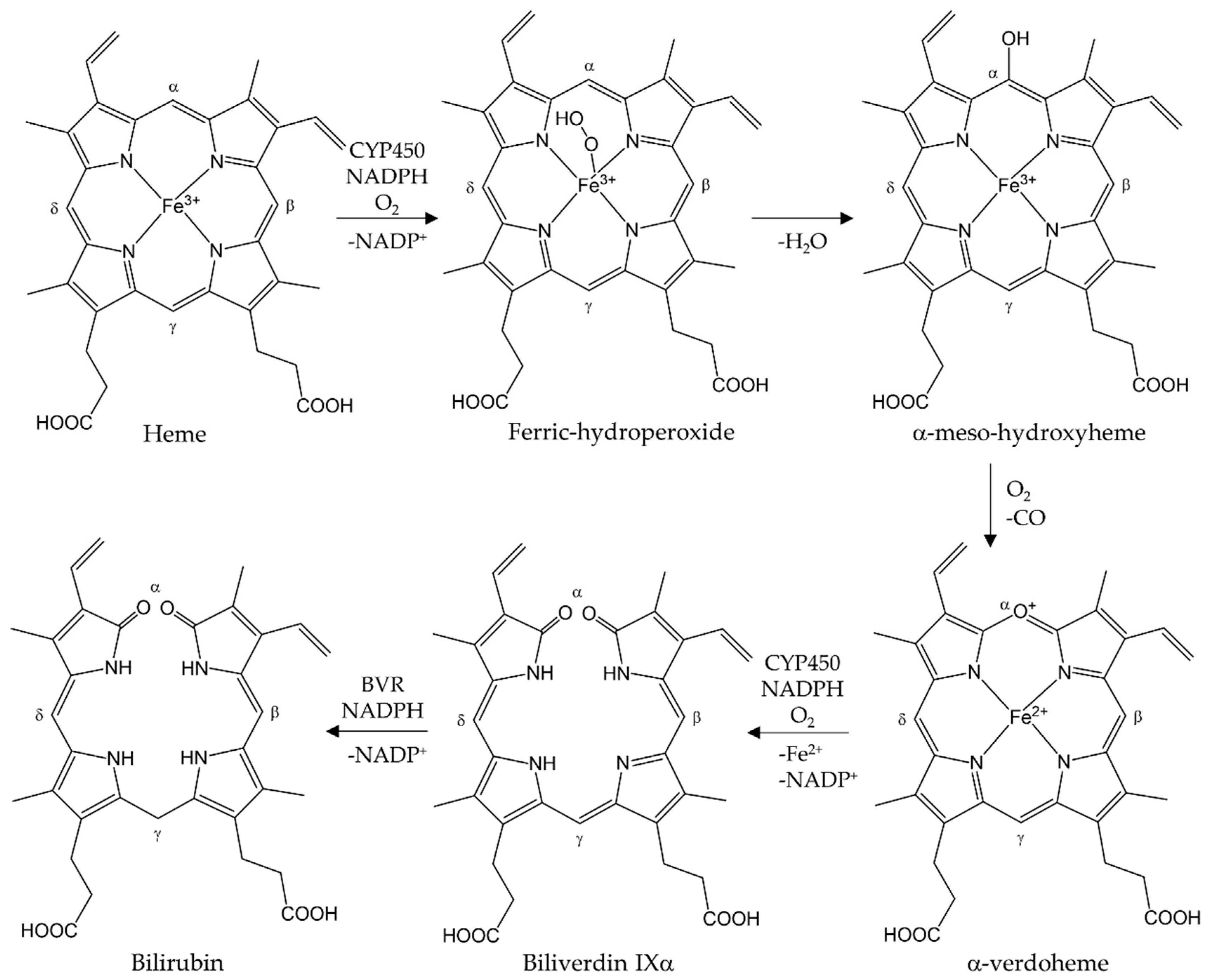
2. Structure and Function of HO-1

HMOX1 is located on chromosome 22 (22q12) in the human genome [24] and encodes for the HO-1 protein with a size of 32 kDa. HO-1 belongs to the class of heme-hydrogen donor-oxygen oxidoreductases (EC 1.14.99.3) and catalyzes the degradation of heme in its oxidized form, i.e., hemin (Fe3+ protoporphyrine) [25]. HO-1 is expressed on the surface of the ER and its catalytic center is oriented towards the cytosol [26]. The single reaction steps catalyzed by HO-1 are illustrated in the figure below (Figure 1).
HO-1 forms a complex with hemin, and ferrous iron is formed through the first electron donation from NADPH by NADPH cytochrome P450 reductase. Molecular oxygen binds to the complex, yielding a metastable oxy-form, after which the iron-bound oxygen is converted to a hydroperoxide intermediate (Fe3+-OOH) through further electron transfer from NADPH cytochrome P450 reductase and a proton from a surrounding water molecule [27]. Terminal oxygen of Fe3+-OOH attacks the α-meso carbon of the porphyrin ring, thereby producing ferric α-meso-hydroxyheme. Subsequently, a ferrous verdoheme-HO complex and CO are formed through the reaction with molecular oxygen. The oxy-verdoheme-HO complex is converted into a ferric iron-biliverdin chelate through a hydroperoxide intermediate that is still bound to HO. The iron in ferric biliverdin is reduced to the ferrous state by the reductase, releasing Fe2+ and BV. Using in vitro experiments, the release of BV from the enzyme was revealed as the rate-determining step [27,28]. BV is then reduced to bilirubin (BR) by biliverdin reductase (BVR), which is glucuronidated in the liver and finally excreted via the bile.

3. Regulation of HO-1 Expression

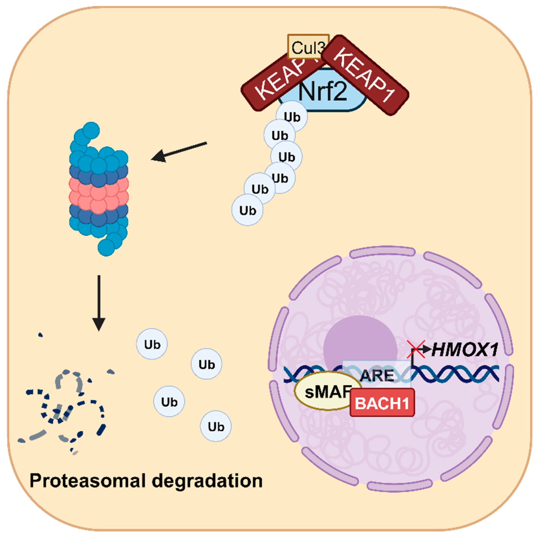
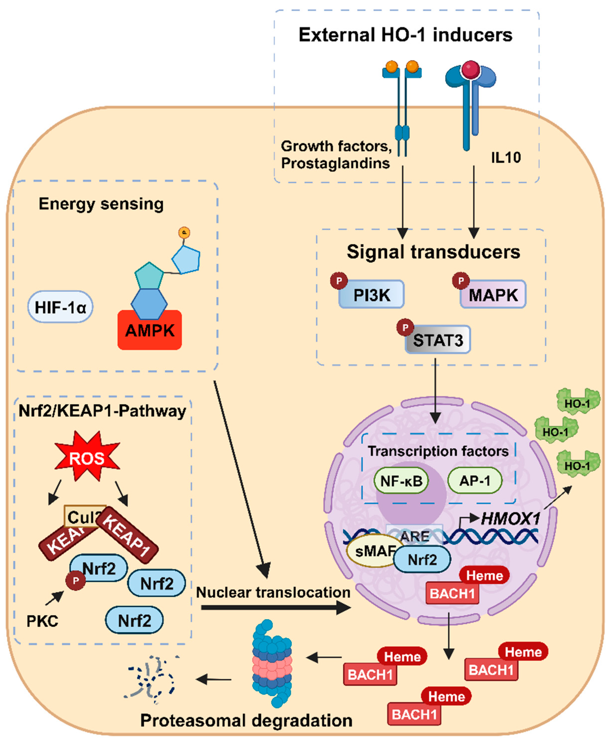
3.1. Transcriptional Regulation of HMOX1

The expression of HMOX1 is mainly regulated on the transcriptional level. Owing to different binding sites in the 5’-flanking region of the promoter, expression of HMOX1 can be regulated by several transcription factors such as nuclear factor erythroid 2-related factor 2 (Nrf2), activator protein-1 (AP-1), and nuclear factor-κB (NF-κB), which bind to specific sequences in the enhancer elements E1 and E2 [29,30].
The cap’n’collar (CNC) subfamily of basic-region leucine zipper (bZIP) transcription factors Nrf2 and BACH1 (BTB and CNC homology 1) are the most important transcriptional regulators of HMOX1 [31,32,33]. Under normal conditions, redox-sensitive Nrf2 resides in the cytosol, where it is marked by Kelch-like ECH-associated protein 1 (KEAP1) for polyubiquitination catalyzed by the ubiquitin E3 ligase cullin 3 (Cul3), leading to its subsequent proteasomal degradation [34] (Figure 2).
In the presence of oxidative or electrophilic reactants as well as other Nrf2 activators, the cysteine residues of KEAP1 are modified, resulting in loss of ubiquitin E3 ligase activity of the KEAP1-CUL3 complex and stabilization of Nrf2 [29,31] (Figure 3). The stabilized Nrf2 then translocates into the nucleus, where it forms a heterodimer with small musculoaponeurotic fibrosarcoma (sMaf) proteins. The Nrf2-sMaf dimer induces transcription of several antioxidant genes such as HMOX1 by binding to the antioxidant response element (ARE) [31,35,36]. Normally, a heterodimer consisting of BACH1 and one of the three sMaf proteins MafF, MafG, or MafK binds to the ARE in two enhancer regions of HMOX1 and represses its transcription [29,31]. However, heme can cause dissociation of the heterodimer by direct binding to BACH1. This leads to the nuclear export of BACH1 into the cytosol, where it is degraded via the proteasome. In turn, this allows Nrf2 to form a Nrf2-sMaf dimer at the enhancer regions and induce HMOX1 transcription [37,38,39,40].
Although Nrf2 and BACH1 are the major regulators of HMOX1 expression, other mechanisms may also be relevant depending on the cellular context. The transcription factors AP-1, NF-κB, and hypoxia-inducible factor-1 (HIF-1α) have been described to induce HMOX1 transcription [41,42,43,44]. AP-1 stimulated the expression of HMOX1 through the cooperation of AP-1 subunits with various transcription factors, including Nrf2 in human endothelial cells [41,42,45]. HIF-1α, a heterodimeric transcription factor induced by low oxygen levels, stimulated HMOX1 expression in human microvascular endothelial cells and in decidual stromal cells, probably via Nrf2 [43,46]. NF-κB was shown to stimulate HMOX1 expression by binding to the HMOX1 promoter in mouse cardiac tissue [44]. Furthermore, the involvement of NF-κB and AP-1 in HMOX1 activation was observed in neonatal ventricular myocytes after hypoxia induction [47].

3.2. Regulation of HMOX1 Expression by Signaling Pathways and Post-Transcriptional Mechanisms

In addition to direct upregulation of HMOX1 via transcription factors, indirect stimulation via signaling pathways triggered by external stimuli has also been described. Several signaling pathways involved in the cellular stress response have been implicated in the regulation of HMOX1 transcription [48,49,50,51]. For example, extracellular stimuli can affect HMOX1 expression via mitogen-activated protein kinases (MAPKs), including extracellular signal-regulated kinase (ERK), c-Jun N-terminal kinase (JNK), and p38 MAPK, which further target transcription factors [48,49,50]. In human hepatocyte L-02 and HepG2 cells, phosphorylation of ERK and p38 has been shown to stimulate HMOX1 transcription via increased Nrf2 expression and nuclear translocation after treatment with either diallyl sulfide or floridoside [48,49]. Furthermore, protein kinase C (PKC) is capable of phosphorylating Nrf2 in response to oxidative stress. This posttranslational modification of Nrf2 promotes its dissociation from KEAP1 and allows Nrf2 to translocate into the nucleus, where it induces HMOX1 expression [52]. Moreover, Signal Transducer and Activator of Transcription (STAT)-3 and phosphatidylinositol 3-kinase (PI3K)-dependent activation of HMOX1 expression in human and mouse myeloid cells by IL-10 has been described [53]. In murine brain cells, PI3K activation was also found to primarily promote translocation of Nrf2 to the nucleus [54]. Furthermore, the enzyme AMP-activated protein kinase (AMPK) stimulates HMOX1 expression by increasing the nuclear localization and phosphorylation of Nrf2 in vivo and in various human epithelial cell lines. AMPK is a serine/threonine protein kinase and energy sensor that is activated by high AMP:ATP ratios as a result of various cellular and metabolic stressors such as hypoxia, ischemia, or heat shock [55,56].
Several studies also indicated that HMOX1 is post-transcriptionally regulated by microRNAs (miRs) [57,58,59,60,61]. The non-coding RNAs showed direct effects on HMOX1 expression in neurons, which was increased by miR-494 under oxidative conditions in a BACH1-independent manner [57]. In contrast, binding of miR-24 or miR-200c to HMOX1 suppressed its expression in human smooth muscle cells [62] and in proximal tubule cells treated with ochratoxin-A [58]. Moreover, control of upstream regulators of HMOX1 by miRs may indirectly affect HMOX1 expression. Whereas miR-153 inhibited the expression of Nrf2 in rats after ischemia-reperfusion (IR) [59], miR-101 increased Nrf2 expression by inhibiting Nrf2 ubiquitination in human umbilical vein endothelial cells (HUVEC) under hypoxic conditions [63]. Moreover, downregulation of BACH-1 translation in HUVECs by miR-155 [60] and degradation of Keap-1 mRNA in breast cancer cells by miR-200a indirectly increase HMOX1 expression [61].

4. Other HO Isoforms

Besides HO-1, which is inducible by the transcription factor Nrf2 as outlined above, another isoform termed HO-2 exists. HO-2 is encoded by HMOX2 located on chromosome 16 (16q12) [24] and gives rise to a 36 kDa protein. It is constitutively expressed in neurons and is also found in other tissues such as the spleen and myocardium [64]. HO-2 breaks down hemin with similar efficiency as HO-1, but it possesses additional heme-regulatory motifs (HRMs) that bind heme and transfer it to the catalytic center of HO-2 [65]. The two HRMs contain cysteine residues that form a disulfide bond; when reduced, these cysteines are available to bind hemin [65]. In total, HO-2 has three heme binding sites: one at its catalytic site and the others at its two HRMs [64].
The activity of HO-2 is regulated by posttranslational modifications. Activation of PKC leads to phosphorylation of casein kinase 2 (CK2), which in turn activates HO-2 through phosphorylation at Ser79 [66]. Currently, only one specific chemical activator of HO-2 is known. Menadione (Vitamin K3) has no effect on HO-1 but can increase the enzymatic activity of HO-2 by up to 30-fold in in vitro models [67]. HO-2 attracted attention due to its various beneficial effects within cells and organs such as the liver, testis, and brain, which express it constitutively. In the testis, HO-2 is involved in the modulation of the ejaculatory activity [68]. In contrast, HO-2 plays a dual role in the brain, which leads on the one hand to antioxidant and anti-inflammatory effects because of products generated by heme catabolism [69,70]. On the other hand, prooxidant and cytotoxic effects are observed due to abundant formation of iron after intracerebral hemorrhage (ICH) [71]. Furthermore, HO-2 is able to suppress inflammatory pathways through the reduction of pro-inflammatory cytokines, such as IL-1b, TNFα and interleukin 6 (IL-6), in macrophages [72]. Many potential therapeutic applications with regard to HO-2 are investigated like preventing renal injury and diabetes [73]. HO-2 was also shown to be involved in the onset of neurodegenerative diseases owing to an HMOX2 polymorphism. This leads to iron release and abnormal iron deposition, and subsequently Alzheimer’s disease, where overexpression of HMOX2 was found [74,75].
A third isoform, HO-3, has been described in rats. It has been identified as a pseudogene processed from HO-2 transcripts, but its function is still unclear [76].

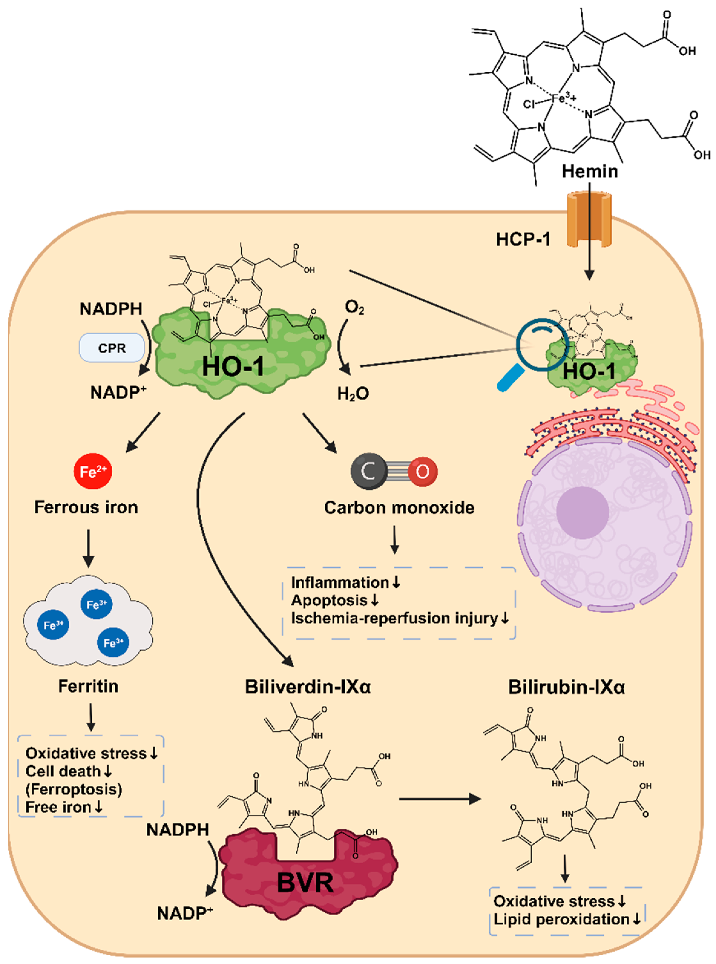
5. Cytoprotective Effects of HO-1

Heme, the substrate of HO-1, is an organic, iron-containing protoporphyrin ring that is essential for numerous biological processes [77,78]. As a prosthetic group of several proteins, heme plays an important role in oxygen transport (e.g., hemoglobin and myoglobin), oxidative metabolism (e.g., catalases, oxygenases), detoxification of xenobiotics (cytochrome P450 enzymes), electron transfer (cytochromes a, b, c), and microRNA processing (DGCR8) [77,79]. On the other hand, free heme is a known catalyst of radical formation, a pro-pathogenic mediator, and a pro-oxidant with cytotoxic effects [2,21,80]. It also induces lipid peroxidation and chronic inflammation [81,82]. The degradation of heme by HO-1 contributes to the maintenance of intracellular homeostasis and represents a direct cytoprotective effect of HO-1. Studies in HO-1-deficient mouse models as well as several cases of HO-1 deficiency in humans caused by HMOX1 mutations highlight the importance of HO-1 for tissue iron and organ homeostasis, protection against oxidative stress, and macrophage function [14,83,84]. The heme degradation products BV/BR and CO have additional cytoprotective effects, which will be detailed below [72,85,86,87,88,89,90,91,92] (Figure 4).
BV and BR are potent antioxidants that show cellular protective effects in vitro and in vivo [72,87,88]. Both BV and BR protected EA.hy926 cells, a standard model of human vascular endothelium, from oxidative stress with an EC50 value equivalent to BR serum concentrations [87]. The protective effects of BV and BR are likely due to the biliverdin-bilirubin redox cycle, in which BR is oxidized to BV by scavenging oxidants. BVR then catalyzes the NADPH-dependent reduction of the generated BV back to BR [85]. The reduction of BV to BR is essential for its antioxidant activity, which was demonstrated in a BVR knockout model. In BVR−/− mice, 100-fold higher BV levels were measured, while BR levels were 25-fold decreased. This was associated with increased serum levels of oxidative stress [72]. BR protects against lipid peroxidation in a more potent manner than the well-known antioxidant glutathione [88]. A study in HeLa cells revealed a protective effect for BR up to a 10,000-fold excess of H2O2, while the depletion of BVR by small interfering RNA (siRNA) led to a significant increase in tissue ROS levels and cell death [85,88]. Moreover, high BR levels negatively correlate with oxidative stress outcomes such as diabetic complications, chronic inflammation, and cardiovascular disease such as atherosclerosis [86,93,94,95]. Zhu and colleagues summarized in a meta-analysis that high BR levels are associated with a protection against diabetic nephropathy, retinopathy, and neuropathy, as well as other diabetic complications, for which oxidative stress is considered as a major cause [93]. A cohort study involving 4196 men and 4648 women also found an association between high BR levels and reduced cardiovascular mortality [86]. In mice with low-density lipoprotein receptor deficiency, BR has also been shown to inhibit atherosclerotic plaque formation by scavenging ROS signaling intermediates, thereby impairing vascular cell adhesion protein 1 (VCAM-1)- and intercellular adhesion molecule 1 (ICAM-1)-mediated endothelial leukocyte migration [94].
While exogenous CO has long been known as a toxic gas, its potential for treating various medical conditions is increasingly recognized [96,97,98]. Endogenous CO is mainly formed due to the degradation of heme by HO-1 [99]. It plays an important physiological role in neuronal signal transduction as well as in the maintenance of vascular tone [97,100,101]. It also exhibits anti-apoptotic, anti-inflammatory, and anti-proliferative effects, thus protecting tissues [89,90,92,96,102,103,104]. In this context, CO acts as a modulator of a variety of signaling pathways, which are detailed below.
CO can downregulate NADP(H)-oxidase-dependent ROS formation in macrophages, thereby inhibiting TLR2,4,5 and 9 signaling pathways. The resulting suppressed migration of TLRs to membrane rafts decreases cytokine production and negatively regulates the proinflammatory cascade [104]. On the other hand, CO can also trigger increased mitochondrial ROS formation in macrophages by inhibiting cytochrome c oxidase. Due to this inhibition, the LPS-induced inflammatory response in macrophages was limited [89]. Such a macrophages-derived ROS burst also leads to an increase in peroxisome proliferator-activated receptor-γ (PPARγ), a nuclear hormone receptor that plays an important role in the expression of genes involved in inflammatory signaling pathways in macrophages [105]. In a mouse model of acute lung injury, CO suppressed the expression of a key inflammatory mediator, early growth response protein 1 (Egr-1), and reduced tissue damage. Inhibition of PPARγ abolished both effects [105]. Via activation of the MKK3/p38β-MAPK pathway, CO triggered anti-inflammatory effects in LPS-induced inflammation models. While the expression of pro-inflammatory cytokines such as TNF-α, IL-1β and macrophage inflammatory protein-1β (MIP-1β) was decreased, there was a concomitant increase in the production of the anti-inflammatory cytokine IL-10 [106]. The anti-inflammatory effect of HO-1/CO was also demonstrated in murine colitis models, in which overexpression of HO-1 as well as inhalation treatment with CO led to a decrease in the pro-inflammatory cytokines TNF-α and interferon-γ (IFN-γ) and tissue-associated myeloperoxidase (MPO) activity in the colonic mucosa [90,107].
In addition to the described anti-inflammatory effects, CO-dependent induction of the p38 MAPK pathway also protects against spontaneous and induced apoptosis [91,96,102,108,109,110]. IR injury is common in organ transplantation and can be attenuated by HO-1 induction via cobalt protoporphyrin IX (CoPP) and exogenous CO administration [96,108,109,110]. CO protected endothelial cells from TNF-α-mediated apoptosis by increasing TNF-β-dependent anti-apoptotic genes such as c-IAP2 in a p38 MAPK-dependent manner [102]. Moreover, CO-induced activation of the MKK3/p38α-MAPK pathway led to the downregulation of Fas ligand (FasL) and suppression of caspase-8 activity in primary rat pulmonary artery endothelial cells and in a mouse lung IR model [108]. Furthermore, an increased expression of anti-apoptotic Bcl-2 family proteins such as Bcl-2 and Bcl-XL with concomitant reduction in expression of pro-apoptotic proteins such as Bid was observed, ultimately preventing release of cytochrome c from mitochondria and thus protecting against IR-induced apoptosis [108]. Similar protective effects of CO against IR-induced apoptosis were also described in lung epithelial cells, which was attributable to STAT3 inhibition via PI3K/Akt- and p38 MAPK-dependent pathways [109].
CO-induced mitochondrial ROS formation through inhibition of NADPH oxidase, as described previously, also leads to a decrease in ERK1/2 MAPK phosphorylation and cyclin D1 expression, which allowed CO to inhibit proliferation in airway smooth muscle cells [92]. In addition to its anti-proliferative effect, CO may also contribute to re-endothelialization and prevent neointima formation after carotid artery balloon injury in male rats. Moreover, proliferation, adhesion and migration of HUVEC were promoted by exogenous CO in a PI3K/Akt/eNOS pathway-dependent manner [111].
Last but not least, the HO-1 breakdown product Fe2+ increases the synthesis of the iron storage protein ferritin [112,113]. Ferritin consists of two subunits, Ferritin heavy-chain (FtH) and Ferritin light-chain (FtL), which have different functions. FtH has a ferroxidase center where Fe2+ is oxidized to the less reactive Fe3+. FtL helps to release Fe3+ from the ferroxidase center and to store it [114]. Several studies indicated the protective role of ferritin against free iron, oxidative stress, and cell death [115,116,117]. In mice with intestinal FtH deletion, intestinal ferritin has been shown to be essential for regulating iron absorption and thus protecting the body from excessive iron levels [115]. In a model of acute kidney injury (AKI), mice with FtH deletion in the proximal renal tubule showed significantly increased mortality, apoptosis rate, and worsening of functional and structural renal injury associated with elevated HO-1 levels. It was also shown that only FtH proficient mice showed an increase in the iron transporter ferroportin (FPN) after glycerol-induced rhabdomyolysis, resulting in an increased export of deleterious ferrous iron across the basolateral membrane into the circulation. These observations suggest that ferritin also promotes the cellular export of toxic free iron, thereby protecting cells [116].
In summary, HO-1 has manifold cytoprotective functions. In addition to the degradation of cytotoxic free heme, the degradation products CO and BV/BR as well as ferritin induction by the released Fe2+ contribute to cell protection via multiple mechanisms. Depending on the tissue and cell type, both the activated signaling pathways and the resulting effects may vary [72,90,91,92,96,111].

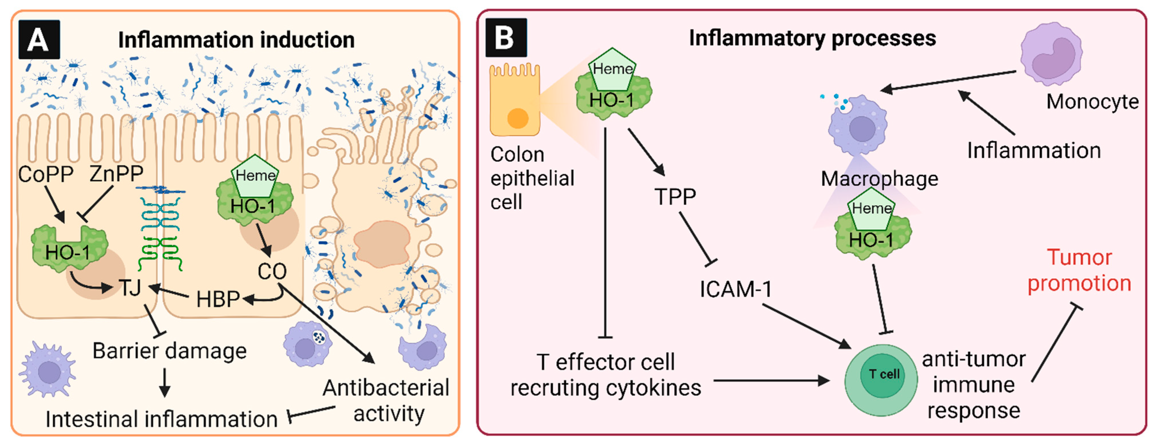
6. Involvement of HO-1 in Intestinal Inflammation and Chronic Diseases

Intestinal inflammation is mainly triggered by disruption of the epithelial barrier function, followed by infiltration of bacteria or bacterial components [118]. It was reported that free heme induced barrier disruption in filter-grown, differentiated human epithelial colorectal adenocarcinoma cells (Caco-2) [18]. This effect was attenuated by induction of HO-1, whereas inhibition of HO-1 resulted in enhanced barrier disruption [18]. In CCl4-treated mice, pathological changes of the colon with reduced colon length, lower levels of tight junction (TJ) proteins and higher TNFα levels were observed, which was influenced by intestinal HO-1 expression (Figure 5A) [118]. CoPP-induced HO-1 expression attenuated the changes of TJ proteins and TNFα levels. In contrast, inhibition of HO-1 by ZnPP increased the pathological changes [118]. The protective effects of HO-1 induction were traced back to CO-releasing molecule-2 (CORM-2) [118]. Due to its high affinity, the heme breakdown product CO can bind to heme-binding proteins that form the tight junction complex, which is crucial for the cellular barrier function, and re-close the barrier by altering protein configuration [119]. Similar to CCl4-treated mice, the induction of HO-1 or CO release by CORM-2 improved intestinal barrier function in rats suffering from cholestasis [120].
Inflammatory bowel disease (IBD) describes a group of chronic inflammatory diseases of the intestinal tract that includes ulcerative colitis (UC) and Crohn’s disease (CD), which show an increasing incidence worldwide [121,122]. A comparison of diseased and healthy intestinal tissue from UC patients revealed an increased HO-1 expression on mRNA and protein level in the inflamed tissue. These observations suggest an important role of HO-1 in IBD [123]. In several IBD models, it was shown that expression of HO-1 as well as exposure to CO resulted in an amelioration of colitis [19,124]. CO suppressed Th17-induced colitis in Il10−/− mice via a HO-1-dependent mechanism. Treatment with CO or the HO inducer CoPP ameliorated chronic intestinal inflammation in T cell receptor-alpha (TCR-α)-deficient mice, in which colitis is driven by Th2 cytokines, similar to human UC. The decrease in intestinal inflammation occurred through inhibition of colonic interleukin-1β (IL-1β), TNF, and IL-4 production and increased IL-10 secretion [124].
In line with these findings, HO-1-dependent reduction of inflammatory processes were reported in HO-1 knockout mice and HO-1-deficient human subjects. Mouse studies with Hmox1+/+, Hmox1+/− and Hmox1−/− genotypes revealed a stronger sensitivity of Hmox1−/− mice towards LPS as inflammatory stimulus, resulting in significantly higher cytokine release and a survival rate of only 7%. In contrast to that, the survival rates of Hmox1+/+ and Hmox1+/− mice were 100% and 94%, respectively [15]. Similar results with a 37-fold increase in IL-6 levels were seen in LPS-treated Hmox1 knockout mice [125]. Case reports of human HO-1-deficient patients showed chronic inflammation and significantly higher ROS-related effects than healthy individuals [83]. Greil and colleagues presented the case of a Turkish male patient with a loss of function mutation in HMOX1 [126]. The patient showed high levels of ROS-induced acrolein-lysine and 8-oxoguanine, an important marker of oxidative DNA damage. Moreover, peripheral blood mononuclear cells obtained from this patient were more sensitive to oxidative stress inducers and inflammatory stimuli [126].
Furthermore, several studies suggest that both the composition of the gut flora and the patient’s protective mechanisms play a crucial role in the development of IBD [127,128]. Colonization of germ-free wild-type (Il10+/+) and Il10−/− mice with a specific pathogen-free (SPF) microbiota resulted in increased TNF and Il12b expression as well as enhanced colonic inflammation in Il10−/− mice. In contrast, increased Hmox1 expression in the colon and elevated colonic HO-1 protein levels were observed in WT animals as compared with Il10−/− animals. Further Nrf2-regulated genes like Fth1 were also highly expressed in WT, but not Il10−/− mice. Pharmacological induction of HO-1 with CoPP in germ-free Il10−/− mice before colonization with SPF microbiota resulted in amelioration of colitis and decreased secretion of IL-12 p40. These observations suggest a protective induction of HO-1 by the microbiome in a Nrf2- and TLR-dependent manner. Furthermore, the treatment of Il10−/− mice with CoPP and the CO-releasing molecule ALF186 resulted in increased bactericidal activity of macrophages against E. coli, E. faecalis, and S. typhimurium in addition to bacterial elimination in mesenteric lymph nodes. This suggests that the anti-inflammatory effect of HO-1 and CO is due to the increased bactericidal activity of macrophages, which are thus better able to eliminate bacteria that may cause or exacerbate colonic inflammation [129]. This hypothesis was later verified by in vitro studies using bone marrow derived macrophages from Hmox1-deficient mice. CO significantly increased the bacterial killing by macrophages and counteracted the knockout-derived defect [20].
Therefore, HO-1 expression in intestinal epithelial cells as well as in macrophages is important to prevent and suppress inflammation, while the time point of HO-1 induction seems to be critical (Figure 5B). Interestingly, HO-1 induction by CoPP prior to colitis induction by dextran sodium sulfate (DSS) reduced the histological degree of intestinal inflammation [130]. It should be emphasized that such an anti-inflammatory effect was only observed, if the HO-1 expression was increased as a preventive treatment, not as a therapeutic approach after the onset of inflammation. The authors hypothesized an HO-1 dependent lower expression of ICAM-1 as a key anti-inflammatory effect. ICAM-1 is an adhesion protein, responsible for the regulation of cell—cell interactions like lymphocyte-mediated cytotoxicity in tumor cells, which is therefore important for the immune surveillance system [131,132]. In this context, HO-1 induction in response to hemin exposure reduced ICAM-1 expression in HT29 and Caco2 CRC cells by regulation of the RNA-binding protein tristetraprolin (TTP). Moreover, hemin reduced the expression of T-effector cell-recruiting cytokines, which further led to anti-inflammatory effects [131]. ICAM-1 is considered as potential oncogene in colorectal carcinogenesis, since it promotes the migration, invasion and expression of epithelial-mesenchymal transition markers [133]. Thus, ICAM-1 could represent a possible link between HO-1 and tumor progression (see Section 9).

7. Colorectal Carcinogenesis and the Role of HO-1

Colorectal cancer (CRC) is one of the most frequent tumor entities worldwide, with an increasing incidence in adults under the age of 50 years [134,135]. CRC is causally linked to genetic predisposition, chronic intestinal inflammation, lifestyle and nutritional risk factors [135,136,137,138]. Sporadic colorectal carcinogenesis is often illustrated with the model developed by Fearon and Vogelstein, which describes a characteristic series of mutations in tumor suppressor genes and proto-oncogenes [139]. The formation of CRC is a multi-step process occurring over decades [140]. The first step is called tumor initiation, in which normal intestinal epithelial cells acquire mutations in critical genes, i.e., tumor suppressor genes and/or proto-oncogenes. These mutations can be induced by various environmental or dietary genotoxins, such as heterocyclic aromatic amines and N-nitroso compounds [21,81,141,142,143]. Furthermore, hereditary or acquired DNA repair defects increase the risk that spontaneous or induced DNA damage in intestinal epithelial cells is fixed into mutations. With regard to CRC formation, defects in mismatch repair, but also O6-methylguanine-DNA methyltransferase (MGMT) and MYH, are of particular relevance [144,145,146]. Mutations in the WNT signaling pathway as the pivotal regulator of intestinal epithelial cell turnover causes hyperproliferation and promotes the formation of aberrant crypt foci [139,140]. These mutations are mainly found in the tumor suppressor gene APC, but also observed in its downstream target ß-catenin [147]. In the next stage termed tumor promotion, a higher cell proliferation together with, e.g., inactivated DNA repair genes such as MGMT, increases the probability for further mutations thereby activating proto-oncogenes such as Kirsten-ras (KRAS) and inactivating tumor suppressor genes like TP53 [139,148,149]. This results in the formation of a benign adenoma. Subsequently, the benign tumor evolves into a malignant carcinoma, with a progressive accumulation of further mutations and epigenetic alterations [139]. At this stage called tumor progression, the tumor grows independently, displaces normal healthy tissue and spreads to distant parts of the body via blood and lymph vessels. This process is called metastasis and is triggered by epithelial-mesenchymal transition (EMT) of cancer cells, whereby the cells are able to migrate and invade into surrounding and distant tissue [150].
In colon (cancer) tissue, HO-1 seems to be involved in tumor induction and progression due to its stage-specific expression. Several independent studies detected a significantly higher HO-1 expression and activity in tumor tissue from CRC patients than in adjacent normal colon tissue [151,152,153]. Moreover, well-differentiated tumors showed the highest HO-1 expression, whereas the content in moderately and poorly differentiated tumors were lower [153]. Concomitant with the overall increase in HO-1 expression, its localization altered during carcinogenesis, with stage-dependent higher nuclear HO-1 levels in polyps and adenocarcinoma as compared to non-tumor tissue [151]. Nuclear localization of HO-1 was also observed in human HCT116 CRC cells. In both untreated and hemin-treated cells, HO-1 localized to the nucleus, while hemin increased the HO-1 expression without affecting its subcellular localization [151]. One possibility for the nuclear translocation of HO-1 might be its passive diffusion through the nuclear pore complex, which has a size limit for proteins with more than 40–60 kDa [154,155]. Furthermore, nuclear localization sequences were identified in HO-1, which can mediate its translocation [156]. Nuclear translocation of HO-1 in mouse fibroblasts and hepatoma cells were attributable to the cleavage of a 52-amino-acids-spanning region from its C-terminus [9]. The truncated HO-1 in the nucleus showed lower enzymatic activity, but mediated the activation of transcription factors involved in the protection from oxidative stress and in cell proliferation, such as AP-1, AP-2 and Brn-3, by alteration of their DNA binding activity [9]. If this specific mechanism is also relevant for colon (cancer) cells, it is not yet known. Interestingly, in non-cancerous human colonic epithelial cells (HCEC), hemin induced the expression of HO-1 in a concentration-dependent manner, which was however exclusively localized in the cytoplasm [21]. To sum up, the expression and localization of HO-1 in colon tissue seems to be associated with CRC formation and might be useful as diagnostic and prognostic marker. In line with this notion, a study pointed out that the HO-1 degradation product CO, which is bound to hemoglobin (COHb) and circulates in the bloodstream of patients, might be a diagnostic marker. The COHb levels in CRC patients were significantly higher than in non-cancer patients [153]. The suitability of COHb as diagnostic marker is supported by a strong correlation of increased serum COHb levels, tumor growth, and HO-1 activity in tumor-bearing mice [153].
While nuclear HO-1 expression positively correlates with CRC formation, it might also influence the survival rate of patients. CRC patients, whose tumor tissue was stained positive for HO-1, showed a significantly higher survival rate as compared to HO-1-negative cancer patients [151,157]. Furthermore, tumors with high HO-1 expression levels were associated with lower lymphatic invasion. Thus, the increased HO-1 levels may be a protective mechanism to suppress colorectal carcinogenesis. In contrast to these studies, a high HO-1 expression in macrophages near colorectal tumors correlated with lower patients’ survival rates as well as higher risks for lymph node metastasis [158]. Alaluf and coworkers demonstrated a strong HO-1 induction in mice as part of the differentiation of monocytes to macrophages by the tumor microenvironment, e.g., the inflammation of gut mucosa. Furthermore, HO-1 expression by these tumor-associated macrophages led to significant immunosuppressive activity, which reduced the T-cell mediated anti-tumor immune response. In turn, HO-1 inactivation enhanced the anti-tumor cell response and suppressed the HO-1-mediated tumor-promoting effects of macrophages [159]. Taken together, the HO-1 expression in colon epithelial cells may reduce tumor formation, while high HO-1 levels in tumor-associated macrophages seem to promote carcinogenesis (see also Figure 5B).
Similar to inflammation and immune reaction, a clear correlation of HO-1 and miR expression was seen in CRC tissue. HO-1 expression is suppressed by its negative regulator BACH1 [29,31], which occurred to a significantly higher extent in non-tumor colon tissue than in cancer tissue [160]. It was hypothesized that the reduction of BACH1 expression during carcinogenesis might be regulated by an increase in miR-135a, because of a documented negative correlation of BACH1 and miR-135a expression in colon (cancer) cells [160]. The underlying mechanism of BACH1 regulation by miR-135a is still unknown. However, the lower BACH1 expression could be mainly responsible for the higher HO-1 levels in colon cancer tissue [160]. In accordance with HO-1, the miR-135a expression is higher in CRC tissue and increases with the CRC tumor stage [151,152,153,160,161]. This was also observed in the CRC cell lines SW480 and SW620, in which the miR-135 expression was higher than in normal mucosa tissue samples [161]. The CRC cell lines were analyzed in more detail by reduction of the miR135a expression using the synthesized oligonucleotide inhibitor antagomiR-135a. The miR-135a-induced promotion in tumor progression-related processes was reduced likewise [161]. Migration and cell invasion are crucial steps in EMT, an important process for tumor progression and metastasis [150,162]. Inhibition of miR-135a expression in CRC cell lines significantly decrease their ability of migration, invasion and proliferation [161]. These cellular properties are relevant for CRC formation and are often accompanied by high HO-1 expression in colon tumor tissue due to reduced cellular BACH1 levels. Cell invasion and anchorage-independent growth of CRC cells can be further directly induced by HO-1 overexpression. In CRC patient tissue the expression of the endothelin-converting enzyme-1 (ECE-1) was significantly higher than in normal tissue and correlates with HO-1 expression [163]. ECE-1 regulates cancer stem cell properties, which was shown in vitro by HO-1 or ECE-1 overexpression [163,164]. In both studies, the transfected cells showed a higher expression of CD44 and CD133 [163,164], known as cancer stem cell markers and essential in tumor progression [165,166,167]. Moreover, high HO-1 or ECE-1 expression increased the formation of spheres and its metastatic potential [163,164].
These studies with human CRC cell models and colon (cancer) tissue samples provided first insights, in which processes HO-1 is involved, with both anti-tumorigenic and tumor-promoting effects described. In the following two chapters, the role of HO-1 in colorectal tumor induction and progression is discussed in more detail.

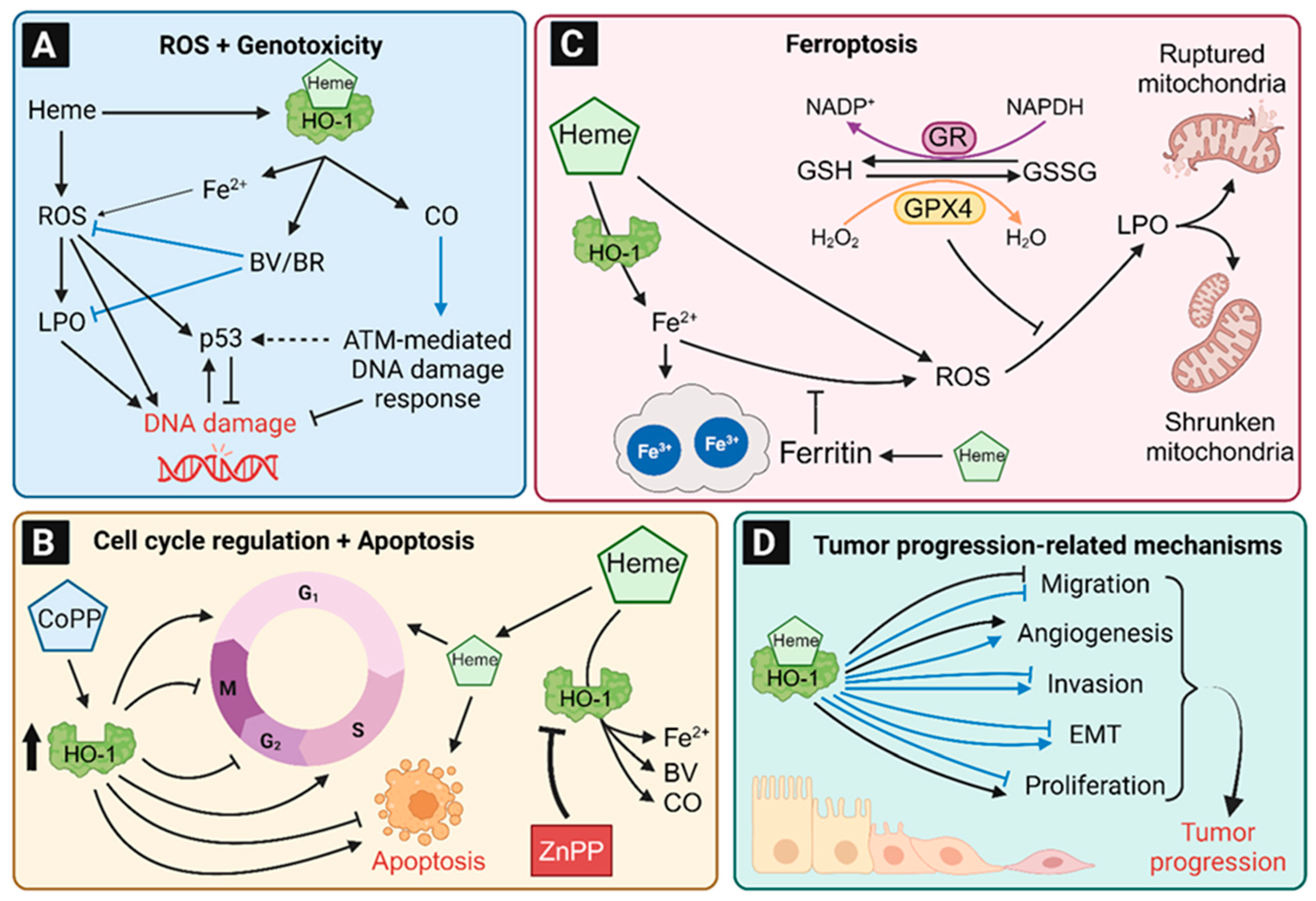
8. Role of HO-1 in Tumor Induction

8.1. HO-1-Dependent Protection against ROS and Lipid Peroxidation

ROS can be involved in tumor formation, e.g., by induction of DNA damage or by other mechanisms like the regulation of immune cells [168]. There is a close relation of ROS and HO-1 expression (summarized in Figure 6). On the one side, the induction of oxidative stress and ROS by H2O2, growth factors or heme, led to higher HO-1 expression in non-tumor and tumor colon cell lines [21,169]. Furthermore, HO-1 was also shown to be upregulated in colon epithelium of mice exposed to dietary heme iron, whereas no induction of HO-1 was observed in the control group receiving non-heme iron [81]. As described in Section 3, ROS induces the release of Nrf2 from the KEAP1-Cul3 complex, followed by phosphorylation and nuclear translocation of Nrf2. In the nucleus, Nrf2 binds to ARE in DNA to upregulate the transcription of genes involved in the antioxidant response like HMOX1 [31,35,36]. On the other side, HO-1 reduces the cellular ROS level by generation of BV and BR, which are known antioxidants by reacting with ROS and lipid peroxidation products [85,170,171]. In line with that, the chemical inhibition of HO-1 activity with ZnPP led to significantly higher ROS levels in treated CRC cells [172,173,174]. In Caco-2 cells, HO-1 expression was increased by both heme and nitrosylated heme in a comparable manner [141], indicating that both heme compounds independent of their ligand are substrates for HO-1.
As mentioned above, the interrelation of heme, ROS and HO-1 was shown for both non-tumor as well as CRC cells. Nevertheless, non-malignant HCEC were more sensitive to hemin than CRC cells (HCT116 cells and others) as demonstrated by higher ROS levels, oxidative DNA damage, and cytotoxicity [21]. The inhibition of HO-1 by ZnPP or its siRNA-mediated knockdown further increased the hemin-induced ROS formation and toxicity. HCT116 CRC cells were characterized as a non-differentiated cell line, classified as stage 3 of the TNM system [175,176] with low basal HO-1 levels, which is in accordance with the above-mentioned association of highly differentiated tumors with higher HO-1 expression levels [153]. Moreover, poorly differentiated colon cell lines like COLO205, HCT-15 and LOVO were more sensitive towards H2O2 as compared to the well-differentiated cell line HT-29 [177]. Unfortunately, HO-1 expression was not analyzed in this setup.

8.2. Role of HO-1 in Genotoxicity and DNA Damage Response

HO-1-derived heme degradation mainly prevents high cellular ROS levels, but it also suppresses lipid peroxidation and DNA damage as biological consequences (Figure 7A) [21,178,179]. Cellular lipid hydroperoxides were formed by the reaction of (e.g., heme-induced) ROS with the unsaturated fatty acids of lipids and can further induce DNA damage, such as DNA adducts and DNA strand breaks [180,181,182]. Several studies using normal or CRC cell lines showed that the HO-1 inhibition by ZnPP alone, as well as in combination with genotoxic stress, increases DNA damage [21,174,183]. Li and colleagues analyzed the influence of HO-1 inhibition on the toxic effects of the chemotherapeutic paclitaxel. Upon inactivation of HO-1 by ZnPP, the CRC cells displayed more DNA damage, as analyzed by γH2AX levels, followed by higher apoptosis and lower cell viability [183]. These effects were already seen for ZnPP without paclitaxel treatment, whereby a combination treatment significantly increased the effects. Thus, the inhibition of HO-1 sensitizes CRC cells for the chemotherapeutic effects of paclitaxel. Another important factor in genome protection by HO-1 is the activation of DNA repair by the heme degradation product CO. HO-1 deficient mice displayed significantly higher levels of γH2AX-positive cells in the kidney, lung, liver and spleen [184]. As mentioned above, yH2AX is an established marker for DNA damage [185,186]. Furthermore, wild-type mice were exposed to CO and showed significantly lower levels of doxorubicin-induced DNA damage in the intestine as well as in the kidney, liver, and lung [184]. A follow-up cell culture study in prostate and kidney cancer cells provided evidence that CO increased the levels of phosphorylated DNA repair proteins like p-BRCA1 and its upstream DNA damage response kinases p-ATM and p-ATR. ATM was revealed as the most important factor in HO-1 and CO-induced DNA repair, since the beneficial effects were abrogated by the ATM inhibitor KU-55933 [184]. Finally, HEK293 kidney tumor cells with HO-1 knockout were analyzed in detail. The HO-1 deficient cells showed significantly lower levels of p-ATM, p-ATR, p-BRCA1, and p-CHK2. Therefore, CO generated by heme breakdown seems to activate DNA double strand break repair with ATM as essential regulator [184].

8.3. Regulation of the Cell Cycle, Apoptosis and Cell Viability by HO-1

ROS formation and DNA damage occur at any time in our cells, while it is hypothesized that each cell produces 50 hydroxyl radicals per second [187]. If the damage levels exceed the cellular DNA repair capacity, apoptosis is induced [188]. Thus, apoptosis is an essential mechanism to prevent carcinogenesis by elimination of initiated cells via controlled cell death induction [188,189]. Interestingly, this tumor-protective mechanism can be affected by HO-1-regulating compounds like CoPP and ZnPP [174,190]. The impact of HO-1 in apoptosis and cell viability was shown in several colon cell lines as well as in mouse studies performed in independent experiments. However, these results are inconsistent and the role of HO-1 in apoptosis is not yet clarified (Figure 7B). The induction of HO-1 expression by CoPP increased the S-population and reduced the G2/M-population of Caco-2 cells, while apoptosis was reduced likewise [190]. Similar results were seen in murine C-26 cells, in which a high HO-1 expression dose-dependently protected the cells against apoptosis [191]. In accordance with these findings, CoPP treatment in mice with DSS-induced colitis led to less apoptotic cells in the colon as compared to untreated animals [130]. While HO-1 induction decreased apoptosis, its inhibition by ZnPP or tin protoporphyrin IX (SnPP) followed by hemin treatment increased the subG1-population, apoptosis and cytotoxicity in different CRC cell lines as well as non-cancerous HCEC [21,172,174,183,192]. In contrast to these findings, there are also studies that report apoptosis induction by high HO-1 expression upon CoPP exposure. In COLO205, HCT15, and LOVO cells the CoPP-induced HO-1 expression was related to higher subG1-population and higher expression levels of the apoptotic markers cleaved caspase-3 and cleaved PARP1. However, there was no increase in apoptosis in HT-29 cells with CoPP-induced high HO-1 expression [193]. Andres and colleagues revealed a significantly higher ratio of cells in the G0/G1-phase as well as higher apoptotic ratios after hemin-triggered HO-1 induction in HCT116 cells, while these effects depended on the p53 status [151]. HCT116 p53−/− cells showed a significantly higher ratio of cells in the G2/M-phase and cell viability after HO-1 induction by hemin. Thus, p53 and/or its downstream targets like p21 seem to be involved and responsible for the HO-1 pro-apoptotic and anti-apoptotic effects. In human MCF-7 breast cancer cells, higher expression of p53 and p21 by hemin-induced HO-1 and its product ferrous iron was confirmed. It was shown that the iron chelators desferrioxamine and 1,10-phenanthroline counteract the p53 induction [63].

8.4. The Contribution of HO-1 to Ferroptosis

Ferroptosis is as an iron-dependent type of programmed, caspase-independent cell death, which is morphologically, biochemically, and genetically distinct from apoptosis, autophagy-dependent cell death, and necroptosis [194,195]. Interestingly, neurodegenerative diseases like Alzheimer and Parkinson are associated with higher cellular iron levels and ferroptosis, which could be a relevant origin of these diseases [196,197]. On the morphological level, ferroptosis is characterized by a shrinkage of mitochondria and a reduced number of mitochondrial cristae [198]. Ferroptosis is triggered by a massive ROS-dependent lipid peroxidation in the plasma membrane and severe mitochondrial damage due to an impaired activity of glutathione peroxidase 4 (GPX4) and its redox cycle (Figure 7C) [194,199]. Biochemical markers of ferroptosis include a reduced expression of GPX4 and SLC7A11 (a subunit of the cysteine/glutamate Xc- Antiporter), an increased expression of ACSL4 (long-chain-fatty-acid-CoA ligase 4) and PTGS2 (prostaglandin-endoperoxide synthase 2; COX-2), depletion of cellular glutathione (GSH), decreased NADPH levels as well as massive lipid peroxidation [195,200]. Iron chelators (e.g., deferoxamin) and lipophilic antioxidants such as Trolox or ferrostatin-1 were shown to inhibit ferroptosis by blocking iron-mediated ROS formation [201,202]. Known inducers of ferroptosis are glutamate, drugs such as erastin and sorafenib, which inhibit the Xc-antiporter and prevent cysteine import, as well as RSL3, which acts as a GPX4 inhibitor [195]. Both the p53 and Nrf2 pathway are also implicated in ferroptosis [195].
Heme degradation by HO-1 generates BV, CO, and ferrous iron. It is thus hypothesized that an increased HO-1 activity could result in ferrous iron accumulation, which may promote ferroptosis via the mechanism described above. Nevertheless, free ferrous iron (Fe2+) is oxidized to ferric iron (Fe3+) and stored by ferritin or directly exported out of the cell by FPN-1, which was shown to be an effective protection mechanism against ferroptosis [115,116]. HO-1 knockdown in non-malignant human colonocytes did not affect the expression of ferritin, but heme as the HO-1 substrate induces the expression of ferritin, which was shown in normal human colonocytes and human CRC cells [21,203]. Heme-treated Caco-2 CRC cells showed higher ferrous iron levels, followed by an increase in lipid peroxidation and signs of ferroptotic cell death [203]. A heme-induced increase in ferroptosis by additional treatment with erastin was also seen in human fibrosarcoma cells [204]. These studies suggest that HO-1 induction by heme and subsequent ferritin upregulation is not sufficient to protect the cells from iron accumulation and to inhibit ferroptosis. Interestingly, the incubation with high doses of ferrous and ferric iron (FeCl3 and FeSO4) did not or only marginally reduce the viability in human CRC cell lines and normal human colonocytes, while heme showed dose-dependent cytotoxicity preferentially in normal human colonocytes [21]. Thus, heme possess a higher cytotoxic potential than ferrous or ferric iron, but the underlying mechanism of heme-induced cell death in human colonocytes is still unclear and currently under investigation.
Even if the HO-1-mediated heme degradation is a detoxification step, the role of HO-1 in iron deposition and ferroptosis in colon epithelial cells is still unknown. Nevertheless, ferroptosis may be able to regulate HO-1 and ferritin expression [205]. In human hepatic cancer cells the induction of ferroptosis by erastin led to significantly higher levels of the p62-KEAP1 complex [205]. The interaction of p62 with its Nrf2 binding site is known to inactivate KEAP1 [206,207] and thus allows Nrf2 to translocate into the nucleus and induce target genes (e.g., HMOX1, Fth1, and NAD(P)H quinone oxidoreductase 1 (NQO1)) [205,207,208]. A detailed overview of Nrf2 and BACH1 target genes in ferroptosis regulation has recently been published [209].
In addition to the direct interaction between HO-1 and ferroptosis, there are some ROS-protective mechanisms, which also influence this mutual interaction. GSH is an effective cellular antioxidant, which can reduce ROS, while at the same time being oxidized to glutathione disulfide (GSSG; Figure 7C) [210]. GPX4 catalyzes the reduction of hydrogen peroxide to water using GSH as a cofactor, whereby GSH is constantly regenerated by the NADPH-dependent glutathione reductase [211,212]. Therefore, both GSH and GPX4 are crucial factors inhibiting ferroptosis. On the one hand, it was shown that Nrf2 induced a significantly higher expression of GPX4 and glutathione synthase, while inhibition of Nrf2 by oxaliplatin reduced the GPX4 expression [172,209]. On the other hand, heme was reported to reduce GPX4 expression, which led to higher ROS levels and lipid peroxidation and further enhanced cellular sensitivity to ferroptosis [203,213]. Such a higher sensitivity was also described for heme treated lung and tubular epithelial cells [214,215]. In Caco-2 CRC cells, the induction of HO-1 by an herbal extract led to ferroptosis, which was characterized by higher ROS levels and lipid peroxidation without changes of the γ-glutamylcysteine synthetase (γ-GCS), a key enzyme in GSH synthesis [216]. To detail the role of HO-1 in ferroptosis of colon cells and tissue, future studies will be necessary by comparing the effects of heme iron versus non-heme iron with or without HO-1 regarding ROS, lipid peroxidation as well as cellular membrane damage.

8.5. Role of HO-1 in Tumor Initiation in Non-Colon Tissues

To sum up, HO-1 has a key function in the protection of colon cells and tissue against ROS, genotoxicity, and inflammation. The regulation of the cell cycle and apoptosis is affected by HO-1 expression, but further depends on the p53 status. Most of these preventive effects by HO-1 were also documented for other tissues. Transgenic mice with reduced HO-1 activity showed significantly higher levels of ROS and alanine transaminase in liver tissue, which are markers for liver injury [217]. The DNA-protective effects of high HO-1 expression was reported in vivo for the liver, kidney, and lung using HO-1-deleted mice as well as in isolated hepatic and peritoneal macrophages with HO-1 deletion [184,218,219]. In vitro, the induction of HO-1 reduced DNA damage as analyzed in monocytes and kidney cells [184,220]. While the impact of HO-1 on apoptosis in colon cells and tissue seems to be context-dependent, its role in other tissues is well established. In monocytes, papillary thyroid carcinoma cells and gastric cancer cells, HO-1 protected the cells from apoptosis and/or increased the G0/G1-population [220,221,222]. Moreover, the ratio of apoptotic cells in liver tissue of HO-1-depleted mice was significantly higher [217]. In turn, an increased HO-1 expression by CoPP led to lower apoptosis and higher cell viability in hepatic tissue of treated mice [223,224]. Similar results were seen in ovarian, breast, and pancreatic cancer cells, in which HO-1 inhibition significantly reduced the cell viability [174]. Even the survival of mice was elongated by high HO-1 expression and prevented the animals from chemically-induced inflammatory liver injuries [225]. The anti-inflammatory effect of HO-1 was shown in several studies using lung carcinoma cells or hepatic tissue of mice after HO-1 induction [218,224,225,226]. Here, HO-1 was able to reduce the expression of inflammation-promoting cytokines, which was also seen by incubation with CO and/or BV [224,225].
Furthermore, the role of HO-1 in liver tumor formation upon diethylnitrosamine (DEN) treatment was studied. DEN is a commonly used hepatic tumor initiator, which causes DNA ethylation adducts and promotes ROS formation after CYP-dependent metabolic activation [142,227,228,229]. Wild-type and transgenic mice with reduced HO-1 activity were treated with DEN followed by the characterization of liver tumors 45 weeks after initial DEN injection. The transgenic mice with reduced HO-1 activity showed a significantly higher relative liver weight, more tumors, as well as higher tumor area and diameter [217]. In another cancer model, liver cancer cells were injected in the liver lobe of mice, followed by siRNA mediated HO-1 knockdown. The treatment was continued for 10 days, while the HO-1 knockdown was induced in all tissue of the animals and not just in the injected liver cells. Interestingly, and in contrast to the first study, the inhibition of HO-1 reduced the formation of hepatic tumors [223]. Furthermore, the authors identified an increased tumor cell apoptosis due to HO-1 downregulation, which could be responsible for the significantly reduced tumor size.
To sum up, HO-1 can prevent cells and tissue from damage, which could lead to tumor formation, while in established tumors and tumor cell lines the well-directed inhibition of HO-1 may increase apoptosis and reduce tumor size.

9. Role of HO-1 in Tumor Progression

As described above, HO-1 is involved in apoptosis induction and cell cycle regulation. Due to the balance between cell cycle arrest and proliferation, HO-1 could further affect cell proliferation and tumor growth (Figure 7D) [230]. When HO-1 was inhibited by ZnPP or SnPP in CRC cell lines (e.g., HCT-15, SW480 and Caco-2), their proliferation was significantly reduced, while HIF-1α level was reduced likewise [183,192,231]. HIF-1α, a subunit of the heterodimeric transcription factor HIF, was shown to be upregulated in more than 50% of CRC tissues samples, and is known to promote angiogenesis and tumor growth under hypoxic condition [231,232] Nevertheless, it is also closely related to cell proliferation and migration [233]. Both are essential properties, which are evolved during EMT, a key process in tumor progression and metastasis [150,162]. In HCT116 cells, hemin significantly reduced cell migration, while the HO-1 inhibitor ZnPP led to higher rates of migration as observed in the wound healing assay [151]. Contrary results were described in HT-29 and DLD-1 cells after HO-1 induction by glucose-related protein 78 (GRP78) knockdown [234]. The underlying mechanisms are still unclear, but it was shown that the GRP78 knockdown activates the Nrf2/HO-1 pathway. Moreover, cell migration, invasion, and the expression of EMT marker proteins such as vimentin were increased similar to HO-1 expression [234]. If these results were directly affected by GRP78 knockdown or by the increased HO-1 level is not clear. Another EMT-promoting effect of HO-1 was shown in ZnPP treated HCT-15 cells. The inhibition of HO-1 reduced tumor growth and angiogenesis in a xenograft mouse model, supporting the notion that HO-1 promotes EMT [231]. Moreover, the proliferation of injected C26 cells in the footpad of mice was slowed down in a dose-dependent manner by ZnPP treatment [174]. In conclusion, there are divergent results about the role of HO-1 during EMT of CRC cells. To clarify its role in EMT and tumor progression, future studies are required, including in vivo experiments, where the whole process of metastasis and tumor progression can be analyzed. Furthermore, the establishment and use of new approached methodologies (NAMs) should be promoted to replace and reduce animal testing [235].
In the case of non-colon tissue, the role of HO-1 in tumor progression is unclear and contrary effects were reported in the literature. In cancer cell lines derived from breast, ovary, and prostate cancer tissues, the induction of HO-1 reduced cell migration, invasion, proliferation as well as the expression of EMT marker proteins and the matrix metallopeptidase 9 (MMP-9) [93,236,237,238,239,240,241,242]. Moreover, the injection of HO-1 overexpressing breast cancer cells in mice led to reduced metastasis, tumor formation, and tumor growth [236]. MMP-9 is able to degrade collagen and gelatin, followed by the release of signal molecules. Therefore, the tight tissue structure becomes disintegrated and facilitates the access of tumor cells to blood vessels, while the signal molecule further promotes cell migration [243]. In lung cancer cells, both a positive and a negative effect of HO-1 on MMP expression was seen. Kang and colleagues showed a significant decrease in cell invasion and MMP-9 expression by HO-1 knockdown, whereas Tertil and coworkers reported a lower cell proliferation, migration and MMP induction by HO-1 overexpression [226,244,245]. The divergent role of HO-1 was also seen in liver cancer cells. On the one hand, HO-1 induction reduced inflammation-induced liver tumor formation in mice [218]. On the other hand, siRNA-mediated knockdown of HO-1 reduced cell proliferation, with lower levels of angiogenesis and tumor volume after injection of hepatic cancer cells in mice [223]. Finally, HO-1 was reported to promote EMT-related mechanisms in cervix cancer and bone osteosarcoma cells [246]. Induction of HO-1 increased cell proliferation, migration and invasion [246]. The results in kidney cancer cells were similar, revealing lower cell proliferation and anchorage independent growth in the colony formation assay after HO-1 inhibition [247]. The role of HO-1 in tumor progression of pancreatic and melanoma cancer cells were studied in vivo. Cancer cells with an overexpression of HO-1 were injected in mice, which resulted in higher levels of angiogenesis, metastasis, and tumor volume [248,249]. For the mice injected with the melanoma cancer cells a significantly lower survival rate was seen in the HO-1 overexpression group compared to the control group with normal HO-1 expression [249]. In conclusion, HO-1 can promote or prevent tumor progression-related mechanisms, which depends on the origin tissue. Breast and prostate cancer cells were inhibited by HO-1, while its function in liver and lung cells is controversially discussed similar to the CRC studies. In conclusion, data on the role of HO-1 in colorectal tumor progression are limited, while tumor-promoting effects like increased tumor growth and angiogenesis as well as inhibiting effects, e.g., a reduced cell migration by HO-1 are documented. Similar contrary results of the contribution of HO-1 in tumor progression were reported for breast, lung, and hepatocellular tissue, highlighting the need for further in vitro and in vivo studies, e.g., with cell-specific HO-1 knockout or HO-1 overexpression.

10. Conclusions

HO-1 is the main enzyme responsible for the cellular heme degradation, thereby forming ferrous iron, CO, and BV. In contrast to its isoform HO-2, which is constitutively expressed in neurons and other tissues, HO-1 occurs in all mammalian tissues and is strongly inducible by Nrf2 activation upon oxidative stress and heme-dependent inactivation of its repressor BACH1. In addition to its cytoprotective role towards toxic free heme and ROS, HO-1 contributes to cellular homeostasis, as shown in different knockout mouse models and human case reports with sporadic HO-1 loss of function mutations. Those patients suffer from chronic inflammation and elevated ROS levels, which can drive tumorigenesis. Several studies provided evidence that the heme degradation product CO can promote DNA repair and exerts anti-inflammatory effects, while BV acts as an antioxidant and may thus prevent ROS-induced genotoxicity. With regard to apoptosis and inflammatory response, the role of HO-1 in colon (cancer) tissue and cells is inconsistent. This also holds true for tumor progression-related mechanisms. On the one hand, HO-1 induction was reported to block cell migration. On the other hand, HO-1 inhibition was found to reduce tumor growth and angiogenesis.
To sum up, the role of HO-1 in colorectal carcinogenesis is insufficiently understood and needs to be analyzed in more detail, e.g., by direct comparison of cancer-related effects in HO-1 overexpressing and HO-1 knockout cell lines as well as appropriate mouse models with tissue-specific HO-1 deletion or overexpression.

Author Contributions

Conceptualization, J.F. and T.K.; writing—original draft preparation, J.F., S.W., A.-C.W. and T.K.; writing—review and editing, J.F. and T.K. All authors have read and agreed to the published version of the manuscript.

Funding

This work was supported by the German Research Foundation DFG (FA1034/3-3, project number 258652740, FA1034/8-1, project number 527664508 and KO6584/1-1, project number 495865234) and TU Nachwuchsring of the RPTU Kaiserslautern-Landau (grant number FF2023-1_Kostka).

Data Availability Statement

Not applicable.

Conflicts of Interest

The authors declare no conflict of interest.

Abbreviations

γ-GCSγ-glutamylcysteine synthetase
ACSL4Long-chain-fatty-acid-CoA ligase 4
AKIAcute kidney injury
AMPKAMP-activated protein kinase
AP-1Activator protein-1
AP-2Activator protein-2
AREAntioxidant response element
BACH1BTB and CNC homology 1
BKCalcium-sensitive potassium
BRBilirubin
BVBiliverdin-IXα
BVRBiliverdin reductase
bZIPBasic-region leucine zipper
Caco-2Human epithelial colorectal adenocarcinoma cells
CDCrohn’s disease
CK2Casein kinase 2
CNCCap’n’collar
CoPPCobalt protoporphyrin IX
CRCColorectal cancer
Cul3Cullin 3
DDS Dextran sodium sulphate
DENDiethylnitrosamine
ECE-1Endothelin-converting enzyme-1
Egr-1Early growth response protein 1
EMTEpithelial-mesenchymal transition
EREndoplasmatic reticulum
ERKExtracellular signal-regulated kinase
FPNFerroportin
FtHFerritin heavy chain
FtLFerritin light chain
GPX4Glutathione peroxidase 4
GRP78Glucose-related protein 78
GSHGlutathione
GSSGGlutathione disulfide
HCECHuman colonic epithelial cells
HIF-1αHypoxia-inducible factor-1
HMEC-1Human microvascular endothelial cell line
HO-1Heme oxygenase-1
HO-2Heme oxygenase-2
HO-3Heme oxygenase-3
HRMsHeme-regulatory motifs
HUVECHuman umbilical vein endothelial cells
IBDInflammatory bowel disease
ICAM-1Intercellular adhesion molecule 1
ICHIntracerebral haemorrhage
IFN-γInterferon-γ
IL-1βInterleukin-1β
IL-6Interleukin 6
IRIschemia-reperfusion
IRIIschemia-reperfusion injury
JNKC-Jun N-terminal kinase
KEAP-1Kelch-like ECH-associated protein 1
KRASKirsten-ras
LPSLipopolysaccharide
MAPKMitogen-activated protein kinase
MIP-1β Macrophage inflammatory protein-1β
miRsMicroRNAs
MMP-9Matrix metallopeptidase 9
MPO Myeloperoxidase
MSCsMesenchymal stem cells
NAMsNew approached methodologies
NF-κB Nuclear factor-κB
NQO1NAD(P)H quinone oxidoreductase 1
Nrf2Nuclear factor erythroid 2-related factor 2
PI3KPhasphatidylinositol 3-kinase
PKCProtein kinase C
PPARγ Peroxisome proliferator-activated receptor-γ
PTGS2Prostaglandin-endoperoxide synthase 2
ROSReactive oxygen species
sMAFSmall musculoaponeurotic fibrosarcoma
SnPPTin protoporphyrin IX
SPFSpecific pathogen-free
STATSignal Transducer and Activator of Transcription
TBARSThiobarbituric acid reactive substances
TCR -α T cell receptor-alpha
TJTight junction
TLRToll-like receptor
TNFαTumor necrosis factor-α
TPPTristetraprolin
UC Ulcerative colitis
VCAM-1Vascular cell adhesion protein 1
ZnPPZinc protoporphyrine IX

References

  1. Tenhunen, R.; Amarver, H.S.; Schmid, R. The enzymatic conversion of heme to bilirubin by microsomal heme oxygenase. Biochemistry 1968, 61, 748–755. [Google Scholar] [CrossRef]
  2. Ryter, S.W. Heme Oxgenase-1, a Cardinal Modulator of Regulated Cell Death and Inflammation. Cells 2021, 10, 515. [Google Scholar] [CrossRef]
  3. Rucker, H.; Amslinger, S. Identification of heme oxygenase-1 stimulators by a convenient ELISA-based bilirubin quantification assay. Free Radic. Biol. Med. 2015, 78, 135–146. [Google Scholar] [CrossRef]
  4. Huber, W.J., 3rd; Backes, W.L. Expression and Characterization of Full-Length Human Heme Oxygenase-1: The Presence of Intact Membrane-Binding Region Leads to Increased Binding Affinity for NADPH Cytochrome P450 Reductase. Biochemistry 2007, 46, 12212–12219. [Google Scholar] [CrossRef]
  5. Prawan, A.; Kundu, J.K.; Surh, Y.J. Molecular basis of heme oxygenase-1 induction: Implications for chemoprevention and chemoprotection. Antioxid. Redox Signal. 2005, 7, 1688–1703. [Google Scholar] [CrossRef]
  6. Maines, M.D.; Trakshel, G.M.; Kutty, R.K. Characterization of two constitutive forms of rat liver microsomal heme oxygenase. Only one molecular species of the enzyme is inducible. J. Biol. Chem. 1986, 261, 411–419. [Google Scholar] [CrossRef]
  7. Ryter, S.W. Heme Oxygenase-1: An Anti-Inflammatory Effector in Cardiovascular, Lung, and Related Metabolic Disorders. Antioxidants 2022, 11, 555. [Google Scholar] [CrossRef]
  8. Kim, H.P.; Wang, X.; Galbiati, F.; Ryter, S.W.; Choi, A.M. Caveolae compartmentalization of heme oxygenase-1 in endothelial cells. FASEB J. 2004, 18, 1080–1089. [Google Scholar] [CrossRef]
  9. Lin, Q.; Weis, S.; Yang, G.; Weng, Y.H.; Helston, R.; Rish, K.; Smith, A.; Bordner, J.; Polte, T.; Gaunitz, F.; et al. Heme oxygenase-1 protein localizes to the nucleus and activates transcription factors important in oxidative stress. J. Biol. Chem. 2007, 282, 20621–20633. [Google Scholar] [CrossRef]
  10. Biswas, C.; Shah, N.; Muthu, M.; La, P.; Fernando, A.P.; Sengupta, S.; Yang, G.; Dennery, P.A. Nuclear heme oxygenase-1 (HO-1) modulates subcellular distribution and activation of Nrf2, impacting metabolic and anti-oxidant defenses. J. Biol. Chem. 2014, 289, 26882–26894. [Google Scholar] [CrossRef]
  11. Yang, G.; Biswasa, C.; Lin, Q.S.; La, P.; Namba, F.; Zhuang, T.; Muthu, M.; Dennery, P.A. Heme oxygenase-1 regulates postnatal lung repair after hyperoxia: Role of beta-catenin/hnRNPK signaling. Redox Biol. 2013, 1, 234–243. [Google Scholar] [CrossRef] [PubMed]
  12. Srivastava, P.; Pandey, V.C. Mitochondrial Heme Oxygenase of lMastomys couclza. Int. J. Biochem. Cell Biol. 1996, 28, 1071–1077. [Google Scholar] [CrossRef] [PubMed]
  13. Consoli, V.; Sorrenti, V.; Grosso, S.; Vanella, L. Heme Oxygenase-1 Signaling and Redox Homeostasis in Physiopathological Conditions. Biomolecules 2021, 11, 589. [Google Scholar] [CrossRef] [PubMed]
  14. Yachie, A.; Niida, Y.; Wada, T.; Igarashi, N.; Kaneda, H.; Toma, T.; Ohta, K.; Kasahara, Y.; Koizumi, S. Oxidative stress causes enhanced endothelial cell injury in human heme oxygenase-1 deficiency. J. Clin. Investig. 1999, 103, 129–135. [Google Scholar] [CrossRef]
  15. Poss, K.D.; Tonegawa, S. Reduced stress defense in heme oxygenase 1-deficient cells. Proc. Natl. Acad. Sci. USA 1997, 94, 10925–10930. [Google Scholar] [CrossRef]
  16. Poss, K.D.; Tonegawa, S. Heme oxygenase 1 is required for mammalian iron reutilization. Proc. Natl. Acad. Sci. USA 1997, 94, 10919–10924. [Google Scholar] [CrossRef]
  17. Fujii, H.; Takahashi, T.; Nakahira, K.; Uehara, K.; Shimizu, H.; Matsumi, M.; Morita, K.; Hirakawa, M.; Akagi, R.; Sassa, S. Protective role of heme oxygenase-1 in the intestinal tissue injury in an experimental model of sepsis. Crit. Care Med. 2003, 31, 893–902. [Google Scholar] [CrossRef]
  18. Akagi, R.; Akagi, M.; Hatori, Y.; Inouye, S. Prevention of Barrier Disruption by Heme Oxygenase-1 in Intestinal Bleeding Model. Biol. Pharm. Bull. 2016, 39, 1007–1012. [Google Scholar] [CrossRef]
  19. Hegazi, R.A.; Rao, K.N.; Mayle, A.; Sepulveda, A.R.; Otterbein, L.E.; Plevy, S.E. Carbon monoxide ameliorates chronic murine colitis through a heme oxygenase 1-dependent pathway. J. Exp. Med. 2005, 202, 1703–1713. [Google Scholar] [CrossRef]
  20. Wegiel, B.; Larsen, R.; Gallo, D.; Chin, B.Y.; Harris, C.; Mannam, P.; Kaczmarek, E.; Lee, P.J.; Zuckerbraun, B.S.; Flavell, R.; et al. Macrophages sense and kill bacteria through carbon monoxide-dependent inflammasome activation. J. Clin. Investig. 2014, 124, 4926–4940. [Google Scholar] [CrossRef]
  21. Seiwert, N.; Wecklein, S.; Demuth, P.; Hasselwander, S.; Kemper, T.A.; Schwerdtle, T.; Brunner, T.; Fahrer, J. Heme oxygenase 1 protects human colonocytes against ROS formation, oxidative DNA damage and cytotoxicity induced by heme iron, but not inorganic iron. Cell Death Dis. 2020, 11, 787. [Google Scholar] [CrossRef] [PubMed]
  22. Vanella, L.; Kim, D.H.; Asprinio, D.; Peterson, S.J.; Barbagallo, I.; Vanella, A.; Goldstein, D.; Ikehara, S.; Kappas, A.; Abraham, N.G. HO-1 expression increases mesenchymal stem cell-derived osteoblasts but decreases adipocyte lineage. Bone 2010, 46, 236–243. [Google Scholar] [CrossRef] [PubMed]
  23. Zwerina, J.; Tzima, S.; Hayer, S.; Redlich, K.; Hoffmann, O.; Hanslik-Schnabel, B.; Smolen, J.S.; Kollias, G.; Schett, G. Heme oxygenase 1 (HO-1) regulates osteoclastogenesis and bone resorption. FASEB J. 2005, 19, 2011–2013. [Google Scholar] [CrossRef] [PubMed]
  24. Kutty, R.K.; Kutty, G.; Rodriguez, I.R.; Chader, G.J.; Wiggert, B. Chromosomal localization of the human heme oxygenase genes: Heme oxygenase-1 (HMOX1) maps to chromosome 22q12 and heme oxygenase-2 (HMOX2) maps to chromosome 16p13.3. Genomics 1994, 20, 513–516. [Google Scholar] [CrossRef]
  25. Montellano, P.R. The mechanism of heme oxygenase. Curr. Opin. Chem. Biol. 2000, 4, 221–227. [Google Scholar] [CrossRef]
  26. Gottlieb, Y.; Truman, M.; Cohen, L.A.; Leichtmann-Bardoogo, Y.; Meyron-Holtz, E.G. Endoplasmic reticulum anchored heme-oxygenase 1 faces the cytosol. Haematologica 2012, 97, 1489–1493. [Google Scholar] [CrossRef]
  27. Zhu, Y.; Silverman, R.B. Revisiting Heme Mechanisms. A Perspective on the Mechanisms of Nitric Oxide Synthase (NOS), Heme Oxygenase (HO), and Cytochrome P450s (CYP450s). Biochemistry 2008, 47, 2231–2243. [Google Scholar] [CrossRef]
  28. Zhang, X.; Fujii, H.; Matera, K.M.; Migita, C.T.; Sun, D.; Sato, M.; Ikeda-Saito, M.; Yoshida, T. Stereoselectivity of each of the three steps of the heme oxygenase reaction: Hemin to meso-hydroxyhemin, meso-hydroxyhemin to verdoheme, and verdoheme to biliverdin. Biochemistry 2003, 42, 7418–7426. [Google Scholar] [CrossRef]
  29. Campbell, N.K.; Fitzgerald, H.K.; Dunne, A. Regulation of inflammation by the antioxidant haem oxygenase 1. Nat. Rev. Immunol. 2021, 21, 411–425. [Google Scholar] [CrossRef]
  30. Ferrándiz, M.L.; Devesa, I. Inducers of Heme Oxygenase-1. Curr. Pharm. Des. 2008, 14, 473–486. [Google Scholar] [CrossRef]
  31. Zhang, A.; Suzuki, T.; Adachi, S.; Naganuma, E.; Suzuki, N.; Hosoya, T.; Itoh, K.; Sporn, M.B.; Yamamoto, M. Distinct Regulations of HO-1 Gene Expression for Stress Response and Substrate Induction. Mol. Cell. Biol. 2021, 41, e0023621. [Google Scholar] [CrossRef] [PubMed]
  32. Itoh, K.; Igarashi, K.; Hayashi, N.; Nishizawa, M.; Yamamoto, M. Cloning and characterization of a novel erythroid cell-derived CNC family transcription factor heterodimerizing with the small Maf family proteins. Mol. Cell. Biol. 1995, 15, 4184–4193. [Google Scholar] [CrossRef] [PubMed]
  33. Ogawa, K.; Sun, J.; Taketani, S.; Nakajima, O.; Nishitani, C.; Sassa, S.; Hayashi, N.; Yamamoto, M.; Shibahara, S.; Fujita, H.; et al. Heme mediates derepression of Maf recognition element through direct binding to transcription repressor Bach1. EMBO J. 2001, 20, 2835–2843. [Google Scholar] [CrossRef] [PubMed]
  34. Thanas, C.; Ziros, P.G.; Chartoumpekis, D.V.; Renaud, C.O.; Sykiotis, G.P. The Keap1/Nrf2 Signaling Pathway in the Thyroid-2020 Update. Antioxidants 2020, 9, 1082. [Google Scholar] [CrossRef] [PubMed]
  35. Itoh, K.; Chiba, T.; Takahashi, S.; Ishii, T.; Igarashi, K.; Katoh, Y.; Oyake, T.; Hayashi, N.; Satoh, K.; Hatayama, I.; et al. An Nrf2/small Maf heterodimer mediates the induction of phase II detoxifying enzyme genes through antioxidant response elements. Biochem. Biophys. Res. Commun. 1997, 236, 312–322. [Google Scholar] [CrossRef] [PubMed]
  36. Katsuoka, F.; Motohashi, H.; Ishii, T.; Aburatani, H.; Engel, J.D.; Yamamoto, M. Genetic evidence that small maf proteins are essential for the activation of antioxidant response element-dependent genes. Mol. Cell. Biol. 2005, 25, 8044–8051. [Google Scholar] [CrossRef]
  37. Sun, J.; Brand, M.; Zenke, Y.; Tashiro, S.; Groudine, M.; Igarashi, K. Heme regulates the dynamic exchange of Bach1 and NF-E2-related factors in the Maf transcription factor network. Proc. Natl. Acad. Sci. USA 2004, 101, 1461–1466. [Google Scholar] [CrossRef]
  38. Suzuki, H.; Tashiro, S.; Hira, S.; Sun, J.; Yamazaki, C.; Zenke, Y.; Ikeda-Saito, M.; Yoshida, M.; Igarashi, K. Heme regulates gene expression by triggering Crm1-dependent nuclear export of Bach1. EMBO J. 2004, 23, 2544–2553. [Google Scholar] [CrossRef]
  39. Zenke-Kawasaki, Y.; Dohi, Y.; Katoh, Y.; Ikura, T.; Ikura, M.; Asahara, T.; Tokunaga, F.; Iwai, K.; Igarashi, K. Heme induces ubiquitination and degradation of the transcription factor Bach1. Mol. Cell. Biol. 2007, 27, 6962–6971. [Google Scholar] [CrossRef]
  40. Wiel, C.; Le Gal, K.; Ibrahim, M.X.; Jahangir, C.A.; Kashif, M.; Yao, H.; Ziegler, D.V.; Xu, X.; Ghosh, T.; Mondal, T.; et al. BACH1 Stabilization by Antioxidants Stimulates Lung Cancer Metastasis. Cell 2019, 178, 330–345.e322. [Google Scholar] [CrossRef]
  41. Wright, M.M.; Kim, J.; Hock, T.D.; Leitinger, N.; Freeman, B.A.; Agarwal, A. Human haem oxygenase-1 induction by nitro-linoleic acid is mediated by cAMP, AP-1 and E-box response element interactions. Biochem. J. 2009, 422, 353–361. [Google Scholar] [CrossRef] [PubMed]
  42. Lu, C.Y.; Yang, Y.C.; Li, C.C.; Liu, K.L.; Lii, C.K.; Chen, H.W. Andrographolide inhibits TNFalpha-induced ICAM-1 expression via suppression of NADPH oxidase activation and induction of HO-1 and GCLM expression through the PI3K/Akt/Nrf2 and PI3K/Akt/AP-1 pathways in human endothelial cells. Biochem. Pharmacol. 2014, 91, 40–50. [Google Scholar] [CrossRef] [PubMed]
  43. Ockaili, R.; Natarajan, R.; Salloum, F.; Fisher, B.J.; Jones, D.; Fowler, A.A., 3rd; Kukreja, R.C. HIF-1 activation attenuates postischemic myocardial injury: Role for heme oxygenase-1 in modulating microvascular chemokine generation. Am. J. Physiol. Heart Circ. Physiol. 2005, 289, H542–H548. [Google Scholar] [CrossRef] [PubMed]
  44. Li, Q.; Guo, Y.; Ou, Q.; Cui, C.; Wu, W.J.; Tan, W.; Zhu, X.; Lanceta, L.B.; Sanganalmath, S.K.; Dawn, B.; et al. Gene transfer of inducible nitric oxide synthase affords cardioprotection by upregulating heme oxygenase-1 via a nuclear factor-kappaB-dependent pathway. Circulation 2009, 120, 1222–1230. [Google Scholar] [CrossRef]
  45. Yang, C.M.; Lin, C.C.; Yang, C.C.; Cho, R.L.; Hsiao, L.D. Mevastatin-Induced AP-1-Dependent HO-1 Expression Suppresses Vascular Cell Adhesion Molecule-1 Expression and Monocyte Adhesion on Human Pulmonary Alveolar Epithelial Cells Challenged with TNF-alpha. Biomolecules 2020, 10, 381. [Google Scholar] [CrossRef]
  46. Shen, H.H.; Wang, C.J.; Zhang, X.Y.; Sheng, Y.R.; Yang, S.L.; Zheng, Z.M.; Shi, J.L.; Qiu, X.M.; Xie, F.; Li, M.Q. HIF1A-induced heme oxygenase 1 promotes the survival of decidual stromal cells against excess heme-mediated oxidative stress. Reproduction 2021, 163, 33–43. [Google Scholar] [CrossRef]
  47. Wu, G.; Marin-Garcia, J.; Rogers, T.B.; Lakatta, E.G.; Long, X. Phosphorylation and hypoxia-induced heme oxygenase-1 gene expression in cardiomyocytes. J. Card. Fail. 2004, 10, 519–526. [Google Scholar] [CrossRef]
  48. Gong, P.; Hu, B.; Cederbaum, A.I. Diallyl sulfide induces heme oxygenase-1 through MAPK pathway. Arch. Biochem. Biophys. 2004, 432, 252–260. [Google Scholar] [CrossRef]
  49. Niu, T.; Fu, G.; Zhou, J.; Han, H.; Chen, J.; Wu, W.; Chen, H. Floridoside Exhibits Antioxidant Properties by Activating HO-1 Expression via p38/ERK MAPK Pathway. Mar. Drugs 2020, 18, 105. [Google Scholar] [CrossRef]
  50. Kietzmann, T.; Samoylenko, A.; Immenschuh, S. Transcriptional regulation of heme oxygenase-1 gene expression by MAP kinases of the JNK and p38 pathways in primary cultures of rat hepatocytes. J. Biol. Chem. 2003, 278, 17927–17936. [Google Scholar] [CrossRef]
  51. Alam, J.; Cook, J.L. How many transcription factors does it take to turn on the heme oxygenase-1 gene? Am. J. Respir. Cell Mol. Biol. 2007, 36, 166–174. [Google Scholar] [CrossRef] [PubMed]
  52. Huang, H.C.; Nguyen, T.; Pickett, C.B. Phosphorylation of Nrf2 at Ser-40 by protein kinase C regulates antioxidant response element-mediated transcription. J. Biol. Chem. 2002, 277, 42769–42774. [Google Scholar] [CrossRef] [PubMed]
  53. Ricchetti, G.A.; Williams, L.M.; Foxwell, B.M. Heme oxygenase 1 expression induced by IL-10 requires STAT-3 and phosphoinositol-3 kinase and is inhibited by lipopolysaccharide. J. Leukoc. Biol. 2004, 76, 719–726. [Google Scholar] [CrossRef] [PubMed]
  54. Xi, Y.D.; Yu, H.L.; Ding, J.; Ma, W.W.; Yuan, L.H.; Feng, J.F.; Xiao, X.Y.; Xiao, R. Flavonoids protect cerebrovascular endothelial cells through Nrf2 and PI3K from β-amyloid peptide-induced oxidative damage. Curr. Neurovasc. Res. 2012, 9, 32–41. [Google Scholar] [CrossRef] [PubMed]
  55. Liu, X.M.; Peyton, K.J.; Shebib, A.R.; Wang, H.; Korthuis, R.J.; Durante, W. Activation of AMPK stimulates heme oxygenase-1 gene expression and human endothelial cell survival. Am. J. Physiol. Heart Circ. Physiol. 2011, 300, H84–H93. [Google Scholar] [CrossRef]
  56. Mo, C.; Wang, L.; Zhang, J.; Numazawa, S.; Tang, H.; Tang, X.; Han, X.; Li, J.; Yang, M.; Wang, Z.; et al. The crosstalk between Nrf2 and AMPK signal pathways is important for the anti-inflammatory effect of berberine in LPS-stimulated macrophages and endotoxin-shocked mice. Antioxid. Redox Signal. 2014, 20, 574–588. [Google Scholar] [CrossRef]
  57. Piras, S.; Furfaro, A.L.; Caggiano, R.; Brondolo, L.; Garibaldi, S.; Ivaldo, C.; Marinari, U.M.; Pronzato, M.A.; Faraonio, R.; Nitti, M. microRNA-494 Favors HO-1 Expression in Neuroblastoma Cells Exposed to Oxidative Stress in a Bach1-Independent Way. Front. Oncol. 2018, 8, 199. [Google Scholar] [CrossRef]
  58. Stachurska, A.; Ciesla, M.; Kozakowska, M.; Wolffram, S.; Boesch-Saadatmandi, C.; Rimbach, G.; Jozkowicz, A.; Dulak, J.; Loboda, A. Cross-talk between microRNAs, nuclear factor E2-related factor 2, and heme oxygenase-1 in ochratoxin A-induced toxic effects in renal proximal tubular epithelial cells. Mol. Nutr. Food Res. 2013, 57, 504–515. [Google Scholar] [CrossRef]
  59. Hou, W.; Zhu, X.; Liu, J.; Ma, J. Inhibition of miR-153 ameliorates ischemia/reperfusion-induced cardiomyocytes apoptosis by regulating Nrf2/HO-1 signaling in rats. Biomed. Eng. Online 2020, 19, 15. [Google Scholar] [CrossRef]
  60. Pulkkinen, K.H.; Yla-Herttuala, S.; Levonen, A.L. Heme oxygenase 1 is induced by miR-155 via reduced BACH1 translation in endothelial cells. Free Radic. Biol. Med. 2011, 51, 2124–2131. [Google Scholar] [CrossRef]
  61. Eades, G.; Yang, M.; Yao, Y.; Zhang, Y.; Zhou, Q. miR-200a regulates Nrf2 activation by targeting Keap1 mRNA in breast cancer cells. J. Biol. Chem. 2011, 286, 40725–40733. [Google Scholar] [CrossRef] [PubMed]
  62. Fiedler, J.; Stohr, A.; Gupta, S.K.; Hartmann, D.; Holzmann, A.; Just, A.; Hansen, A.; Hilfiker-Kleiner, D.; Eschenhagen, T.; Thum, T. Functional microRNA library screening identifies the hypoxamir miR-24 as a potent regulator of smooth muscle cell proliferation and vascularization. Antioxid. Redox Signal. 2014, 21, 1167–1176. [Google Scholar] [CrossRef] [PubMed]
  63. Kim, D.H.; Song, N.Y.; Kim, E.H.; Na, H.K.; Joe, Y.; Chung, H.T.; Surh, Y.J. 15-deoxy-Delta12,14-prostaglandin J(2) induces p53 expression through Nrf2-mediated upregulation of heme oxygenase-1 in human breast cancer cells. Free Radic. Res. 2014, 48, 1018–1027. [Google Scholar] [CrossRef]
  64. Liu, L.; Dumbrepatil, A.B.; Fleischhacker, A.S.; Marsh, E.N.G.; Ragsdale, S.W. Heme oxygenase-2 is post-translationally regulated by heme occupancy in the catalytic site. J. Biol. Chem. 2020, 295, 17227–17240. [Google Scholar] [CrossRef] [PubMed]
  65. Fleischhacker, A.S.; Gunawan, A.L.; Kochert, B.A.; Liu, L.; Wales, T.E.; Borowy, M.C.; Engen, J.R.; Ragsdale, S.W. The heme-regulatory motifs of heme oxygenase-2 contribute to the transfer of heme to the catalytic site for degradation. J. Biol. Chem. 2020, 295, 5177–5191. [Google Scholar] [CrossRef]
  66. Boehning, D.; Moon, C.; Sharma, S.; Hurt, K.J.; Hester, L.D.; Ronnett, G.V.; Shugar, D.; Snyder, S.H. Carbon Monoxide Neurotransmission Activated by CK2 Phosphorylation of Heme Oxygenase-2. Neuron 2003, 40, 129–137. [Google Scholar] [CrossRef]
  67. Vukomanovic, D.; McLaughlin, B.E.; Rahman, M.N.; Szarek, W.A.; Brien, J.F.; Jia, Z.; Nakatsu, K. Selective activation of heme oxygenase-2 by menadione. Can. J. Physiol. Pharmacol. 2011, 89, 861–864. [Google Scholar] [CrossRef]
  68. Burnetti, A.L.; Johns, D.G.; Kriegsfeld, L.J.; Klein, S.L.; Calvin, D.C.; Demas, G.E.; Schramm, L.P.; Tonegawa, S.; Nelson, R.J.; Synder, S.H.; et al. Ejaculatory abnormalities in mice with targeted disruption of the gene for heme oxygenase-2. Nat. Med. 1998, 4, 84–87. [Google Scholar] [CrossRef]
  69. Stocker, R.; Yamamoto, Y.; McDonagh, A.F.; Glazer, A.N.; Ames, B.N. Bilirubin Is an Antioxidant of Possible Physiological Importance. Science 1987, 235, 1043–1046. [Google Scholar] [CrossRef]
  70. Wang, J.; Zhuang, H.; Dore, S. Heme oxygenase 2 is neuroprotective against intracerebral hemorrhage. Neurobiol. Dis. 2006, 22, 473–476. [Google Scholar] [CrossRef]
  71. Qu, Y.; Chen-Roetling, J.; Benvenisti-Zarom, L.; Regan, R.F. Attenuation of oxidative injury after induction of experimental intracerebral hemorrhage in heme oxygenase–2 knockout mice. J. Neurosurg. 2007, 106, 428–435. [Google Scholar] [CrossRef] [PubMed]
  72. Chen, R.; Wang, Z.; Chen, Q.; Zhang, T.; Zhu, Y.; Xian, X.; Han, X. Heme oxygenase-2 suppresses acute inflammation and improves the survival of skin allografts. Int. Immunopharmacol. 2018, 63, 191–197. [Google Scholar] [CrossRef] [PubMed]
  73. Goodman, A.I.; Chander, P.N.; Rezzani, R.; Schwartzman, M.L.; Regan, R.F.; Rodella, L.; Turkseven, S.; Lianos, E.A.; Dennery, P.A.; Abraham, N.G. Heme oxygenase-2 deficiency contributes to diabetes-mediated increase in superoxide anion and renal dysfunction. J. Am. Soc. Nephrol. 2006, 17, 1073–1081. [Google Scholar] [CrossRef] [PubMed]
  74. Xing, S.; Shen, D.; Chen, C.; Wang, J.; Yu, Z. Early induction of oxidative stress in a mouse model of Alzheimer’s disease with heme oxygenase activity. Mol. Med. Rep. 2014, 10, 599–604. [Google Scholar] [CrossRef]
  75. Ayuso, P.; Martinez, C.; Lorenzo-Betancor, O.; Pastor, P.; Luengo, A.; Jimenez-Jimenez, F.J.; Alonso-Navarro, H.; Villalba, M.T.; Agundez, J.A.; Garcia-Martin, E. A polymorphism located at an ATG transcription start site of the heme oxygenase-2 gene is associated with classical Parkinson’s disease. Pharmacogeneti. Genom. 2011, 21, 565–571. [Google Scholar] [CrossRef]
  76. Hayashi, S.; Omata, Y.; Sakamoto, H.; Higashimoto, Y.; Hara, T.; Sagara, Y.; Noguchi, M. Characterization of rat heme oxygenase-3 gene. Implication of processed pseudogenes derived from heme oxygenase-2 gene. Gene 2004, 336, 241–250. [Google Scholar] [CrossRef]
  77. Dutt, S.; Hamza, I.; Bartnikas, T.B. Molecular Mechanisms of Iron and Heme Metabolism. Annu. Rev. Nutr. 2022, 42, 311–335. [Google Scholar] [CrossRef]
  78. Chambers, I.G.; Willoughby, M.M.; Hamza, I.; Reddi, A.R. One ring to bring them all and in the darkness bind them: The trafficking of heme without deliverers. Biochim. Biophys. Acta Mol. Cell Res. 2021, 1868, 118881. [Google Scholar] [CrossRef]
  79. Korolnek, T.; Hamza, I. Like iron in the blood of the people: The requirement for heme trafficking in iron metabolism. Front. Pharmacol. 2014, 5, 126. [Google Scholar] [CrossRef]
  80. Kumar, A.; Ganini, D.; Deterding, L.J.; Ehrenshaft, M.; Chatterjee, S.; Mason, R.P. Immuno-spin trapping of heme-induced protein radicals: Implications for heme oxygenase-1 induction and heme degradation. Free Radic. Biol. Med. 2013, 61, 265–272. [Google Scholar] [CrossRef]
  81. Seiwert, N.; Adam, J.; Steinberg, P.; Wirtz, S.; Schwerdtle, T.; Adams-Quack, P.; Hövelmeyer, N.; Kaina, B.; Foersch, S.; Fahrer, J. Chronic intestinal inflammation drives colorectal tumor formation triggered by dietary heme iron in vivo. Arch. Toxicol. 2021, 95, 2507–2522. [Google Scholar] [CrossRef] [PubMed]
  82. Jeney, V.; Balla, J.; Yachie, A.; Varga, Z.; Vercellotti, G.M.; Eaton, J.W.; Balla, G. Pro-oxidant and cytotoxic effects of circulating heme. Blood 2002, 100, 879–887. [Google Scholar] [CrossRef] [PubMed]
  83. Yachie, A. Heme Oxygenase-1 Deficiency and Oxidative Stress: A Review of 9 Independent Human Cases and Animal Models. Int. J. Mol. Sci. 2021, 22, 1514. [Google Scholar] [CrossRef] [PubMed]
  84. Kovtunovych, G.; Ghosh, M.C.; Ollivierre, W.; Weitzel, R.P.; Eckhaus, M.A.; Tisdale, J.F.; Yachie, A.; Rouault, T.A. Wild-type macrophages reverse disease in heme oxygenase 1-deficient mice. Blood 2014, 124, 1522–1530. [Google Scholar] [CrossRef] [PubMed]
  85. Baranano, D.E.; Rao, M.; Ferris, C.D.; Snyder, S.H. Biliverdin reductase: A major physiologic cytoprotectant. Proc. Natl. Acad. Sci. USA 2002, 99, 16093–16098. [Google Scholar] [CrossRef]
  86. Suh, S.; Cho, Y.R.; Park, M.K.; Kim, D.K.; Cho, N.H.; Lee, M.K. Relationship between serum bilirubin levels and cardiovascular disease. PLoS ONE 2018, 13, e0193041. [Google Scholar] [CrossRef]
  87. Ziberna, L.; Martelanc, M.; Franko, M.; Passamonti, S. Bilirubin is an Endogenous Antioxidant in Human Vascular Endothelial Cells. Sci. Rep. 2016, 6, 29240. [Google Scholar] [CrossRef]
  88. Sedlak, T.W.; Saleh, M.; Higginson, D.S.; Paul, B.D.; Juluri, K.R.; Snyder, S.H. Bilirubin and glutathione have complementary antioxidant and cytoprotective roles. Proc. Natl. Acad. Sci. USA 2009, 106, 5171–5176. [Google Scholar] [CrossRef]
  89. Zuckerbraun, B.S.; Chin, B.Y.; Bilban, M.; d’Avila, J.C.; Rao, J.; Billiar, T.R.; Otterbein, L.E. Carbon monoxide signals via inhibition of cytochrome c oxidase and generation of mitochondrial reactive oxygen species. FASEB J. 2007, 21, 1099–1106. [Google Scholar] [CrossRef]
  90. Takagi, T.; Naito, Y.; Mizushima, K.; Hirai, Y.; Harusato, A.; Okayama, T.; Katada, K.; Kamada, K.; Uchiyama, K.; Handa, O.; et al. Heme oxygenase-1 prevents murine intstinal inflammation. J. Clin. Biochem. Nutr. 2018, 63, 169–174. [Google Scholar] [CrossRef]
  91. Kim, H.S.; Loughran, P.A.; Rao, J.; Billiar, T.R.; Zuckerbraun, B.S. Carbon monoxide activates NF-kappaB via ROS generation and Akt pathways to protect against cell death of hepatocytes. Am. J. Physiol. Gastrointest. Liver Physiol. 2008, 295, G146–G152. [Google Scholar] [CrossRef] [PubMed]
  92. Taille, C.; El-Benna, J.; Lanone, S.; Boczkowski, J.; Motterlini, R. Mitochondrial respiratory chain and NAD(P)H oxidase are targets for the antiproliferative effect of carbon monoxide in human airway smooth muscle. J. Biol. Chem. 2005, 280, 25350–25360. [Google Scholar] [CrossRef] [PubMed]
  93. Zhu, X.; Huang, S.; Zeng, L.; Ma, J.; Sun, S.; Zeng, F.; Kong, F.; Cheng, X. HMOX-1 inhibits TGF-beta-induced epithelial-mesenchymal transition in the MCF-7 breast cancer cell line. Int. J. Mol. Med. 2017, 40, 411–417. [Google Scholar] [CrossRef] [PubMed]
  94. Vogel, M.E.; Idelman, G.; Konaniah, E.S.; Zucker, S.D. Bilirubin Prevents Atherosclerotic Lesion Formation in Low-Density Lipoprotein Receptor-Deficient Mice by Inhibiting Endothelial VCAM-1 and ICAM-1 Signaling. J. Am. Heart Assoc. 2017, 6, e004820. [Google Scholar] [CrossRef]
  95. Zhou, Z.X.; Chen, J.K.; Hong, Y.Y.; Zhou, R.; Zhou, D.M.; Sun, L.Y.; Qin, W.L.; Wang, T.C. Relationship between the Serum Total Bilirubin and Inflammation in Patients with Psoriasis Vulgaris. J. Clin. Lab. Anal. 2016, 30, 768–775. [Google Scholar] [CrossRef]
  96. Akamatsu, Y.; Haga, M.; Tyagi, S.; Yamashita, K.; Graca-Souza, A.V.; Ollinger, R.; Czismadia, E.; May, G.A.; Ifedigbo, E.; Otterbein, L.E.; et al. Heme oxygenase-1-derived carbon monoxide protects hearts from transplant associated ischemia reperfusion injury. FASEB J. 2004, 18, 771–772. [Google Scholar] [CrossRef]
  97. Verma, A.; Hirsch, D.J.; Glatt, C.E.; Ronnett, G.V.; Snyder, S.H. Carbon monoxide: A putative neural messenger. Science 1993, 259, 381–384. [Google Scholar] [CrossRef]
  98. Nikolic, I.; Saksida, T.; Mangano, K.; Vujicic, M.; Stojanovic, I.; Nicoletti, F.; Stosic-Grujicic, S. Pharmacological application of carbon monoxide ameliorates islet-directed autoimmunity in mice via anti-inflammatory and anti-apoptotic effects. Diabetologia 2014, 57, 980–990. [Google Scholar] [CrossRef]
  99. Wu, L.; Wang, R. Carbon monoxide: Endogenous production, physiological functions, and pharmacological applications. Pharmacol. Rev. 2005, 57, 585–630. [Google Scholar] [CrossRef]
  100. Sylvester, J.T.; McGowan, C. The effects of agents that bind to cytochrome P-450 on hypoxic pulmonary vasoconstriction. Circ. Res. 1978, 43, 429–437. [Google Scholar] [CrossRef]
  101. Wang, R.; Wang, Z.; Wu, L. Carbon monoxide-induced vasorelaxation and the underlying mechanisms. Br. J. Pharmacol. 1997, 121, 927–934. [Google Scholar] [CrossRef] [PubMed]
  102. Brouard, S.; Otterbein, L.E.; Anrather, J.; Tobiasch, E.; Bach, F.H.; Choi, A.M.; Soares, M.P. Carbon monoxide generated by heme oxygenase 1 suppresses endothelial cell apoptosis. J. Exp. Med. 2000, 192, 1015–1026. [Google Scholar] [CrossRef] [PubMed]
  103. Morita, T.; Kourembanas, S. Endothelial cell expression of vasoconstrictors and growth factors is regulated by smooth muscle cell-derived carbon monoxide. J. Clin. Investig. 1995, 96, 2676–2682. [Google Scholar] [CrossRef] [PubMed]
  104. Nakahira, K.; Kim, H.P.; Geng, X.H.; Nakao, A.; Wang, X.; Murase, N.; Drain, P.F.; Wang, X.; Sasidhar, M.; Nabel, E.G.; et al. Carbon monoxide differentially inhibits TLR signaling pathways by regulating ROS-induced trafficking of TLRs to lipid rafts. J. Exp. Med. 2006, 203, 2377–2389. [Google Scholar] [CrossRef]
  105. Bilban, M.; Bach, F.H.; Otterbein, S.L.; Ifedigbo, E.; d’Avila, J.C.; Esterbauer, H.; Chin, B.Y.; Usheva, A.; Robson, S.C.; Wagner, O.; et al. Carbon monoxide orchestrates a protective response through PPARgamma. Immunity 2006, 24, 601–610. [Google Scholar] [CrossRef]
  106. Otterbein, L.E.; Bach, F.H.; Alam, J.; Soares, M.; Lu, H.T.; Wysk, M.; Davis, R.J.; Flavell, R.A.; Choi, A.M. Carbon monoxide has anti-inflammatory effects involving the mitogen-activated protein kinase pathway. Nat. Med. 2000, 6, 422–428. [Google Scholar] [CrossRef]
  107. Takagi, T.; Naito, Y.; Mizushima, K.; Akagiri, S.; Suzuki, T.; Hirata, I.; Omatsu, T.; Handa, O.; Kokura, S.; Ichikawa, H.; et al. Inhalation of carbon monoxide ameliorates TNBS-induced colitis in mice through the inhibition of TNF-alpha expression. Dig. Dis. Sci. 2010, 55, 2797–2804. [Google Scholar] [CrossRef]
  108. Zhang, X.; Shan, P.; Alam, J.; Davis, R.J.; Flavell, R.A.; Lee, P.J. Carbon monoxide modulates Fas/Fas ligand, caspases, and Bcl-2 family proteins via the p38alpha mitogen-activated protein kinase pathway during ischemia-reperfusion lung injury. J. Biol. Chem. 2003, 278, 22061–22070. [Google Scholar] [CrossRef]
  109. Zhang, X.; Shan, P.; Alam, J.; Fu, X.Y.; Lee, P.J. Carbon monoxide differentially modulates STAT1 and STAT3 and inhibits apoptosis via a phosphatidylinositol 3-kinase/Akt and p38 kinase-dependent STAT3 pathway during anoxia-reoxygenation injury. J. Biol. Chem. 2005, 280, 8714–8721. [Google Scholar] [CrossRef]
  110. Chin, B.Y.; Jiang, G.; Wegiel, B.; Wang, H.J.; MacDonald, T.; Zhang, X.C.; Gallo, D.; Cszimadia, E.; Bach, F.H.; Lee, P.J.; et al. Hypoxia-inducible factor 1α stabilization by carbon monoxide results in cytoprotective preconditioning. Proc. Natl. Acad. Sci. USA 2007, 104, 5109–5114. [Google Scholar] [CrossRef]
  111. Hu, Q.S.; Chen, Y.X.; Huang, Q.S.; Deng, B.Q.; Xie, S.L.; Wang, J.F.; Nie, R.Q. Carbon Monoxide Releasing Molecule Accelerates Reendothelialization after Carotid Artery Balloon Injury in Rat. Biomed. Environ. Sci. 2015, 28, 253–262. [Google Scholar] [CrossRef]
  112. Sheftel, A.D.; Kim, S.F.; Ponka, P. Non-heme Induction of Heme Oxygenase-1 Does Not Alter Cellular Iron Metabolism. J. Biol. Chem. 2007, 282, 10480–10486. [Google Scholar] [CrossRef]
  113. Eisenstein, R.S.; Garcia-Mayol, D.; Pettingell, W.; Munro, H.N. Regulation of ferritin and heme oxygenase synthesis in rat fibroblasts by different forms of iron. Proc. Natl. Acad. Sci. USA 1991, 88, 688–692. [Google Scholar] [CrossRef]
  114. Koorts, A.M.; Viljoen, M. Ferritin and ferritin isoforms I: Structure-function relationships, synthesis, degradation and secretion. Arch. Physiol. Biochem. 2007, 113, 30–54. [Google Scholar] [CrossRef] [PubMed]
  115. Vanoaica, L.; Darshan, D.; Richman, L.; Schumann, K.; Kuhn, L.C. Intestinal ferritin H is required for an accurate control of iron absorption. Cell Metab. 2010, 12, 273–282. [Google Scholar] [CrossRef] [PubMed]
  116. Zarjou, A.; Bolisetty, S.; Joseph, R.; Traylor, A.; Apostolov, E.O.; Arosio, P.; Balla, J.; Verlander, J.; Darshan, D.; Kuhn, L.C.; et al. Proximal tubule H-ferritin mediates iron trafficking in acute kidney injury. J. Clin. Investig. 2013, 123, 4423–4434. [Google Scholar] [CrossRef] [PubMed]
  117. Pham, C.G.; Bubici, C.; Zazzeroni, F.; Papa, S.; Jones, J.; Alvarez, K.; Jayawardena, S.; De Smaele, E.; Cong, R.; Beaumont, C.; et al. Ferritin heavy chain upregulation by NF-kappaB inhibits TNFalpha-induced apoptosis by suppressing reactive oxygen species. Cell 2004, 119, 529–542. [Google Scholar] [CrossRef] [PubMed]
  118. Zhang, Z.; Zhang, L.; Zhang, Q.; Liu, B.; Li, F.; Xin, Y.; Duan, Z. HO-1/CO Maintains Intestinal Barrier Integrity through NF-kappaB/MLCK Pathway in Intestinal HO-1(−/−) Mice. Oxid. Med. Cell Longev. 2021, 2021, 6620873. [Google Scholar] [CrossRef]
  119. Akagi, R. Role of Heme Oxygenase in Gastrointestinal Epithelial Cells. Antioxidants 2022, 11, 1323. [Google Scholar] [CrossRef]
  120. Zhang, L.; Zhang, Z.; Liu, B.; Jin, Y.; Tian, Y.; Xin, Y.; Duan, Z. The Protective Effect of Heme Oxygenase-1 against Intestinal Barrier Dysfunction in Cholestatic Liver Injury Is Associated with NF-κB Inhibition. Mol. Med. 2017, 23, 215–224. [Google Scholar] [CrossRef]
  121. Glassner, K.L.; Abraham, B.P.; Quigley, E.M.M. The microbiome and inflammatory bowel disease. J. Allergy Clin. Immunol. 2020, 145, 16–27. [Google Scholar] [CrossRef]
  122. Kaplan, G.G. The global burden of IBD: From 2015 to 2025. Nat. Rev. Gastroenterol. Hepatol. 2015, 12, 720–727. [Google Scholar] [CrossRef]
  123. Takagi, T.; Naito, Y.; Mizushima, K.; Nukigi, Y.; Okada, H.; Suzuki, T.; Hirata, I.; Omatsu, T.; Okayama, T.; Handa, O.; et al. Increased intestinal expression of heme oxygenase-1 and its localization in patients with ulcerative colitis. J. Gastroenterol. Hepatol. 2008, 23 (Suppl. S2), S229–S233. [Google Scholar] [CrossRef]
  124. Sheikh, S.Z.; Hegazi, R.A.; Kobayashi, T.; Onyiah, J.C.; Russo, S.M.; Matsuoka, K.; Sepulveda, A.R.; Li, F.; Otterbein, L.E.; Plevy, S.E. An anti-inflammatory role for carbon monoxide and heme oxygenase-1 in chronic Th2-mediated murine colitis. J. Immunol. 2011, 186, 5506–5513. [Google Scholar] [CrossRef]
  125. Kapturczak, M.H.; Wasserfall, C.; Brusko, T.; Campbell-Thompson, M.; Ellis, T.M.; Atkinson, M.A.; Agarwal, A. Heme Oxygenase-1 Modulates Early Inflammatory Responses. Am. J. Pathol. 2004, 165, 1045–1053. [Google Scholar] [CrossRef]
  126. Greil, J.; Verga-Falzacappa, M.V.; Echner, N.E.; Behnisch, W.; Bandapalli, O.R.; Pechanska, P.; Immenschuh, S.; Vijayan, V.; Balla, J.; Tsukahara, H.; et al. Mutating heme oxygenase-1 into a peroxidase causes a defect in bilirubin synthesis associated with microcytic anemia and severe hyperinflammation. Haematologica 2016, 101, e437. [Google Scholar] [CrossRef] [PubMed]
  127. Gionchetti, P.; Rizzello, F.; Helwig, U.; Venturi, A.; Lammers, K.M.; Brigidi, P.; Vitali, B.; Poggioli, G.; Miglioli, M.; Campieri, M. Prophylaxis of pouchitis onset with probiotic therapy: A double-blind, placebo-controlled trial. Gastroenterology 2003, 124, 1202–1209. [Google Scholar] [CrossRef] [PubMed]
  128. Sutherland, L.; Singleton, J.; Sessions, J.; Hanauer, S.; Krawitt, E.; Rankin, G.; Summers, R.; Mekhjian, H.; Greenberger, N.; Kelly, M. Double blind, placebo controlled trial of metronidazole in Crohn’s disease. Gut 1991, 32, 1071–1075. [Google Scholar] [CrossRef] [PubMed]
  129. Onyiah, J.C.; Sheikh, S.Z.; Maharshak, N.; Steinbach, E.C.; Russo, S.M.; Kobayashi, T.; Mackey, L.C.; Hansen, J.J.; Moeser, A.J.; Rawls, J.F.; et al. Carbon monoxide and heme oxygenase-1 prevent intestinal inflammation in mice by promoting bacterial clearance. Gastroenterology 2013, 144, 789–798. [Google Scholar] [CrossRef]
  130. Paul, G.; Bataille, F.; Obermeier, F.; Bock, J.; Klebl, F.; Strauch, U.; Lochbaum, D.; Rummele, P.; Farkas, S.; Scholmerich, J.; et al. Analysis of intestinal haem-oxygenase-1 (HO-1) in clinical and experimental colitis. Clin. Exp. Immunol. 2005, 140, 547–555. [Google Scholar] [CrossRef]
  131. Seo, G.S.; Jiang, W.-Y.; Chi, J.H.; Jin, H.; Park, W.-C.; Sohn, D.H.; Park, P.-H.; Lee, S.H. Heme oxygenase-1 promotes tumor progression and metastasis of colorectal carcinoma cells by inhibiting antitumor immunity. Oncotarget 2015, 6, 19792–19806. [Google Scholar] [CrossRef] [PubMed]
  132. Schreiber, R.D.; Old, L.J.; Smyth, M.J. Cancer Immunoediting: Integrating Immunity’s Roles in Cancer Suppression and Promotion. Science 2011, 331, 1565–1570. [Google Scholar] [CrossRef] [PubMed]
  133. Lim, E.J.; Kang, J.H.; Kim, Y.J.; Kim, S.; Lee, S.J. ICAM-1 promotes cancer progression by regulating SRC activity as an adapter protein in colorectal cancer. Cell Death Dis. 2022, 13, 417. [Google Scholar] [CrossRef] [PubMed]
  134. Spaander, M.C.W.; Zauber, A.G.; Syngal, S.; Blaser, M.J.; Sung, J.J.; You, Y.N.; Kuipers, E.J. Young-onset colorectal cancer. Nat. Rev. Dis. Primers 2023, 9, 21. [Google Scholar] [CrossRef] [PubMed]
  135. Keum, N.; Giovannucci, E. Global burden of colorectal cancer: Emerging trends, risk factors and prevention strategies. Nat. Rev. Gastroenterol. Hepatol. 2019, 16, 713–732. [Google Scholar] [CrossRef] [PubMed]
  136. Murphy, N.; Moreno, V.; Hughes, D.J.; Vodicka, L.; Vodicka, P.; Aglago, E.K.; Gunter, M.J.; Jenab, M. Lifestyle and dietary environmental factors in colorectal cancer susceptibility. Mol. Asp. Med. 2019, 69, 2–9. [Google Scholar] [CrossRef]
  137. Seiwert, N.; Heylmann, D.; Hasselwander, S.; Fahrer, J. Mechanism of colorectal carcinogenesis triggered by heme iron from red meat. Biochim. Biophys. Acta. Rev. Cancer 2020, 1873, 188334. [Google Scholar] [CrossRef]
  138. Dörsam, B.; Seiwert, N.; Foersch, S.; Stroh, S.; Nagel, G.; Begaliew, D.; Diehl, E.; Kraus, A.; McKeague, M.; Minneker, V.; et al. PARP-1 protects against colorectal tumor induction, but promotes inflammation-driven colorectal tumor progression. Proc. Natl. Acad. Sci. USA 2018, 115, E4061–E4070. [Google Scholar] [CrossRef]
  139. Fearon, E.R.; Vogelstein, B.; Fearon, E.R.; Vogelstein, B. A Genetic Model for Colorectal Tumorigenesis. Cell 1990, 61, 759–767. [Google Scholar] [CrossRef]
  140. Vargas, A.J.; Thompson, P.A.; Vargas, A.J.; Thompson, P.A. Diet and Nutrient Factors in Colorectal Cancer Risk. Nutr. Clin. Pract. 2012, 27, 613–623. [Google Scholar] [CrossRef]
  141. Kostka, T.; Fohrer, J.; Guigas, C.; Briviba, K.; Seiwert, N.; Fahrer, J.; Steinberg, P.; Empl, M.T. Synthesis and in vitro characterization of the genotoxic, mutagenic and cell-transforming potential of nitrosylated heme. Arch. Toxicol. 2020, 94, 3911–3927. [Google Scholar] [CrossRef]
  142. Fahrer, J.; Christmann, M. DNA Alkylation Damage by Nitrosamines and Relevant DNA Repair Pathways. Int. J. Mol. Sci. 2023, 24, 4684. [Google Scholar] [CrossRef]
  143. Mimmler, M.; Peter, S.; Kraus, A.; Stroh, S.; Nikolova, T.; Seiwert, N.; Hasselwander, S.; Neitzel, C.; Haub, J.; Monien, B.H.; et al. DNA damage response curtails detrimental replication stress and chromosomal instability induced by the dietary carcinogen PhIP. Nucleic Acids Res. 2016, 44, 10259–10276. [Google Scholar] [CrossRef] [PubMed]
  144. Lynch, H.T.; Lynch, J.F.; Lynch, P.M.; Attard, T. Hereditary colorectal cancer syndromes: Molecular genetics, genetic counseling, diagnosis and management. Fam. Cancer 2008, 7, 27–39. [Google Scholar] [CrossRef] [PubMed]
  145. Jones, S.; Emmerson, E.; Maynard, J.; Best, J.M.; Jordan, S.; Williams, G.T.; Sampson, J.R.; Cheadle, J.P. Biallelic germline mutations in MYH predispose to multiple colorectal adenoma and somatic G:C-->T:A mutations. Hum. Mol. Genet. 2002, 11, 2961–2967. [Google Scholar] [CrossRef] [PubMed]
  146. Fahrer, J.; Kaina, B. O6-methylguanine-DNA methyltransferase in the defense against N-nitroso compounds and colorectal cancer. Carcinogenesis 2013, 34, 2435–2442. [Google Scholar] [CrossRef] [PubMed]
  147. Markowitz, S.D.; Bertagnolli, M.M. Molecular origins of cancer: Molecular basis of colorectal cancer. N. Engl. J. Med. 2009, 361, 2449–2460. [Google Scholar] [CrossRef]
  148. Kinzler, K.W.; Vogelstein, B.; Kinzler, K.W.; Vogelstein, B. Lessons from Hereditary Colorectal Cancer. Cell 1996, 87, 159–170. [Google Scholar] [CrossRef]
  149. Tomasetti, C.; Marchionni, L.; Nowak, M.A.; Parmigiani, G.; Vogelstein, B.; Tomasetti, C.; Marchionni, L.; Nowak, M.A.; Parmigiani, G.; Vogelstein, B. Only three driver gene mutations are required for the development of lung and colorectal cancers. Proc. Natl. Acad. Sci. USA 2015, 112, 118–123. [Google Scholar] [CrossRef]
  150. Harlozinska, A. Progress in Molecular Mechanisms of Tumor Metastasis and Angiogenesis. Anticancer Res. 2005, 25, 3327–3334. [Google Scholar]
  151. Andres, N.C.; Fermento, M.E.; Gandini, N.A.; Romero, A.L.; Ferro, A.; Donna, L.G.; Curino, A.C.; Facchinetti, M.M. Heme oxygenase-1 has antitumoral effects in colorectal cancer: Involvement of p53. Exp. Mol. Pathol. 2014, 97, 321–331. [Google Scholar] [CrossRef] [PubMed]
  152. Kang, K.A.; Maeng, Y.H.; Zhang, R.; Yang, Y.R.; Piao, M.J.; Kim, K.C.; Kim, G.Y.; Kim, Y.R.; Koh, Y.S.; Kang, H.K.; et al. Involvement of heme oxygenase-1 in Korean colon cancer. Tumour Biol. 2012, 33, 1031–1038. [Google Scholar] [CrossRef] [PubMed]
  153. Yin, H.; Fang, J.; Liao, L.; Maeda, H.; Su, Q. Upregulation of heme oxygenase-1 in colorectal cancer patients with increased circulation carbon monoxide levels, potentially affects chemotherapeutic sensitivity. BMC Cancer 2014, 14, 436. [Google Scholar] [CrossRef] [PubMed]
  154. Timney, B.L.; Raveh, B.; Mironska, R.; Trivedi, J.M.; Kim, S.J.; Russel, D.; Wente, S.R.; Sali, A.; Rout, M.P. Simple rules for passive diffusion through the nuclear pore complex. J. Cell Biol. 2016, 215, 57–76. [Google Scholar] [CrossRef]
  155. Ma, J.; Goryaynov, A.; Sarma, A.; Yang, W. Self-regulated viscous channel in the nuclear pore complex. Proc. Natl. Acad. Sci. USA 2012, 109, 7326–7331. [Google Scholar] [CrossRef]
  156. Vanella, L.; Barbagallo, I.; Tibullo, D.; Forte, S.; Zappalà, A.; Volti, G.L. The non-canonical functions of the heme oxygenases. Oncotarget 2016, 7, 69075–69086. [Google Scholar] [CrossRef]
  157. Becker, J.C.; Fukui, H.; Imai, Y.; Sekikawa, A.; Kimura, T.; Yamagishi, H.; Yoshitake, N.; Pohle, T.; Domschke, W.; Fujimori, T. Colonic expression of heme oxygenase-1 is associated with a better long-term survival in patients with colorectal cancer. Scand. J. Gastroenterol. 2007, 42, 852–858. [Google Scholar] [CrossRef]
  158. Kimura, S.; Aung, N.Y.; Ohe, R.; Yano, M.; Hashimoto, T.; Fujishima, T.; Kimura, W.; Yamakawa, M. Increasing Heme Oxygenase-1-Expressing Macrophages Indicates a Tendency of Poor Prognosis in Advanced Colorectal Cancer. Digestion 2020, 101, 401–410. [Google Scholar] [CrossRef]
  159. Alaluf, E.; Vokaer, B.; Detavernier, A.; Azouz, A.; Splittgerber, M.; Carrette, A.; Boon, L.; Libert, F.; Soares, M.; Le Moine, A.; et al. Heme oxygenase-1 orchestrates the immunosuppressive program of tumor-associated macrophages. JCI Insight 2020, 5, e133929. [Google Scholar] [CrossRef]
  160. Jiang, Z.; Tao, G.; Zhu, Y. The Expression and Clinical Significance of miRNA-135a and Bach1 in Colorectal Cancer. Int. Surg. 2021, 105, 649–654. [Google Scholar] [CrossRef]
  161. Zhou, W.; Li, X.; Liu, F.; Xiao, Z.; He, M.; Shen, S.; Liu, S. MiR-135a promotes growth and invasion of colorectal cancer via metastasis suppressor 1 in vitro. Acta Biochim. Biophys. Sin. 2012, 44, 838–846. [Google Scholar] [CrossRef] [PubMed]
  162. Novikov, N.M.; Zolotaryova, S.Y.; Gautreau, A.M.; Denisov, E.V. Mutational drivers of cancer cell migration and invasion. Br. J. Cancer 2021, 124, 102–114. [Google Scholar] [CrossRef] [PubMed]
  163. Rios-Arrabal, S.; Puentes-Pardo, J.D.; Moreno-SanJuan, S.; Szuba, A.; Casado, J.; Garcia-Costela, M.; Escudero-Feliu, J.; Verbeni, M.; Cano, C.; Gonzalez-Puga, C.; et al. Endothelin-1 as a Mediator of Heme Oxygenase-1-Induced Stemness in Colorectal Cancer: Influence of p53. J. Pers. Med. 2021, 11, 509. [Google Scholar] [CrossRef] [PubMed]
  164. Perez-Moreno, P.; Indo, S.; Niechi, I.; Huerta, H.; Cabello, P.; Jara, L.; Aguayo, F.; Varas-Godoy, M.; Burzio, V.A.; Tapia, J.C. Endothelin-converting enzyme-1c promotes stem cell traits and aggressiveness in colorectal cancer cells. Mol. Oncol. 2020, 14, 347–362. [Google Scholar] [CrossRef]
  165. Huang, E.H.; Hynes, M.J.; Zhang, T.; Ginestier, C.; Dontu, G.; Appelman, H.; Fields, J.Z.; Wicha, M.S.; Boman, B.M. Aldehyde dehydrogenase 1 is a marker for normal and malignant human colonic stem cells (SC) and tracks SC overpopulation during colon tumorigenesis. Cancer Res. 2009, 69, 3382–3389. [Google Scholar] [CrossRef]
  166. Du, L.; Wang, H.; He, L.; Zhang, J.; Ni, B.; Wang, X.; Jin, H.; Cahuzac, N.; Mehrpour, M.; Lu, Y.; et al. CD44 is of functional importance for colorectal cancer stem cells. Clin. Cancer Res. 2008, 14, 6751–6760. [Google Scholar] [CrossRef]
  167. Todaro, M.; Gaggianesi, M.; Catalano, V.; Benfante, A.; Iovino, F.; Biffoni, M.; Apuzzo, T.; Sperduti, I.; Volpe, S.; Cocorullo, G.; et al. CD44v6 is a marker of constitutive and reprogrammed cancer stem cells driving colon cancer metastasis. Cell Stem Cell 2014, 14, 342–356. [Google Scholar] [CrossRef]
  168. Tavassolifar, M.J.; Vodjgani, M.; Salehi, Z.; Izad, M. The Influence of Reactive Oxygen Species in the Immune System and Pathogenesis of Multiple Sclerosis. Autoimmune Dis. 2020, 2020, 5793817. [Google Scholar] [CrossRef]
  169. Lien, G.S.; Wu, M.S.; Bien, M.Y.; Chen, C.H.; Lin, C.H.; Chen, B.C. Epidermal growth factor stimulates nuclear factor-kappaB activation and heme oxygenase-1 expression via c-Src, NADPH oxidase, PI3K, and Akt in human colon cancer cells. PLoS ONE 2014, 9, e104891. [Google Scholar] [CrossRef]
  170. Foresti, R.; Goatly, H.; Green, C.J.; Motterlini, R. Role of heme oxygenase-1 in hypoxia-reoxygenation: Requirement of substrate heme to promote cardioprotection. Am. J. Physiol.—Heart C 2001, 281, H1976–H1984. [Google Scholar] [CrossRef]
  171. Cuypers, H.T.M.; Ter Haar, E.M.; Jansen, P.L. Microsomal conjugation and oxidation of bilirubin. Biochim. Biophys. Acta 1983, 758, 135–143. [Google Scholar] [CrossRef] [PubMed]
  172. Liu, B.; Wang, H. Oxaliplatin induces ferroptosis and oxidative stress in HT29 colorectal cancer cells by inhibiting the Nrf2 signaling pathway. Exp. Ther. Med. 2022, 23, 394. [Google Scholar] [CrossRef] [PubMed]
  173. Fang, J.; Sawa, T.; Akaike, T.; Akuta, T.; Sahoo, S.K.; Khaled, G.; Hamada, A.; Maeda, H. In Vivo Antitumor Activity of Pegylated Zinc Protoporphyrin: Targeted Inhibition of Heme Oxygenase in Solid Tumor. Cancer Res. 2003, 63, 3567–3574. [Google Scholar]
  174. Nowis, D.; Bugajski, M.; Winiarska, M.; Bil, J.; Szokalska, A.; Salwa, P.; Issat, T.; Was, H.; Jozkowicz, A.; Dulak, J.; et al. Zinc protoporphyrin IX, a heme oxygenase-1 inhibitor, demonstrates potent antitumor effects but is unable to potentiate antitumor effects of chemotherapeutics in mice. BMC Cancer 2008, 8, 197. [Google Scholar] [CrossRef] [PubMed]
  175. Olejniczak, A.; Szarynska, M.; Kmiec, Z. In vitro characterization of spheres derived from colorectal cancer cell lines. Int. J. Oncol. 2018, 52, 599–612. [Google Scholar] [CrossRef] [PubMed]
  176. Luk, I.Y.; Jenkins, L.J.; Schoffer, K.L.; Ng, I.; Tse, J.W.T.; Mouradov, D.; Kaczmarczyk, S.; Nightingale, R.; Burrows, A.D.; Anderson, R.L.; et al. Epithelial de-differentiation triggered by co-ordinate epigenetic inactivation of the EHF and CDX1 transcription factors drives colorectal cancer progression. Cell Death Differ. 2022, 29, 2288–2302. [Google Scholar] [CrossRef]
  177. Wu, M.S.; Chien, C.C.; Chang, J.; Chen, Y.C. Pro-apoptotic effect of haem oxygenase-1 in human colorectal carcinoma cells via endoplasmic reticular stress. J. Cell Mol. Med. 2019, 23, 5692–5704. [Google Scholar] [CrossRef]
  178. Pierre, F.; Peiro, G.; Tache, S.; Cross, A.J.; Bingham, S.A.; Gasc, N.; Gottardi, G.; Corpet, D.E.; Gueraud, F.; Pierre, F.; et al. New marker of colon cancer risk associated with heme intake: 1,4-dihydroxynonane mercapturic acid. Cancer Epidemiol. Biomark. Prev. 2006, 15, 2274–2279. [Google Scholar] [CrossRef]
  179. Ijssennagger, N.; Rijnierse, A.; Wit, N.J.W.d.; Boekschoten, M.V.; Dekker, J.; Schonewille, A.; Müller, M.; van der Meer, R. Dietary heme induces acute oxidative stress, but delayed cytotoxicity and compensatory hyperproliferation in mouse colon. Carcinogenesis 2013, 34, 1628–1635. [Google Scholar] [CrossRef]
  180. Cheng, Z.; Li, Y. What Is Responsible for the Initiating Chemistry of Iron-Mediated Lipid Peroxidation: An Update. Chem. Rev. 2007, 107, 748–766. [Google Scholar] [CrossRef]
  181. Gaschler, M.M.; Stockwell, B.R. Lipid peroxidation in cell death. Biochem. Biophys. Res. Commun. 2017, 482, 419–425. [Google Scholar] [CrossRef]
  182. Guéraud, F.; Atalay, M.; Bresgen, N.; Cipak, A.; Eckl, P.M.; Huc, L.; Jouanin, I.; Siems, W.; Uchida, K. Chemistry and biochemistry of lipid peroxidation products. Free Radic. Res. 2010, 44, 1098–1124. [Google Scholar] [CrossRef] [PubMed]
  183. Li, Q.; Xia, T.; Yang, G.; Yang, H.; Zhang, J. Zinc protoporphyrin IX improves the sensitivity of colorectal cancer cells to paclitaxel by inactivating AKT/mTOR pathway via HO-1. Trop. J. Pharm. Res. 2022, 21, 2249–2253. [Google Scholar] [CrossRef]
  184. Otterbein, L.E.; Hedblom, A.; Harris, C.; Csizmadia, E.; Gallo, D.; Wegiel, B. Heme oxygenase-1 and carbon monoxide modulate DNA repair through ataxia-telangiectasia mutated (ATM) protein. Proc. Natl. Acad. Sci. USA 2011, 108, 14491–14496. [Google Scholar] [CrossRef]
  185. Nikolova, T.; Dvorak, M.; Jung, F.; Adam, I.; Kramer, E.; Gerhold-Ay, A.; Kaina, B. The gammaH2AX assay for genotoxic and nongenotoxic agents: Comparison of H2AX phosphorylation with cell death response. Toxicol. Sci. 2014, 140, 103–117. [Google Scholar] [CrossRef] [PubMed]
  186. Fahrer, J.; Frisch, J.; Nagel, G.; Kraus, A.; Dörsam, B.; Thomas, A.D.; Reißig, S.; Waisman, A.; Kaina, B. DNA repair by MGMT, but not AAG, causes a threshold in alkylation-induced colorectal carcinogenesis. Carcinogenesis 2015, 36, 1235–1244. [Google Scholar] [CrossRef]
  187. Ayala, A.; Munoz, M.F.; Arguelles, S.; Ayala, A.; Muñoz, M.F.; Argüelles, S. Lipid Peroxidation: Production, Metabolism, and Signaling Mechanisms of Malondialdehyde and 4-Hydroxy-2-Nonenal. Oxid. Med. Cell Longev. 2014, 2014, 360438. [Google Scholar] [CrossRef] [PubMed]
  188. Thomas, A.D.; Fahrer, J.; Johnson, G.E.; Kaina, B. Theoretical considerations for thresholds in chemical carcinogenesis. Mutat. Res. 2015, 765, 56–67. [Google Scholar] [CrossRef]
  189. Lowe, S.W.; Cepero, E.; Evan, G. Intrinsic tumour suppression. Nature 2004, 432, 307–315. [Google Scholar] [CrossRef]
  190. Busserolles, J.; Megias, J.; Terencio, M.C.; Alcaraz, M.J. Heme oxygenase-1 inhibits apoptosis in Caco-2 cells via activation of Akt pathway. Int. J. Biochem. Cell Biol. 2006, 38, 1510–1517. [Google Scholar] [CrossRef]
  191. Nowis, D.; Legat, M.; Grzela, T.; Niderla, J.; Wilczek, E.; Wilczynski, G.M.; Glodkowska, E.; Mrowka, P.; Issat, T.; Dulak, J.; et al. Heme oxygenase-1 protects tumor cells against photodynamic therapy-mediated cytotoxicity. Oncogene 2006, 25, 3365–3374. [Google Scholar] [CrossRef] [PubMed]
  192. Uc, A.; Britigan, B.E. Does Heme Oxygenase-1 Have a Role in Caco-2 Cell Cycle Progression? Exp. Biol. Med. 2003, 228, 590–595. [Google Scholar] [CrossRef] [PubMed]
  193. Wu, W.-K.; Chen, C.-C.; Panyod, S.; Chen, R.-A.; Wu, M.-S.; Sheen, L.-Y.; Chang, S.-C. Optimization of fecal sample processing for microbiome study—The journey from bathroom to bench. J. Formos. Med. Assoc.=Taiwan. Yi Zhi 2019, 118, 545–555. [Google Scholar] [CrossRef] [PubMed]
  194. Dixon, S.J.; Lemberg, K.M.; Lamprecht, M.R.; Skouta, R.; Zaitsev, E.M.; Gleason, C.E.; Patel, D.N.; Bauer, A.J.; Cantley, A.M.; Yang, W.S.; et al. Ferroptosis: An iron-dependent form of nonapoptotic cell death. Cell 2012, 149, 1060–1072. [Google Scholar] [CrossRef]
  195. Li, J.; Cao, F.; Yin, H.L.; Huang, Z.J.; Lin, Z.T.; Mao, N.; Sun, B.; Wang, G. Ferroptosis: Past, present and future. Cell Death Dis. 2020, 11, 88. [Google Scholar] [CrossRef]
  196. Zukor, H.; Song, W.; Liberman, A.; Mui, J.; Vali, H.; Fillebeen, C.; Pantopoulos, K.; Wu, T.D.; Guerquin-Kern, J.L.; Schipper, H.M. HO-1-mediated macroautophagy: A mechanism for unregulated iron deposition in aging and degenerating neural tissues. J. Neurochem. 2009, 109, 776–791. [Google Scholar] [CrossRef]
  197. Ryan, S.K.; Ugalde, C.L.; Rolland, A.S.; Skidmore, J.; Devos, D.; Hammond, T.R. Therapeutic inhibition of ferroptosis in neurodegenerative disease. Trends Pharmacol. Sci. 2023, 44, 674–688. [Google Scholar] [CrossRef]
  198. Friedmann Angeli, J.P.; Schneider, M.; Proneth, B.; Tyurina, Y.Y.; Tyurin, V.A.; Hammond, V.J.; Herbach, N.; Aichler, M.; Walch, A.; Eggenhofer, E.; et al. Inactivation of the ferroptosis regulator Gpx4 triggers acute renal failure in mice. Nat. Cell Biol. 2014, 16, 1180–1191. [Google Scholar] [CrossRef]
  199. Yan, B.; Ai, Y.; Sun, Q.; Ma, Y.; Cao, Y.; Wang, J.; Zhang, Z.; Wang, X. Membrane Damage during Ferroptosis Is Caused by Oxidation of Phospholipids Catalyzed by the Oxidoreductases POR and CYB5R1. Mol. Cell 2021, 81, 355–369.e310. [Google Scholar] [CrossRef]
  200. Macias-Rodriguez, R.U.; Inzaugarat, M.E.; Ruiz-Margain, A.; Nelson, L.J.; Trautwein, C.; Cubero, F.J. Reclassifying Hepatic Cell Death during Liver Damage: Ferroptosis-A Novel Form of Non-Apoptotic Cell Death? Int. J. Mol. Sci. 2020, 21, 1651. [Google Scholar] [CrossRef]
  201. Song, Y.Q.; Yan, X.D.; Wang, Y.; Wang, Z.Z.; Mao, X.L.; Ye, L.P.; Li, S.W. Role of ferroptosis in colorectal cancer. World J. Gastrointest. Oncol. 2023, 15, 225–239. [Google Scholar] [CrossRef] [PubMed]
  202. Miotto, G.; Rossetto, M.; Di Paolo, M.L.; Orian, L.; Venerando, R.; Roveri, A.; Vuckovic, A.M.; Bosello Travain, V.; Zaccarin, M.; Zennaro, L.; et al. Insight into the mechanism of ferroptosis inhibition by ferrostatin-1. Redox Biol. 2020, 28, 101328. [Google Scholar] [CrossRef] [PubMed]
  203. Dang, D.; Meng, Z.; Zhang, C.; Li, Z.; Wei, J.; Wu, H. Heme induces intestinal epithelial cell ferroptosis via mitochondrial dysfunction in transfusion-associated necrotizing enterocolitis. FASEB J. 2022, 36, e22649. [Google Scholar] [CrossRef]
  204. Kwon, M.-Y.; Park, E.; Lee, S.-J.; Chung, S.W. Heme oxygenase-1 accelerates erastin-induced ferroptotic cell death. Oncotarget 2015, 6, 24393–24403. [Google Scholar] [CrossRef] [PubMed]
  205. Hino, K.; Yanatori, I.; Hara, Y.; Nishina, S. Iron and liver cancer: An inseparable connection. FEBS J. 2022, 289, 7810–7829. [Google Scholar] [CrossRef] [PubMed]
  206. Ichimura, Y.; Waguri, S.; Sou, Y.S.; Kageyama, S.; Hasegawa, J.; Ishimura, R.; Saito, T.; Yang, Y.; Kouno, T.; Fukutomi, T.; et al. Phosphorylation of p62 activates the Keap1-Nrf2 pathway during selective autophagy. Mol. Cell 2013, 51, 618–631. [Google Scholar] [CrossRef] [PubMed]
  207. Komatsu, M.; Kurokawa, H.; Waguri, S.; Taguchi, K.; Kobayashi, A.; Ichimura, Y.; Sou, Y.-S.; Ueno, I.; Sakamoto, A.; Tong, K.I.; et al. The selective autophagy substrate p62 activates the stress responsive transcription factor Nrf2 through inactivation of Keap1. Nat. Cell Biol. 2010, 12, 213–223. [Google Scholar] [CrossRef]
  208. Sun, X.; Ou, Z.; Chen, R.; Niu, X.; Chen, D.; Kang, R.; Tang, D. Activation of the p62-Keap1-NRF2 pathway protects against ferroptosis in hepatocellular carcinoma cells. Hepatology 2016, 63, 173–184. [Google Scholar] [CrossRef]
  209. Nishizawa, H.; Yamanaka, M.; Igarashi, K. Ferroptosis: Regulation by competition between NRF2 and BACH1 and propagation of the death signal. FEBS J. 2023, 290, 1688–1704. [Google Scholar] [CrossRef]
  210. Chen, E.I.; Hewel, J.; Krueger, J.S.; Tiraby, C.; Weber, M.R.; Kralli, A.; Becker, K.; Yates, J.R., 3rd; Felding-Habermann, B. Adaptation of energy metabolism in breast cancer brain metastases. Cancer Res. 2007, 67, 1472–1486. [Google Scholar] [CrossRef]
  211. Trachootham, D.; Alexandre, J.; Huang, P. Targeting cancer cells by ROS-mediated mechanisms: A radical therapeutic approach? Nat. Rev. Drug Discov. 2009, 8, 579–591. [Google Scholar] [CrossRef] [PubMed]
  212. Brigelius-Flohe, R.; Maiorino, M. Glutathione peroxidases. Biochim. Biophys. Acta 2013, 1830, 3289–3303. [Google Scholar] [CrossRef] [PubMed]
  213. Singhal, R.; Mitta, S.R.; Das, N.K.; Kerk, S.A.; Sajjakulnukit, P.; Solanki, S.; Andren, A.; Kumar, R.; Olive, K.P.; Banerjee, R.; et al. HIF-2alpha activation potentiates oxidative cell death in colorectal cancers by increasing cellular iron. J. Clin. Investig. 2021, 131, e143691. [Google Scholar] [CrossRef] [PubMed]
  214. Almahi, W.A.; Yu, K.N.; Mohammed, F.; Kong, P.; Han, W. Hemin enhances radiosensitivity of lung cancer cells through ferroptosis. Exp. Cell Res. 2022, 410, 112946. [Google Scholar] [CrossRef]
  215. Gao, Z.; Zhang, Z.; Gu, D.; Li, Y.; Zhang, K.; Dong, X.; Liu, L.; Zhang, J.; Chen, J.; Wu, D.; et al. Hemin mitigates contrast-induced nephropathy by inhibiting ferroptosis via HO-1/Nrf2/GPX4 pathway. Clin. Exp. Pharmacol. Physiol. 2022, 49, 858–870. [Google Scholar] [CrossRef] [PubMed]
  216. Malfa, G.A.; Tomasello, B.; Acquaviva, R.; Genovese, C.; La Mantia, A.; Cammarata, F.P.; Ragusa, M.; Renis, M.; Di Giacomo, C. Betula etnensis Raf. (Betulaceae) Extract Induced HO-1 Expression and Ferroptosis Cell Death in Human Colon Cancer Cells. Int. J. Mol. Sci. 2019, 20, 2723. [Google Scholar] [CrossRef]
  217. Jin, J.; Wang, D.; Xiao, H.; Wei, H.; Matunda, C.; Zhang, H.; Li, X.; Wang, C.; Zou, C.; Gao, X.; et al. Enhancement of DEN-induced liver tumorigenesis in heme oxygenase-1 G143H mutant transgenic mice. Biochem. Biophys. Res. Commun. 2016, 481, 169–175. [Google Scholar] [CrossRef]
  218. Barikbin, R.; Berkhout, L.; Bolik, J.; Schmidt-Arras, D.; Ernst, T.; Ittrich, H.; Adam, G.; Parplys, A.; Casar, C.; Krech, T.; et al. Early heme oxygenase 1 induction delays tumour initiation and enhances DNA damage repair in liver macrophages of Mdr2(−/−) mice. Sci. Rep. 2018, 8, 16238. [Google Scholar] [CrossRef]
  219. Hedblom, A.; Hejazi, S.M.; Canesin, G.; Choudhury, R.; Hanafy, K.A.; Csizmadia, E.; Persson, J.L.; Wegiel, B. Heme detoxification by heme oxygenase-1 reinstates proliferative and immune balances upon genotoxic tissue injury. Cell Death Dis. 2019, 10, 72. [Google Scholar] [CrossRef]
  220. Lang, D.; Reuter, S.; Buzescu, T.; August, C.; Heidenreich, S. Heme-induced heme oxygenase-1 (HO-1) in human monocytes inhibits apoptosis despite caspase-3 up-regulation. Int. Immunol. 2005, 17, 155–165. [Google Scholar] [CrossRef]
  221. Chen, G.G.; Liu, Z.M.; Vlantis, A.C.; Tse, G.M.; Leung, B.C.; van Hasselt, C.A. Heme oxygenase-1 protects against apoptosis induced by tumor necrosis factor-alpha and cycloheximide in papillary thyroid carcinoma cells. J. Cell Biochem. 2004, 92, 1246–1256. [Google Scholar] [CrossRef] [PubMed]
  222. Liu, Z.M.; Chen, G.G.; Ng, E.K.; Leung, W.K.; Sung, J.J.; Chung, S.C. Upregulation of heme oxygenase-1 and p21 confers resistance to apoptosis in human gastric cancer cells. Oncogene 2004, 23, 503–513. [Google Scholar] [CrossRef] [PubMed]
  223. Sass, G.; Leukel, P.; Schmitz, V.; Raskopf, E.; Ocker, M.; Neureiter, D.; Meissnitzer, M.; Tasika, E.; Tannapfel, A.; Tiegs, G. Inhibition of heme oxygenase 1 expression by small interfering RNA decreases orthotopic tumor growth in livers of mice. Int. J. Cancer 2008, 123, 1269–1277. [Google Scholar] [CrossRef] [PubMed]
  224. Sass, G.; Soares, M.C.P.; Yamashita, K.; Seyfried, S.; Zimmermann, W.-H.; Eschenhagen, T.; Kaczmarek, E.; Ritter, T.; Volk, H.-D.; Tiegs, G. Heme oxygenase-1 and its reaction product, carbon monoxide, prevent inflammation-related apoptotic liver damage in mice. Hepatology 2003, 38, 909–918. [Google Scholar] [CrossRef] [PubMed]
  225. Sass, G.; Seyfried, S.; Parreira Soares, M.; Yamashita, K.; Kaczmarek, E.; Neuhuber, W.L.; Tiegs, G. Cooperative effect of biliverdin and carbon monoxide on survival of mice in immune-mediated liver injury. Hepatology 2004, 40, 1128–1135. [Google Scholar] [CrossRef]
  226. Tertil, M.; Golda, S.; Skrzypek, K.; Florczyk, U.; Weglarczyk, K.; Kotlinowski, J.; Maleszewska, M.; Czauderna, S.; Pichon, C.; Kieda, C.; et al. Nrf2-heme oxygenase-1 axis in mucoepidermoid carcinoma of the lung: Antitumoral effects associated with down-regulation of matrix metalloproteinases. Free Radic. Biol. Med. 2015, 89, 147–157. [Google Scholar] [CrossRef]
  227. Qi, Y.; Chen, X.; Chan, C.Y.; Li, D.; Yuan, C.; Yu, F.; Lin, M.C.; Yew, D.T.; Kung, H.F.; Lai, L. Two-dimensional differential gel electrophoresis/analysis of diethylnitrosamine induced rat hepatocellular carcinoma. Int. J. Cancer 2008, 122, 2682–2688. [Google Scholar] [CrossRef]
  228. Tolba, R.; Kraus, T.; Liedtke, C.; Schwarz, M.; Weiskirchen, R. Diethylnitrosamine (DEN)-induced carcinogenic liver injury in mice. Lab. Anim. 2015, 49, 59–69. [Google Scholar] [CrossRef]
  229. Verna, L.; Whysner, J.; Wiliams, G.M. N-Nitrosodiethylamine Mechanistic Data and Risk Assessment: Bioactivation, DNA-Adduct Formation, Mutagenicity, and Tumor Initiation. Pharmacol. Ther. 1996, 71, 57–81. [Google Scholar] [CrossRef]
  230. Gerard, C.; Goldbeter, A. The balance between cell cycle arrest and cell proliferation: Control by the extracellular matrix and by contact inhibition. Interface Focus 2014, 4, 20130075. [Google Scholar] [CrossRef]
  231. Cheng, C.C.; Guan, S.S.; Yang, H.J.; Chang, C.C.; Luo, T.Y.; Chang, J.; Ho, A.S. Blocking heme oxygenase-1 by zinc protoporphyrin reduces tumor hypoxia-mediated VEGF release and inhibits tumor angiogenesis as a potential therapeutic agent against colorectal cancer. J. Biomed. Sci. 2016, 23, 18. [Google Scholar] [CrossRef] [PubMed]
  232. Neitzel, C.; Demuth, P.; Wittmann, S.; Fahrer, J. Targeting Altered Energy Metabolism in Colorectal Cancer: Oncogenic Reprogramming, the Central Role of the TCA Cycle and Therapeutic Opportunities. Cancers 2020, 12, 1731. [Google Scholar] [CrossRef]
  233. Peng, J.K.; Shen, S.Q.; Wang, J.; Jiang, H.W.; Wang, Y.Q. Hypoxia-inducible factor 1-alpha promotes colon cell proliferation and migration by upregulating AMPK-related protein kinase 5 under hypoxic conditions. Oncol. Lett. 2018, 15, 3639–3645. [Google Scholar] [CrossRef] [PubMed]
  234. Chang, Y.J.; Chen, W.Y.; Huang, C.Y.; Liu, H.H.; Wei, P.L. Glucose-regulated protein 78 (GRP78) regulates colon cancer metastasis through EMT biomarkers and the NRF-2/HO-1 pathway. Tumour Biol. 2015, 36, 1859–1869. [Google Scholar] [CrossRef] [PubMed]
  235. Audebert, M.; Assmann, A.S.; Azqueta, A.; Babica, P.; Benfenati, E.; Bortoli, S.; Bouwman, P.; Braeuning, A.; Burgdorf, T.; Coumoul, X.; et al. New approach methodologies to facilitate and improve the hazard assessment of non-genotoxic carcinogens-a PARC project. Front. Toxicol. 2023, 5, 1220998. [Google Scholar] [CrossRef]
  236. Li, D.; Yun, Y.; Gao, R. Oxygenated Polycyclic aromatic hydrocarbons (Oxy-PAHs) facilitate lung cancer metastasis by epigenetically regulating the epithelial-to-mesenchymal transition (EMT). Environ. Pollut. 2019, 255, 113261. [Google Scholar] [CrossRef] [PubMed]
  237. Coló, G.P.; Schweitzer, K.; Oresti, G.M.; Alonso, E.G.; Chávez, L.F.; Mascaró, M.; Giorgi, G.; Curino, A.C.; Facchinetti, M.M. Proteomic analysis of the effect of hemin in breast cancer. Sci. Rep. 2022, 13, 10091. [Google Scholar] [CrossRef]
  238. Park, S.Y.; Jin, M.L.; Kim, Y.H.; Lee, S.J.; Park, G. Sanguinarine inhibits invasiveness and the MMP-9 and COX-2 expression in TPA-induced breast cancer cells by inducing HO-1 expression. Oncol. Rep. 2014, 31, 497–504. [Google Scholar] [CrossRef]
  239. Gueron, G.; De Siervi, A.; Ferrando, M.; Salierno, M.; De Luca, P.; Elguero, B.; Meiss, R.; Navone, N.; Vazquez, E.S. Critical role of endogenous heme oxygenase 1 as a tuner of the invasive potential of prostate cancer cells. Mol. Cancer Res. 2009, 7, 1745–1755. [Google Scholar] [CrossRef]
  240. Gueron, G.; Giudice, J.; Valacco, P.; Paez, A.; Elguero, B.; Toscani, M.; Jaworski, F.; Leskow, F.C.; Cotignola, J.; Marti, M.; et al. Heme-oxygenase-1 implications in cell morphology and the adhesive behavior of prostate cancer cells. Oncotarget 2014, 5, 4087. [Google Scholar] [CrossRef]
  241. Kwok, S.C. Zinc Protoporphyrin Upregulates Heme Oxygenase-1 in PC-3 Cells via the Stress Response Pathway. Int. J. Cell Biol. 2013, 2013, 162094. [Google Scholar] [CrossRef]
  242. Zhao, Z.; Zhao, J.; Xue, J.; Zhao, X.; Liu, P. Autophagy inhibition promotes epithelial-mesenchymal transition through ROS/HO-1 pathway in ovarian cancer cells. Am. J. Cancer Res. 2016, 6, 2162. [Google Scholar] [PubMed]
  243. Jacob, A.; Prekeris, R. The regulation of MMP targeting to invadopodia during cancer metastasis. Front. Cell Dev. Biol. 2015, 3, 4. [Google Scholar] [CrossRef] [PubMed]
  244. Kang, X.; Kong, F.; Wu, X.; Ren, Y.; Wu, S.; Wu, K.; Jiang, Z.; Zhang, W. High glucose promotes tumor invasion and increases metastasis-associated protein expression in human lung epithelial cells by upregulating heme oxygenase-1 via reactive oxygen species or the TGF-beta1/PI3K/Akt signaling pathway. Cell Physiol. Biochem. 2015, 35, 1008–1022. [Google Scholar] [CrossRef] [PubMed]
  245. Tertil, M.; Skrzypek, K.; Florczyk, U.; Weglarczyk, K.; Was, H.; Collet, G.; Guichard, A.; Gil, T.; Kuzdzal, J.; Jozkowicz, A.; et al. Regulation and novel action of thymidine phosphorylase in non-small cell lung cancer: Crosstalk with Nrf2 and HO-1. PLoS ONE 2014, 9, e97070. [Google Scholar] [CrossRef]
  246. Lin, P.H.; Lan, W.M.; Chau, L.Y. TRC8 suppresses tumorigenesis through targeting heme oxygenase-1 for ubiquitination and degradation. Oncogene 2013, 32, 2325–2334. [Google Scholar] [CrossRef]
  247. Mucha, O.; Podkalicka, P.; Czarnek, M.; Biela, A.; Mieczkowski, M.; Kachamakova-Trojanowska, N.; Stepniewski, J.; Jozkowicz, A.; Dulak, J.; Loboda, A. Pharmacological versus genetic inhibition of heme oxygenase-1—The comparison of metalloporphyrins, shRNA and CRISPR/Cas9 system. Acta Biochim. Pol. 2018, 65, 277–286. [Google Scholar] [CrossRef]
  248. Sunamura, M.; Duda, D.G.; Ghattas, M.H.; Lozonschi, L.; Motoi, F.; Yamauchi, J.-I.; Matsuno, S.; Shibahara, S.; Abraham, N.G. Heme oxygenase-1 accelerates tumor angiogenesis of human pancreatic cancer. Angiogenesis 2003, 6, 15–24. [Google Scholar] [CrossRef]
  249. Was, H.; Cichon, T.; Smolarczyk, R.; Rudnicka, D.; Stopa, M.; Chevalier, C.; Leger, J.J.; Lackowska, B.; Grochot, A.; Bojkowska, K.; et al. Overexpression of heme oxygenase-1 in murine melanoma: Increased proliferation and viability of tumor cells, decreased survival of mice. Am. J. Pathol. 2006, 169, 2181–2198. [Google Scholar] [CrossRef]
Figure 1. Degradation of heme catalyzed by HOs. The final products are CO, Fe2+ and biliverdin IXα, which is reduced to bilirubin by biliverdin reductase (BVR). Created with BioRender.com, accessed on 26 October 2023.
Antioxidants 12 01989 g001
Figure 2. KEAP1/Nrf2-pathway under homeostatic conditions and HMOX1. Under normal conditions, Nrf2 in the cytosol is tagged with ubiquitin units by a complex consisting of Kelch-like ECH-associated protein 1 (KEAP1) and the ubiquitin E3 ligase Cullin 3 (Cul3). Cul3-catalyzed polyubiquitination of Nrf2 leads to its proteasomal degradation and prevents its nuclear translocation. In the nucleus, Nrf2-mediated HMOX1 transcription is repressed (indicated by the red cross) by a heterodimer of small musculoaponeurotic fibrosarcoma protein (sMaf) and BTB and CNC homology 1 (BACH1) that binds to two enhancer sequences of the antioxidant response element (ARE). Created with BioRender.com, accessed on 25 October 2023.
Antioxidants 12 01989 g002
Figure 3. Multiple layers of HMOX1 transcriptional regulation under cellular stress. Reactive oxygen species (ROS) react with cysteine residues of KEAP1, leading to the dissociation of the Kelch-like ECH-associated protein 1 (KEAP1)-ubiquitin E3 ligase cullin 3 (Cul3) complex from Nrf2. In response to oxidative stress, protein kinase C (PKC) is able to phosphorylate Nrf2, which also promotes its dissociation from KEAP1. Stabilized Nrf2 translocates into the nucleus, where it forms a heterodimer with small musculoaponeurotic fibrosarcoma protein (sMaf) proteins. This heterodimer binds to the enhancer region of the antioxidant response element (ARE) and induces HMOX1 transcription. Heme can bind to the negative regulator BTB and CNC homology 1 (BACH1), leading to its dissociation from the BACH1-sMAF dimer, followed by its nuclear export and proteasomal degradation. The transcription factor hypoxia-inducible factor-1 (HIF-1α) and the enzyme AMP-activated protein kinase (AMPK) were shown to activate HMOX1 expression in an Nrf2-dependent manner. In addition, extracellular stimuli such as interleukin-10 (IL-10) can induce HMOX1 transcription via mitogen-activated protein kinases (MAPKs), signal transducer and activator of transcription (STAT)-3 and phosphatidylinositol 3-kinase (PI3K) pathways. Transcription factors such as activator protein-1 (AP-1) and nuclear factor-κB (NF-κB) can bind directly to specific sequences in the enhancer element to promote HMOX1 gene expression. Created with BioRender.com, accessed on 25 October 2023.
Antioxidants 12 01989 g003
Figure 4. Cytoprotective effects of HO-1 and its breakdown products. Ferric heme (hemin) is internalized into the cell via heme carrier protein 1 (HCP-1). Intracellular ferric heme is degraded in the cytosol by heme oxygenase 1 (HO-1) located at the endoplasmic reticulum (ER). This reaction requires an electron transfer by NADPH-cytochrome P450 oxidoreductase (CPR), which catalyzes the conversion of NADPH and O2 into NADP+ and H2O. The released ferrous iron (Fe2+) is oxidized to ferric iron (Fe3+) by ferritin and then stored. The protoporphyrin ring is broken down to carbon monoxide (CO) and biliverdin-IXα, which is further converted to bilirubin-IXα by biliverdin reductase (BVR). HO-1 as well as its degradation products CO and biliverdin display multiple cytoprotective and antioxidative functions (please refer to the main text for further details). Created with BioRender.com, accessed on 25 October 2023.
Antioxidants 12 01989 g004
Figure 5. Role of HO-1 in prevention of an inflammation response due to intestinal epithelial barrier damage (A) and regulation of inflammatory processes (B). Depending on the cell-type specific HO-1 expression the anti-tumor immune response is boosted or inhibited. HBP: heme binding protein; ICAM-1: Intercellular adhesion molecule 1; TJ: Tight junction; TPP: tristetraprolin. Created with BioRender.com, accessed on 26 October 2023.
Antioxidants 12 01989 g005
Figure 6. HO-1-dependent protection against heme-induced oxidative stress and genotoxicity in human colon epithelial cells (A) and human CRC cells (B). Non-cancer colon epithelial cells are more sensitive to heme toxicity, showing higher levels of reactive oxygen species (ROS), lipid peroxidation (LPO) and DNA damage. BR: bilirubin; BV: biliverdin; ER: endoplasmic reticulum; HCP-1: heme carrier protein-1; Nrf2: nuclear factor erythroid 2-related factor 2. Created with BioRender.com, accessed on 8 November 2023.
Antioxidants 12 01989 g006
Figure 7. Impact of HO-1 in cancer-related mechanisms. (A) The protective role of HO-1 in ROS formation and reduction of genotoxic effects. (B) Regulation of the cell cycle and apoptosis by HO-1. (C) Contribution of HO-1, ferritin and antioxidant enzymes in ferroptosis. (D) Induction and inhibition of tumor progression-related mechanisms. Blue arrows and lines indicate mechanisms, which were documented in non-colon cancer cell lines; ATM: ATM serine/threonine kinase; BR: bilirubin; BV: biliverdin; GPX4: glutathione peroxidase 4; GR: glutathione reductase; GSH: glutathione; GSSG: glutathione disulfide; LPO: lipid peroxidation; p53: Tumor protein p53; ROS: reactive oxygen species. Created with BioRender.com, accessed on 25 October 2023.
Antioxidants 12 01989 g007
Disclaimer/Publisher’s Note: The statements, opinions and data contained in all publications are solely those of the individual author(s) and contributor(s) and not of MDPI and/or the editor(s). MDPI and/or the editor(s) disclaim responsibility for any injury to people or property resulting from any ideas, methods, instructions or products referred to in the content.

