Article

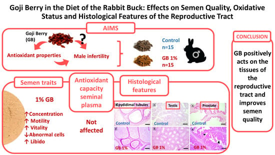
Goji Berry in the Diet of the Rabbit Buck: Effects on Semen Quality, Oxidative Status and Histological Features of the Reproductive Tract

by
Gabriele Brecchia
1,†,
Gerald Muça
2,†,
Albana Munga
2,
Laura Menchetti
3,*,
Livio Galosi
3,*,
Giacomo Rossi
3,
Olimpia Barbato
4,
Grazia Pastorelli
1,
Stella Agradi
1,
Valentina Serra
1,
Majlind Sulçe
2,
Enkeleda Ozuni
2,
Luigj Turmalaj
2,
Marta Castrica
5,
Maria Rachele Ceccarini
6,
Federica Riva
1,
Bernard Fioretti
7,
Alda Quattrone
1,
Maria Laura Marongiu
8 and
Giulio Curone
1
1
Department of Veterinary Medicine and Animal Sciences, University of Milan, Via dell’Università 6, 26900 Lodi, Italy
2
Faculty of Veterinary Medicine, Agricultural University of Tirana, Kodër Kamëz, 1029 Tirana, Albania
3
School of Biosciences and Veterinary Medicine, University of Camerino, Via Circonvallazione 93/95, 62024 Matelica, Italy
4
Department of Veterinary Medicine, University of Perugia, Via San Costanzo 4, 06126 Perugia, Italy
5
Department of Comparative Biomedicine and Food Science, University of Padova, Agripolis, Viale dell’Università 16, 35020 Legnaro, Italy
6
Department of Pharmaceutical Science, University of Perugia, 06123 Perugia, Italy
7
Department of Chemistry, Biology and Biotechnologies, University of Perugia, Via dell’Elce di Sotto 8, 06123 Perugia, Italy
8
Department of Veterinary Medicine, University of Sassari, Via Vienna 2, 07100 Sassari, Italy
*
Authors to whom correspondence should be addressed.
These authors contributed equally to this work and share first authorship.
Antioxidants 2023, 12(11), 1959; https://doi.org/10.3390/antiox12111959
Submission received: 29 September 2023 / Revised: 25 October 2023 / Accepted: 30 October 2023 / Published: 2 November 2023

Abstract

:
Goji berry (GB) shows beneficial effects on human health, although its effects on the male rabbit have been little investigated. This study examines the impact of GB dietary supplementation on the semen traits, antioxidant capacity of seminal plasma, and histological features of the reproductive tract of rabbit buck. Eighteen rabbits were distributed into two dietary groups: one receiving a commercial feed (Control), and the other a feed supplemented with 1% of GB (Goji). After a nutritional adaptation period of 60 days, the animals were subjected to semen collection every 15 days. The semen traits, libido, antioxidant, and inflammatory parameters were collected and analyzed. The rabbits were sacrificed after 60 days, and tissues of the genital tract were analyzed. Compared to the Control group, the Goji group showed higher spermatozoa concentration, motility, and vitality (p < 0.05), as well as fewer abnormal spermatozoa and a higher libido (p < 0.1). Histological features such as functional activity and hyperplasia were improved by GB and correlated with some semen traits (p < 0.05). Conversely, antioxidant and anti-inflammatory parameters were unaffected by the diet. These findings suggest that GB acts on the tissues of the reproductive tract positively influencing semen quality, although further studies are needed to understand the effect on oxidative stress.

Graphical Abstract

1. Introduction

Goji berry (GB) (Lycium barbarum) has been used for thousands of years as food and medicine in Asian countries, especially in China [1]. In recent decades, this fruit has become popular also in Western countries where it is marketed as a functional food [2]. The berry is rich in protein, carbohydrates, dietary fiber, macro- and micronutrients while containing low levels of fat [3]. GB exhibits a wide range of health promoting effects for humans, including anti-aging [4], anticancer [5], immune enhancement [6], protective for various organs [7], and therapeutic against different diseases [8]. Moreover, GB shows significant antioxidant potential, preventing the production of reactive oxygen species (ROS) and, as a consequence, DNA, lipids, and protein damage, by metal chelation or interactions with other antioxidant molecules [9]. The antioxidant properties of GB can be attributed to the presence of several biologically active compounds including polysaccharides, flavonoids, quercetin in particular, and carotenoids such as lycopene [10,11,12].
These health-protective benefits of GB can be exerted in many contexts. Indeed, ROS are generated under various physiochemical conditions, and their balance with antioxidants is always necessary. In particular, the imbalance between ROS and antioxidants is also implicated in the alteration of reproductive functions, and it is linked to male infertility, a health issue that significantly increased in recent decades [13]. Several risk factors can induce oxidative stress and, thus, have a role in the reduction of fertility, such as lifestyle [14], work environment [15,16], dietary behaviors with an imbalance of the n-6/n-3 fatty acids ratio [17,18,19], stress [20], chronic diseases [21], and environmental pollutants [22,23].
The spermatozoa and testis of mammalian species are rich in long-chain polyunsaturated fatty acids that are essential for several physiological processes, such as spermatozoa maturation, motility, and acrosome reaction [24,25]. However, the high content of polyunsaturated fatty acids in the spermatozoa membrane and mitochondria makes it particularly vulnerable to free radical attack that initiates lipid peroxidation and results in ROS generation. ROSs intervene in various physiological processes such as spermatozoa maturation, motility, chemotaxis, capacitation, hyperactivation, and acrosome reaction. Nonetheless, when produced in excessive concentration, ROS can lead to several types of oxidative damage, resulting in the loss of functionality and, ultimately, the death of spermatozoa [26]. Thus, the oxidation of polyunsaturated fatty acids in spermatozoa can cause deleterious effects on semen quality (spermatozoa damage, compromised DNA integrity, and increased apoptosis) and functions (altered spermatozoa motility, morphological abnormalities, and compromised sperm–oocyte interactions), ultimately resulting in reduced fertility [14,27,28].
New innovative strategies can be employed to mitigate the risk of oxidative stress, thereby improving reproductive functions in both humans and animals. There is evidence that fruits and vegetables within the Mediterranean diet [29] and substances with antioxidant potential, such as polyphenols, flavonoids, carotenoids, l-carnitine, selenium, coenzyme Q10, ubiquinol, resveratrol, and vitamin C, can improve seminal plasma oxidative stress and semen quality [30,31,32]. Recently, studies have been performed in various animal models using diets supplemented with nutraceutical substances such as flaxseed, algae [19], bovine colostrum [33], and GB [34,35,36,37]. Regarding GB, the effect of its derived products has been investigated in vitro [38,39], while dietary supplements with GB have been primarily studied in rodent models [40,41,42,43,44]. However, the in vitro studies are not completely satisfactory because the tissue lacks innervation and vascularization, and the effect on spermatogenesis is not considered. On the other hand, standardization of diets in humans is difficult and, therefore, the utilization of experimental models for in vivo nutritional studies is essential. The rabbit is an excellent animal model to study different physiological functions, including the impact of nutritional supplementations and aspects related to reproduction [45,46,47,48,49]. It has already been employed in experimental trials to evaluate the effects of dietary supplementation with GB on reproductive performance, energy balance, and meat quality [50,51,52,53]. Conversely, only a few studies have evaluated the effect of GB on the male reproductive system of different species [54,55,56,57,58,59] and, in particular, the research activity in the rabbit buck is very limited. However, the rabbit buck is a good model to study mature spermatozoa because the semen can be easily and continuously collected using an artificial vagina without the sacrifice of the animal. In addition, the rabbit is also a valid model for studying semen alterations caused by infection and/or inflammation [60].
We hypothesized that GB supplementation may influence the male reproductive tract and have an impact on the oxidative stress of semen and its quality. Accordingly, for the first time, this study investigated the effect of GB dietary supplementation on the reproductive parameters of the rabbit buck using a multidisciplinary approach that included different analytical techniques. Semen quality parameters, total antioxidant capacity, the activity of antioxidant enzymes, and inflammatory cytokines in the seminal plasma, in addition to the histological features of the reproductive tract, were investigated.

2. Materials and Methods

2.1. Animals and Experimental Design

The experiment was performed at the Agricultural University of Tirana, Faculty of Veterinary Medicine, Albania. The experimental protocol was conducted with the permission of the Ministry of Agriculture and Rural Development, National Authority of Veterinary and Plants Protection (prot. 824/2021) of Albania. All efforts were made to minimize animal suffering and to use only the number of animals necessary to produce consistent results. Moreover, the responsible veterinarian for the farm assessed the health and welfare of the rabbits on a daily basis.
Thirty New Zealand white rabbit bucks, aged 7 months and weighing between 3.4 and 4.4 kg, were individually housed under controlled environmental conditions with a lighting schedule of 16 h of light and 8 h of darkness.
The rabbit bucks were randomly divided into two experimental groups (n = 15/group) according to their diets: Control group received a commercial feed, while the Goji group was fed a commercial feed supplemented with 1% of GB. The rabbits were provided 150 g/d of feed and had water ad libitum. The composition of the diets supplied to the rabbits is described in Table 1 and has been previously used in other studies [52,53,61].
Figure 1 schematizes the experimental design. Thirty animals were divided into two groups (n = 15) and subjected to a nutritional adaptation period of 60 days during which the bucks were also trained for semen collection using an artificial vagina. At the end of this period, 18 males skilled in semen collection were randomly selected from the two groups (n = 9 for each group). During a subsequent experimental period of 60 days (a full spermatogenic cycle), semen samples were collected every 15 days to evaluate several traits (i.e., volume, color, concentration, motility, live spermatozoa, and anomalies), as well as libido. Body weight (BW) measurements were taken before the beginning of the nutritional adaptation and training period, at the beginning of the experimental period (0 d), and at the time of slaughter (60 d).

2.2. Sampling of Semen and Reproductive Organs

Throughout the experimental period, a total of five semen samples per rabbit buck were collected at specific time intervals (days 0, 15, 30, 45, and 60; Figure 1). The semen samples were collected using an artificial vagina with a maintained temperature range of 38–40 °C, regularly monitored and adjusted after each ejaculation. Following collection, the samples were divided into two aliquots: one was used to perform the spermatozoa quality evaluation (spermiogram) and the second was centrifuged at 3500 rpm for 15 minutes (Centrifuge 5418 R, Hamburg, Germany). Seminal plasma was recovered, placed in Eppendorf tubes (Eppendorf, Hamburg, Germany), and stored at −20 °C until the determination of the oxidative capacity and inflammatory status. Libido was calculated as “reaction time” by measuring the time (in seconds) between the introduction of the “teaser” doe into the male’s cage and the first ejaculation [63].
At the end of the trial, the rabbits were sacrificed. The testes, epididymis, vesicle glands, prostate, and bulbourethral glands were accurately collected. The samples were fixed in 10% buffered formalin for histological evaluation.

2.3. Seminal Quality Assessment

After collection, the semen was immediately subjected to analyses to determine the following seminal traits:
  • Volume (mL): determined by graduated tubes;
  • Color: defined as milky (normal), cream, yellowish, or transparent;
  • Presence of gel (yes or no);
  • Concentration (106 spermatozoa/mL): measured with the direct cell count method using a Burker chamber with a 40× objective after dilution of the semen 1:100;
  • Motility (%): evaluated by placing two drops of fresh semen on a warm microscope slide and covering it with a glass cover slip. The percentage of motility was calculated after the evaluation of at least 10 microscopic fields with 100× magnification and 10 microscopic fields with 400× magnification;
  • Live spermatozoa (%): determined using an eosin–nigrosin blue staining mixture and counting 200 cells. Specifically, 10 µL of undiluted semen was combined with 10 µL of eosin–nigrosin in a 1 mL Eppendorf tube (Eppendorf, Hamburg, Germany), gently mixed, and placed on a microscopic slide for evaluation. Cells that excluded the eosin stain, appearing white, were classified as live cells, while those with compromised or damaged membranes, colored with eosin and appearing pink, were considered dead;
  • Abnormal spermatozoa (%): determined using an eosin–nigrosin blue staining mixture and counting 200 cells. Abnormalities were evaluated in all cases, focusing on the abnormal head and tail defects.

2.4. Oxidative and Inflammatory Status of Seminal Plasma

The total antioxidant capacity to ROS (mM Trolox equivalents) was analyzed in the seminal plasma samples using the commercial kit Antioxidant Assay Kit (catalog no. 709001; Cayman Chemical, Ann Arbor, MI, USA). The principle of the assay is the ability of antioxidants present within a sample to inhibit the oxidation of ABTS® (2,2’-azino-di-3-ethylbenzthiazoline sulphonate) to ABTS®+ by metmyoglobin. The antioxidants cause suppression of absorbance at 750 nm or 405 nm proportionally to their concentration; this capacity to prevent ABTS® oxidation is compared to that of standard Trolox, a water-soluble tocopherol analogue, and is expressed as mM Trolox equivalent (mM TE eq.). The range of detection for this test was 0.068–0.495 mM TE eq.
The activity of three antioxidant enzymes in rabbit seminal plasma was determined with an enzyme-linked immunosorbent assay (ELISA) using commercial kits following the manufacturer’s instructions: catalase (CAT; Catalase Assay Kit, catalog no. 707002; Cayman Chemical, Ann Arbor, MI, USA), superoxide dismutase (SOD; Superoxide Dismutase Assay Kit, catalog no. 706002; Cayman Chemical, Ann Arbor, MI, USA), and glutathione peroxidase (GPx; catalog no. 703102; Cayman Chemical, Ann Arbor, MI, USA).
The Catalase Assay Kit is based on the reaction of the enzyme CAT with methanol in the presence of an optimal H2O2 concentration. This reaction produces formaldehyde which is measured colorimetrically with the chromogen Purpald that forms a bicyclic heterocycle with aldehydes, changing from colorless to purple upon oxidation.
The Superoxide Dismutase Assay Kit measures all three types of SOD (Cu/Zn, mn, and FeSOD) using a tetrazolium salt for the detection of superoxide radicals generated by xanthine oxidase and hypoxanthine. One unit of SOD is defined as the amount of enzyme needed to exhibit 50% dismutation of the superoxide radical. The range of detection for this test was 0.005–0.050 U/mL.
The Glutathione Peroxidase Assay Kit measures the activity of GPx in the sample indirectly by a coupled reaction with glutathione reductase; the reduction of hydroperoxide by GPx produces oxidized glutathione which is recycled to its reduced state by glutathione reductase and NADPH (subsequently oxidized to NADP+). One unit of GPx is defined as the amount of enzyme that oxidizes 1.0 nmol of NADPH to NADP+ per 1 min at 25 °C.
The concentration of interleukin-1β (IL-1β) was detected using a commercial ELISA test (Rabbit IL-1β ELISA Kit; FineTest, Wuhan Fine Biotech Co., Ltd., Wuhan, China) based on a sandwich binding in which capture antibody was pre-adsorbed on the surface of microtiter wells. The colorimetric reaction, catalyzed by streptavidin-conjugated horseradish peroxidase (HRP), produced a yellow product, which was proportional to the amount of target molecules present in the sample. The range of detection for this test was 15.625–1000 pg/mL.
The absorbance values of all assays were measured using a spectrophotometer (Epoch Biotek, Agilent Technologies Inc., Santa Clara, CA, USA) at the wavelengths established in each manufacturer’s protocol.

2.5. Histological Analysis

The histological examination was performed on the male genital tracts and specifically on the testis, epididymis, seminal vesicles, prostate, and bulbourethral glands. Samples from one rabbit belonging to the Goji group were not evaluated because of technical problems. Briefly, after the rabbits’ sacrifice, the abovementioned organs were extracted and fixed in a 10% neutral buffered formalin solution. The tissue samples were then processed for histological examination with paraffin embedding. Three micrometer-thick sections from paraffin blocks of each sample were stained by hematoxylin and eosin technique. Stained preparations were investigated with an optical microscope (Leica DM2500, Wetzlar, Germany) using 20× magnification. The microscopic evaluation of each sample was conducted by a pathologist who was totally unaware of the division of the two experimental groups, having no information regarding whether the observed sample belonged to the group of treated rabbits or the control group. For each organ sampled, the histological score took into consideration the following parameters: phlogosis, degeneration, functional activity, hyperplasia, and necrosis. To assess the severity of each parameter, a scoring system ranging from 0 to 3 points was employed. This scale allowed for the evaluation of the progressive severity of each parameter, as indicated below.
Phlogosis: 0 = 5–19; 1 = 20–49; 2 = 50–100; 3 = >100 leucocytes per high-power field (40×).
Degeneration: 0 = absence of cellular damage; 1 = presence of cytoplasmic vacuolization in some epithelial or neuronal cells; 2 = diffuse cellular vacuolization; 3 = diffuse vacuolization and necrosis/with areas of epithelial cells detachment (groups of cells with microerosive status)/neuronal cells loss.
Functional activity:
(A) For prostate, bulbourethral glands, and spermatic vesicles: 0 = glands (tubule-alveolar) with alveoli lined by cuboidal epithelium, collecting ducts lined by cuboidal or columnar epithelium without any trace of secretum; 1 = alveoli lined with cuboidal epithelium, collecting ducts lined with cuboidal epithelium, with a trace of secretum inside some of the alveoli; 2 = alveoli lined with columnar epithelium, hyperplastic epithelium, collecting ducts lined with columnar epithelium with the presence of secretum; 3 = alveoli lined with hyperplastic columnar epithelium, collecting ducts lined with columnar epithelium, with abundant secretum and corpora amylacea.
(B) For testes and epididymis: 0 = Developing spermatogenic cells are only seen in several tubules, and the cell density is increased in the intratubular space. In other tubules, Sertoli cells line the base of the tubules, and spermatogonia are rarely seen in the tubules. The epididymis appears to be characterized as having over 30% empty tubules. The tubules with spermatozoa appear to be characterized by cells that are not very concentrated and localized in the central part of the tubules. 1 = Developing spermatogenic cells are seen in more than 50% of tubules, and the cell density is increased in the intratubular space. The epididymis appears to be characterized by having approximately 20% empty tubules. The tubules with spermatozoa appear to be characterized by cells that are not very concentrated and localized in the central part of the tubules. 2 = Developing spermatogenic cells are seen in more than 60% of tubules, and the cell density is increased in the intratubular space. The epididymis appears to be characterized as having approximately 10% empty tubules. The tubules with spermatozoa appear to be characterized by very concentrated cells and homogeneously localized within the tubules. 3 = Developing spermatogenic cells are seen in all spermatogenic tubules of the rete testis, and the cell density is very abundant in the intratubular space. The epididymis appears to be characterized by tubules that are all full, and the spermatozoa form a very concentrated ball of sperm in the tubule, which appears totally full. The Leydig cells that are located between the seminiferous tubules are evident and gathered in small groups, leaving only a few irregular polyhedral cells in rows along with the blood vessels.
Hyperplasia:
(A) For prostate, bulbourethral glands, and spermatic vesicles: 0 = normal aspect of the epithelium, cells in monolayer; 1 = epithelial cells from cuboidal to columnar, with a trace of secretum; 2 = focal area of stratified columnar epithelium and a diffuse presence of secretum; 3 = diffuse increase in the number of columnar and stratified glandular epithelial cells, with an abundance of secretum. The proliferating epithelium may form papillary structures with supporting stroma and an extension into the glandular lumen. In both scores 2, and 3, there is nuclear crowding, occasional mitotic figures, piling up, and slight basophilia of the hyperplastic epithelium.
(B) For testes and epididymis: 0 = Normal Sertoli cells line the base of the tubules, and spermatogonia are rarely seen in the tubules. The epididymal tubules show a normal cuboidal or flattened epithelium. 1 = Focal areas of Sertoli hyperplastic cells line the base of the tubules, and spermatogonia are normally seen in the tubules. The epididymis tubules show a normal cuboidal epithelium. 2 = Diffuse hyperplastic Sertoli cells line the base of the tubules, and spermatogonia and spermatids are normally seen in the tubules. The epididymis tubules show a normal cuboidal epithelium with areas of columnar epithelium. 3 = Diffuse hyperplastic Sertoli cells line the base of the tubules, and spermatogonia and spermatids are normally seen in the tubules. The epididymis tubules show a diffuse columnar hyperplastic epithelium.
Necrosis: 0 = absence of necrosis; 1 = few and small areas of necrosis; 2 = diffuse but well-defined areas of tissue necrosis; 3 = diffuse and coalescing areas of tissue necrosis.

2.6. Statistical Analyses

The normality was verified with the Kolmogorov–Smirnov test, while diagnostic graphs were used to identify outliers. Root square transformation was used for CAT, GPx, IL-1β, abnormal spermatozoa, and reaction time; arcsine transformation was used for motility, while the complement to 100 was calculated for live cells percentage. The raw data are reported in the tables as the means and standard errors (SEs). Two values for the CAT, 1 for GPx, 1 for IL-1β, and 2 values for the reaction time were considered outliers and eliminated. Data were then analyzed using generalized estimating equations assuming an exchangeable working correlation matrix and evaluating the effect of group (2 levels: Control and Goji), time (5 levels), and their interaction. The time was included as a repeated measure and rabbits as subjects. Sidak correction was used for multiple comparisons. The normal distribution with identity link was used for the parameters related to the oxidative status, volume, concentration, motile cells, and abnormal spermatozoa; gamma with log link was used for reaction time, whereas negative binomial distribution with log link was used to analyze live cells. Color differences of the semen were analyzed at each time using Fisher’s exact tests.
The associations between the group and histological scores were evaluated with the eta coefficient. The eta is a measure of nominal-by-interval association that ranges from 0 to 1, with 0 indicating no association among the variables, and values close to 1 indicating a high degree of association [64]. In particular, as a measure of the effect size, the eta was interpreted as a small association if it was <0.3, medium if 0.3 ≤ eta < 0.5, and large if eta was ≥ 0.5 [65]. Moreover, eta2 (the percent of variance in the dependent variable explained by the group variable [64] was calculated for large effect sizes. The results of the Fisher’s exact and z tests (that compare the column proportions) are also reported.
Finally, the correlations between seminal parameters, TAC, antioxidant enzymes, interleukin, and histological scores were evaluated using Spearman’s rho coefficient (ρ). The correlation was considered poor if ρ < |0.3|, medium if |0.3|≤ ρ < |0.5|, and large if ρ ≥|0.5| [66]. Histological scores were correlated with the mean values of the other parameters. Necrosis and phlogosis were not included because they were constant.
Statistical analyses were performed with SPSS Statistics version 25 (IBM, SPSS Inc., Chicago, IL, USA). We defined p < 0.05 as significant and 0.05 ≤ p < 0.1 as a trend.

3. Results

3.1. Clinical Evaluation and Body Weight

The clinical status and welfare of rabbit bucks, daily evaluated by the responsible veterinarian, were good during the entire trial period (120 days: nutritional adaptation and experimental period) in both groups. The rabbit’s body weights increased over time (p < 0.001), and at slaughter they were higher in the Goji group than in the Control (4.3 ± 0.1 kg and 4.6 ± 0.1 kg for Control and Goji groups, respectively; p = 0.007). We assumed that the palatability of the feed supplemented with GB was the same as the Control feed, given that rabbits from both groups consumed the entire daily administered feed.

3.2. Semen Quality Assessment

The semen volume was not influenced by either group or time, while a significant group and/or time effect was found for the other parameters (Table 2). Pairwise comparisons showed no differences between groups within each time point. However, the Goji group showed higher marginal means for concentration, motility, and live spermatozoa, whereas the percentage of abnormal spermatozoa was lower than the Control group (for all: p < 0.05). Abnormal spermatozoa, regardless of the group, also increased over time (p < 0.001). Finally, the reaction time progressively reduced over time (p < 0.001) and tended to be the lowest in the Goji group (p < 0.1), indicating an improvement in libido.
Regarding the color, no differences between the groups were found at any time point (Figure S1).

3.3. Antioxidant and Inflammatory Parameters in Seminal Plasma

The values for TAC, antioxidant enzymes, and IL-1β at different time points are reported in Table 3. No significant differences due to the nutritional treatment were found for any analyzed parameter. The interaction group × time was significant for TAC, CAT, and SOD (p < 0.01). The TAC values for the Control group increased at the last time point (p < 0.05), while in the Goji group, the increase was not significant; however, pairwise comparisons did not show differences between groups at any time point. Similarly, CAT and SOD showed a significant interaction effect but, perhaps due to the high variability of the values, the pairwise comparisons did not show significant differences between the groups at any time point.

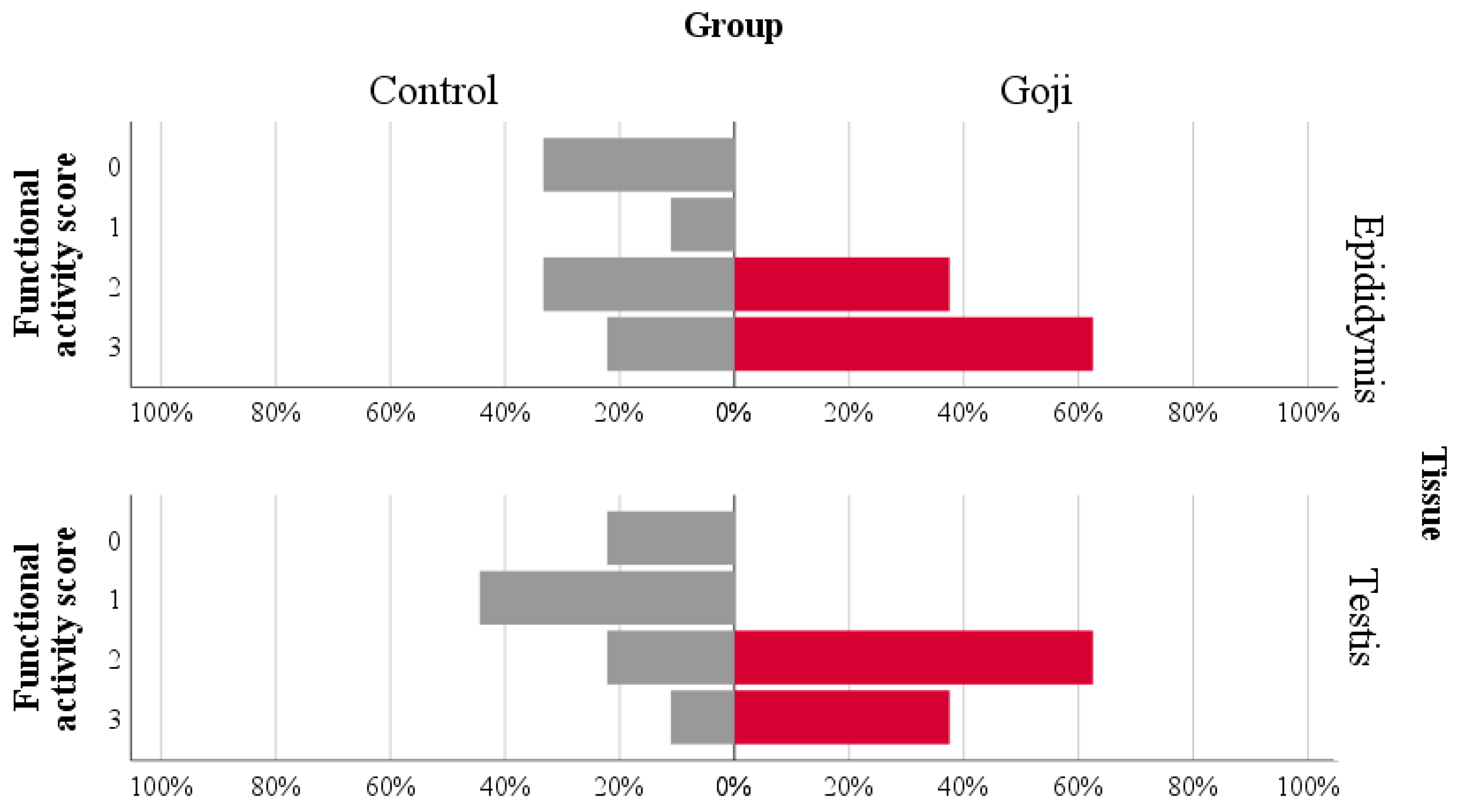
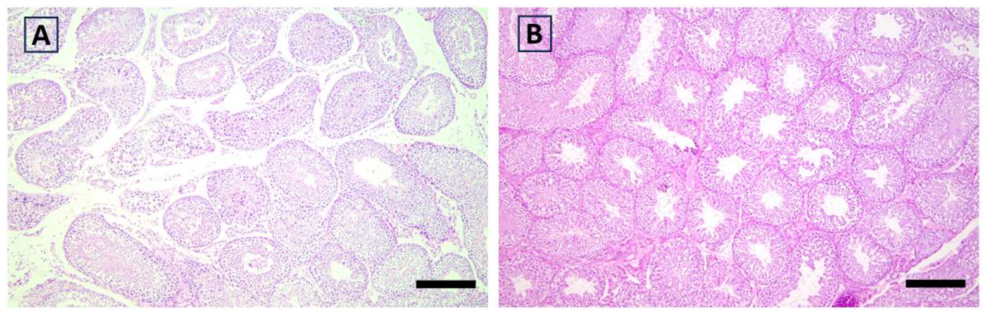
3.4. Histological Analysis

The eta coefficient indicated a large association of functional activity scores with the group in the epididymis (eta = 0.543) and testis (eta = 0.611; Figure 2, Figure 3, Figure 4 and Figure 5). The greater proportion of the highest scores in the Goji group explained 74% and 78% of the variance in the epididymis and testis, respectively. In the testis, a medium association with the group was also found for hyperplasia (eta = 0.457), as most of the samples (78%) in the Control group had scores = 0, while a more heterogeneous distribution between the highest scores was found in the Goji group (Table S1). No other parameters were associated with nutritional treatment (Table S1).

3.5. Correlations between Seminal Parameters, TAC, Antioxidant Enzymes, and Interleukin-1β

A poor (ρ < 0.3) although significant positive correlation was found between TAC and volume, GPx and concentration, GPx and rate of live spermatozoa, and IL-1β and percentage of live spermatozoa. Conversely, GPx was negatively correlated with the percentage of abnormal spermatozoa (Table 4).
Table 5 shows the correlations between seminal quality, oxidative stress, inflammatory status parameters and histological scores of the epididymis and testes. The epididymal degeneration score was positively correlated with volume and live spermatozoa, whereas those of the testis was negatively correlated with the volume. Functional activity was negatively correlated with abnormal spermatozoa both in the epididymis and testis. The functional activity of the testes also showed a positive correlation with motility and a negative one with SOD. Finally, hyperplasia of the epididymis was positively correlated with live spermatozoa, TAC, and CAT (for all p < 0.05). The correlations with histological scores of the other glands are shown in Table S2. Among these, the positive correlation of the functional activity score of the bulbourethral glands with motility and live spermatozoa, as well as the negative correlation of the functional activity score of the prostate with abnormal spermatozoa, can be mentioned (for all p < 0.05).

4. Discussion

This study investigated, for the first time, the effect of GB on semen quality, antioxidant and inflammatory capacity of seminal plasma, and histological structure of reproductive organs in rabbit bucks. The findings suggest that GB dietary supplementation can improve several semen traits (concentration, motile cells, live cells, and abnormal spermatozoa), although no effect on parameters related to oxidative status was found. However, the histological analyses showed noticeable effects of the GB-supplemented diet on the functional activity of the testis, as well as on the degree of hyperplasia of the epididymis and prostate, which could be linked to the better semen quality. These preliminary results suggest that GB could affect rabbit fertility and can be used for animal nutrition, although further studies are required to understand its mechanism of action and determine the optimal dose to be used.
In this pioneering work regarding male rabbits, the selection of the dose to be administered was based on previous studies conducted on female rabbits, which had shown interesting results on various aspects of reproduction and productivity [50,51,52]. The primary notable outcome of this study is thus that the 1% integration of GB did not induce any health issues in the animals. The feed proved to be palatable and was consumed by the rabbits without any problem. These findings confirm previous studies, as GB appeared to be safe at various dosages [11]. Only mild toxicity [8] and adverse effects such as urticarial-like reactions related to its consumption have been reported in humans [67]. Furthermore, the histological results demonstrated that, at the level of the genital organs of rabbits, GB not only does not cause alterations but also increases functional activity. The effects of the berry on the male reproductive tract histology under physiological conditions are little known. On the contrary, experimental models of various pathological conditions, especially in rodents, show that GB supplementation can improve the histological characteristics of the reproductive tissues, as well as the spermatic functions [36,43,44,68,69]. In the streptozotomycin-induced diabetic rats model, GB treatment ameliorated the damage in the spermatogenic tubules, increased the number of spermatogenic cells, and suppressed apoptosis in the testes [68]. Lycium barbarum polysaccharides ameliorated the damage in the seminiferous tubules induced by cadmium both in rats and mice [36,44]. The pretreatment with GB mitigated the degenerative changes in seminiferous tubules and improved hormonal secretion, spermatozoa concentration, and motility, as well as oxidative capacity in doxorubicin-induced testicular toxicity in rats [43]. Finally, our findings are in agreement with other studies performed on the rabbit does and fattening rabbits [50,51,61]. The findings from these studies highlight an improvement in the mitigation of the damage caused by various stressors on the reproductive system tissues; however, it remains uncertain whether this improvement is due to a direct action of the active biological substances of the berry or if it is an indirect effect, perhaps mediated by an increase in antioxidant activity within the reproductive tract, or a combination of both.
Regardless of the mechanism of action, our findings demonstrate that parameters related to functional activity, hyperplasia, and tissue degeneration in the reproductive tract of bucks are correlated with several semen traits such as motility, number of live and abnormal spermatozoa. As a matter of fact, rabbits supplemented with GB also had better semen quality in terms of concentration, motility, vitality, and abnormal spermatozoa. Although as a trend, libido also improved. To our knowledge, the effects of GB dietary supplementation on the characteristics of semen in physiological conditions have been previously studied only by Yang et al. [58]. These authors evaluated a dried GB supplementation for 160 days in boars, finding an improvement in spermatozoa motility, concentration, and abnormality rate. On the other hand, several studies have assessed both in vivo and in vitro the impact of GB in different pathological models and various animal species, including mice, rats, and goats [59,70,71].
The positive effects of GB on seminal quality are often attributed to its antioxidant effect [14,18,23,72]. However, our results do not support this hypothesis as no differences between groups were found neither in the parameters indicating oxidative status nor in the cytokine. Specifically, the nutritional treatment did not affect the TAC, CAT, SOD, GPx, and IL-1β concentrations in the seminal plasma of the rabbits. Some differences between groups were found for the TAC, CAT, and SOD patterns over time, although these appear weak and inconclusive. The disagreement with previous studies may be attributed to the dose of the berry and/or the nutritional adaptation period applied in our study that may not have been sufficient to induce specific and significant effects on the oxidative status. Furthermore, species-specific characteristics and the use of the whole fruit rather than its derivatives could be influential factors. It has also been demonstrated that the GB polysaccharides administered for 5 days with dosages of 200, 400, and 600 mg/Kg improved spermatozoa mobility, concentration, percentage of normal spermatozoa morphology, and SOD concentration in mice treated with cyclophosphamide [73]. GB polysaccharides administered at a dose of 200 mg/Kg for 7 days induced an increase in antioxidant enzyme concentrations and profound protective effects against spermatogenic injury induced by bisphenol A in mice [70]. Luo et al. [55] demonstrated that GB polysaccharides, administered at a dosage of 200 mg/Kg for 15 days, enhanced sexual ability, increased antioxidant activity, and improved seminal quality in rat testes models subjected to heat-induced damage. In many studies, the improvement in semen quality of animals receiving GB was also associated with an increase in the concentration of reproductive hormones such as testosterone and LH and a reduction in the expression of genes promoting apoptosis such as BAX [40,44,55,70,74,75].
Goji is rich in polyphenols, flavonoids, carotenoids, and vitamin C, and it also exhibits anti-inflammatory, antisclerotic, protective, and regulatory properties, and t could exert epigenetic and gut-microbiota modulations [29,76,77]. In particular, it has been demonstrated that flavonoids, such as quercetin, improve male performance by acting on crucial mitochondrial processes of spermatozoa and enhancing the activity of the hypothalamic–pituitary–testicular axis [76,78,79]. Lycopene, a carotenoid also found in other fruits, such as ripe tomatoes and F. sellowiana [12,80], accumulates in seminal prostasomes and seems to improve sperm count and reduce the number of abnormal spermatozoa [80,81], playing a role in several physiological processes, mainly with protective effects (antioxidants and anti-inflammatory) [80,82]. Moreover, beyond the specific effects of GB, the enhancement in reproductive functions could reflect the overall improved health associated with the consumption of these substances [29]. It could be speculated that GB supplementation may influence the secretion of reproductive hormones and/or their receptors, consequently, impacting the histological conditions of the male reproductive tract tissues and improving the spermatozoa concentration and mobility, apoptosis rate, and number of abnormal cells. Hormonal changes could also explain the improved libido in the Goji group. In the present study, an evaluation of the concentration of reproductive hormones was not conducted, although the histological results clearly demonstrate that the diet supplemented with GB improves the histological characteristics of the tissues of the reproductive organs and associated glands.
Thus, the main limitation of the study is the lack of evaluation of hormones, receptors, enzymes, and genes directly involved in reproductive functions. Moreover, a higher supplementation dosage of the berry and/or a longer nutritional adaptation period can be employed to evaluate the antioxidant and anti-inflammatory effect of GB and to understand any alternative mechanisms of action on the reproductive system of rabbit bucks. Finally, we recognize that the incorporation of goji in animal feed may be intended for a niche market; however, a cost–benefit analysis should still be carried out.

5. Conclusions

The dietary administration of GB improved several semen traits, including spermatozoa motility and concentration, as well as live and abnormal cells. Histological changes were also observed, including increased functional activity in the testis, hyperplasia of the epididymis and prostatic epithelium (with increased intra-acinar content of corpora amylacea), along with a higher concentration of spermatozoa within dilated epididymal tubules. Conversely, its effect on the antioxidative and inflammatory capacity of seminal plasma has not been demonstrated. Further studies may involve a higher percentage of GB diet integration and a longer nutritional adaptation period as the antioxidant effect could be dose and time dependent.
The use of this natural and nutraceutical product is safe and does not have particular side effects both at the systemic and local levels. For this reason, GB could potentially be a new ingredient for the rabbit feed. Dietary supplementation with the berry could be an innovative strategy to increase the fertility and welfare of animals, as well as the profitability of farmers, although a cost analysis should be performed. Further studies are in progress to understand GB’s mechanism of action including its effects on the antioxidant and inflammatory capacity of reproductive tract tissues and reproductive hormones.

Supplementary Materials

The following supporting information can be downloaded at: https://www.mdpi.com/article/10.3390/antiox12111959/s1, Figure S1: Evaluation of the color of the semen samples. The values are the percentages of the samples for each time point; Table S1: Crosstabulation of the histological scores by group according to the tissue and parameter; Table S2: Spearman’s rho coefficient for the correlations between histological scores evaluated in urethral bulb glands, prostate and seminal vesicles, and seminal parameters, total antioxidant capacity, antioxidant enzymes, and interleukin-1β.

Author Contributions

Conceptualization, G.B., G.M. and M.S.; Data curation, L.M. and V.S.; Formal analysis, G.B., L.M., L.G., G.R., G.P., V.S. and M.C.; Funding acquisition, G.B., G.M., F.R. and M.L.M.; Investigation, G.M., A.M., L.G., G.R., M.S., E.O., L.T. and M.R.C.; Methodology, G.B., A.M., L.M. and G.C.; Project administration, G.B., M.S., B.F. and G.C.; Resources, G.B., G.P., F.R. and M.L.M.; Software, L.M., O.B. and V.S.; Supervision, G.B., G.M., M.S. and G.C.; Validation, L.M., L.G., F.R. and B.F.; Visualization, L.M., L.G., G.R. and M.C.; Writing—original draft, G.B., G.M., L.M., O.B., S.A. and G.C.; Writing—review and editing, A.M., L.G., G.R., V.S., E.O., L.T., B.F., A.Q. and M.L.M. All authors have read and agreed to the published version of the manuscript.

Funding

This research was partially supported by FAR 2020 (Fondo di Ateneo per la Ricerca 2020) of University of Sassari.

Institutional Review Board Statement

The experimental protocol was conducted with the permission of the Ministry of Agriculture and Rural Development, National Authority of Veterinary and Plants Protection (prot. 824/2020) of Albania.

Informed Consent Statement

Not applicable.

Data Availability Statement

The data presented in this study are available on request from the corresponding author.

Conflicts of Interest

The authors declare no conflict of interest.

References

  1. Cheng, J.; Zhou, Z.W.; Sheng, H.P.; He, L.J.; Fan, X.W.; He, Z.X.; Sun, T.; Zhang, X.; Zhao, R.J.; Gu, L.; et al. An Evidence-Based Update on the Pharmacological Activities and Possible Molecular Targets of Lycium barbarum Polysaccharides. Drug Des. Devel. Ther. 2015, 9, 33–78. [Google Scholar] [CrossRef] [PubMed]
  2. Xin, T.; Yao, H.; Gao, H.; Zhou, X.; Ma, X.; Xu, C.; Chen, J.; Han, J.; Pang, X.; Xu, R.; et al. Super Food Lycium barbarum (Solanaceae) Traceability via an Internal Transcribed Spacer 2 Barcode. Food Res. Int. 2013, 54, 1699–1704. [Google Scholar] [CrossRef]
  3. Tian, X.; Liang, T.; Liu, Y.; Ding, G.; Zhang, F.; Ma, Z. Extraction, Structural Characterization, and Biological Functions of Lycium barbarum Polysaccharides: A Review. Biomolecules 2019, 9, 389. [Google Scholar] [CrossRef] [PubMed]
  4. Gao, Y.; Wei, Y.; Wang, Y.; Gao, F.; Chen, Z. Lycium Barbarum: A Traditional Chinese Herb and A Promising Anti-Aging Agent. Aging Dis. 2017, 8, 778–792. [Google Scholar] [CrossRef]
  5. Ceccarini, M.R.; Vannini, S.; Cataldi, S.; Moretti, M.; Villarini, M.; Fioretti, B.; Albi, E.; Beccari, T.; Codini, M. Effect of Lycium barbarum Berries Cultivated in Umbria (Italy) on Human Hepatocellular Carcinoma Cells. J. Biotechnol. 2016, 231, S26–S27. [Google Scholar] [CrossRef]
  6. Bo, R.; Sun, Y.; Zhou, S.; Ou, N.; Gu, P.; Liu, Z.; Hu, Y.; Liu, J.; Wang, D. Simple Nanoliposomes Encapsulating Lycium barbarum Polysaccharides as Adjuvants Improve Humoral and Cellular Immunity in Mice. Int. J. Nanomed. 2017, 12, 6289–6301. [Google Scholar] [CrossRef]
  7. Lian, Y.Z.; Lin, I.H.; Yang, Y.C.; Chao, J.C.J. Gastroprotective Effect of Lycium barbarum Polysaccharides and C-Phyocyanin in Rats with Ethanol-Induced Gastric Ulcer. Int. J. Biol. Macromol. 2020, 165, 1519–1528. [Google Scholar] [CrossRef]
  8. Kwok, S.S.; Bu, Y.; Lo, A.C.Y.; Chan, T.C.Y.; So, K.F.; Lai, J.S.M.; Shih, K.C. A Systematic Review of Potential Therapeutic Use of Lycium barbarum Polysaccharides in Disease. Biomed. Res. Int. 2019, 2019, 4615745. [Google Scholar] [CrossRef]
  9. Ma, Z.F.; Zhang, H.; Teh, S.S.; Wang, C.W.; Zhang, Y.; Hayford, F.; Wang, L.; Ma, T.; Dong, Z.; Zhang, Y.; et al. Goji Berries as a Potential Natural Antioxidant Medicine: An Insight into Their Molecular Mechanisms of Action. Oxid. Med. Cell Longev. 2019, 2019, 2437397. [Google Scholar] [CrossRef]
  10. Vidović, B.B.; Milinčić, D.D.; Marčetić, M.D.; Djuriš, J.D.; Ilić, T.D.; Kostić, A.; Pešić, M.B. Health Benefits and Applications of Goji Berries in Functional Food Products Development: A Review. Antioxidants 2022, 11, 248. [Google Scholar] [CrossRef]
  11. Amagase, H.; Farnsworth, N.R. A Review of Botanical Characteristics, Phytochemistry, Clinical Relevance in Efficacy and Safety of Lycium barbarum Fruit (Goji). Food Res. Int. 2011, 44, 1702–1717. [Google Scholar] [CrossRef]
  12. Piscopo, M.; Tenore, G.C.; Notariale, R.; Maresca, V.; Maisto, M.; de Ruberto, F.; Heydari, M.; Sorbo, S.; Basile, A. Antimicrobial and Antioxidant Activity of Proteins from Feijoa sellowiana Berg. Fruit before and after in Vitro Gastrointestinal Digestion. Nat. Prod. Res. 2020, 34, 2607–2611. [Google Scholar] [CrossRef] [PubMed]
  13. Mannucci, A.; Argento, F.R.; Fini, E.; Coccia, M.E.; Taddei, N.; Becatti, M.; Fiorillo, C. The Impact of Oxidative Stress in Male Infertility. Front. Mol. Biosci. 2021, 8, 799294. [Google Scholar] [CrossRef] [PubMed]
  14. Agarwal, A.; Virk, G.; Ong, C.; du Plessis, S.S. Effect of Oxidative Stress on Male Reproduction. World J. Mens. Health 2014, 32, 1088727. [Google Scholar] [CrossRef]
  15. Zorgniotti, A.W.; Sealfon, A.I.; Toth, A. Further Clinical Experience with Testis Hypothermia for Infertility Due to Poor Semen. Urology 1982, 19, 636–640. [Google Scholar] [CrossRef]
  16. Lancranjan, I.; Măicănesgu, M.; Rafailaj, E.; Klepsgh, I.; Popesgu, H.I. Gonadic Function in Workmen with Long-Term Exposure to Microwaves. Health Phys. 1975, 29, 381–383. [Google Scholar] [CrossRef] [PubMed]
  17. Safarinejad, M.R.; Safarinejad, S. The Roles of Omega-3 and Omega-6 Fatty Acids in Idiopathic Male Infertility. Asian J. Androl. 2012, 14, 514–515. [Google Scholar] [CrossRef]
  18. Gharagozloo, P.; Aitken, R.J. The Role of Sperm Oxidative Stress in Male Infertility and the Significance of Oral Antioxidant Therapy. Hum. Reprod. 2011, 26, 1628–1640. [Google Scholar] [CrossRef]
  19. Castellini, C.; Mattioli, S.; Signorini, C.; Cotozzolo, E.; Noto, D.; Moretti, E.; Brecchia, G.; Dal Bosco, A.; Belmonte, G.; Durand, T.; et al. Effect of Dietary N-3 Source on Rabbit Male Reproduction. Oxid. Med. Cell Longev. 2019, 2019, 3279670. [Google Scholar] [CrossRef]
  20. De Gennaro, L.; Balistreri, S.; Lenzi, A.; Lombardo, F.; Ferrara, M.; Gandini, L. Psychosocial Factors Discriminate Oligozoospermic from Normozoospermic Men. Fertil. Steril. 2003, 79, 1571–1576. [Google Scholar] [CrossRef]
  21. Glazer, C.H.; Bonde, J.P.; Eisenberg, M.L.; Giwercman, A.; Hærvig, K.K.; Rimborg, S.; Vassard, D.; Pinborg, A.; Schmidt, L.; Bräuner, E.V. Male Infertility and Risk of Nonmalignant Chronic Diseases: A Systematic Review of the Epidemiological Evidence. Semin. Reprod. Med. 2017, 35, 282–290. [Google Scholar] [CrossRef]
  22. Whorton, D.; Krauss, R.M.; Marshall, S.; Milby, T.H. Infertility in Male Pesticide Workers. Lancet 1977, 310, 1259–1261. [Google Scholar] [CrossRef]
  23. Selevan, S.G.; Borkovec, L.; Slott, V.L.; Zudová, Z.; Rubeš, J.; Evenson, D.P.; Perreault, S.D. Semen Quality and Reproductive Health of Young Czech Men Exposed to Seasonal Air Pollution. Environ. Health Perspect. 2000, 108, 887–894. [Google Scholar] [CrossRef] [PubMed]
  24. Retterstøl, K.; Hauger, T.B.; Tran, T.N.; Christophersen, B.O. Studies on the Metabolism of Essential Fatty Acids in Isolated Human Testicular Cells. Reproduction 2001, 121, 881–887. [Google Scholar] [CrossRef] [PubMed]
  25. Björkgren, I.; Alvarez, L.; Blank, N.; Balbach, M.; Turunen, H.; Laajala, T.D.; Toivanen, J.; Krutskikh, A.; Wahlberg, N.; Huhtaniemi, I.; et al. Targeted Inactivation of the Mouse Epididymal Beta-Defensin 41 Alters Sperm Flagellar Beat Pattern and Zona Pellucida Binding. Mol. Cell Endocrinol. 2016, 427, 143–154. [Google Scholar] [CrossRef]
  26. Kothari, S.; Thompson, A.; Agarwal, A.; du Plessis, S.S. Free Radicals: Their Beneficial and Detrimental Effects on Sperm Function. Indian. J. Exp. Biol. 2010, 48, 425–435. [Google Scholar]
  27. Aitken, R.J.; Baker, M.A. Oxidative Stress, Sperm Survival and Fertility Control. Mol. Cell Endocrinol. 2006, 250, 66–69. [Google Scholar] [CrossRef] [PubMed]
  28. Barati, E.; Nikzad, H.; Karimian, M. Oxidative Stress and Male Infertility: Current Knowledge of Pathophysiology and Role of Antioxidant Therapy in Disease Management. Cell. Mol. Life Sci. 2020, 77, 93–113. [Google Scholar] [CrossRef]
  29. Montano, L.; Maugeri, A.; Volpe, M.G.; Micali, S.; Mirone, V.; Mantovani, A.; Navarra, M.; Piscopo, M. Mediterranean Diet as a Shield against Male Infertility and Cancer Risk Induced by Environmental Pollutants: A Focus on Flavonoids. Int. J. Mol. Sci. 2022, 23, 1568. [Google Scholar] [CrossRef] [PubMed]
  30. Gambera, L.; Stendardi, A.; Ghelardi, C.; Fineschi, B.; Aini, R. Effects of Antioxidant Treatment on Seminal Parameters in Patients Undergoing in Vitro Fertilization. Arch. Ital. Urol. E Androl. 2019, 91, 187. [Google Scholar] [CrossRef] [PubMed]
  31. Majzoub, A.; Agarwal, A. Systematic Review of Antioxidant Types and Doses in Male Infertility: Benefits on Semen Parameters, Advanced Sperm Function, Assisted Reproduction and Live-Birth Rate. Arab. J. Urol. 2018, 16, 113–124. [Google Scholar] [CrossRef] [PubMed]
  32. Dutta, S.; Majzoub, A.; Agarwal, A. Oxidative Stress and Sperm Function: A Systematic Review on Evaluation and Management. Arab. J. Urol. 2019, 17, 87–97. [Google Scholar] [CrossRef] [PubMed]
  33. Castrica, M.; Menchetti, L.; Agradi, S.; Curone, G.; Vigo, D.; Pastorelli, G.; Di Giancamillo, A.; Modina, S.C.; Riva, F.; Serra, V.; et al. Effect of Bovine Colostrum Dietary Supplementation on Rabbit Meat Quality. Foods 2022, 11, 3433. [Google Scholar] [CrossRef] [PubMed]
  34. Liu, C.; Gu, J.; Ma, W.; Zhang, Q.; Song, M.; Ha, L.; Xu, X.; Jiao, H.; Huo, Z. Lycium Barbarum Polysaccharide Protects against Ethanol-Induced Spermiotoxicity and Testicular Degeneration in Immp2l+/− Mice. Andrologia 2020, 52, e13554. [Google Scholar] [CrossRef]
  35. Jeong, H.C.; Jeon, S.H.; Guan Qun, Z.; Bashraheel, F.; Choi, S.W.; Kim, S.J.; Bae, W.J.; Cho, H.J.; Ha, U.S.; Hong, S.H.; et al. Lycium chinense Mill Improves Hypogonadism via Anti-Oxidative Stress and Anti-Apoptotic Effect in Old Aged Rat Model. Aging Male 2020, 23, 287–296. [Google Scholar] [CrossRef]
  36. Varoni, M.V.; Gadau, S.D.; Pasciu, V.; Baralla, E.; Serra, E.; Palomba, D.; Demontis, M.P. Investigation of the Effects of Lycium barbarum Polysaccharides against Cadmium Induced Damage in Testis. Exp. Mol. Pathol. 2017, 103, 26–32. [Google Scholar] [CrossRef]
  37. Huang, X.; Yang, M.; Wu, X.; Yan, J. Study on Protective Action of Lycium barbarum Polysaccharides on DNA Imparments of Testicle Cells in Mice. Wei Sheng Yan Jiu 2003, 32, 599–601. [Google Scholar]
  38. Yang, Z.J.; Wang, Y.X.; Zhao, S.; Hu, N.; Chen, D.M.; Ma, H.M. SIRT 3 Was Involved in Lycium barbarum Seed Oil Protection Testis from Oxidative Stress: In Vitro and in Vivo Analyses. Pharm. Biol. 2021, 59, 1314–1325. [Google Scholar] [CrossRef] [PubMed]
  39. Wang, Y.; Zhao, H.; Sheng, X.; Gambino, P.E.; Costello, B.; Bojanowski, K. Protective Effect of Fructus lycii Polysaccharides against Time and Hyperthermia-Induced Damage in Cultured Seminiferous epithelium. J. Ethnopharmacol. 2002, 82, 169–175. [Google Scholar] [CrossRef]
  40. Shi, G.J.; Zheng, J.; Wu, J.; Qiao, H.Q.; Chang, Q.; Niu, Y.; Sun, T.; Li, Y.X.; Yu, J.Q. Protective Effects of Lycium barbarum Polysaccharide on Male Sexual Dysfunction and Fertility Impairments by Activating Hypothalamic Pituitary Gonadal Axis in Streptozotocin-Induced Type-1 Diabetic Male Mice. Endocr. J. 2017, 64, 907–922. [Google Scholar] [CrossRef]
  41. Luo, Q.; Cui, X.; Yan, J.; Yang, M.; Liu, J.; Jiang, Y.; Li, J.; Zhou, Y. Antagonistic Effects of Lycium barbarum Polysaccharides on the Impaired Reproductive System of Male Rats Induced by Local Subchronic Exposure to 60Co-γ Irradiation. Phytother. Res. 2011, 25, 694–701. [Google Scholar] [CrossRef]
  42. Yang, F.L.; Wei, Y.X.; Liao, B.Y.; Wei, G.J.; Qin, H.M.; Pang, X.X.; Wang, J.L. Effects of Lycium barbarum Polysaccharide on Endoplasmic Reticulum Stress and Oxidative Stress in Obese Mice. Front. Pharmacol. 2020, 11, 742. [Google Scholar] [CrossRef] [PubMed]
  43. Xin, Y.-F.; You, Z.-Q.; Gao, H.; Zhou, G.-L.; Chen, Y.; Yu, J.; Xuan, Y.-X. Protective Effect of Lycium barbarum Polysaccharides against Doxorubicin-Induced Testicular Toxicity in Rats. Phytother. Res. 2012, 26, 716–721. [Google Scholar] [CrossRef] [PubMed]
  44. Zhang, L.; Li, Q.; Zheng, G.; Chen, Y.; Huang, M.; Zhang, L.; Lin, X. Protective Effect of Lycium barbarum Polysaccharides against Cadmium-Induced Testicular Toxicity in Male Mice. Food Funct. 2017, 8, 2322–2330. [Google Scholar] [CrossRef]
  45. Zerani, M.; Boiti, C.; Dall’Aglio, C.; Pascucci, L.; Maranesi, M.; Brecchia, G.; Mariottini, C.; Guelfi, G.; Zampini, D.; Gobbetti, A. Leptin Receptor Expression and in Vitro Leptin Actions on Prostaglandin Release and Nitric Oxide Synthase Activity in the Rabbit Oviduct. J. Endocrinol. 2005, 185, 319–325. [Google Scholar] [CrossRef] [PubMed]
  46. Boiti, C.; Guelfi, G.; Zerani, M.; Zampini, D.; Brecchia, G.; Gobbetti, A. Expression Patterns of Cytokines, P53 and Nitric Oxide Synthase Isoenzymes in Corpora lutea of Pseudopregnant Rabbits during Spontaneous luteolysis. Reproduction 2004, 127, 229–238. [Google Scholar] [CrossRef]
  47. Cremonesi, P.; Curone, G.; Biscarini, F.; Cotozzolo, E.; Menchetti, L.; Riva, F.; Marongiu, M.L.; Castiglioni, B.; Barbato, O.; Munga, A.; et al. Dietary Supplementation with Goji Berries (Lycium barbarum) Modulates the Microbiota of Digestive Tract and Caecal Metabolites in Rabbits. Animals 2022, 12, 121. [Google Scholar] [CrossRef]
  48. Boiti, C.; Guelfi, G.; Zampini, D.; Brecchia, G.; Gobbetti, A.; Zerani, M. Regulation of Nitric Oxide Synthase Isoforms and Role of Nitric Oxide during Prostaglandin F2alpha-Induced Luteolysis in Rabbits. Reproduction 2003, 125, 807–816. [Google Scholar] [CrossRef]
  49. Zerani, M.; Parillo, F.; Brecchia, G.; Guelfi, G.; Dall’Aglio, C.; Lilli, L.; Maranesi, M.; Gobbetti, A.; Boiti, C. Expression of Type I GNRH Receptor and in Vivo and in Vitro GNRH-I Effects in Corpora lutea of Pseudopregnant Rabbits. J. Endocrinol. 2010, 207, 289–300. [Google Scholar] [CrossRef]
  50. Andoni, E.; Curone, G.; Agradi, S.; Barbato, O.; Menchetti, L.; Vigo, D.; Zelli, R.; Cotozzolo, E.; Ceccarini, M.R.; Faustini, M.; et al. Effect of Goji Berry (Lycium barbarum) Supplementation on Reproductive Performance of Rabbit Does. Animals 2021, 11, 1672. [Google Scholar] [CrossRef]
  51. Brecchia, G.; Sulce, M.; Curone, G.; Barbato, O.; Canali, C.; Troisi, A.; Munga, A.; Polisca, A.; Agradi, S.; Ceccarini, M.R.; et al. Goji Berry (Lycium barbarum) Supplementation during Pregnancy Influences Insulin Sensitivity in Rabbit Does but Not in Their Offspring. Animals 2022, 12, 39. [Google Scholar] [CrossRef] [PubMed]
  52. Menchetti, L.; Curone, G.; Andoni, E.; Barbato, O.; Troisi, A.; Fioretti, B.; Polisca, A.; Codini, M.; Canali, C.; Vigo, D.; et al. Impact of Goji Berries (Lycium barbarum) Supplementation on the Energy Homeostasis of Rabbit Does: Uni- and Multivariate Approach. Animals 2020, 10, 2000. [Google Scholar] [CrossRef] [PubMed]
  53. Castrica, M.; Menchetti, L.; Balzaretti, C.M.; Branciari, R.; Ranucci, D.; Cotozzolo, E.; Vigo, D.; Curone, G.; Brecchia, G.; Miraglia, D. Impact of Dietary Supplementation with Goji Berries (Lycium barbarum) on Microbiological Quality, Physico-Chemical, and Sensory Characteristics of Rabbit Meat. Foods 2020, 9, 1480. [Google Scholar] [CrossRef] [PubMed]
  54. Jo, J.; Lee, S.H.; Lee, J.M.; Jerng, U.M. Semen Quality Improvement in a Man with Idiopathic Infertility Treated with Traditional Korean Medicine: A Case Report. Explor. J. Sci. Health 2015, 11, 320–323. [Google Scholar] [CrossRef] [PubMed]
  55. Luo, Q.; Li, Z.; Huang, X.; Yan, J.; Zhang, S.; Cai, Y.Z. Lycium barbarum Polysaccharides: Protective Effects against Heat-Induced Damage of Rat Testes and H2O2-Induced DNA Damage in Mouse Testicular Cells and Beneficial Effect on Sexual Behavior and Reproductive Function of Hemicastrated Rats. Life Sci. 2006, 79, 613–621. [Google Scholar] [CrossRef]
  56. Shi, G.J.; Zheng, J.; Wu, J.; Qiao, H.Q.; Chang, Q.; Niu, Y.; Sun, T.; Li, Y.X.; Yu, J.Q. Beneficial Effects of Lycium barbarum Polysaccharide on Spermatogenesis by Improving Antioxidant Activity and Inhibiting Apoptosis in Streptozotocin-Induced Diabetic Male Mice. Food Funct. 2017, 8, 1215–1226. [Google Scholar] [CrossRef]
  57. Liu, C.L.; Zhang, Q.; Zhang, S.H.; Mu, C.L.; Yao, P.; Jiao, H.Y.; Xu, X.; Huo, Z.H. Lycium Barbarum Polysaccharide Reduces Testicular Spermatogenic Injury in Immp2l−/− mice through GPX4 and AIF Pathways. Zhonghua Nan Ke Xue 2021, 27, 387–393. [Google Scholar]
  58. Yang, Q.; Xing, Y.; Qiao, C.; Liu, W.; Jiang, H.; Fu, Q.; Zhou, Y.; Yang, B.; Zhang, Z.; Chen, R. Semen Quality Improvement in Boars Fed with Supplemental Wolfberry (Lycium barbarum). Anim. Sci. J. 2019, 90, 1517–1522. [Google Scholar] [CrossRef]
  59. Ren, F.; Fang, Q.; Feng, T.; Li, Y.; Wang, Y.; Zhu, H.; Hu, J. Lycium Barbarum and Laminaria Japonica Polysaccharides Improve Cashmere Goat Sperm Quality and Fertility Rate after Cryopreservation. Theriogenology 2019, 129, 29–36. [Google Scholar] [CrossRef]
  60. Castellini, C.; Ruggeri, S.; Mattioli, S.; Bernardini, G.; Macchioni, L.; Moretti, E.; Collodel, G. Long-Term Effects of Silver Nanoparticles on Reproductive Activity of Rabbit Buck. Syst. Biol. Reprod. Med. 2014, 60, 143–150. [Google Scholar] [CrossRef]
  61. Menchetti, L.; Brecchia, G.; Branciari, R.; Barbato, O.; Fioretti, B.; Codini, M.; Bellezza, E.; Trabalza-Marinucci, M.; Miraglia, D. The Effect of Goji Berries (Lycium barbarum) Dietary Supplementation on Rabbit Meat Quality. Meat Sci. 2020, 161, 108018. [Google Scholar] [CrossRef]
  62. Maertens, L.; Moermans, R.; De Groote, G. Prediction of the Apparent Digestible Energy Content of Commercial Pelleted Feeds for Rabbits. J. Appl. Rabbit. Res. 1988, 11, 60–67. [Google Scholar]
  63. Fallas-López, M.; Rodríguez-De Lara, R.; Bárcena-Gama, R.; Sánchez-Torres Esqueda, M.T.; Hernández-Sánchez, D.; Martínez-Hernández, P.A.; Aguilar-Romero, O. Rabbit Sexual Behavior, Semen and Sperm Characteristics When Supplemented with Sprouted Wheat. Anim. Reprod. Sci. 2011, 129, 221–228. [Google Scholar] [CrossRef] [PubMed]
  64. Garson, G.D. Measures of Association (Statistical Associates “Blue Book” Series 8) (English Edition), 1st ed.; Statistical Associates Publishers: Washington, DC, USA, 2012. [Google Scholar]
  65. Field, A.; Miles, J.; Field, Z. Discovering Statistics Using SPSS, 3rd ed.SAGE Publications: London, UK, 2009; Volume 81, ISBN 9781847879066. [Google Scholar]
  66. Field, A. Discovering Statistics Using IBM SPSS Statistics; SAGE Publications: London, UK, 2013; Volume 58. [Google Scholar]
  67. Carnés, J.; De Larramendi, C.H.; Ferrer, A.; Huertas, A.J.; López-Matas, M.A.; Pagán, J.A.; Navarro, L.A.; García-Abujeta, J.L.; Vicario, S.; Peña, M. Recently Introduced Foods as New Allergenic Sources: Sensitisation to Goji Berries (Lycium barbarum). Food Chem. 2013, 137, 130–135. [Google Scholar] [CrossRef]
  68. Lei, X.; Huo, P.; Wang, Y.; Xie, Y.; Shi, Q.; Tu, H.; Yao, J.; Mo, Z.; Zhang, S. Lycium Barbarum Polysaccharides Improve Testicular Spermatogenic Function in Streptozotocin-Induced Diabetic Rats. Front. Endocrinol. 2020, 11, 164. [Google Scholar] [CrossRef]
  69. Dursun, R.; Zengin, Y.; Gündüz, E.; İçer, M.; Durgun, H.M.; Dağgulli, M.; Kaplan, I.; Alabalık, U.; Güloğlu, C. The Protective Effect of Goji Berry Extract in Ischemic Reperfusion in Testis Torsion. Int. J. Clin. Exp. Med. 2015, 8, 2727–2733. [Google Scholar] [PubMed]
  70. Zhang, C.; Wang, A.; Sun, X.; Li, X.; Zhao, X.; Li, S.; Ma, A. Protective Effects of Lycium barbarum Polysaccharides on Testis Spermatogenic Injury Induced by Bisphenol a in Mice. Evid. Based Complement. Altern. Med. 2013, 2013, 690808. [Google Scholar] [CrossRef]
  71. Luo, Q.; Li, J.; Cui, X.; Yan, J.; Zhao, Q.; Xiang, C. The Effect of Lycium Barbarum Polysaccharides on the Male Rats’ Reproductive System and Spermatogenic Cell Apoptosis Exposed to Low-Dose Ionizing Irradiation. J. Ethnopharmacol. 2014, 154, 249–258. [Google Scholar] [CrossRef]
  72. Rasooli, A.; Taha Jalali, M.; Nouri, M.; Mohammadian, B.; Barati, F. Effects of Chronic Heat Stress on Testicular Structures, Serum Testosterone and Cortisol Concentrations in Developing Lambs. Anim. Reprod. Sci. 2010, 117, 55–59. [Google Scholar] [CrossRef]
  73. Qian, L.; Yu, S. Protective Effect of Polysaccharides from Lycium Barbarum on Spermatogenesis of Mice with Impaired Reproduction System Induced by Cyclophosphamide. Am. J. Reprod. Immunol. 2016, 76, 383–385. [Google Scholar] [CrossRef]
  74. Dalton, J.C. Semen Quality Factors Associated with Fertility. In Proceedings of the Applied Reproductive Strategies in Beef Cattle—Northwest, Boise, ID, USA, 30 September–1 October 2011. [Google Scholar]
  75. Salas-Huetos, A.; Rosique-Esteban, N.; Becerra-Tomás, N.; Vizmanos, B.; Bulló, M.; Salas-Salvadó, J. The Effect of Nutrients and Dietary Supplements on Sperm Quality Parameters: A Systematic Review Andmeta-Analysis of Randomized Clinical Trials. Adv. Nutr. 2018, 9, 833–848. [Google Scholar] [CrossRef] [PubMed]
  76. Duracka, M.; Lukac, N.; Kacaniova, M.; Kantor, A.; Hleba, L.; Ondruska, L.; Tvrda, E. Antibiotics Versus Natural Biomolecules: The Case of In Vitro Induced Bacteriospermia by Enterococcus Faecalis in Rabbit Semen. Molecules 2019, 24, 4329. [Google Scholar] [CrossRef]
  77. Tuttolomondo, A.; Simonetta, I.; Daidone, M.; Mogavero, A.; Ortello, A.; Pinto, A. Metabolic and Vascular Effect of the Mediterranean Diet. Int. J. Mol. Sci. 2019, 20, 4716. [Google Scholar] [CrossRef] [PubMed]
  78. Taepongsorat, L.; Tangpraprutgul, P.; Kitana, N.; Malaivijitnond, S. Stimulating Effects of Quercetin on Sperm Quality and Reproductive Organs in Adult Male Rats. Asian J. Androl. 2008, 10, 249–258. [Google Scholar] [CrossRef]
  79. Diao, R.; Gan, H.; Tian, F.; Cai, X.; Zhen, W.; Song, X.; Duan, Y. In Vitro Antioxidation Effect of Quercetin on Sperm Function from the Infertile Patients with Leukocytospermia. Am. J. Reprod. Immunol. 2019, 82, e13155. [Google Scholar] [CrossRef] [PubMed]
  80. Tvrdá, E.; Benko, F.; Slanina, T.; du Plessis, S.S. The Role of Selected Natural Biomolecules in Sperm Production and Functionality. Molecules 2021, 26, 5196. [Google Scholar] [CrossRef]
  81. Goyal, A.; Delves, G.H.; Chopra, M.; Lwaleed, B.A.; Cooper, A.J. Can Lycopene Be Delivered into Semen via Prostasomes? In Vitro Incorporation and Retention Studies. Int. J. Androl. 2006, 29, 528–533. [Google Scholar] [CrossRef]
  82. Tvrdá, E.; Kováčik, A.; Tušimová, E.; Paál, D.; Mackovich, A.; Alimov, J.; Lukáč, N. Antioxidant Efficiency of Lycopene on Oxidative Stress—Induced Damage in Bovine Spermatozoa. J. Anim. Sci. Biotechnol. 2016, 7, 50. [Google Scholar] [CrossRef]
Figure 1. Design of the experimental trial. The green line shows the adaptation and training period (i.e., the animals were fed different diets and trained for semen collection); the black line shows the experimental period (i.e., semen samples were collected every 15 days, and tissues of the reproductive tract were collected after slaughter).
Antioxidants 12 01959 g001
Figure 2. Functional activity: a population pyramid showing the relative distribution by groups (Control: gray bars on the left; Goji: red bars on the right) of functional activity scores in the epididymis (top panel) and testis (bottom panel). The eta (0.543 and 0.611 for epididymis and testis, respectively) indicated a strong association between the group and these scores.
Antioxidants 12 01959 g002
Figure 3. Rabbit histological aspect of the epididymis in which the difference between the Control (A,B) and Goji (C,D) groups is evident: (A,B) epididymal tubules are almost empty; (C,D) a “ball” of spermatozoa can be observed in the tubular lumen (arrow). Hematoxylin and eosin; scale bar = 150 µm.
Antioxidants 12 01959 g003
Figure 4. Rabbit histological aspect of the testes in the Control (A) and Goji (B) groups showing different patterns of spermatogenesis, with intense activity in the testis of (B). Hematoxylin and eosin; scale bar = 150 µm.
Antioxidants 12 01959 g004
Figure 5. Rabbit histological aspect of the prostate in the Control (A) and Goji (B,C) groups showing a hyperplastic structure, with some foci of proliferating epithelium forming papillary structures with supporting stroma and extension into the glandular lumen (arrow heads). Corpora amylacea are also seen (arrows). Hematoxylin and eosin; scale bar = 150 µm.
Antioxidants 12 01959 g005
Table 1. Formulation and chemical composition (as fed) of control and experimental diet supplemented with 1% GB.
Ingredients/Analytical DataDiet
Control Goji
Ingredients 1
Wheat bran 30.029.5
Dehydrated alfalfa meal42.041.5
Barley9.69.6
Sunflower meal4.64.6
Rice bran4.04.0
Soybean meal4.04.0
Calcium carbonate2.02.0
Cane molasses2.02.0
Dicalcium phosphate0.70.7
Vitamin–mineral premix 20.40.4
Soybean oil0.40.4
Salt0.30.3
Goji berries-1
Analytical data 1
Crude protein15.7415.64
Ether extract2.252.23
Ash9.289.36
Starch16.8617.07
NDF38.0538.55
ADF19.5419.60
ADL4.014.31
Digestible Energy 324642463
1 As a percentage (%). 2 Per kg diet: vitamin A 11,000 IU; vitamin D3 2000 IU; vitamin B1 2.5 mg; vitamin B2 4 mg; vitamin B6 1.25 mg; vitamin B12 0.01 mg; alpha-tocopherol acetate 50 mg; biotin 0.06 mg; vitamin K 2.5 mg; niacin 15 mg; folic acid 0.30 mg; D-pantothenic acid 10 mg; choline 600 mg; Mn 60 mg; Fe 50 mg; Zn 15 mg; I 0.5 mg; Co 0.5 mg. 3 As Kcal/kg. Estimated by Maertens et al. [62].
Table 2. Descriptive statistics and generalized linear model results for the seminal parameters. The values are the means and standard error (SE) at each time point, as well as the marginal means according to the group.
Parameter Time Group p-Value
ControlGojiGroupTimeGroup × Time
Volume (mL)T10.95 ± 0.190.97 ± 0.070.7910.7330.413
T20.98 ± 0.120.80 ± 0.12
T30.83 ± 0.101.01 ± 0.09
T40.98 ± 0.081.03 ± 0.17
T50.96 ± 0.161.03 ± 0.06
Marginal means ± SE0.94 ± 0.060.97 ± 0.05
Concentration (spermatozoa/mL; 106)T1189 ± 36263 ± 360.0400.1400.339
T2176 ± 33309 ± 49
T3253 ± 45302 ± 23
T4249 ± 47310 ± 33
T5268 ± 50354 ± 45
Marginal means ± SE227 a ± 19308 b ± 17
Motility (%)T181 ± 695 ± 10.0010.0810.096
T287 ± 494 ± 1
T388 ± 394 ± 1
T487 ± 392 ± 3
T593 ± 194 ± 2
Marginal means ± SE87 a ± 294 b ± 1
Live
spermatozoa (%)
T191.1 ± 4.097.0 ± 0.60.0100.2830.003
T295.0 ± 1.592.4 ± 2.8
T393.2 ± 1.895.1 ± 1.1
T495.2 ± 1.097.6 ± 0.5
T593.1 ± 1.394.1 ± 2.0
Marginal means ± SE93.5 a ± 1.095.2 b ± 0.8
Abnormal spermatozoa (%)T17.6 ± 1.25.3 ± 1.1<0.001<0.0010.277
T27.9 ± 0.96.6 ± 1.1
T38.6 ± 2.24.6 ± 1.5
T419.2 ± 2.111.2 ± 1.5
T513.2 ± 0.89.9 ± 1.3
Marginal means ± SE11.3 b ± 0.97.5 a ± 0.7
Reaction time (s) *T111.11 ± 2.896.52 ± 1.740.066<0.0010.340
T24.50 ± 1.334.27 ± 2.81
T35.59 ± 1.881.88 ± 0.44
T42.99 ± 1.751.89 ± 0.66
T53.23 ± 0.990.99 ± 0.23
Marginal means ± SE5.48 ± 0.923.02 ± 0.77
For each row, the values followed by the same letter do not have statistically significant differences (at the level of 0.05). If no letters are present, the differences were not statistically significant. p-Values in bold denote statistical significance at the level of 0.05. * The interval from the introduction of the “teaser” doe into the male’s cage to the first ejaculation was considered an indication of libido [63].
Table 3. Antioxidant and inflammatory parameters were evaluated in the seminal plasma of rabbits at different sampling times (values are given as the means ± SE).
ParameterTimeGroup p-Value
ControlGojiGroupTimeGroup × Time
TACT14.32 ± 0.745.78 ± 1.000.808<0.001<0.001
T24.79 ± 0.803.71 ± 0.59
T34.51 ± 0.456.38 ± 0.45
T45.88 ± 0.214.52 ± 0.32
T57.42 ± 0.677.17 ± 0.46
Marginal means ± SE5.38 ± 0.315.51 ± 0.33
CATT1161.58 ± 36.27294.60 ± 82.540.2960.004<0.001
T2109.96 ± 19.63169.99 ± 37.63
T3151.16 ± 27.85190.70 ± 23.51
T4150.00 ± 17.72130.47 ± 33.58
T5190.94 ± 39.92171.74 ± 34.39
Marginal means ± SE152.73 ± 13.22192.59 ± 22.40
SODT1150.42 ± 19.61173.36 ± 15.260.1150.2610.008
T2169.52 ± 19.06165.70 ± 28.08
T3194.43 ± 27.69161.23 ± 20.48
T4195.05 ± 16.33161.41 ± 11.13
T5212.45 ± 31.38158.22 ± 15.65
Marginal means ± SE184.38 ± 10.55163.98 ± 8.10
GPx (U/mL)T147.54 ± 24.85182.42 ± 54.690.0840.5650.094
T244.15 ± 12.0786.23 ± 21.33
T3137.53 ± 44.84210.12 ± 98.47
T451.79 ± 19.01135.62 ± 35.89
T575.56 ± 16.6785.00 ± 38.92
Marginal means ± SE71.31 ± 12.32141.26 ± 25.78
IL-1β (pg/mL)T11698.47 ± 200.791901.66 ± 199.640.6300.5610.205
T21594.85 ± 264.491834.93 ± 250.40
T31742.88 ± 274.361887.11 ± 229.48
T41998.12 ± 184.931740.87 ± 208.84
T51833.63 ± 183.011945.94 ± 150.24
Marginal means ± SE 1775.30 ± 98.661862.10 ± 89.86
TAC: total antioxidant capacity; CAT: catalase; GPx: glutathione peroxidase; SOD: superoxide dismutase; IL-1 β: interleukin-1 β. CAT, GPx, and IL-1β were analyzed after root square transformation. Letters indicating differences between the groups are not present because pairwise comparisons were not significant. p-Values in bold denote statistical significance at the level of 0.05.
Table 4. Spearman’s rho coefficient for the correlations between seminal parameters, total antioxidant capacity, antioxidant enzymes, and interleukin-1β.
Concentration Motility Live Spermatozoa Abnormal Spermatozoa Reaction Time TACCATSODGPxIL-1β
Volume0.1820.1310.233 *−0.1180.0250.219 *−0.0740.0310.0700.091
Concentration 0.572 **0.236 *−0.195−0.256 *−0.0250.045−0.0080.233 *0.213
Motility 0.348 **−0.214 *0.0270.0150.065−0.0570.1710.086
Live spermatozoa −0.382 **0.020−0.0140.1180.0200.237 *0.288 **
Abnormal spermatozoa −0.2140.077−0.108−0.056−0.279 *−0.143
Reaction time −0.082−0.011−0.171−0.205−0.178
TAC 0.422 **0.1500.1210.133
CAT 0.262 *0.365 **0.131
SOD 0.346 **0.162
GPx 0.265 *
TAC: total antioxidant capacity; CAT: catalase; GPx: glutathione peroxidase; SOD: superoxide dismutase; IL-1β: interleukin-1β. The values in bold denote the statistical significance of the correlation (2-tailed): ** At the 0.01 level; * At the 0.05 level.
Table 5. Spearman’s rho coefficient for the correlations between histological scores evaluated on epididymis and testis and seminal parameters, total antioxidant capacity, antioxidant enzymes, and interleukin-1β.
TissueParameterVolumeConcentration Motility Live Spermatozoa Abnormal Spermatozoa Reaction Time TACCATSODGPxIL-1β
EpididymisDegeneration0.260.489 *0.3240.568 *−0.251−0.2930.2160.399−0.1750.1920.011
Functional activity−0.1330.2290.4530.164−0.507 *−0.130.120.284−0.2480.3950.113
Hyperplasia0.2220.2140.4430.639 **−0.313−0.1380.542 *0.535 *−0.3740.3740.351
TestisDegeneration−0.606 **−0.046−0.334−0.1640.299−0.12−0.253−0.0170.148−0.24−0.413
Functional activity−0.0370.2420.595 *0.273−0.501 *−0.2770.0880.327−0.493 *0.452−0.121
Hyperplasia−0.2770.1080.1500.276−0.389−0.0550.2840.482−0.1610.2230.169
TAC: total antioxidant capacity; CAT: catalase; GPx: glutathione peroxidase; SOD: superoxide dismutase; IL-1β: interleukin-1β. The values in bold denote the statistical significance of the correlation (2-tailed): ** at the 0.01 level; * at the 0.05 level.
Disclaimer/Publisher’s Note: The statements, opinions and data contained in all publications are solely those of the individual author(s) and contributor(s) and not of MDPI and/or the editor(s). MDPI and/or the editor(s) disclaim responsibility for any injury to people or property resulting from any ideas, methods, instructions or products referred to in the content.

