Article

The Azadirachta indica (Neem) Seed Oil Reduced Chronic Redox-Homeostasis Imbalance in a Mice Experimental Model on Ochratoxine A-Induced Hepatotoxicity

by
Galina Nikolova
1,*,
Julian Ananiev
2,
Veselin Ivanov
1,
Kamelia Petkova-Parlapanska
1,
Ekaterina Georgieva
1 and
Yanka Karamalakova
1
1
Department of Chemistry and Biochemistry, Medical Faculty, Trakia University, 11 Armeiska Str., 6000 Stara Zagora, Bulgaria
2
Department of General and Clinical Pathology, Forensic Medicine and Deontology, Faculty of Medicine, Trakia University, 11 Armeiska Str., 6000 Stara Zagora, Bulgaria
*
Author to whom correspondence should be addressed.
Antioxidants 2022, 11(9), 1678; https://doi.org/10.3390/antiox11091678
Submission received: 18 July 2022 / Revised: 16 August 2022 / Accepted: 25 August 2022 / Published: 28 August 2022
(This article belongs to the Special Issue Oxidative Stress in Hepatic Injury)

Abstract

:
Liver damage severity depends on both the dose and the exposure duration. Oxidative stress may increase the Ochratoxine-A (OTA) hepatotoxicity and many antioxidants may counteract toxic liver function. The present study aims to investigate the hepatoprotective potential of Azadirachta indica A (A. indica; neem oil) seed oil to reduce acute oxidative disorders and residual OTA toxicity in a 28-day experimental model. The activity of antioxidant and hepatic enzymes, cytokines and the levels of oxidative stress biomarkers –MDA, GSPx, Hydroxiproline, GST, PCC, AGEs, PGC-1, and STIR-1 were analyzed by ELISA. The free radicals ROS and RNS levels were measured by EPR. The protective effects were studied in BALB/C mice treated with A. indica seed oil (170 mg/kg), alone and in combination with OTA (1.25 mg/kg), by gavage daily for 28 days. At the end of the experiment, mice treated with OTA showed changes in liver and antioxidant enzymes, and oxidative stress parameters in the liver and blood. A. indica oil significantly reduced oxidative stress and lipid peroxidation compared to the OTA group. In addition, the hepatic histological evaluation showed significant adipose tissue accumulation in OTA-treated tissues, while treatment with 170 mg/kg A. indica oil showed moderate adipose tissue accumulation.

1. Introduction

The liver, a major detoxifying organ in the human body, is involved in the biosynthesis and metabolism of macromolecules such as proteins, lipids, and carbohydrates [1]. Redox imbalance in macromolecular biosynthesis leads to liver diseases which can be inflammatory, non-inflammatory, and degenerative, all with the possibility of liver fibrosis progression [1]. Chronic liver damage caused by mycotoxins induces redox-homeostatic up-regulation in liver cells and interrupts liver function and hepatocyte stimulation that activates apoptotic signaling [2]. The synthesis of different mycotoxins genera is carried out by sequential oxidative, alkylating, or condensing enzymatic reactions in the metabolism of malonate, mevalonate, acetate, or amino acids [3,4]. The most toxic fungal agent produced by Aspergillus ochraceus and Penicillium verrucosum fungi is Ochratoxin A (OTA) [5,6]. Once in the organs, OTA disrupts the antioxidant-prooxidant balance, causing oxidative stress (OS) disorders, which in turn activate key mechanisms in gene expression, and genetically determine the physiological cell death processes [7]. Due to accumulated cellular mycotoxicosis, OTA is associated with hepatotoxic, carcinogenic, and other effects affecting biological systems and cells [8,9]. According to Pfohl-Leszkowicz et al. [10], OTA affects blood coagulation and carbohydrate metabolism. The research of Petkova-Bocharova et al. [11] provided evidence for the involvement of OTA in Balkan endemic nephropathy, and the development of urinary tract tumors. Imaoka et al. [12] reported acute hepatotoxicity induced by OTA and concluded that OTA metabolism varies among different tissues. An in vivo study by Gagliano et al. [13] highlighted the fact that the overall early hepatic OTA effects are different from the nephro effects, as the toxin does not play a fibrogenic role. OTA-induced acute liver injury contributes to the weakening of the body’s ability to counteract the redox-homeostatic imbalance and, in particular, to stop altered gap junctional intercellular communication (GJIC). In vitro studies discussed the fact that GJIC is directly affected by cellular OS disorders, and changes in the expression of GJIC and E-cadherin contribute to the progression of tumorigenesis [13]. In addition, chronic exposure to OTA leads to hepatocarcinoma development [12,13].
Azadirachta indica A. (A. indica; neem) leaves, bark, fruits, flowers, resin, and oil contain valuable components (alkaloids, flavonoids, phenols, etc.) that are used in the treatment of hypertension, heart disease, diabetes, and cancer [14]. A. indica oil has a high percentage of antioxidant activity due to its total phenolic content and great free radical scavenger ability [15]. The combined use of A. indica seed oil with mycotoxins may reduce redox homeostatic imbalances and related side effects and possibly provide symptomatic relief. Moreover, mycotoxin accumulated OS in biological systems can be neutralized by the active ingredients contained in A. indica seed oil [16]. The increased hepatic redox-homeostatic imbalance produced by reactive oxygen species (ROS) overproduction leads to a decrease in the endogenous and exogenous antioxidant protection of the hepatocyte cell, which has a multistage system.
This cellular “vulnerability” is expressed in the presence of low molecular weight intra- and extracellular antioxidants, and the limited activity of antioxidant enzymes. [17]. Oxidative disorders induce damage to the structure of nucleic acids, lipids, and proteins, and impair the synthesis of prostaglandins, leukotrienes, and thromboxanes [18]. In addition, the generation of secondary active forms stimulates radical development [19]. Free radical production is part of normal cellular function but increased redox-homeostatic imbalance damages all types of macromolecules [20], and produces hepatotoxicity [21], and liver fibrosis [22].
The main hypothesis is that A. indica seed oil regulates the OS redox-homeostatic imbalance and the immunomodulatory response after OTA-induced acute liver inflammation in experimental mouse models, in part, through its protective effects on liver tissue and full restoration of liver function. Sirtuin 1 (SIRT1) is a member of the proteins from Sirtuins family. SIRT1 functions together with peroxisome proliferator-activated receptor gamma coactivator 1-α (PGC-1α) and encode the nicotinamide adenine dinucleotide (NAD+)-dependent histone deacetylase. SIRT1 interacts with PGC-1α and deacetylates it, resulting in increased activity, which induces hepatic gene transcription. Moreover, SIRT1 plays a central role in metabolic function, and increased ROS levels as a consequence of mitochondrial function [23]. According to Lagouge et al., SIRT1 regulates liver metabolism by peroxisome proliferator-activated receptor-gamma (PPARγ)-coactivator 1 alpha (PGC-1α) [24]. SIRT1 protects against OS by activating PGC-1α gene transcription and regulates the expression of antioxidant enzymes (SOD and glutathione peroxidase) and the inflammatory response. In addition, SIRT1 can control the transcription of genes, such as interleukin (IL)-1, tumor necrosis factor α (TNF-α), IL-10, IL-6, and other inflammatory factors by regulating the level of acetylation [25].
The specific objectives were to investigate the protective effect of A. indica seed oil on biomarkers of oxidative stress through the oxidation products of proteins and lipids, the activity of antioxidant enzymes, and the production ROS and RNS in the liver and blood. A correlation analysis was performed between the OS biomarkers and the studied biochemical parameters. We hypothesize that A. indica oil prevented the OTA-induced liver disorders by regulating the protein carbonyl content (PC), protein peroxidation, and protein expression of the SIRT1-mediated PGC-1α pathway, inhibiting the production of cytokines and decreasing oxidative disorders.

2. Materials and Methods

2.1. Plant Material

A. indica (seeds) was collected from the Eastern province (Mechi Anchal) of Nepal in the end of 2021. The A. indica seeds were decorticated manually to obtain kernels, which were then spread over filter paper and left to air dry for about 96 h to a constant weight. The dry kernels were ground in a coffee mill and portions of the material were used for further investigation. The oil was extracted by an 8 h Soxhlet extraction with petroleum ether. A. indica oil was extracted from the Azadirachta indica A seed according to the methodology of Deng et al. [26]. All chemicals were analytical grade (AR > 99.7%). The used OTA was donated by prof. St Stoev, Trakia University, Stara Zagora, Bulgaria [27].

2.2. Animals, Experimental Design, and Ethical Approval

BALB/C male mice (n = 24) aged 6 weeks (mean weight 27–36 ± 2.0 g) were purchased from the Institute of Animal Science, Slivnitsa, Bulgaria. After transport, the animals had a 10-day cycle of adaptive feeding and acclimatization. During the experiment, they had ad libitum free access to fresh water and food, maintained 12 h/d light/dark cycles, 23 ± 2 °C and humidity 55% in the vivarium of the Faculty of Medicine of Trakia University. The work procedures were approved by the Trakia University Animal Ethics Commission, Stara Zagora, Bulgaria, and the Bulgarian Food Safety Agency, Sofia, Bulgaria with a license (325/6000-0333/09.12.2021) following the Directive 2010/63/EU on the animals’ protection used for experimental and other scientific work.
Scheme 1 shows the protocol of the experiment where the experimental animals were divided into four groups (n = 6) under controlled environmental conditions: (1) control group (n = 6) on a basic diet (containing 19.6% crude protein, 4.03% crude fat, 6.89% crude fiber, 10.71% moisture and 8.97% ash); (2) A. indica oil group (n = 6) administered by gavage for 28 consecutive days (170 mg/kg b.w.); (3) OTA group (n = 6) administered by gavage (1.25 mg/kg b.w.) per day given by gavage via a feeding needle for 28 consecutive days; (4) A. indica seed oil (n = 6) (170 mg/kg b.w.) and OTA (1.25 mg/kg b.w.) once daily for 28 days. The used A. indica seed oil and OTA were mixed with d.H2O and crude olive oil (Lekkas Farm, Mikro Horio, Greece) before treating the mice. The animal feed was tested, and no other mycotoxins were found. The physiological state and behavior of the experimental animals were monitored daily.

2.3. Dissection Procedure

Mice were individually daily weighed for a period of 28 days (presented as mean ± SEM for each group) and were measured for food intake and body weight (B.W.). Their general physical condition and food intake were monitored. The animals were sacrificed under anesthesia (Nembutal 50 mg/kg i.p.) on day 29 from the beginning of the experiment. Blood samples were collected by intracardiac standard technique and fresh blood was collected with vacutainer serum tubes. Serum samples were prepared by centrifugation (4000 rpm, 10 min, 4 °C). Liver samples were stored in ice-cold PBS (4 °C), weighed (SW), and homogenized.

2.4. Histological Analysis for Visualization of Liver Changes

The preparation of the liver samples for histopathological examination included removal and perfusing of the right liver, followed by immersing the tissues in a fixative 10% aqueous formalin solution for 24 h. After dehydration in a graduated series of ethanolic concentrations, the blocks are clarified in xylene and embedded in paraffin. The tissue 4 µm-sections were mounted on gelatin-coated slides dewaxed twice in xylene and rehydrated by a series of decreasing ethanol concentrations. The histological evaluation was performed after staining the sections with a standard hematoxylin/eosin-based method (0.1% H&E).

2.5. Analysis of Hydroxyproline (Hyp) in Liver Tissue

The spectrophotometric analysis for Hyp levels in liver was used as an indirect measure of tissue collagen content and to quantify the liver damage by a Woessner method [28] with slight modifications.

2.6. Determination of Glutathione S-Transferase (GST)

The glutathione-S-transferase activity (GST) in liver homogenates was determined using a 1-chloro-2,4-dinitrobenzene reagent [29]. The chemical mixture included 0.1 M phosphate buffer (pH = 8.2), 15 mM GSH, and 15 mM CDNB which are added to a 10 µL tissue homogenate sample. A control sample contained only a chemical reaction mixture without tissue homogenate. The enzyme production and, respectively, the activity of GST in each investigated sample, were evaluated through the spectrophotometric sample investigation with absorption at 340 nm.

2.7. Experimental Protocol for Functional Markers Measurement of Hepatic Damage

For evaluation of the functional hepatic damage biomarkers [30,31], the aspartate aminotransferase (AST), alanine aminotransferase (ALT), and alkaline phosphatase (ALP) levels were tracked. The serum fractions were collected after centrifugation of blood samples at 4000 rpm for 10 min (4 °C). A quantified assessment of the biochemical parameters AST, ALT, and ALP levels were made by commercial enzymatic kits (Sigma-Aldrich Pty Ltd., Merck KGaA, Sofia, Bulgaria).

2.8. Lipid Peroxidation Analysis

Total ROS overproduction in liver tissue and serum was examined by the EPR method (X-Band, Emxmicro, Bruker) according to the method by Shi et al. [32].

2.9. NO Radical Formations

The •NO radical formations in liver homogenate and serum were evaluated by EPR methods of Yoshioka et al. [33] and Yokoyama et al. [34].

2.10. 3-Maleimido proxyl (5-MSL) Protein Oxidation in the Liver

The degree of protein/albumin damage in liver tissue was assessed by the EPR method using spin-conjugation with 3-maleimido proxyl (5-MSL), according to Takeshita et.al. [35].
All EPR measurements were in arbitrary units (a.u.).

2.11. Immunoenzyme Assays

2.11.1. Determination of the Activity of Antioxidant Enzyme System and Products of Oxidation of Proteins and Lipids in the Liver

To determine the enzyme activity of superoxide dismutase (SOD) and glutathione peroxidase (GSPx), and oxidative stress parameters malondialdehyde (MDA), protein carbonyl content (PCC) and glycation end products (AGEs) were used ELISA methods.

2.11.2. Measurement of Proinflammatory Markers in Liver Tissue and Blood

The proteins (pro-collagen I alpha 1 (PGC-1) and gene SIRT1) and cytokine levels (IL-1β, IL-10, IL-6, ITF-γ, and TNF-α) were measured with ELISA kits on the 14th day and on the 28th day from the beginning of the experiment.

2.12. Statistical Analysis

The obtained results were processed on Statistic 8.0 (Stasoft, Inc., Tulsa, OK, USA), to determine statistically significant differences between groups. Student-t-tests were used. Results were expressed as mean ± standard error (SE), and p < 0.05 was considered statistically significant.

3. Results

3.1. Physiological Status

Mice were weighed daily during the experimental period of 28 days; their general physical condition and food intake were also monitored. Table 1 shows the changes in body weight in OTA-induced hepatotoxicity and in the other three groups measured on the 5th, 8th, 14th and 28th day.

3.2. Liver Histopathology

The liver tissue examination from the three groups of experimental animals showed varying degrees of morphological changes. We used our own protocol to account for pathomorphological changes in different groups according to their severity (Table 2).
Mild degenerative changes characteristic of the first minutes and hours after death were observed in the liver tissue of the control group and group protected with A. indica oil. The changes in the group treated with OTA alone, as well as in the group treated with a combination of A. indica oil and OTA, are significantly more pronounced, with degenerative, necrotic, inflammatory, and vascular changes.
In animals treated with OTA alone, more pronounced inflammatory changes were observed, consisting of pronounced infiltration of mononuclear inflammatory cells and proliferation of Kupffer cells, these changes being more pronounced here than in the group treated with the combination A. indica oil and OTA. In addition, more pronounced vascular congestion was reported, as well as areas of degenerative and necrotic changes. Regarding nuclear changes in hepatocytes, only the group with OTA showed such in isolated cases (Figure 1).

3.3. Functional Markers of Hepatic Damage

The mice treated with OTA alone compared to the control group had statistically significant increased serum levels of hepatic enzymes AST (364.5 ± 16 (U/L) vs. 219.9 ± 10 (U/L), p < 0.05, Figure 2A), ALT (86.2 ± 1.1 (U/L) vs. 26.1 ± 0.7 (U/L), p < 0.05, Figure 2B), and ALP (69.4 ± (U/L) vs. 31.7 ± 3.5 (U/L), p < 0.05, Figure 2C), confirming hepatic cell injury. The enzyme levels in the group treated with OTA alone were statistically significantly higher compared to the group protected with A. indica oil + OTA, AST (364.5 ± 16 (U/L) vs. 242.7 ± 8.6 (U/L), p < 0.05, Figure 2A), ALT (86.2 ± 1.1 (U/L) vs. 45.9 ± 0.6 (U/L), p < 0.05, Figure 2B), and ALP (69.4 ± (U/L) vs. 51.4 ± 2.1 (U/L), p < 0.05 (Figure 2C).
Statistically significant differences were also observed in the results of the OTA group compared to the group protected only with A. indica oil: AST (364.5 ± 16 (U/L) vs. 216.7 ± 5.9 (U/L), p < 0.05, Figure 2A), ALT (86.2 ± 1.1 (U/L) vs. 24.8 ± 0.8 (U/L), p < 0.05, Figure 2B), and ALP (69.4 ± (U/L) vs. 32.1 ± 3.5 (U/L), p < 0.05, Figure 2C). Furthermore, in the group treated with a combination of A. indica and OTA, the levels of hepatic enzymes were effectively reduced and showed hepatoprotective properties. The hepatoprotective properties of A. indica oil were confirmed in Figure 1 and Table 1. Figure 1a–h showed no significant pathological changes in the liver samples. Table 1 reported no drastic differences in BW between the control group and mice treated with A. indica oil alone and in combination with A. indica oil and OTA.

3.4. The Effects on SIRT1/PGC-1α Pathway

SIRT1 mediates lipogenesis checkpoint responses and directly inhibits ROS/RNS generation (Figure 3A). The SIRT1-mediated PGC-1α pathway plays a key role in regulating the antioxidant status in OTA-injured mice. In this case, the extent PGC-1α and SIRT1 are involved in liver damage resulting from OTA intoxication was investigated. Figure 3A,B shows that the protein expressions of SIRT1 and PGC-1α in the OTA group significantly decreased (p < 0.05).
After protection with A. indica oil alone and in combination with A. indica oil and OTA, an increase in levels of SIRT1 and PGC-1α was observed compared to the group treated with OTA alone on both day 14 and day 28 (p < 0.001). The group treated only with A. indica oil had SIRT1 (Figure 3A) and PGC-1α (Figure 3B) statistically insignificant differences to the control (p > 0.05).

3.5. Analysis of Hydroxyproline (Hyp) and MDA in Liver Tissue

Figure 4 presents the OTA toxicity by measuring the hydroxyproline content in liver tissue and lipid peroxidation levels measured as MDA substances.
OTA long-term administration statistically significantly increased the Hyp levels vs. controls (849.18 ± 108.4 mg/g tissue vs. 405.56 ± 74.8 mg/g tissue, p < 0.05). The results presented in Figure 4A show that OTA-induced liver damage was compensated by A. indica oil protection. The Hyp level was statistically significantly reduced in the group treated with the combination of A. indica and OTA (849.18 ± 108.4 mg/g tissue vs. 549.09 ± 81.9 mg/g tissue, p < 0.05). The group protected for 28 days with A. indica oil only showed lower levels with no statistically significant difference compared to controls (Figure 4A).
In OTA-treated mice, the MDA levels were significantly increased in the liver compared to controls (4.55 ± 0.8 µmol/mL vs. 2.16 ± 0.44 µmol/mL, p < 0.05, Figure 4B). Pre-treatment with A. indica oil effectively brought down liver MDA levels compared to the OTA-treated group (2.74 ± 0.57 µmol/mL vs. 4.55 ± 0.8 µmol/mL, p < 0.05, Figure 4B) and close to the control (2.74 ± 0.57 µmol/mL vs. 2.16 ± 0.44 µmol/mL, p > 0.05, Figure 4B). The results mean that A. indica oil reduced the OTA toxicity and possesses hepatoprotective properties.

3.6. Effect of Antioxidant Enzymes

GST activity (Figure 5A) was statistically significantly increased in the liver of OTA-injured mice compared to the control (692.12 ± 55.14 nmol/gPr vs. 502.3 ± 58.59 nmol/gPr, p < 0.05) vs. the protected with A. indica oil group (692.12 ± 55.14 nmol/gPr vs. 509.4 ± 62.78 nmol/gPr, p < 0.05). The group treated with a combination of A. indica oil and OTA showed a statistically insignificant decrement in GST activity compared to the OTA group (601.8 ± 59.14 nmol/gPr vs. 692.12 ± 55.14 nmol/gPr, p > 0.05, Figure 5A).
The SOD activity (Figure 5B) in the OTA group was significantly decreased compared to the control after 28-day exposure (1.3 ± 0.02 U/gPr vs. 2.44 ± 0.07 U/gPr, p < 0.05), whereas in the group with a combination of A. indica oil protection and OTA, the levels of SOD activity were markedly elevated (1.3 ± 0.02 U/gPr vs. 1.9 ± 0.03 U/gPr, p < 0.05). SOD activity in the group protected with A. indica oil only was similar to the controls (2.47 ±0.04 U/gPr vs. 2.44 ± 0.07 U/gPr, p < 0.05, Figure 5B).
Statistically significant lower GSPx liver activity was measured in mice treated with OTA compared to the healthy controls (20.31 ± 1.2 U/gPr vs. 66.14 ± 3.44 U/gPr, p < 0.05, Figure 5C), and to the group treated with a combination of A. indica oil and OTA (20.31 ± 1.2 U/gPr vs. 51.35 ± 4.14 U/gPr, p < 0.05). The liver GSPx activity in A. indica-protected mice were significantly elevated compared to the OTA group (68.48 ± 3.07 U/gPr vs. 20.31 ± 1.2 U/gPr, p < 0.05).
No significant changes in the activity of SOD, GSPx, and GST were indicated between the mice treated with A. indica alone and the control group.

3.7. Determination of Oxidative Protein Remodeling in Liver Tissue

Liver tissue is highly vulnerable to oxidative injury and OTA-induced mycotoxicity. The AGEs levels (Figure 6A) were statistically significantly increased in the OTA-treated group compared to the healthy group (802.59 ± 23.47 mg/mL vs. 243.35 ± 21.99 mg/mL, p < 0.05). A statistically insignificant increase compared to the controls was observed in the group protected with A. indica oil only (251.76 ± 20.19 mg/mL vs. 243.35 ± 21.99 mg/mL, p > 0.05) and in the group treated with a combination of A. indica oil + OTA (359.7 ± 18.86 mg/mL vs. 243.35 ± 21.99 mg/mL, p > 0.05). A statistically significant reduction compared to the OTA group in AGEs levels was detected in A. indica oil only (251.76 ± 20.19 mg/mL vs. 802.59 ± 23.47 mg/mL p < 0.05) and the group treated with the combination A. indica + OTA (359.7 ± 18.86 mg/mL vs. 802.59 ± 23.47 mg/mL, p < 0.05, t-test).
The liver homogenate expression of 5-MSL conjugated proteins (Figure 6B) was statistically significantly higher after controlled OTA administration compared to the control group (p < 0.05). The A. indica oil significantly reduced hepatic protein up-regulation in the liver of the OTA-treated group (p < 0.05). Nitroxides were spread to the liver tissue and used for the evaluation of conformational changes in albumin [35]. Reading available oxidative proteins remodeling is assessed by EPR [36], with a 5-MSL spin-trap.
The PCC levels (Figure 6C) in the group treated with OTA were statistically significantly increased compared to healthy mice (10.89 ± 1.12 nmol/mL vs. 5.02 ± 0.81 nmol/mL, p < 0.05). A statistically significant difference was observed in the OTA-treated group compared to the protected group with the combination A. indica oil and OTA (10.89 ± 1.12 nmol/mL vs. 6.23 ± 0.75 nmol/mL p < 0.05) and the group treated only with A. indica oil (10.89 ± 1.12 nmol/mL vs. 4.78 ± 0.5 nmol/mL p < 0.05).

3.8. Parameters of Oxidative Damage in Liver Tissue

As a result of the significant depletion in the activity of the antioxidant enzymes SOD and GSH, as well as Hyp and GST, the EPR method showed the overproduction of free radicals in both liver homogenate and blood serum. The statistically significant increase in the NO radical levels (Figure 7A) and ROS levels (Figure 7C) measured in the liver is due to OTA exposure (p < 0.05). A statistically significant reduction in NO radical level was measured in the group treated with the combination A. indica oil+ OTA compared to the OTA group (p < 0.05) in serum (Figure 7B). Moreover, the values registered in the group with combined therapy are close to the control group and the group treated with A. indica oil, only. The decrease in ROS levels measured in serum indicated its restorative effect against OTA-induced damage in the oil and OTA-protected group, compared to the OTA-only group (p < 0.05) (Figure 7D).

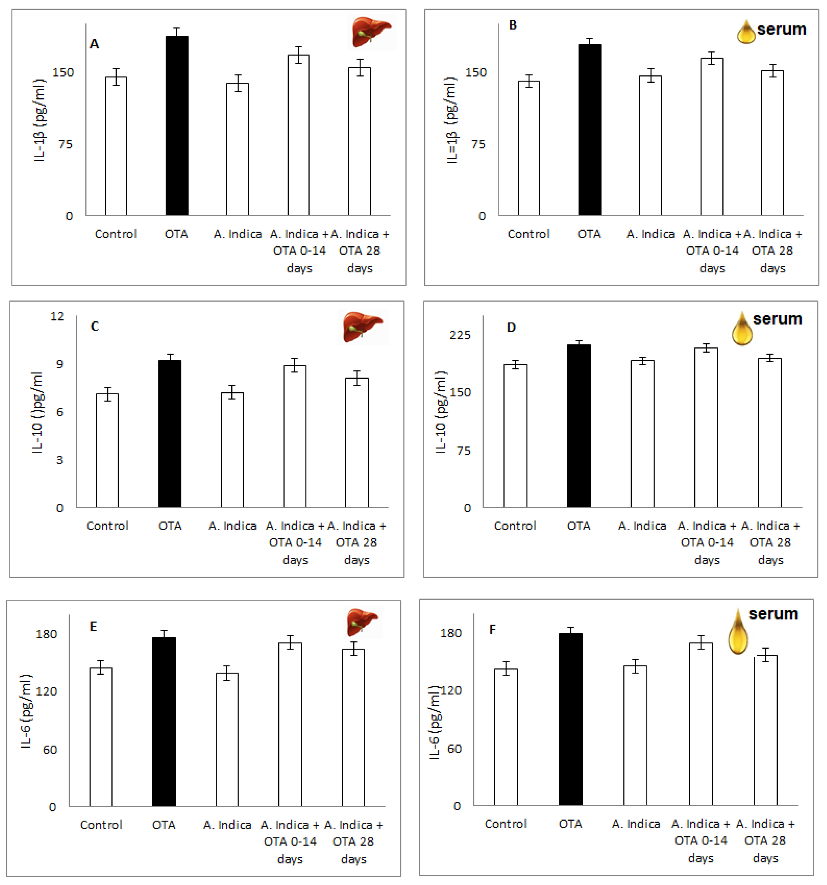
3.9. IL-1b, IL-6, IL-10 in Liver Homogenate/Serum of OTA-Injured Group, Compare to Group Protected with A. indica Oil Alone, Group with Combination Oil + OTA and Control

Compared to the healthy mice, OTA treatment increased interleukins levels in liver homogenate IL-1β (187.5 ± 12.5 pg/mL vs. 144.7 ± 10.01 pg/mL, p < 0.05), IL-10 (9.2 ± 0.8 vs. 7.1 ± 0.9; p < 0.05), IL-6 (176.6 ± 15.8 pg/mL vs. 145.2 ± 21 pg/mL; p < 0.05) and serum IL-1β (178.2 ± 21.8 pg/mL vs. 140.5 ± 15.67 pg/mL, p < 0.05), IL-10 (212.6 ± 17.55 vs. 186.4 ± 2.8; p < 0.05), IL-6 (179.2 ± 21.3 pg/mL vs. 142.8 ± 27.1 pg/mL; p < 0.05), (Figure 8A–F). The group protected with A. indica oil alone and the combination of A. indica + OTA (14 days) and (28 days) showed results similar to the controls. In addition, the oil-protected groups (alone and in combination with OTA) showed a significant decrease in interleukin levels compared to the OTA group in the liver homogenate (p < 0.05) and serum (p < 0.05).

3.10. TNF-a and INF-γ Concentration of A. indica Oil Protected Group Treated with OTA, with A. indica oil Alone and OTA-Damaged Group Compared to Healthy Mice

OTA administration increased the concentrations of TNF-α (12.9 ± 1.2 pg/mL vs. 9.01 ± 0.12 pg/mL in liver homogenate, (p > 0.05) and 79.45 ± 27.01 pg/mL vs. 63.24 ± 14.77 pg/mL in serum (p > 0.05)), and INF-γ (10.71 ± 0.05 pg/mL vs. 8.2 ± 0.07 p > 0.05 in liver homogenate (p > 0.05) and serum 189.8 ± 24.88 pg/mL vs. 145.7± 26.14 pg/mL, p > 0.05) compared to the control in liver homogenate and serum on the 28-day course (Figure 9A–D).
A. indica oil hepatoprotection markedly decreased the concentration of TNF-α (p < 0.05) and INF-γ (p > 0.05) compared to the OTA-induced toxicity group (p < 0.05) on the 28-day course, respectively. The A. indica oil therapy (120 mg/mL) insignificantly lowered the serum INF-α and TNF-γ expression on the 14th day and the last 28th day (Figure 9B,D), compared to OTA-administration. The OTA-induced liver and serum modifications in the concentration of TNF-γ (p > 0.05; Figure 9A,B) and INF-α (p > 0.05; Figure 9C,D) were significantly attenuated with A. indica protection on day 28 (p < 0.05). Moreover, the group treated with A. indica oil showed protection from serum and liver inflammations and the results were almost analogous to the controls.

3.11. Correlations between Parameters

In an analysis of the results of the markers examined in this study, a positive correlation was found in PCC vs. AGEs (r = 0.96, p < 0.05); PCC vs. MDA (r = 0.91, p < 0.05); PCC vs. 5-MSL (r = 0.91, p < 0.05); MDA vs. AGEs (r = 0.92, p < 0.05); MDA vs. NO radicals (r = 0.89, p < 0.05); MDA vs. ROS (r = 0.94, p < 0.05); GSH vs. ROS (r = 0.91, p < 0.05); GSH vs. SIRT1 (r = 0.91, p < 0.05); GSH vs. PGC-1α (r = 0.91, p < 0.05); GSH vs. 5-MSL (r = 0.92, p < 0.05), IL-1β vs. TNF-γ (r = 0.81, p < 0.05); and PCC vs. IL-6 (r = 0.85, p < 0.05).

4. Discussion

The analysis of the results presented in this study shows that the group treated with OTA showed increased oxidative stress by depleting the activity of antioxidant enzymes and showing a drastic increase in ROS/RNS levels, leading to protein damage and increased levels of inflammatory cytokines. Cells quality control systems that the homeostasis of oxidative stress were reduced in mycotoxicosis due to an increase in inflammatory cytokines, leading to the accumulation of oxidized biomolecules [37]. Damaged proteins and lipids accumulated in stressed cells alter the structural and potential function of protein adducts, leading to changes in cellular metabolism and functioning in a state of mycotoxicosis [38]. According to Tao et al. [39], OTA stimulates both NADPH-dependent and ascorbate-dependent lipid peroxidation with co-factor ferrous ions (Fe3+). Therefore, the OTA-Fe3+ complex in the presence of NADPH-cytochrome P450 reductase system facilitates the reduction in ferric ions to iron ions, and the resulting OTA-Fe2+ complex generates •OH, which increases MDA and ROS levels [40]. In addition, plasma membrane permeability and calcium homeostasis are impaired, affecting calcium-sensitive channels and leading to increased ROS and RNS free radical initiation [41]. The involvement of ROS/RNS in the development of oxidative stress due to mycotoxicosis is a proven fact, and the presented results show that the intensity of pathomorphological and biochemical changes and deterioration of oxidative status is best expressed in the group treated with OTA [42].
Cells have various mechanisms to prevent ROS-induced damage, which include enzyme systems (SOD, CAT), glutathione liver enzymes, and endogenous antioxidant molecules such as glutathione (GSH). Glutathione is the most important antioxidant synthesized in cells and helps to remove peroxides and many xenobiotic compounds [43]. In addition, GSH is a cofactor and substrate for glutaredoxins (Grx), which catalyzes disulfide reductions in the presence of NADPH and glutathione reductase GR. Protein and lipid damage is prevented by enzymes such as GR, multiple glutaredoxins (GSPx), and glutathione-S-transferases (GST). For example, the antioxidant enzyme SOD degrades the superoxide anion radical (•O2) to H2O and O2, while GPx reduces H2O2 to water and GSH, and the glutathione reductase GR regenerates GSH [44]. The antioxidant protection mechanism against peroxides involves glutathione, which is involved in the physiological control of free radical generation and increased oxidative stress through the interaction and expression of GR, GPx, and GST [44,45]. Glutathione reductase (GR) is a homodimeric enzyme that catalyzes the oxidized glutathione (GSSG) reduction process to GSH, using FAD and NADPH as cofactors, thus participating in the maintenance of redox balance in living organisms and helping to remove peroxides and many xenobiotic compounds. Glutathione peroxidases (GPXs) are differentially expressed in cells and tissues and are involved in H2O2 detoxification; members of this family reduce peroxides produced by peroxisomes and mitochondria in cells in the cytosol. The activity of glutathione-S-transferases (GST) depends on the presence of GSH, whose role is to detoxify reactive electrophilic compounds, including toxins and products of oxidative stress. The mechanism involves the conjugation of free radicals and non-radical molecules with reduced glutathione GSH conjugated products, which are removed from the cell by specific transporters [46]. OTA induces ROS, thereby modulating the inflammatory response by up-regulating pro-inflammatory cytokines, down-regulating anti-inflammatory IL-4 expression, cytochrome P450 activity, and increasing polyunsaturated fatty acid metabolism [45]. Furthermore, OTA mycotoxicity depletes antioxidant enzymes (GSPx and SOD) and GST at the cellular level and increases the 8-oxoguanine formation and protein carbonylation [47]. In addition, a dose-dependent increase in ROS/RNS due to OTA intoxication in primary proximal tubular cells of experimental animals and cell lines leads to the depletion of intracellular glutathione and cell death [48]. The increased MDA and ROS/RNS in the OTA group suggested that the damage caused was related to impaired oxidative pathways [48]. The presented results reported significant changes in antioxidant enzyme activity and hydroxyproline concentration in the OTA (1.25 mg/kg)-treated group, suggesting oxidative tissue damage and impaired antioxidant status. The elevated liver enzymes (AST, ALP, and ALT) indicate increased oxidative stress and OTA toxicity [48]. The high ROS and RNS led to a significant increase in peroxidase and enlarged phenoxyl radical production [49]. Furthermore, under the GSH action, the phenoxyl radicals were converted back to reactive species, which increased the formation of •O2 as a by-product. This led to the depletion of the activity of the antioxidant enzymes (SOD, GSRx) and GST and oxidative damage to biomacromolecules (lipids and proteins).
The A. indica oil possesses a phytoremediation capability and acts as a protector that directly inhibits high ROS and RNS, regulates protein oxidation, collagen deposition, and reduces mycotoxicity OTA [44,45]. Analysis of the results in the group protected with the combination of A. indica and OTA showed the hepatoprotective role of the oil [50]. These results are consistent with studies reporting the protective effect of A. indica oil against induced liver damage after intoxication with CCl4 and Cisplatin [51,52]. Yanpallewar et al. [53] reported liver damage in rats after paracetamol poisoning. A. indica shows high hepatoprotection, demonstrated by stabilizing the serum activity of liver enzymes ALP, ALT, AST, and histopathological observations of liver tissue. Bhanwra [54] reported protection by A. indica leaf extract in paracetamol-damaged rats, resulting in normal liver appearance and histology, significantly decreased MDA and increased antioxidant and detoxifying enzymes. The oil isolated from A. indica seeds contains active ingredients and secondary metabolites, which have antioxidant, antifungal, antiviral, and anti-inflammatory properties [55,56].
According to Gossé et al. [57] A. indica oil is rich in polyunsaturated fatty acids (PUFA) and improves antioxidant protection by significantly increasing the body’s activity against viruses and mycotoxins. The PUFAs act directly or through their metabolites on signaling pathways and affect many metabolites, reducing TBARS production, superoxide anion secretion, and LDL peroxidation [58]. Based on these data, we treated a group of mice (n = 6) with A. indica seed oil (170 mg/kg), both alone and in combination with OTA (1.25 mg/kg) and monitored the protective effect of the oil against mycotoxicosis. The analysis of the results shows that A. indica seed oil in combination with OTA has a strong hepatoprotective and antioxidant effect. The activities of serum liver enzymes AST, ALP, and ALT were statistically significantly reduced (Figure 3), while SIRT1 (Figure 4A) and PGC-1 (Figure 4B), Hydroxyproline (Figure 5A), MDA (Figure 5B), and the activity of antioxidant enzymes GSPx, SOD and GST were increased (Figure 6A–C).
In the group protected with A. indica oil, no degenerative changes in the liver were observed (Figure 1). The cell depletion in the immunocompetent organs of experimental animals treated with a combination of A. indica oil and the OTA showed oil immunostimulatory, hepatoprotective, and immunoprotective properties. Baligar et al. reported a hepatoprotective role of A. indica and its active constituents in hepatotoxicity in rats [59]. Histology and ultrastructure results confirmed that A. indica dose-dependent treatment reduced hepatocellular necrosis and moderately restored rat liver to normal [59]. The study investigating the protective effect of A. indica oil against CCl4-induced hepatotoxicity in rats demonstrated a hepatoprotective effect of the A. indica oil with an efficacy close to that of silymarin [60].
Our 28-day protection with A. indica oil in OTA mycotoxin food-fed mice reported a significant decrease in hepatocyte density, and the results were close to the controls. Moreover, the combined use of A. indica oil and OTA statistically significantly reduced mycotoxicosis, extracellular matrix, and protein modification. Therefore, this led to an improvement in the oxidative status of the oil-treated mice compared to the OTA-only treated mice. Protecting with A. indica oil has a beneficial effect on OTA-induced hepatotoxic processes and probably participates in the re-modulation of the Th2 immune response. Furthermore, damaged liver tissue and increased levels of inflammatory enzymes resulting from the development of protein dysfunction induce carbonyl stress due to OTA mycotoxicosis [48,60,61].
SIRT1 is correlated with mitochondrial function, energy metabolism, autophagy, apoptosis, and oxidative stress processes and plays a significant role in liver diseases. SIRT1 overexpression leads to increased levels of PPARα and its coactivator PPARγ by a mechanism including acetylation of the peroxisome proliferator-activated receptor-γ coactivator-1α, PGC-1α, which can regulate a function of the multi-functional transcriptional coactivator [62]. In the present study, we analyzed the gene and protein expressions of SIRT1 and PGC-1α in the liver. The results showed that OTA significantly down-regulated the protein levels of SIRT1 and PGC-1α, which suggested that inhibition of the SIRT1/PGC-1α pathway will impair the quality of mitochondrial synthesis, thereby exacerbating oxidative stress. Activating the transcription of PGC-1α is related to enhancing tissue antioxidant capacity of SIRT1 and the expression of superoxide [23,24], and the SIRT1/PGC-1α cascade may act as an activator of the Nrf2 signaling pathway and reduce OTA-oxidative injury. Activated Nrf2 provoked an antioxidant response, resulting in the activation of the endogenous antioxidant enzymes [23,24]. A. indica showed a positive effect on OTA-induced hepatic injury by increasing the levels of SIRT1 and PPAR-α. Therefore, we hypothesize that pretreatment of mice with A. indica attenuates OTA-induced liver injury by expressing SIRT1, stimulating PGC-1α production, increasing essential fatty acid levels, reducing lipogenesis, and improving lipid metabolism.
Moreover, the mice treated with OTA, the AGEs products (Figure 7A) and carbonylated proteins (Figure 7B) were statistically significantly increased, and the reduction in nitroxide protein (Figure 7B) was almost twice delayed and liver damage was registered. Protection with A. indica oil in a combination treatment restores these parameters almost to control levels. Therefore, A. indica oil modulates OTA-induced inflammation and progressive hepatocyte processes in the liver by lowering PCC, reducing protein dysfunction, and completely inhibiting the concentrations of •O2ˉ, •OH, NO• radicals. Furthermore, A. indica oil rapidly inhibits AGEs glycation and the action of proteins and thiols [63]. The active compounds from A. indica deactivated the protein and lipid oxidation and suppressed procoagulant activity in hepatic cells, which equilibrated collagen turnover during an exchange in antioxidant-prooxidant balance [52]. The spin-labeled 5-MSL scavenged membrane proteins alkylated as a result of the reaction between the maleimide group with the SH sites, i.e., by rapid rotation, giving an isotropic triplet spectrum with sharp lines (not shown); nitroxyl captures the weakened SH regions of the corresponding amino acid [35] and due to limitations in movement, the EPR spectrum changes shape as a result of anisotropy [36]. Long-term protection with A. indica (170 mg/kg) against OTA-induced mycotoxicosis completely remodulates the concentration of 5-MSL, reduces membrane proteins, and probably further reduces the levels of protein aggregates and, thus, restores antioxidant status to levels close to the controls.
In the OTA-induced mycotoxicosis model, ROS/RNS generation resulted in progressive lipid peroxidation that affected the reductive protein metabolism, increased collagen synthesis by increasing hydroxyproline and directly affected the antioxidant defense system. OTA leads to oxidation, and oxidation reactions occur through the cytochrome protein family. Among the isoforms of CYP450 enzymes, cytochrome expression is relatively high in the liver. When toxic substances enter the body, transcription factors migrate from the cytoplasm to the nucleus and increase the expression of cytochrome proteins, leading to oxidation. Antioxidant enzymes are involved in the process to provide protection against oxidative stress [63].
An anti-inflammatory IL-10 is cytokine produced mainly by macrophages and Th2 cells. The reported biological activities of IL-10 that may be interrelated include the inhibition of macrophage-mediated cytokine synthesis, inhibition of delayed-type hypersensitivity reactions, and stimulation of the Th2 cellular response, leading to increased antibody production [63,64]. IL-10 functions by inhibiting proinflammatory cytokines produced by macrophages and regulatory T cells, including IFN-gamma and TNF-alpha. In the present study, A. indica was shown to significantly inhibit OTA-induced levels of TNF-α, IL-1β, IL-6, IL-10, and IFN-gamma in mice. Rodrigues et al. [65] reported that the suppressive inflammatory effects of A. indica extract completely inhibited OTA production. Schumacher et al. [66] reported that A. indica leaf extract induced apoptosis in leukemia cells and inhibited proliferation in prostate cancer. The leaf extract also stimulates immune function by activating killer cells and has an antiproliferative effect, which is probably due to the active compound in A. indica oil.

5. Conclusions

In the present study, we reported significantly elevated cytocines, interferon-gamma (IFN-γ), tumor necrosis factor-alpha (TNF-α), and collagen synthesis in the OTA-treated mice. Protection with A. indica oil (170 mg/mL) induced cell proliferation of normal hepatocytes and a probable anti-inflammatory therapeutic effect. The results presented above suggest inhibition of OTA-induced inflammation by A. indica oil and ROS reduction influx by regulating IL-1β, IL-6, IL-10, IFN-γ, and TNF-α balance after a 28-day course. OTA is a mycotoxin with a hepatotoxic effect, and toxicity resulted in statistically significant increases in biomarkers of oxidative stress, abnormal hepatic and antioxidant enzymes, and cytokine levels. A. indica oil has a potential restorative and protective effect, as a result of which hepatic and antioxidant enzymes are restored. In addition, the protective effects of A. indica seed oil have led to functional recovery in the OTA-induced mice model. Therefore, A. indica oil can be used as a practical approach to safe OTA-contaminated feed use.

Author Contributions

Conceptualization, G.N. and Y.K.; methodology, G.N., Y.K. and J.A.; software, E.G.; validation, K.P.-P., V.I. and E.G.; formal analysis, E.G., G.N. and Y.K.; investigation, G.N. and Y.K.; resources, G.N. and Y.K.; data curation, G.N. and Y.K.; writing—original draft preparation, J.A., K.P.-P., G.N. and Y.K.; writing, G.N. and Y.K.; visualization, E.G. and G.N.; supervision, G.N.; project administration, G.N.; funding acquisition, G.N. and Y.K. All authors have read and agreed to the published version of the manuscript.

Funding

This research received no external funding.

Institutional Review Board Statement

The study was conducted in accordance with the Declaration of Helsinki and approved by the Animal Ethics Commission of the Trakia University, Stara Zagora, Bulgaria and the Bulgarian Food Safety Agency, Sofia, Bulgaria with a license (325/6000-0333/9 December 2021).

Informed Consent Statement

Not applicable.

Data Availability Statement

Not applicable.

Acknowledgments

The present research was funded by scientific project №3/2020, Medical Faculty, Trakia University, Stara Zagora, Bulgaria.

Conflicts of Interest

The authors declare no conflict of interest.

References

  1. Bhukan, P.G.; Bangar, N.P.; Ramesh, D. Review on Hepatoprotective Medicinal. Plants 2020, 9, 531–549. [Google Scholar] [CrossRef]
  2. Huang, L.; Liu, J.; Bie, C.; Liu, H.; Ji, Y.; Chen, D.; Zhu, M.; Kuang, W. Advances in cell death-related signaling pathways in acute-on-chronic liver failure. Clin. Res. Hepatol. Gastroenterol. 2022, 46, 101783. [Google Scholar] [CrossRef] [PubMed]
  3. Atanasova-Penichon, V.; Legoahec, L.; Bernillon, S.; Deborde, C.; Maucourt, M.; Verdal-Bonnin, M.; Richard-Forget, F. Mycotoxin biosynthesis and central metabolism are two interlinked pathways in Fusarium graminearum, as demonstrated by the extensive metabolic changes induced by caffeic acid exposure. Appl. Environ. Microb. 2018, 84, 01705–01717. [Google Scholar] [CrossRef] [PubMed]
  4. Agriopoulou, S.; Stamatelopoulou, E.; Varzakas, T. Advances in occurrence, importance, and mycotoxin control strategies: Prevention and detoxification in foods. Foods 2020, 9, 137. [Google Scholar] [CrossRef]
  5. Wang, L.; Jin, J.; Liu, X.; Wang, Y.; Liu, Y.; Zhao, Y.; Xing, F. Effect of cinnamaldehyde on morphological alterations of Aspergillus ochraceus and expression of key genes involved in ochratoxin A biosynthesis. Toxins 2018, 10, 340. [Google Scholar] [CrossRef]
  6. Zeidan, R.; Ul-Hassan, Z.; Al-Thani, R.; Migheli, Q.; Jaoua, S. In-Vitro Application of a Qatari Burkholderia cepacia strain (QBC03) in the Biocontrol of Mycotoxigenic Fungi and in the Reduction of Ochratoxin A biosynthesis by Aspergillus carbonarius. Toxins 2019, 11, 700. [Google Scholar] [CrossRef]
  7. Benkerroum, N. Chronic and Acute Toxicities of Aflatoxins: Mechanisms of Action. Int. J. Environ. Res. Public Health 2020, 17, 423. [Google Scholar] [CrossRef]
  8. Khaneghah, M.A.; Ismail, E.; Raeisi, S.; Fakhri, Y. Aflatoxins in cereals: State of the art. J. Food Saf. 2018, 38, e12532. [Google Scholar] [CrossRef]
  9. Petzinger, E.; Ziegler, K. Ochratoxin A from a toxicological perspective. J. Vet. Pharmacol. Ther. 2000, 23, 91–98. [Google Scholar] [CrossRef]
  10. Pfohl-Leszkowicz, A.; Bartsc, H.; Azémar, B. MESNA protects rats against nephrotoxicity but not carcinogenicity induced by ochratoxin A, implicating two separate pathways. Facta Univ. Ser. Med. Biol. 2002, 9, 57–63. [Google Scholar]
  11. Petkova-Bocharova, T.; Castegnaro, M. Ochratoxin A contamination of cereals in an area of high incidence of Balkan endemic nephropathy in Bulgaria. Food Addit. Contam. 1985, 2, 267–270. [Google Scholar] [CrossRef]
  12. Imaoka, T.; Yang, J.; Wang, L.; McDonald, M.; Afsharinejad, Z.; Bammler, T.; Kelly, E. Microphysiological system modeling of ochratoxin A-associated nephrotoxicity. Toxicology 2020, 444, 152582. [Google Scholar] [CrossRef]
  13. Gagliano, N.; Donne, I.D.; Torr, C.; Migliori, M.; Grizzi, F.; Milzani, A.; Filippi, C.; Annoni, G.; Colombo, P.; Costa, F. Early cytotoxic effects of ochratoxin A in rat liver: A morphological, biochemical and molecular study. Toxicology 2006, 15, 214–224. [Google Scholar] [CrossRef]
  14. Biney, E.E.; Nkoom, M.; Darkwah, W.K.; Puplampu, J.B. High-Performance Liquid Chromatography Analysis and Antioxidant Activities of Extract of Azadirachta indica (Neem) Leaves. Pharmacogn. Res. 2021, 12, 29–34. [Google Scholar] [CrossRef]
  15. Bakr, A. Changes of hemoglobin content and glucose levels in the blood of Rattus norvegicus by water extracts of Azadirachta indica. Chin. J. Nat. Med. 2012, 10, 135–137. [Google Scholar] [CrossRef]
  16. Sen, S.; Chakraborty, R. The role of antioxidants in human health. In Oxidative Stress: Diagnostics, Prevention, and Therapy; ACS Publications: Washington, DC, USA, 2011; pp. 1–37. [Google Scholar] [CrossRef]
  17. Parcheta, M.; Świsłocka, R.; Orzechowska, S.; Akimowicz, M.; Choińska, R.; Lewandowski, W. Recent Developments in Effective Antioxidants: The Structure and Antioxidant Properties. Materials 2021, 14, 1984. [Google Scholar] [CrossRef]
  18. Ayala, A.; Muñoz, M.F.; Argüelles, S. Lipid peroxidation: Production, metabolism, and signaling mechanisms of malondialdehyde and 4-hydroxy-2-nonenal. Oxidative Med. Cell. Longev. 2014, 2014, 360438. [Google Scholar] [CrossRef]
  19. Di Meo, S.; Reed, T.T.; Venditti, P.; Victor, V.M. Role of ROS and RNS Sources in Physiological and Pathological Conditions. Oxidative Med. Cell. Longev. 2016, 2016, 1245049. [Google Scholar] [CrossRef]
  20. Lobo, V.; Patil, A.; Phatak, A.; Chandra, N. Free radicals, antioxidants and functional foods: Impact on human health. Pharmacogn. Rev. 2010, 4, 118–126. [Google Scholar] [CrossRef]
  21. Smirne, C.; Croce, E.; Di Benedetto, D.; Cantaluppi, V.; Comi, C.; Sainaghi, P.P.; Minisini, R.; Grossini, E.; Pirisi, M. Oxidative Stress in Non-Alcoholic Fatty Liver Disease. Livers 2022, 2, 30–76. [Google Scholar] [CrossRef]
  22. Spector, A. Oxidative stress and disease. J. Ocular Pharmacol. Ther. 2000, 16, 193–201. [Google Scholar] [CrossRef] [PubMed]
  23. Yan, T.; Huang, J.; Nisar, M.F.; Wan, C.; Huang, W. The beneficial roles of SIRT1 in drug-induced liver injury. Oxidative Med. Cell. Longev. 2019, 2019, 8506195. [Google Scholar] [CrossRef] [PubMed]
  24. Lagouge, M.; Argmann, C.; Gerhart-Hines, Z.; Meziane, H.; Lerin, C.; Daussin, F.; Auwerx, J. Resveratrol improves mitochondrial function and protects against metabolic disease by activating SIRT1 and PGC-1α. Cell 2006, 127, 1109–1122. [Google Scholar] [CrossRef] [PubMed]
  25. Ren, Z.; He, H.; Zuo, Z.; Xu, Z.; Wei, Z.; Deng, J. The role of different SIRT1-mediated signaling pathways in toxic injury. Cell. Mol. Biol. Lett. 2019, 24, 36. [Google Scholar] [CrossRef]
  26. Deng, Y.X.; Cao, M.; Shi, D.X.; Yin, Z.Q.; Jia, R.Y.; Xu, J.; Zhao, J. Toxicological evaluation of neem (Azadirachta indica) oil: Acute 690 and subacute toxicity. Environ. Toxicol. Pharmacol. 2013, 35, 240–246. [Google Scholar] [CrossRef]
  27. Stoev, S.D.; Dimitrov, K.; Zarkov, I.; Mircheva, T.; Zapryanova, D.; Valchev, I.; Arora, R. Some Indian herbs have protective effects against deleterious effects of ochratoxin A in broiler chicks. World Mycotoxin J. 2021, 14, 525–538. [Google Scholar] [CrossRef]
  28. Woessner, J.B. The determination of hidroxyproline in tissue and protein samples containing small proportions of this amino acid. Arch. Biochem. Biophys. 1961, 93, 440–447. [Google Scholar] [CrossRef]
  29. Habig, W.H.; Pabst, M.J.; Jakoby, W.B. Glutathione S-transferases: The first enzymatic step in mercapturic acid formation. J. Biol. Chem. 1974, 249, 7130–7139. [Google Scholar] [CrossRef]
  30. Novaes, R.D.; Gonçalves, R.V.; Marques, D.C.; Cupertino, M.C.; Peluzio, M.C.; Leite, J.P.; Maldonado, I.R. Effect of bark extract of Bathysa cuspidata on hepatic oxidative damage and blood glucose kinetics in rats exposed to paraquat. Toxicol. Pathol. 2012, 40, 62–70. [Google Scholar] [CrossRef]
  31. Novaes, R.D.; Santos, E.C.; Cupertino, M.C.; Bastos, D.S.; Oliveira, J.M.; Carvalho, T.V.; Neves, M.M.; Oliveira, L.L.; Talvani, A. Trypanosoma cruzi infection and benznidazole therapy independently stimulate oxidative status and structural pathological remodeling of the liver tissue in mice. Parasitol. Res. 2015, 114, 2873–2881. [Google Scholar] [CrossRef]
  32. Shi, H.; Sui, Y.; Wang, X.; Luo, Y.; Ji, L. Hydroxyl radical production and oxidative damage induced by cadmium and naphthalene in liver of Carassius auratus. Comp. Biochem. Phys. Part C Toxicol. Pharmacol. 2005, 140, 115–121. [Google Scholar] [CrossRef]
  33. Yokoyama, K.; Hashiba, K.; Wakabayashi, H.; Hashimoto, K.; Saton, K.; Kurihara, T.; Motohashi, N.; Sakagami, H. Inhibition of LPS-stimulated NO production in mouse macrophage-like cells by Tropolones. Anticancer Res. 2004, 24, 3917–3922. [Google Scholar]
  34. Yoshioka, T.; Iwamoto, N.; Lto, K. An applicationof Electron Paramagnetic Resonance to evaluate nitric oxide and its quenchers. J. Am. Soc. Nephrol. 1996, 7, 961–965. [Google Scholar] [CrossRef]
  35. Takeshita, K.; Saito, K.; Ueda, J.I.; Anzai, K.; Ozawa, T. Kinetic study on ESR signal decay of nitroxyl radicals, potent redox probes for in vivo ESR spectroscopy, caused by reactive oxygen species. Biochim. Et Biophys. Acta (BBA)-Gen. Subj. 2002, 1573, 156–164. [Google Scholar] [CrossRef]
  36. Momchilova, S.; Antonova, D.; Marekov, I.; Kuleva, L.; Nikolova-Damyanova, B. Fatty acids, triacylglycerols, and sterols in neem oil (Azadirachta indica a. juss) as determined by a combination of chromatographic and spectral techniques. J. Liq. Chromatogr. Relat. Technol. 2007, 30, 11–25. [Google Scholar] [CrossRef]
  37. Mavrommatis, A.; Giamouri, E.; Tavrizelou, S.; Zacharioudaki, M.; Danezis, G.; Simitzis, P.E.; Feggeros, K. Impact of mycotoxins on animals’ oxidative status. Antioxidants 2021, 10, 214. [Google Scholar] [CrossRef]
  38. Chienwichai, P.; Reamtong, O.; Pisitkun, T.; Somparn, P.; Tharnpoophasiam, P.; Worakhunpiset, S.; Topanurak, S. Hepatic protein Carbonylation profiles induced by lipid accumulation and oxidative stress for investigating cellular response to non-alcoholic fatty liver disease in vitro. Proteome Sci. 2019, 17, 1–13. [Google Scholar] [CrossRef]
  39. Tao, Y.; Xie, S.; Xu, F.; Liu, A.; Wang, Y.; Chen, D.; Yuan, Z. Ochratoxin A: Toxicity, oxidative stress and metabolism. Food Chem. Toxicol. 2018, 112, 320–331. [Google Scholar] [CrossRef]
  40. Gray, J.P.; Mishin, V.; Heck, D.E.; Laskin, D.L.; Laskin, J.D. Inhibition of NADPH cytochrome P450 reductase by the model sulfur mustard vesicant 2-chloroethyl ethyl sulfide is associated with increased production of reactive oxygen species. Toxicol. Appl. Pharmacol. 2010, 247, 76–82. [Google Scholar] [CrossRef]
  41. Robinson, M.W.; Harmon, C.; O’Farrelly, C. Liver immunology and its role in inflammation and homeostasis. Cell Molecimmunol. 2016, 13, 267–276. [Google Scholar] [CrossRef]
  42. Ighodaro, O.M.; Akinloye, O. First line defence antioxidants-superoxide dismutase (SOD), catalase (CAT) and glutathione peroxidase (GPx): Their fundamental role in the entire antioxidant defence grid. Alex. J. Med. 2018, 54, 287–293. [Google Scholar] [CrossRef] [Green Version]
  43. Kennedy, L.; Sandhu, J.K.; Harper, M.E.; Cuperlovic-Culf, M. Role of glutathione in cancer: From mechanisms to therapies. Biomolecules 2020, 10, 1429. [Google Scholar] [CrossRef] [PubMed]
  44. Ansari, M.A.; Scheff, S.W. Oxidative stress in the progression of Alzheimer disease in the frontal cortex. J. Neuropathol. Exp. Neurol. 2010, 69, 155–167. [Google Scholar] [CrossRef] [PubMed]
  45. Dhanasekaran, M.; Ignacimuthu, S.; Agastian, P. Potential hepatoprotective activity of ononitol monohydrate isolated from Cassia tora L. on carbon tetrachloride induced hepatotoxicity in wistar rats. Phytomedicine 2009, 16, 891–895. [Google Scholar] [CrossRef]
  46. Snezhkina, A.V.; Kudryavtseva, A.V.; Kardymon, O.L.; Savvateeva, M.V.; Melnikova, N.V.; Krasnov, G.S.; Dmitriev, A.A. ROS Generation and Antioxidant Defense Systems in Normal and Malignant Cells. Oxidative Med. Cell Longev. 2019, 5, 6175804. [Google Scholar] [CrossRef]
  47. Nabi, S.; Tanveer, S.; Ganie, S.A. Glutathione-S-transferase, Superoxide Dismutase (GST, SOD) Levels, Protein Content and Lipid Peroxidation in Schizothorax plagiostomus under the Infection of Pomphorhynchus in Nallah Sukhnag of Kashmir Valley. Pak. J. Biol. Sci. 2017, 20, 442–446. [Google Scholar] [CrossRef]
  48. Zhu, L.; Yu, T.; Qi, X.; Gao, J.; Huang, K.; He, X.; Luo, H.; Xu, W. Limited link between oxidative stress and ochratoxin A—Induced renal injury in an acute toxicity rat model. Toxins 2016, 8, 373. [Google Scholar] [CrossRef]
  49. Marin-Kuan, M.; Ehrlich, V.; Delatour, T.; Cavin, C.; Schilter, B. Evidence for a role of oxidative stress in the carcinogenicity of ochratoxin A. J. Toxicol. 2011, 2011, 645361. [Google Scholar] [CrossRef]
  50. Schrenk, D.; Bodin, L.; Chipman, J.K.; del Mazo, J.; Grasl-Kraupp, B.; Bignami, M. Risk assessment of ochratoxin A in food. EFSA J. 2020, 18, e06113. [Google Scholar] [CrossRef]
  51. Baligar, N.S.; Aladakatti, R.H.; Ahmed, M.; Hiremath, M.B. Hepatoprotective activity of the neem-based constituent azadirachtin-A in carbon tetrachloride intoxicated Wistar rats. Can. J. Physiol. Pharmacol. 2014, 92, 267–277. [Google Scholar] [CrossRef]
  52. Dkhil, M.A.; Al-Quraishy, S.; Aref, A.M.; Othman, M.S.; El-Deib, K.M.; Abdel Moneim, A.E. The potential role of Azadirachta indica treatment on cisplatin-induced hepatotoxicity and oxidative stress in female rats. Oxidative Med. Cell Longev. 2013, 2013, 741817. [Google Scholar] [CrossRef] [Green Version]
  53. Yanpallewar, S.U.; Sen, S.; Tapas, S.; Kumar, M.; Raju, S.S.; Acharya, S.B. Effect of Azadirachta indica on paracetamol-induced hepatic damage in albino rats. Phytomedicine 2003, 10, 391–396. [Google Scholar] [CrossRef]
  54. Bhanwra, S.; Singh, J.; Khosla, P. Effect of Azadirachta indica (neem) leaf aqueous extract on paracetamol induced liver damage in rats. Ind. J. Physiol. Pharmacol. 2000, 44, 64–68. [Google Scholar]
  55. Alzohairy, M.A. Therapeutics Role of Azadirachta indica (Neem) and Their Active Constituents in Diseases Prevention and Treatment. Evid.-Based Complementary Altern. Med. 2016, 2016, 7382506. [Google Scholar] [CrossRef]
  56. Rao, N.G.; Rao, P.P.G.; Satyanarayana, A. Chemical, fatty acid, volatile oil composition and antioxidant activity of shade dried neem (Azadirachta indica L.) flower powder. Int. Food Res. J. 2014, 21, 807–813. [Google Scholar]
  57. Gossé, B.; Amissa, A.A.; Adjé, A.F.; Niamké, B.F.; Ollivier, D.; Ito, Y. Analysis of components of neem (Azadirachta indica) oil by diverse chromatographic techniques. J. Liquid Chromatogr. Relat. Technol. 2005, 28, 2225–2233. [Google Scholar] [CrossRef]
  58. Papackova, Z.; Cahova, M. Fatty acid signaling: The new function of intracellular lipases. Int. J. Mol. Sci. 2015, 16, 3831–3855. [Google Scholar] [CrossRef]
  59. Baligar, N.; Aladakatti, R.; Ahmed, M.; Hiremath, M.B. Evaluation of acute toxicity of neem active constituent, nimbolide and its hepatoprotective activity against acute dose of carbon tetrachloride treated albino rats. Int. J. Pharm. Sci. Res. 2014, 5, 3455–3466. [Google Scholar] [CrossRef]
  60. Janik, E.; Niemcewicz, M.; Ceremuga, M.; Stela, M.; Saluk-Bijak, J.; Siadkowski, A.; Bijak, M. Molecular Aspects of Mycotoxins-A Serious Problem for Human Health. Int. J. Mol. Sci. 2020, 21, 8187. [Google Scholar] [CrossRef]
  61. Niaz, K.; Shah, S.Z.A.; Khan, F.; Bule, M. Ochratoxin A–induced genotoxic and epigenetic mechanisms lead to Alzheimer disease: Its modulation with strategies. Environ. Sci. Pollut. Res. 2020, 27, 44673–44700. [Google Scholar] [CrossRef]
  62. Shin, M.-R.; Lee, J.A.; Kim, M.; Lee, S.; Oh, M.; Moon, J.; Nam, J.-W.; Choi, H.; Mun, Y.-J.; Roh, S.-S. Gardeniae Fructus Attenuates Thioacetamide-Induced Liver Fibrosis in Mice via Both AMPK/SIRT1/NF-κB Pathway and Nrf2 Signaling. Antioxidants 2021, 10, 1837. [Google Scholar] [CrossRef]
  63. Dalle-Donne, I.; Rossi, R.; Giustarini, D.; Milzani, A. Colombo R Protein carbonyl groups as biomarkers of oxidative stress. Clin. Chim. Acta 2003, 329, 23–38. [Google Scholar] [CrossRef]
  64. Shin, H.S.; Lee, H.J.; Pyo, M.C.; Ryu, D.; Lee, K.-W. Ochratoxin A-Induced Hepatotoxicity through Phase I and Phase II Reactions Regulated by AhR in Liver Cells. Toxins 2019, 11, 377. [Google Scholar] [CrossRef] [Green Version]
  65. Rodrigues, M.P.; Astoreca, A.L.; Oliveira, Á.A.D.; Salvato, L.A.; Biscoto, G.L.; Keller, L.A.M.; Keller, K.M. In vitro activity of neem (Azadirachta indica) oil on growth and ochratoxin A production by Aspergillus carbonarius Isolates. Toxins 2019, 11, 579. [Google Scholar] [CrossRef]
  66. Schumacher, M.; Cerella, C.; Reuter, S.; Dicato, M.; Diederich, M. Anti-inflammatory, pro-apoptotic, and anti-proliferative effects of a methanolic neem (Azadirachta indica) leaf extract are mediated via modulation of the nuclear factor-κB pathway. Genes Nutr. 2011, 6, 149–160. [Google Scholar] [CrossRef] [Green Version]
Scheme 1. Experimental protocol design.
Antioxidants 11 01678 sch001
Figure 1. 1—control mice; 2—group protected with A. indica; 3—group protected with A. indica in combination with OTA; 4—treated with OTA only; (a) normal appearance of the liver without significant pathological changes (×100); (b) normal appearance of the liver without significant pathological changes (×400); (c) normal appearance of the liver without significant pathological changes (×100); (d) normal appearance of the liver without significant pathological changes (×200); (e) mild degenerative changes in hepatocytes (×100); (f) weak mononuclear infiltrate and mild degenerative changes in hepatocytes (×400); (g) mild vascular congestion (×100); (h) area with hepatocytes with marked karyomegaly and mild degenerative changes in hepatocytes (×400); (i) pronounced degenerative and necrotic changes in hepatocytes (×400); (j) moderate mononuclear inflammatory infiltrate (×400); (k) markable vascular congestion (×100); (l) area with hepatocytes with karyomegaly and polychromasia, part of a vessel with vascular congestion (×400).
Antioxidants 11 01678 g001
Figure 2. Levels of hepatic enzymes AST (A), ALT (B), ALP (C) measured in serum of mice (n = 6) on a diet included: OTA alone, A. indica oil, and combination of A. indica oil + OTA compared to the healthy controls; * p <0.05 vs. control group; ** p < 0.05 vs. OTA group.
Antioxidants 11 01678 g002
Figure 3. The effects on SIRT1 (A) and PGC-1α pathway (B) in four group of mice (n = 6) treated with A. indica oil, OTA, and combination of A. indica + OTA all results were compared to controls (non-treated mice). The results are expressed in µg/g tissue hydroxyproline. Values are presented as mean ± SEM. The statistically significant difference used in relation to controls (*); p < 0.05 vs. controls, (**) p < 0.05 vs. OTA treated mice.
Antioxidants 11 01678 g003
Figure 4. (A) The A. indica oil effects on OTA-induced oxidative changes in liver hydroxyproline content and (B)—lipid peroxidation measured as MDA (μmol/mL). The results are presented as mean ± SEM. The significant difference used in relation to controls (*) p < 0.05 vs. controls, (**) p < 0.05 vs. OTA treated mice.
Antioxidants 11 01678 g004
Figure 5. The activity of antioxidant enzymes SOD (A), (GSPx) (B), and GST (C) was presented in control mice, mice treated with OTA, A. indica oil, and a combination of A. indica + OTA. The presented data as means with SEM. (*) indicate significant differences from (* p < 0.05 vs. control, ** p < 0.05 vs. OTA).
Antioxidants 11 01678 g005
Figure 6. (A) AGEs in the group treated with OTA, with A. indica and combination A. indica + OTA, vs. non-treated controls, p < 0.05; (B) 5-MSL in the liver of treated with OTA, with A. indica oil only and combination A. indica oil+ mycotoxin vs. healthy controls, p < 0.05; (C) PCC in OTA group, A. indica oil group, and group protected with A. indica oil against the mycotoxin OTA, vs. non-treated controls. The presented data as means with SEM. (*) indicate significant differences from (* p < 0.05 vs. control, ** p < 0.05 vs. OTA).
Antioxidants 11 01678 g006
Figure 7. (A,B) NO radicals measured in arbitrary units (A.U.) in serum/liver of control mice, mice treated with OTA, mice treated with A. indica oil and with combination A. indica oil + OTA; (C,D) ROS (A.U.) measured in serum/liver of OTA treated mice, group protected with A. indica oil only and pretreatment with A. indica oil + OTA compared to healthy mice. The presented data as means with SEM. (*) indicate significant differences from (* p < 0.05 vs. control, ** p < 0.05 vs. OTA).
Antioxidants 11 01678 g007
Figure 8. The A. indica effects on OTA-induced mycotoxicity and the expressions of IL-1b (A,B), IL-6 (C,D), IL-10 (E,F) in mouse liver homogenate/serum samples; mean ± S.E. p < 0.05.
Antioxidants 11 01678 g008
Figure 9. The concentration of TNF-α (A,B) and INF-γ (C,D) in liver homogenate/serum of OTA-injured group, compared to a group protected with A. indica oil alone, the group with A. indica oil + OTA and control; mean ± S.E. p < 0.05; (*) vs. controls; (**) vs. OTA.
Antioxidants 11 01678 g009
Table 1. Body weight (BW) was varied by group during the experiment; The mice from all four groups were measured on 5th, 8th, 14th and 28th days during the 28 days experiment; controls (standard diet, n = 6); OTA (n = 6); A. indica oil (n = 6); A. indica oil + OTA in combination (n = 6). The quantitative data were expressed as the means ± SE. * p < 0.05 vs. control group; ** p < 0.05 vs. OTA treated group.
AdministrationBW (g)BW (g)BW (g)BW (g)
(n = 6)5th day8th day14th day28th day
Control205.3 ± 2.98221.7 ± 1.87233.1 ± 2.11255.4 ± 2.82
OTA189.1± 1.99 *175.4± 2.14 *165.4 ± 2.03 *157.8 ± 3.01 *
A. indica oil204.8 ± 3.11224.4 ± 2.01232.1 ± 2.25254.9 ± 2.14
A. indica oil + OTA199.1 ± 188218.1 ± 2.31 **231.9 ± 2.09 **252.9 ± 1.05 **
Table 2. Comparative pathomorphological changes in four groups: control-non-treated mice; A. indica oil-mice are protected only with oil; the group with combined therapy A. indica + OTA; a group treated only with OTA. Legend: 0—No changes; 1—Weak changes; 2—Moderate changes; 3—Strong changes.
ControlsA. indicaA. indica + OTAOTA
Fat degeneration0000
Hyaline drops0000
Inflammation0012
Necrosis0001
Vascular congestion0012
Karyomegaly0012
Aneuploidy0001
Polychromasia0002
Publisher’s Note: MDPI stays neutral with regard to jurisdictional claims in published maps and institutional affiliations.

