Article

Preparation of Mangosteen Peel Extract Microcapsules by Fluidized Bed Spray-Drying for Tableting: Improving the Solubility and Antioxidant Stability

by
Sriwidodo Sriwidodo
1,*,
Reza Pratama
2,
Abd. Kakhar Umar
1,
Anis Yohana Chaerunisa
1,
Afifah Tri Ambarwati
1 and
Nasrul Wathoni
1
1
Department of Pharmaceutics and Pharmaceutical Technology, Faculty of Pharmacy, Universitas Padjadjaran, Sumedang 45363, Indonesia
2
Department of Pharmaceutics and Pharmaceutical Technology, Faculty of Pharmacy, Universitas Bhakti Kencana, Bandung 40614, Indonesia
*
Author to whom correspondence should be addressed.
Antioxidants 2022, 11(7), 1331; https://doi.org/10.3390/antiox11071331
Submission received: 29 April 2022 / Revised: 29 June 2022 / Accepted: 4 July 2022 / Published: 6 July 2022

Abstract

:
Mangosteen fruit has been widely consumed and used as a source of antioxidants, either in the form of fresh fruit or processed products. However, mangosteen peel only becomes industrial waste due to its bitter taste, low content solubility, and poor stability. Therefore, this study aimed to design mangosteen peel extract microcapsules (MPEMs) and tablets to overcome the challenges. The fluidized bed spray-drying method was used to develop MPEM, with hydroxypropyl methylcellulose (HPMC) as the core mixture and polyvinyl alcohol (PVA) as the coating agent. The obtained MPEM was spherical with a hollow surface and had a size of 411.2 µm. The flow rate and compressibility of MPEM increased significantly after granulation. A formula containing 5% w/w polyvinyl pyrrolidone K30 (PVP K30) as a binder had the best tablet characteristics, with a hardness of 87.8 ± 1.398 N, friability of 0.94%, and disintegration time of 25.75 ± 0.676 min. Microencapsulation of mangosteen peel extract maintains the stability of its compound (total phenolic and α-mangosteen) and its antioxidant activity (IC50) during the manufacturing process and a month of storage at IVB zone conditions. According to the findings, the microencapsulation is an effective technique for improving the solubility and antioxidant stability of mangosteen peel extract during manufacture and storage.

Graphical Abstract

1. Introduction

Indonesia is one of the world’s leading producers of mangosteen fruit (Garcinia mangostana L.), both fresh and processed into canned products and juices [1,2]. During mangosteen processing, the peel is separated and discarded as industrial waste, even though it offers numerous benefits. Mangosteen peel can be used as a food, supplement, cosmetic, and medicine because it contains many therapeutic compounds and antioxidants [3,4,5,6,7,8,9]. Mangosteen peel is rarely ingested raw due to its astringent taste. The major antioxidant components in mangosteen peel, xanthones, have poor water solubility, and direct usage reduces their efficiency since they can be degraded or oxidized during processing and storage. According to reports, the microencapsulation technique can overcome these challenges [10,11,12].
Microencapsulation is the process of encapsulating tiny solid, liquid, or gaseous particles in a polymeric substance. The polymer material can modulate odor, taste, volatility, and reactivity, as well as enhance the extract material’s stability and solubility [13,14,15,16]. Many factors affect the quality of microcapsules, including the preparation technique and the type of coating material [17]. Fluidized bed microencapsulation is the prevalent approach and offers several advantages, including low cost, minimal dust pollution, high uniformity, high encapsulation efficiency, and excellent stability [18,19]. The polymer utilized further enhances the quality and stability of the microencapsulation; hence, choosing the proper base polymer should be performed [17,20].
Several natural polymers have been used as core and coating materials in microencapsulation. One of them is HPMC, which is known to be biodegradable, biocompatible, renewable, and low in toxicity [21,22]. HPMC has been used as a mixture with active ingredients to increase solubility and extend drug contact time due to its hydrophilic and mucoadhesive properties [23,24,25]. This cellulose derivate has also demonstrated substantial potential to alleviate GI irritation caused by APIs known to induce diarrhea, ulcers, nausea, or vomiting [23]. Since HPMC is hygroscopic, particularly after drying, it is typically mixed with other polymers [26,27] or coated [23]. As a coating material, PVA offers several advantages, including biodegradability, biocompatibility, and good mechanical properties [28]. The PVA film is both water- and heat-resistant, which contributes to the core material’s stability [29]. The study by Wu et al. reveals that employing PVA as a coating can minimize the size of the microencapsulation while also providing protection against coalescence [30]. The encapsulation efficiency of PVA is also reported to be high [31]. As a result, we employed HPMC as the core material’s binder and PVP as the microspheres’ coating. In this work, we investigated the stability and antioxidant activity of mangosteen peel extract during its transformation into microcapsules and, lastly, tablets. The characteristics of microcapsules, granules, and tablets were also studied.

2. Materials and Methods

2.1. Materials

We used the dry mangosteen peel powder obtained from Subang, Indonesia, ethanol 96% (PT. Brataco, Indonesia), polyvinyl alcohol (PVA) (Sigma Aldrich, St. Louis, MO, USA), polyvinylpyrrolidone (PVP K30) (Sigma Aldrich, St. Louis, MO, USA), citric acid (PT. Brataco, Indonesia), sodium citrate (PT. Brataco, Indonesia), xanthan gum (Sigma Aldrich, St. Louis, MO, USA), sucrose (PT. Brataco, Indonesia), carboxymethylcellulose sodium (Sigma Aldrich, St. Louis, MO, USA), hydroxypropylmethylcellulose (HPMC) (Sigma Aldrich, St. Louis, MO, USA), 1,1-diphenyl-2-picryl-hydrazyl (DPPH) (Sigma Aldrich, St. Louis, MO, USA), Mg stearate (Sigma Aldrich, St. Louis, MO, USA), Amprotab (Sigma Aldrich, St. Louis, MO, USA), talcum (Sigma Aldrich, St. Louis, MO, USA), and lactose (PT. Brataco, Indonesia).

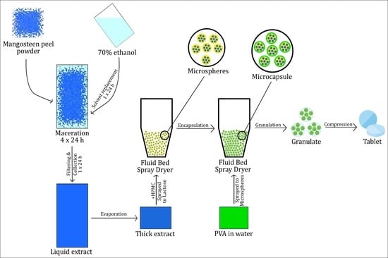
2.2. Mangosteen Peel Extraction

The 25 kg of fine dry mangosteen peel powder was placed in the macerator (Extraction and Concentration Machine TD-300, China) and soaked in aqueous ethanol solution 70% (v/v) until all the powder was wetted, then ethanol was added again until the powder was completely submerged. This procedure was carried out for 4 days, with a replacement of solvent every 24 h. Each macerate was filtered and mixed before being evaporated to make a thick extract in a rotary evaporator (BUCHI Rotavapor R-300, PT. BUCHI, Indonesia) at 40 °C and 30 rpm.

2.3. Phytochemical Screening, Standardization, Thin-Layer Chromatography Profile, and Antioxidant Activity of Mangosteen Peel Extract

Phytochemical screening was conducted to determine the secondary metabolite content of mangosteen peel extract (MPE). The phytochemical screening included alkaloids, tannins, polyphenols, flavonoids, quinones, saponins, monoterpenes, sesquiterpenes, steroids, and triterpenoids. Standardization was also carried out to determine the quality of MPE. Types of inspection performed were organoleptic, ethanol-soluble extract content, water-soluble extract content, total ash content, acid-insoluble ash content, and drying shrinkage.
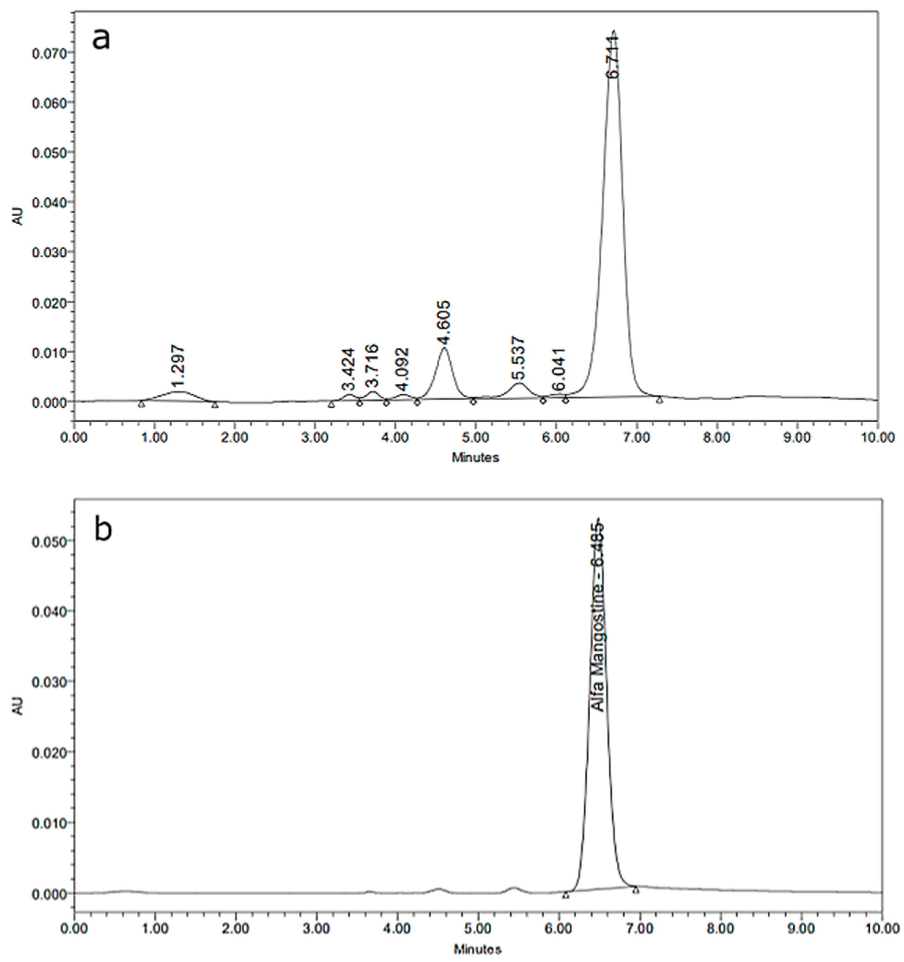
The thin-layer chromatography profile of MPE was examined using a silica plate GF 254 with a mixed mobile phase of chloroform and ethyl acetate (9:1). After the chamber was saturated, 5% v/v MPE solution in methanol was applied to the starting line on the silica plate and then waited for the mobile phase to reach the finish line on the silica plate. The retention factor value of each spot was recorded.
The antioxidant activity of MPE was determined using the DPPH reagent. The sample and positive control (ascorbic acid) were prepared to as much as 100 µL with a concentration range of 10–50 and 1–5 ppm, respectively. A total of 0.1 mL of DPPH reagent (0.2 mg/mL in ethanol) was added to the sample and the positive control. The sample and positive control were allowed to stand for 30 min in the dark (25 °C), after which the absorbance was measured at a wavelength of 517 nm (Epoch™ Microplate Spectrophotometer, BioTek Instrument, Inc., Winooski, VT, USA). The IC50 value was calculated from the linear equation of %inhibition vs. concentrations, where %inhibition was obtained using the following equation:
%   inhibition = A C o n t r o l A S a m p l e A C o n t r o l × 100 %

2.4. Total Phenolic and α-Mangosteen Content of Mangosteen Peel Extract

Total phenolic of MPE was determined using gallic acid as the standard. The standard was made into several variations of concentration (200–600 ppm). The extract solution was prepared by dissolving 10 mg of MPE in 10 mL of methanol. One mL of the extract solution and each concentration of the standard solution was put into a dilution tube, then 5 mL of Folin–Ciocalteau reagent was added. The preparations were mixed for 8 min, then 4 mL of 1% v/v NaOH was added. The absorbance of the extract solution and the standard were measured at a wavelength of 256 nm (Epoch™ Microplate Spectrophotometer, BioTek Instrument, Inc., Winooski, VT, USA).
Determination of the α-mangosteen content was carried out using high-performance liquid chromatography (HPLC) (Waters Alliance HPLC, Waters Corporation, USA). The standard solution of α-mangosteen was made into several concentrations in the range of 10–50 ppm to create the standard curve. The extract solution (10 mg/mL in methanol) was filtered using a syringe filter and then inserted into the HPLC sample tube. The column used was C18. The mobile phase used was methanol and aquades (95:5 v/v). The injection volume was set to 10 L with a flow rate of 1 mL/min. The retention time of α-mangosteen was 10 min and was measured at a wavelength of 318 nm using a UV detector.

2.5. Mangosteen Peel Extract Microencapsulation

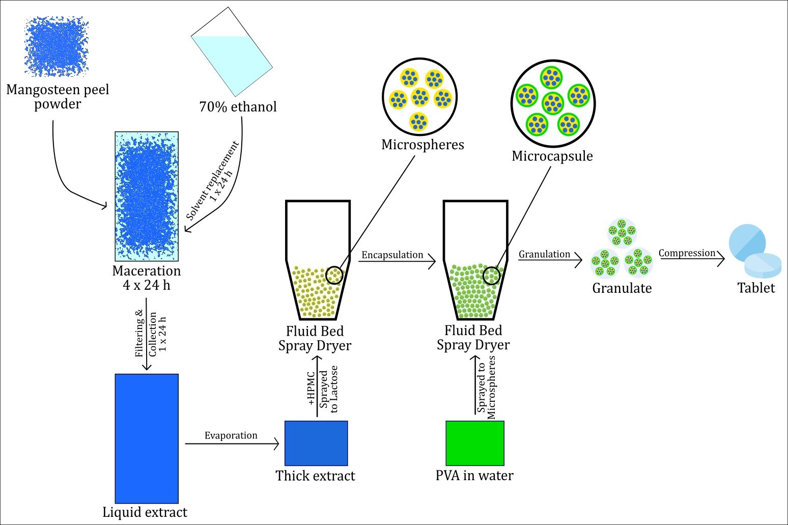
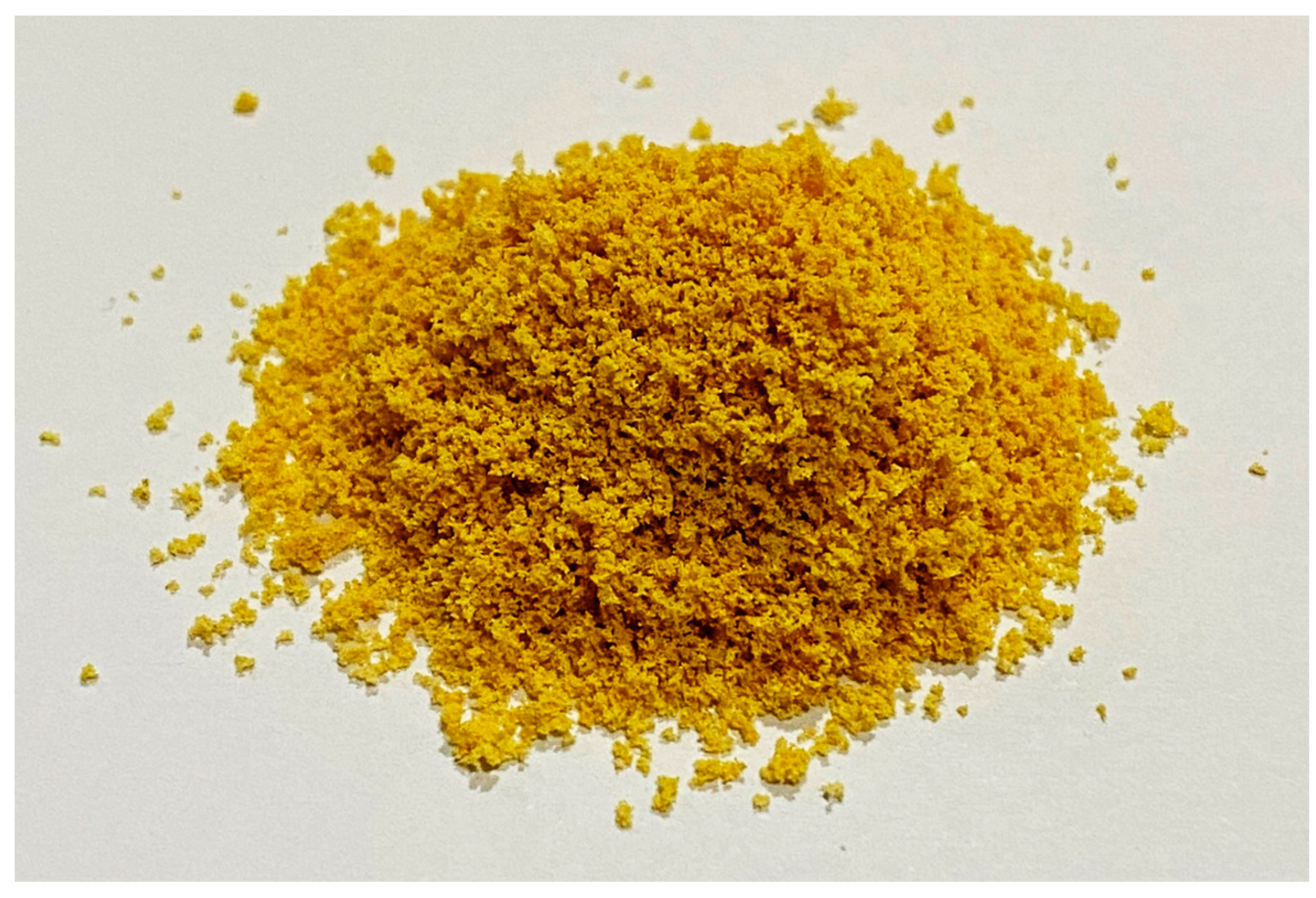
Microcapsules of MPE (MPEM) were prepared using a Fluid Bed Spray Dryer (PMS FBD5, Armitec, Thailand). Outlet temperature was set at 37.2 °C, with a spray interval of 10 s, product temperature of 37.8 °C, inlet temperature of 37.8 °C, and spray rate of 20.5 rpm. The core material (microspheres) was prepared by spraying a mixture of 20% w/v HPMC and 33.3% v/v MPE (600 mL) onto 1.5 kg of lactose. After the mixture was dry and homogeneous, the microspheres were again sprayed with 15% w/v polyvinyl alcohol in 450 mL of water as a coating agent. The thin-layer chromatography profile and IC50 of MPEM were then determined using the previously described method.

2.6. Characterization of Mangosteen Peel Extract Microcapsule

2.6.1. Moisture Content

A total of 1 g of MPEM was placed in a dish on a moisture balance device (Moisture Analyzer MA 50.R, Radwag, Miami, FL, USA). The temperature was set at 105 °C. Drying loss was recorded after the tool showed constant weight during heating.

2.6.2. Flow Rate and Angle of Repose

A total of 25 g of MPEM was placed in the funnel of the flowmeter (GTB Series, Erweka, Langen, Germany). The MPEM flow rate was determined by observing the time it takes for MPEM to pass through the funnel until it runs out. The angle of repose was obtained by measuring the diameter and height of the MPEM pile formed.

2.6.3. Compressibility Index

A total of 25 g of MPEM was put into a measuring cup contained in the volumenometer (Tapped Density Tester, Erweka SVM 221, Erweka). The compressibility index of MPEM was determined by the final volume of the microcapsule after 500 beats.

2.6.4. Shape, Morphology, and Particle Size

The shape and surface morphology of MPEM were observed using a scanning electron microscope (SEM) (JSM-6360, Jeol, Tokyo, Japan) with 500× magnification. The particle size of MPEM was then determined using a particle size analyzer (Horiba SZ-100, Horiba Ltd., Kyoto, Japan). The test was carried out by dispersing the sample in a phosphate buffer at pH 6.8, after which a 1 mL sample was taken for testing.

2.6.5. Solubility or Sedimentation Volume

A total of 5 g of MPEM was dissolved in 100 mL of distilled water and then stirred for 20 s. Sedimentation volume was measured after 15 min.

2.7. Tablet Formulation and Evaluation

MPEM tablets were made by mixing MPEM and internal phase materials (binder, disintegrant, and filler). The wet granulation method was used for the granulation. After the granules were formed, the external phase (lubricant, glidant, and filler residue) was added to the granule mass. The optimal binder was chosen using a formula optimization method, as indicated in Table 1. The amount of MPEM used was 435 mg, equivalent to 30 mg of ascorbic acid (based on the IC50 value). The granule mass was then molded with a diameter of 0.9 cm and a thickness of 0.33 cm. The total weight per tablet was 750 mg.
The characteristics of the granules and tablets were evaluated to determine the quality of the tablets in each test formula. The thin-layer chromatography profile and IC50 of the best formulas were determined using the previously described method. The examined granules’ properties were drying shrinkage, flow rate, angle of repose, and compressibility index using the same method for evaluating the quality of the microcapsules. The tablets evaluations were as follows.

2.7.1. Organoleptic

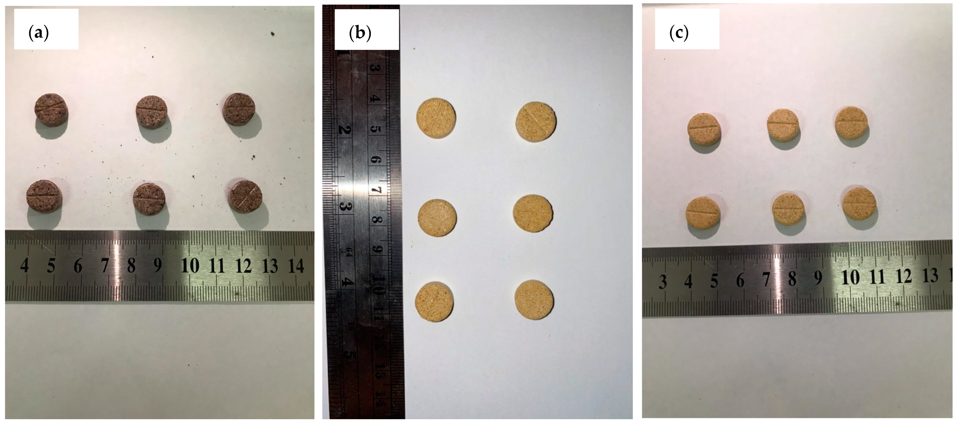
Organoleptic examination of MPEM tablets was carried out by observing the shape, color, and aroma of the 10 whole tablets. To assess the taste, the tablet was crushed into a powder and a certain amount of the powder was then tasted. This test was assessed by 8 people of the research project team.

2.7.2. Hardness Test

A hardness test was performed by placing the tablet on the hardness tester (Monsato VMT, Vinsyst Technologies, Mumbai, India). The hardness tester applied increasing pressure periodically until the tablet cracked. The tablet hardness was then recorded in the N unit. The number of samples used in this procedure was 10 tablets.

2.7.3. Size and Weight Uniformity

A total of 20 tablets were prepared to check the uniformity of the tablets’ size and weight. Tablet dimension (diameter and thickness) was measured using a caliper (Vernier Caliper, Tricle Brand, Shanghai, China), while the tablet weight was weighed using an analytical balance (Mettler Toledo ME204, PT. Mettler-Toledo, Bekasi City, Indonesia). Average dimensions and weight were then calculated along with the standard deviation.

2.7.4. Friability Test

The friability test was carried out by placing 20 weighed tablets into the friability tester (Biobase TFT-2, Biobase Group, Jinan, China). The rotational speed of the friabilator was set at 400 rpm for 15 min. After that, the tablets were removed and reweighed to obtain the average weight.

2.7.5. Disintegration Test

The disintegration test was carried out using a disintegration tester (Disintegration Tester TDT-2IM, Zhengzhou, China) with a water medium at 37 ± 2 °C. Disintegration time was recorded when the tablets in the basket were completely crushed.

2.8. Statistical Analysis

The paired T-test method was used to assess the significance of the change in antioxidant activity (IC50) in each intermediate product. The data were statistically analyzed using the Statistical Package for the Social Sciences (SPSS) version 22 (IBM Corporation, New York, NY, USA).

3. Results and Discussion

3.1. Phytochemical Screening and Standardization of Mangosteen Peel Extract

The yield of the extract obtained was 12.16% w/w (weight of simplicia 25 kg, extract weight 2973 kg). Eight secondary metabolites were successfully discovered in the simplicia of mangosteen peel. Following the methanol extraction method, it was found that MPE includes six secondary metabolites (see Table 2). It has been reported that MPE does contain many alkaloids, polyphenols, flavonoids, tannins, saponins, and quinones [32]. Therefore, MPE is known to have high antioxidant properties [3,33]. The extract was a thick dark liquid with a chelate odor and a bitter taste. The high tannin content is responsible for the bitter taste and chelate [34].
Based on the standardization process, it was found that the obtained MPE followed the desired specifications (see Table 3).

3.2. Total Phenolic and α-Mangosteen Content of Mangosteen Peel Extract

MPE had a higher total phenolic content than that in the reported literature [34,36]. After being made into tablets, the phenolic content did not change significantly. The concentration of α-mangosteen in MPEM tablets was also high, see Table 4. This is higher than the reported data (12.06% α-mangosteen in ethyl acetate extract of mangosteen peel) [37]. Based on this percentage, the amount of α-mangosteen contained in one MPEM tablet was ~70 mg. The chromatogram of MPE and standard α-mangosteen can be seen in Figure 1.

3.2.1. Physical Properties

The obtained MPEM had acceptable physical properties, see Table 5. Based on the angle of repose value, the flowability and compressibility of MPEM were fair (>30° and >15, respectively) and could be improved through granulation [38]. The MPEM also showed good solubility in water (5 g/100 mL) [39] and did not show any precipitate from 15 min to 1 day of storage. The appearance of MPEM can be seen in Figure 2. Although the PVA coating was water-resistant when dry, it did not hinder the solubility of MPEM. PVA has hydrophilic and hydrophobic groups that can act as surfactants to improve solubility [40].

3.2.2. Scanning Electron Microscope (SEM) and Particle Size

The morphology of the mangosteen rind extract microcapsules was observed using a scanning electron microscope (SEM) with 500× magnification. Figure 3 shows that the particle size after coating (right) did not increase appreciably when compared to before coating (left). In both images, it can be seen that the MPEM surface was rough, hollow, and not crystalline. This situation may affect the solubility of MPEM. The particles tend to be round and connected. As reported by Ekdahl et al., the particles formed from the spray-drying process have surface characteristics that are hollow and amorphous. This might be owing to the evaporation of the solvent that was previously bonded to the particle surface and evaporated during the heating and drying process in fluidized bed spray-drying, causing voids to appear on the surface [41]. The particle size of MPEM was measured using a particle size analyzer (PSA) (Horiba SZ-100, Horiba Ltd., Kyoto, Japan) and the particle size of the mangosteen rind extract microcapsules was 411.2 µm.

3.3. Characterization of Granules

Drying shrinkage was used to ensure the stability of the preparation storage, and the smaller the water content, the better the quality of the granules. The smallest water content contained in MPEM granules was found in formula F3 with a LOD of 1.74%.
The flow rate is the time required for the granules to flow through a funnel-shaped device (flowmeter). Based on the test result, the MPEM granules in formula F3 had excellent flow properties (see Table 6) because they had an angle of repose of <30° [42] and flow rate of >10 g/s [43].
The compressibility test was intended to see the decrease in granule volume due to tapping accompanied by vibration. MPEM granules in the F3 formula had a good compressibility index with a value below 15 [44]. Based on the four test parameters, the formula with the best granule properties was a formula containing 5% w/w PVP K30 as a binder. This granulation process has also been shown to increase the flowability and compressibility of MPEM.

3.4. Tablet Evaluations

The three test formulas produced uniform tablet colors. However, in formula F1, the tablet was dark brown and less attractive (see Figure 4a), while formulas F2 and F3 produced a yellowish-orange color (see Figure 4b or Figure 4c). Based on its shape, the F3 formula produced a rounded shape that was neater and firmer than the other formulas. This may be due to the low hardness and friability of the tablets in formulas F2 and F3 (see Table 5).
The hardness test was carried out to determine whether the tablet can survive and avoid damage during packaging and distribution [36]. Tablet hardness should be in the range of 4–8 kg or 39.2–78.4 N (where 1 kg = 9.8 N) [45]. The results of the hardness and friability test show that the MPEM tablets in the F3 formula have good mechanical integrity [37]. A friability value below 1% indicates that the tablet can withstand abrasion during packaging, transportation, and handling [38]. Tablets must not only be hard and not brittle, but they must also be easily dissolved to provide an immediate effect. This can be proven through the disintegration test. For immediate release tablets, the disintegration time must be under 15 min [46]. The properties of the formulas can be seen in Table 7.
Based on the evaluation results, the formula that produced the best tablet characteristics was the F3 formula. For this reason, the evaluation of the stability and effectiveness of antioxidants was only carried out on the F3 formula.

3.5. Stability and Antioxidant Activity of Mangosteen Peel Extract during Preparation and Storage

The stability of α-mangosteen in the fabrication process can be observed through the TLC profile. The retention factor value of α-mangosteen was 0.75, with a blue coloration under the UV 254 lamp and green color under the UV 366 lamp. Based on the TLC profile, it can be seen that MPE, MPEM, and their tablets still contain α-mangosteen, which was indicated by the presence of the same spot at a retention factor value of 0.75 (see Table 8). This indicates that the method employed to transform mangosteen peel powder into tablets does not affect the stability of α-mangosteen.
Antioxidant stability in each intermediate product was also maintained (see Table 7). This was confirmed by statistical analysis using the paired t-test method, where the IC50 value for each intermediate product did not change significantly (p > 0.05). This shows that the microencapsulation method can maintain antioxidant stability from thermal and mechanical exposure in the tableting process. The IC50 of the products can be seen in Table 9.
MPEM tablets showed good stability with maintained α-mangosteen levels for a month of storage at zone IVB conditions (temperature of 40 °C and RH of 75%). The levels of α-mangosteen on days 0, 15, and 30 were 15.87%, 15.75%, and 15.66%, respectively.

4. Conclusions

The microencapsulation method using HMPC as a core mixture and PVA as a coating can maintain stability and increase the solubility of mangosteen peel extract in water. This is demonstrated by the α-mangosteen content and IC50 values that were maintained during tablet production and one month of storage at zone IVB conditions. The obtained microcapsule was 411.2 µm in size and has a shape that tends to be round with a rough surface texture. The flow properties and compressibility of MPEM were fair and significantly improved through granulation. The best granule and tablet formula was a formula containing 5% w/w PVP K30 as a binder, with the characteristics of the tablet being yellowish-orange, hardness of 87.8 ± 1.398 N, friability of 0.94%, and disintegration time of 25.75 ± 0.676 min. According to the findings, the microencapsulation is an effective technique for improving the solubility and antioxidant stability of mangosteen peel extract during manufacture and storage.

Author Contributions

Conceptualization, A.K.U., A.Y.C. and S.S.; data curation, A.K.U., S.S., A.Y.C., R.P., A.T.A. and N.W.; formal analysis, A.K.U., S.S., A.Y.C., R.P., A.T.A. and N.W.; funding acquisition, S.S. and A.K.U.; investigation, A.K.U., R.P. and A.T.A.; methodology, A.K.U., S.S., A.Y.C., N.W., R.P. and A.T.A.; project administration, A.K.U., S.S. and A.Y.C.; resources, R.P., A.T.A., S.S. and N.W.; software, A.K.U.; supervision, A.K.U., S.S. and A.Y.C.; validation, A.K.U., S.S., A.Y.C. and N.W.; visualization, R.P. and A.T.A.; writing—original draft, A.K.U., S.S., A.Y.C., R.P., A.T.A. and N.W.; writing—review and editing, A.K.U., S.S., A.Y.C., R.P., A.T.A. and N.W. All authors have read and agreed to the published version of the manuscript.

Funding

This work was supported by research grants from the Ministry of Research and Technology/National Research and Innovation Agency (1827/UN6.3.1/LT/2020).

Institutional Review Board Statement

Not applicable.

Informed Consent Statement

Not applicable.

Data Availability Statement

Not applicable.

Conflicts of Interest

The authors declare no conflict of interest.

References

  1. Mamondol, M.R. Investment feasibility and marketing of mangosteen commodity in Central Sulawesi, Indonesia. J. Socioecon. Dev. 2020, 3, 115. [Google Scholar] [CrossRef]
  2. Poerwanto, R.; Efendi, D.; Sobir; Suhartanto, R. Improving Productivity and Quality of indonesian mangosteen. Acta Hortic. 2008, 769, 285–288. [Google Scholar] [CrossRef]
  3. Suttirak, W.; Manurakchinakorn, S. In Vitro antioxidant properties of mangosteen peel extract. J. Food Sci. Technol. 2014, 51, 3546–3558. [Google Scholar] [CrossRef] [Green Version]
  4. Huang, X.; Zhou, X.; Dai, Q.; Qin, Z. Antibacterial, Antioxidation, UV-Blocking, and Biodegradable Soy Protein Isolate Food Packaging Film with Mangosteen Peel Extract and ZnO Nanoparticles. Nanomaterials 2021, 11, 3337. [Google Scholar] [CrossRef] [PubMed]
  5. Widowati, W.; Ginting, C.N.; Lister, I.N.E.; Girsang, E.; Amalia, A.; Wibowo, S.H.B.; Kusuma, H.S.W. Rizal Anti-aging Effects of Mangosteen Peel Extract and Its Phytochemical Compounds: Antioxidant Activity, Enzyme Inhibition and Molecular Docking Simulation. Trop. Life Sci. Res. 2020, 31, 127–144. [Google Scholar] [CrossRef] [PubMed]
  6. Widyarman, A.S.; Lay, S.H.; Wendhita, I.P.; Tjakra, E.E.; Murdono, F.I.; Binartha, C.T.O. Indonesian Mangosteen Fruit (Garcinia mangostana L.) Peel Extract Inhibits Streptococcus mutans and Porphyromonas gingivalis in Biofilms In Vitro. Contemp. Clin. Dent. 2019, 10, 123–128. [Google Scholar] [CrossRef]
  7. Kusmayadi, A.; Bachtiar, K.R.; Prayitno, C.H. The effects of mangosteen peel (Garcinia mangostana L.) and Turmeric (Curcuma domestica Val) flour dietary supplementation on the growth performance, lipid profile, and abdominal fat content in Cihateup ducks. Vet. World 2019, 12, 402–408. [Google Scholar] [CrossRef] [PubMed]
  8. Polyorach, S.; Wanapat, M.; Phesatcha, K.; Kang, S. Effect of different levels of mangosteen peel powder supplement on the performance of dairy cows fed concentrate containing yeast fermented cassava chip protein. Trop. Anim. Health Prod. 2015, 47, 1473–1480. [Google Scholar] [CrossRef]
  9. Foiklang, S.; Wanapat, M.; Norrapoke, T. Effect of Grape Pomace Powder, Mangosteen Peel Powder and Monensin on Nutrient Digestibility, Rumen Fermentation, Nitrogen Balance and Microbial Protein Synthesis in Dairy Steers. Asian-Australas. J. Anim. Sci. 2016, 29, 1416–1423. [Google Scholar] [CrossRef]
  10. Sitti, R.H.S.; Sugita, P.; Ambarsari, L.; Rahayu, D.U.C. Antibacterial Mangosteen (Garcinia mangostana Linn.) peel extract encapsulated in Chitosan. J. Phys. Conf. Ser. 2018, 1116, 042037. [Google Scholar] [CrossRef]
  11. Pratami, D.K.; Mun’im, A.; Hermansyah, H.; Gozan, M.; Sahlan, M. Microencapsulation Optimization of Propolis Ethanolic Extract from Tetragonula spp Using Response Surface Methodology. Int. J. Appl. Pharm. 2020, 12, 197–206. [Google Scholar] [CrossRef]
  12. Jamdar, F.; Ali Mortazavi, S.; Reza Saiedi Asl, M.; Sharifi, A. Physicochemical properties and enzymatic activity of wheat germ extract microencapsulated with spray and freeze drying. Food Sci. Nutr. 2021, 9, 1192–1201. [Google Scholar] [CrossRef]
  13. Rodrigues, R.; Bilibio, D.; Plata-Oviedo, M.S.V.; Pereira, E.A.; Mitterer-Daltoé, M.L.; Perin, E.C.; Carpes, S.T. Microencapsulated and Lyophilized Propolis Co-Product Extract as Antioxidant Synthetic Replacer on Traditional Brazilian Starch Biscuit. Molecules 2021, 26, 6400. [Google Scholar] [CrossRef] [PubMed]
  14. Vonghirundecha, P.; Chusri, S.; Meunprasertdee, P.; Kaewmanee, T. Microencapsulated functional ingredients from a Moringa oleifera leaf polyphenol-rich extract: Characterization, antioxidant properties, in vitro simulated digestion, and storage stability. LWT 2022, 154, 112820. [Google Scholar] [CrossRef]
  15. Nori, M.P.; Favaro-Trindade, C.S.; Matias de Alencar, S.; Thomazini, M.; de Camargo Balieiro, J.C.; Contreras Castillo, C.J. Microencapsulation of propolis extract by complex coacervation. LWT Food Sci. Technol. 2011, 44, 429–435. [Google Scholar] [CrossRef]
  16. Anwar, S.H.; Weissbrodt, J.; Kunz, B. Microencapsulation of Fish Oil by Spray Granulation and Fluid Bed Film Coating. J. Food Sci. 2010, 75, E359–E371. [Google Scholar] [CrossRef]
  17. Jyothi, N.V.N.; Prasanna, P.M.; Sakarkar, S.N.; Prabha, K.S.; Ramaiah, P.S.; Srawan, G.Y. Microencapsulation techniques, factors influencing encapsulation efficiency. J. Microencapsul. 2010, 27, 187–197. [Google Scholar] [CrossRef]
  18. Frey, C. Fluid Bed Coating-Based Microencapsulation. In Microencapsulation in the Food Industry; Elsevier: Amsterdam, The Netherlands, 2014; pp. 65–79. [Google Scholar]
  19. Aguilera-Chávez, S.L.; Gallardo-Velázquez, T.; Meza-Márquez, O.G.; Osorio-Revilla, G. Spray Drying and Spout-Fluid Bed Drying Microencapsulation of Mexican Plum Fruit (Spondias purpurea L.) Extract and Its Effect on In Vitro Gastrointestinal Bioaccessibility. Appl. Sci. 2022, 12, 2213. [Google Scholar] [CrossRef]
  20. Parente, J.F.; Sousa, V.I.; Marques, J.F.; Forte, M.A.; Tavares, C.J. Biodegradable Polymers for Microencapsulation Systems. Adv. Polym. Technol. 2022, 2022, 4640379. [Google Scholar] [CrossRef]
  21. Ngwuluka, N.C.; Abu-Thabit, N.Y.; Uwaezuoke, O.J.; Erebor, J.O.; Ilomuanya, M.O.; Mohamed, R.R.; Soliman, S.M.A.; Abu Elella, M.H.; Ebrahim, N.A.A. Natural Polymers in Micro- and Nanoencapsulation for Therapeutic and Diagnostic Applications: Part I: Lipids and Fabrication Techniques. In Nano- and Microencapsulation—Techniques and Applications; IntechOpen: London, UK, 2021. [Google Scholar]
  22. Cilurzo, F. Characterization and physical stability of fast-dissolving microparticles containing nifedipine. Eur. J. Pharm. Biopharm. 2008, 68, 579–588. [Google Scholar] [CrossRef]
  23. Rogers, T.L.; Wallick, D. Reviewing the use of ethylcellulose, methylcellulose and hypromellose in microencapsulation. Part 3: Applications for microcapsules. Drug Dev. Ind. Pharm. 2012, 38, 521–539. [Google Scholar] [CrossRef] [PubMed]
  24. Sheikh, M.A.A. Formulation and evaluation of Bacillus coagulans-loaded hypromellose mucoadhesive microspheres. Int. J. Nanomed. 2011, 6, 619–629. [Google Scholar] [CrossRef] [PubMed] [Green Version]
  25. da Silva Júnior, W.F.; de Oliveira Pinheiro, J.G.; Moreira, C.D.L.F.A.; de Souza, F.J.J.; de Lima, Á.A.N. Alternative Technologies to Improve Solubility and Stability of Poorly Water-Soluble Drugs. In Multifunctional Systems for Combined Delivery, Biosensing and Diagnostics; Elsevier: Amsterdam, The Netherlands, 2017; pp. 281–305. [Google Scholar] [CrossRef]
  26. Hiremath, P.; Nuguru, K.; Agrahari, V. Material Attributes and Their Impact on Wet Granulation Process Performance. In Handb Pharm Wet Granulation Theory Pract a Qual by Des Paradig; Academic Press: Cambridge, MA, USA, 2019; pp. 263–315. [Google Scholar] [CrossRef]
  27. Eyjolfsson, R. Introduction. Design and Manufacture of Pharmaceutical Tablets; Academic Press: Cambridge, MA, USA, 2015; pp. 1–28. [Google Scholar] [CrossRef]
  28. Ghoshal, S.; Denner, P.; Stapf, S.; Mattea, C. Study of the Formation of Poly (vinyl alcohol) Films. Macromolecules 2012, 45, 1913–1923. [Google Scholar] [CrossRef]
  29. Wu, K.; Song, L.; Wang, Z.; Hu, Y. Microencapsulation of ammonium polyphosphate with PVA-melamine-formaldehyde resin and its flame retardance in polypropylene. Polym. Adv. Technol. 2008, 19, 1914–1921. [Google Scholar] [CrossRef]
  30. Jaswir, I.; Noviendri, D.; Taher, M.; Mohamed, F.; Octavianti, F.; Lestari, W.; Mukti, A.; Nirwandar, S.; Hamad Almansori, B. Optimization and Formulation of Fucoxanthin-Loaded Microsphere (F-LM) Using Response Surface Methodology (RSM) and Analysis of Its Fucoxanthin Release Profile. Molecules 2019, 24, 947. [Google Scholar] [CrossRef] [Green Version]
  31. Sahlan, M.; Rahman, M.R. Optimization of Microencapsulation Composition of Menthol, Vanillin, and Benzyl Acetate inside Polyvinyl Alcohol with Coacervation Method for Application in Perfumery. IOP Conf. Ser. Mater. Sci. Eng. 2017, 214, 012005. [Google Scholar] [CrossRef] [Green Version]
  32. Sinaga, R.N.; Siregar, N.S. Phytochemical Screening and Test of Antioxidant Activity in the Extract of Mangosteen Rind. In In Accelerating the Achievement of Sustainable Development Goals for the Improvement and Equitable Distribution of Population Health; Graduate Studies in Public Health, Graduate Program; Sebelas Maret University: Surakarta, Indonesia, 2016; Available online: www.theicph.com (accessed on 13 January 2022).
  33. Aizat, W.M.; Ahmad-Hashim, F.H.; Syed Jaafar, S.N. Valorization of mangosteen, “The Queen of Fruits,” and new advances in postharvest and in food and engineering applications: A review. J. Adv. Res. 2019, 20, 61–70. [Google Scholar] [CrossRef]
  34. Megawati; Ginting, R.R.; Kusumaningtyas, R.D.; Sediawan, W.B. Mangosteen Peel Antioxidant Extraction and Its Use to Improve the Stability of Biodiesel B20 Oxidation; Springer: Cham, Switzerland, 2020; pp. 29–61. [Google Scholar]
  35. Kementerian Kesehatan Republik Indonesia. Farmakope Herbal Inddonesia. In Pocket Handbook of Nonhuman Primate Clinical Medicine; Kementerian Kesehatan Republik Indonesia: Jakarta Selatan, Indonesia, 2017. [Google Scholar]
  36. Zadernowski, R.; Czaplicki, S.; Naczk, M. Phenolic acid profiles of mangosteen fruits (Garcinia mangostana). Food Chem. 2009, 112, 685–689. [Google Scholar] [CrossRef]
  37. Ghasemzadeh, A.; Jaafar, H.; Baghdadi, A.; Tayebi-Meigooni, A. Alpha-Mangostin-Rich Extracts from Mangosteen Pericarp: Optimization of Green Extraction Protocol and Evaluation of Biological Activity. Molecules 2018, 23, 1852. [Google Scholar] [CrossRef] [Green Version]
  38. Shanmugam, S. Granulation techniques and technologies: Recent progresses. BioImpacts 2017, 5, 55–63. [Google Scholar] [CrossRef] [Green Version]
  39. Savjani, K.T.; Gajjar, A.K.; Savjani, J.K. Drug Solubility: Importance and Enhancement Techniques. ISRN Pharm. 2012, 2012, 195727. [Google Scholar] [CrossRef] [PubMed] [Green Version]
  40. Li, X.; Chen, K.; Ji, X.; Yuan, X.; Lei, Z.; Ullah, M.W.; Xiao, J.; Yang, G. Microencapsulation of Poorly Water-soluble Finasteride in Polyvinyl Alcohol/chitosan Microspheres as a Long-term Sustained Release System for Potential Embolization Applications. Eng. Sci. 2020, 13, 106–120. [Google Scholar] [CrossRef]
  41. Ekdahl, A.; Mudie, D.; Malewski, D.; Amidon, G.; Goodwin, A. Effect of Spray-Dried Particle Morphology on Mechanical and Flow Properties of Felodipine in PVP VA Amorphous Solid Dispersions. J. Pharm. Sci. 2019, 108, 3657–3666. [Google Scholar] [CrossRef] [PubMed]
  42. Šimek, M.; Grünwaldová, V.; Kratochvíl, B. Comparison of Compression and Material Properties of Differently Shaped and Sized Paracetamols. KONA Powder Part. J. 2017, 34, 197–206. [Google Scholar] [CrossRef] [Green Version]
  43. Murini, T.; Wahyuningsih, M.S.H.; Fudholi, A.; Satoto, T.B.T. Optimization of Formula Granule of Lempuyang Gajah (Zingiber zerumbet (L) J.E.Smith) Rhizome Purified Extract as a Larvicide. Maj. Obat Tradis. 2020, 25, 34. [Google Scholar] [CrossRef]
  44. Svonja-Parezanovic, G.; Lalic-Popovic, M.; Golocorbin-Kon, S.; Todorovic, N.; Pavlovic, N.; Jovicic-Bata, J. The effect of magnesium stearate and sodium starch glycolate on powder flowability. Acta. Period. Technol. 2019, 50, 304–310. [Google Scholar] [CrossRef] [Green Version]
  45. Sari, R.N.; Irianto, H.E.; Hastarini, E. Production of Unsaturated Fatty Acids Concentrate Tablets from Sardinella sp. Oil. In E3S Web Conference, Proceedings of the 3rd International Symposium on Marine and Fisheries Research (3rd ISMFR), Yogyakarta, Indonesia, 8–9 July 2019; Isnansetyo, A., Dwi Jayanti, A., Wahyu Kartika Sari, D., Dewi Puspita, I., Prima Putra, M.M., Huda, N., Eds.; EDP Sciences: Les Ulis Cedex, France, 2020; Volume 10, p. 03008. [Google Scholar] [CrossRef] [Green Version]
  46. Chen, H.; Aburub, A.; Sun, C.C. Direct Compression Tablet Containing 99% Active Ingredient—A Tale of Spherical Crystallization. J. Pharm. Sci. 2019, 108, 1396–1400. [Google Scholar] [CrossRef]
Figure 1. Chromatogram of mangosteen peel extract (a) and standard α-mangosteen (b).
Antioxidants 11 01331 g001
Figure 2. Mangosteen peel extract microcapsules.
Antioxidants 11 01331 g002
Figure 3. Morphological observation of mangosteen peel extract microcapsules.
Antioxidants 11 01331 g003
Figure 4. Tablet appearance of formula F1 (a), formula F2 (b), and formula F3 (c).
Antioxidants 11 01331 g004
Table 1. The optimized tablet formulas.
CompositionAmount (%)Function
F1F2F3
Amprotab 555Disintegrant
NaCMC5--Binder
Starch-5-Binder
PVP K30--5Binder
Mg stearate 222Lubricants
Talcum 222Glidant
Lactose Ad 100Ad 100Ad 100Filler
Table 2. Phytochemical contents of mangosteen peel extract.
CompoundMangosteen Peel
SimpliciaExtract
Alkaloids++
Flavonoids++
Tannins++
Polyphenol++
Saponins++
Quinone++
Monoterpenes-Sequiterpenes+
Triterpenoids-Steroids+
Description: (+) detected, (−) not detected.
Table 3. Standard parameters of mangosteen peel extract.
Parameters Result
SampleReference [35]
Moisture content (% w/v) 7.83 <10.8
Total ash content (%) 0.244 <4.4
Acid insoluble ash content (%) 0.069 <0.2
Drying shrinkage (%) 8.49 <10
Specific gravity (g/cm3)0.81 <1
Table 4. Total phenolic and α-mangosteen content of mangosteen peel extract, mangosteen peel extract microcapsules, and their tablets.
No.ParameterValue
1.Total phenolic content of mangosteen peel extract39.87% ± 1.840%
2.Total phenolic content of MPEM tablets34.73% ± 0.617%
3.α-mangosteen content of MPEM tablets15.68% ± 0.332%
4.Amount of α-mangosteen in MPEM tablets~70 mg
Note: the significance of changes in phenolic levels in samples before and after tableting was analyzed using the paired t-test (p > 0.05).
Table 5. Physical properties of mangosteen peel extract microcapsules.
No.ParameterValue
1.Flow properties32.46 ± 1.73
2.Angle of repose43.45 ± 1.79°
3.Compressibility21.51 ± 0.59
4.Loss on drying0.74% ± 0.11%
Table 6. Granule properties of the optimized formulas.
NoParametersFormula
F1F2F3
1.Loss on drying (%)3.64 ± 0.352.14 ± 0.721.74 ± 0.05
2.Flowability (g/s)3.79 ± 0.297.04 ± 0.5810.74 ± 0.56
3.Angle of repose (°)24.21 ± 1.7225.06 ± 4.1725.06 ± 0.72
4.Carr’s index18.00 ± 3.6017.00 ± 2.089.00 ± 1.52
Table 7. Tablet properties of the optimized formulas.
NoParametersFormula
F1F2F3
1.Organoleptic Dark brown, still bitter, typical mangosteen aroma, and non-uniform shape Yellowish orange, less bitter, typical mangosteen aroma, and more uniform shapeYellowish orange, less bitter, typical mangosteen aroma, and uniform shape
2.Hardness test (N)22.45 ± 3.3836.40 ± 7.1887.80 ± 1.39
3.Weight (mg)0.81 ± 0.000.71 ± 0.000.77 ± 0.01
4.Diameter (cm)0.90 ± 0.000.90 ± 0.000.90 ± 0.00
5.Thickness (cm)0.34 ± 0.010.34 ± 0.040.33 ± 0.00
6.Friability test (%)6026.390.94
7.Disintegration test (min) 43.04 ± 1.7210.86 ± 0.4514.29 ± 0.67
Table 8. Thin-layer chromatography profile of each fabrication raw product.
Rf ValueSpots’ Color
UV 254UV 366
α-MMPEMPEMTabletα-MMPEMPEMTablet
0.2-Blue---GreenGreen-
0.25-Blue---Green--
0.3--BlackBlack--GreenGreen
0.5-----Green--
0.75BlueBlueBlueBlueGreenGreenGreenGreen
0.87-----Green--
0.97-----Green--
Note: α-M = α-mangosteen standard, MPE = mangosteen peel extract, MPEM = mangosteen peel extract microcapsule.
Table 9. The IC50 value of each intermediate product compared to ascorbic acid.
SampleIC50 (µg/mL)
Ascorbic acid 1.81 ± 0.09
Mangosteen peel extract34.64 ± 6.58
Mangosteen peel extract microcapsule40.68 ± 0.17
Tablet41.16 ± 0.69
Note: the significance of changes in IC50 values of samples was analyzed using the paired t-test (p > 0.05).
Publisher’s Note: MDPI stays neutral with regard to jurisdictional claims in published maps and institutional affiliations.

