Article

Histidine Deficiency Inhibits Intestinal Antioxidant Capacity and Induces Intestinal Endoplasmic-Reticulum Stress, Inflammatory Response, Apoptosis, and Necroptosis in Largemouth Bass (Micropterus salmoides)

1
Key Laboratory of Integrated Rice-Fish Farming Ecology, Ministry of Agriculture and Rural Affairs, Freshwater Fisheries Research Center, Chinese Academy of Fishery Sciences, Wuxi 214081, China
2
Wuxi Fisheries College, Nanjing Agricultural University, Wuxi 214081, China
3
Healthy Aquaculture Key Laboratory of Sichuan Province, Tongwei Co., Ltd., Chengdu 610093, China
*
Authors to whom correspondence should be addressed.
Antioxidants 2022, 11(12), 2399; https://doi.org/10.3390/antiox11122399
Submission received: 19 October 2022 / Revised: 12 November 2022 / Accepted: 29 November 2022 / Published: 2 December 2022
(This article belongs to the Special Issue Natural Antioxidants in Animal Immunity)

Abstract

:
This 56-day study aimed to evaluate the effects of histidine levels on intestinal antioxidant capacity and endoplasmic-reticulum stress (ERS) in largemouth bass (Micropterus salmoides). The initial weights of the largemouth bass were (12.33 ± 0.01) g. They were fed six graded levels of histidine: 0.71% (deficient group), 0.89%, 1.08%, 1.26%, 1.48%, and 1.67%. The results showed that histidine deficiency significantly suppressed the intestinal antioxidant enzyme activities, including SOD, CAT, GPx, and intestinal level of GSH, which was supported by significantly higher levels of intestinal MDA. Moreover, histidine deficiency significantly lowered the mRNA level of nrf2 and upregulated the mRNA level of keap1, which further lowered the mRNA levels of the downstream genes sod, cat, and gpx. Additionally, histidine-deficiency-induced intestinal ERS, which was characterized by activating the PEPK-signalling pathway and IRE1-signalling pathway, including increased core gene expression of pepk, grp78, eif2α, atf4, chopα, ire1, xbp1, traf2, ask1, and jnk1. Dietary histidine deficiency also induced apoptosis and necroptosis in the intestine by upregulating the expressions of proapoptotic genes, including caspase 3, caspase 8, caspase 9, and bax, and necroptosis-related genes, including mlkl and ripk3, while also lowering the mRNA level of the antiapoptotic gene bcl-2. Furthermore, histidine deficiency activated the NF-κB-signalling pathway to induce an inflammatory response, improving the mRNA levels of the proinflammatory factors tnf-α, hepcidin 1, cox2, cd80, and cd83 and lowering the mRNA levels of the anti-inflammatory factors tgf-β1 and ikbα. Similarly, dietary histidine deficiency significantly lowered the intestinal levels of the anti-inflammatory factors TGF-β and IL-10 and upregulated the intestinal levels of the proinflammatory factor TNF-α, showing a trend similar to the gene expression of inflammatory factors. However, dietary histidine deficiency inhibited only the level of C3, and no significant effects were observed for IgM, IgG, HSP70, or IFN-γ. Based on the MDA and T-SOD results, the appropriate dietary histidine requirements of juvenile largemouth bass were 1.32% of the diet (2.81% dietary protein) and 1.47% of the diet (3.13% dietary protein), respectively, as determined by quadratic regression analysis.

1. Introduction

The intestine, as one of the most important tissues in fish, plays a role in the digestion and absorption of nutrients; hence, intestinal health is a prerequisite for healthy animal growth [1,2]. The intestinal tract is also the largest mucosal immune organ and the most vulnerable immune barrier in fish [3]. However, fish intestinal walls are thin and easily damaged [2]. Additionally, the intestinal polyunsaturated fatty acid content in fish is high and easily damaged by oxidation [1,2,3]. In addition, the intestinal tract is one of the parts of the body most closely related to the external environment and is prone to environmental stress. Therefore, it is important to construct nutritional programs to maintain the intestinal health of fish. Numerous studies have shown that healthy intestinal development depends on the composition of the feed formulation, including elements such as amino acids [4,5], tributyrin [6], and vitamins [7,8]. These studies suggested that balanced nutrition may have beneficial effects on the intestinal health of fish.
Histidine, an essential amino acid for fish, has important functions in improving fish growth [9]. According to research reports, histidine acts as an antioxidant and plays an important role in scavenging oxidative free radicals in fish and mammals [10,11]. Histidine has other significant antioxidant activities in addition to scavenging free radicals, such as binding divalent metal ions and resisting glycation [10,12]. In grass carp, dietary histidine has a positive effect on lowering lipid peroxidation and protein oxidation in the intestines and gills [4,13]. In addition, histidine has important functions in inhibiting lipid peroxidation in the sarcoplasmic reticulum of fish muscle [14]. The antioxidant functions of histidine are mainly achieved through the nuclear factor erythroid 2-related factor 2 (Nrf2) signalling pathway [13,15]. Furthermore, the endoplasmic reticulum (ER), as one of the most important organelles in eukaryotic cells, is closely related to cell homeostasis, and ER homeostasis is maintained through unfolded protein reactions involving three signalling pathways: the PEPK-signalling pathway, IRE-1-signalling pathway, and ATF6-signalling pathway [16]. In aquatic animals, numerous studies have reported that adverse factors could induce endoplasmic-reticulum stress (ERS) by affecting the mRNA levels of core genes in the IRE1-signalling pathway, PERK-signalling pathway, and ATF6-signalling pathway, such as high-fat induction in blunt snout bream (Megalobrama amblycephala) [17], hydrogen peroxide exposure and ammonia stress in common carp (Cyprinus carpio) [18,19], waterborne cadmium exposure in Gibel carp (Carassius gibelio) [20], and nitrite induction in grass carp (Ctenopharyngodon idella) [21]. When ERS occurs or is persistent and exceeds the range of cellular self-regulation, it leads to cell apoptosis and death and induces disease [22]. Furthermore, ERS might activate the NF-κB-signalling pathway and regulate downstream inflammatory factors to induce an inflammatory response [23]. Hence, reducing ERS and oxidative stress is crucial to improving animal health and immunity.
The largemouth bass (Micropterus salmoides) is native to freshwater rivers and large lakes in America, especially the Great Lakes in the United States. These fish have been widely raised around the world because of their tender meat and delicious taste that is loved by consumers. Largemouth bass sell well on the international market and are known as freshwater grouper. However, due to the problems of breeding density and nutrient level, largemouth bass often suffer from physiological problems such as metabolic disorders and intestinal inflammation, which could reduce their growth and induce diseases, resulting in great economic losses. In our previous study, the appropriate dietary histidine level was 1.26% of their diet [24], which showed the best growth performance. However, how histidine, an important antioxidant, regulates the intestinal health needs to be investigated. Hence, this study is to assess the functions of histidine in the intestinal health of largemouth bass.

2. Materials and Methods

2.1. Ethical Statement

This experiment was based strictly on the requirements of the Institutional Animal Care and Ethics Committee of Nanjing Agricultural University, Nanjing, China [Permit number: SYXK (Su) 2011-0036].

2.2. Experimental Diet

The experimental formulation referred to our previous study [24]. A combination of crystalline amino acids, excluding histidine, was supplemented to simulate the 47%-whole-body-amino-acid pattern of largemouth bass [24]. The additional level of histidine was designed with an arithmetic difference of 0.2%. The compositional analysis of histidine levels in feed were 0.71%, 0.89%, 1.08%, 1.26%, 1.48%, and 1.67%. The basic formula levels are shown in Table 1. All ingredients were fully crushed to completely pass through an 80 μm mesh screen, and then the feed was prepared according to the experimental formulas and thoroughly mixed with water and oil. Lastly, the mixed ingredients were made into 1 mm expanded pellets with an aquatic feed puffing machine (Jiangsu Muyang Holdings Co., Ltd., Yangzhou, China). The feed was dried and stored for breeding experiments.

2.3. Breeding Experiment

Juvenile largemouth bass ((12.33 ± 0.01) g) were randomly divided into 18 experimental cages (3 replicates for each treatment group and 20 fish in each experimental cage). The fish were fed two times daily (at 7:00 and 17:00) with the appropriate DHL feed according to the standard of apparent satiety. The indices of the aquaculture water during the experiment were (28 ± 2) °C (water temperature), 7–7.5 (pH), >6.0 mg/L (dissolved oxygen), and <0.01 mg/L (total ammonia nitrogen).

2.4. Sample Collection

At the end of 56 days of feeding, the fish were starved for 24 h. After anesthetization with MS-222 (200 mg/L), three fish from each cage were dissected to obtain intestine samples, and a total of 9 samples in each experimental group were used for antioxidant indices, immune cytokines, and qRT-PCR analysis.. These samples were stored at −80 °C for subsequent analysis.

2.5. Chemical Analysis, RNA Extraction, and Quantitative Real-Time PCR

Measurements of the moisture, crude protein, ash, and crude lipid of the raw materials and diets were conducted using the established methods of AOAC [25]. After pretreatment, all samples were analyzed using an Agilent-1100 amino acid assay system (Agilent Technologies Co., Ltd., Santa Clara, CA, USA). Tryptophan hydrolysis was carried out in 5 N NaOH at 110 °C for 20 h, which referred to our previous study [24]. The gross energy of diets were analyzed by combustion using a IKA C6000 oxygen bomb calorimeter (IKA WORKS GUANGZHOU, Guangzhou, China). The contents of glutathione (GSH) and malondialdehyde (MDA), as well as the activities of catalase (CAT), glutathione peroxidase (GPx), total superoxide dismutase (T-SOD), and total antioxidant capacity (T-AOC), were analyzed with commercial kits (Nanjing Jiancheng Institutes, Nanjing, China). Enzyme-linked immunosorbent assays (ELISAs) were used to evaluate the contents of intestinal tumour necrosis factor-α (TNF-α), interleukin 10 (IL-10), transforming growth factor-β (TGF-β), heat stress protein 70 (HSP70), interleukin 1β (IL-1β), component 3 (C3), immunoglobulin M (IgM), interleukin 8 (IL-8), and interferon-γ (IFN-γ) by the double-antibody sandwich method (with a test wavelength of 450 nm) according to our previous study [6].
The gene mRNA levels were analysed by qRT-PCR. The main processes included extracting tissue RNA, checking the quantity and quality of the RNA, and proceeding with qRT–PCR analysis on a 7500 Real Time PCR System (Applied Biosystems, Foster city, CA, USA), which was described in our previous study [26]. The designed primers for the qRT-PCR analysis are shown in Table 2. The internal reference gene is glyceraldehyde-3-phosphate dehydrogenase (gapdh), and the mRNA levels were calculated based on the standard curve and quantified using a relative standard curve method.

2.6. Statistical Analysis

The data were evaluated by one-way analysis of variance (ANOVA) with Tukey’s multiple comparisons test. The results are presented as the means with the standard deviation. Results with p-values less than 0.05 indicate that the results are significantly different.

3. Results

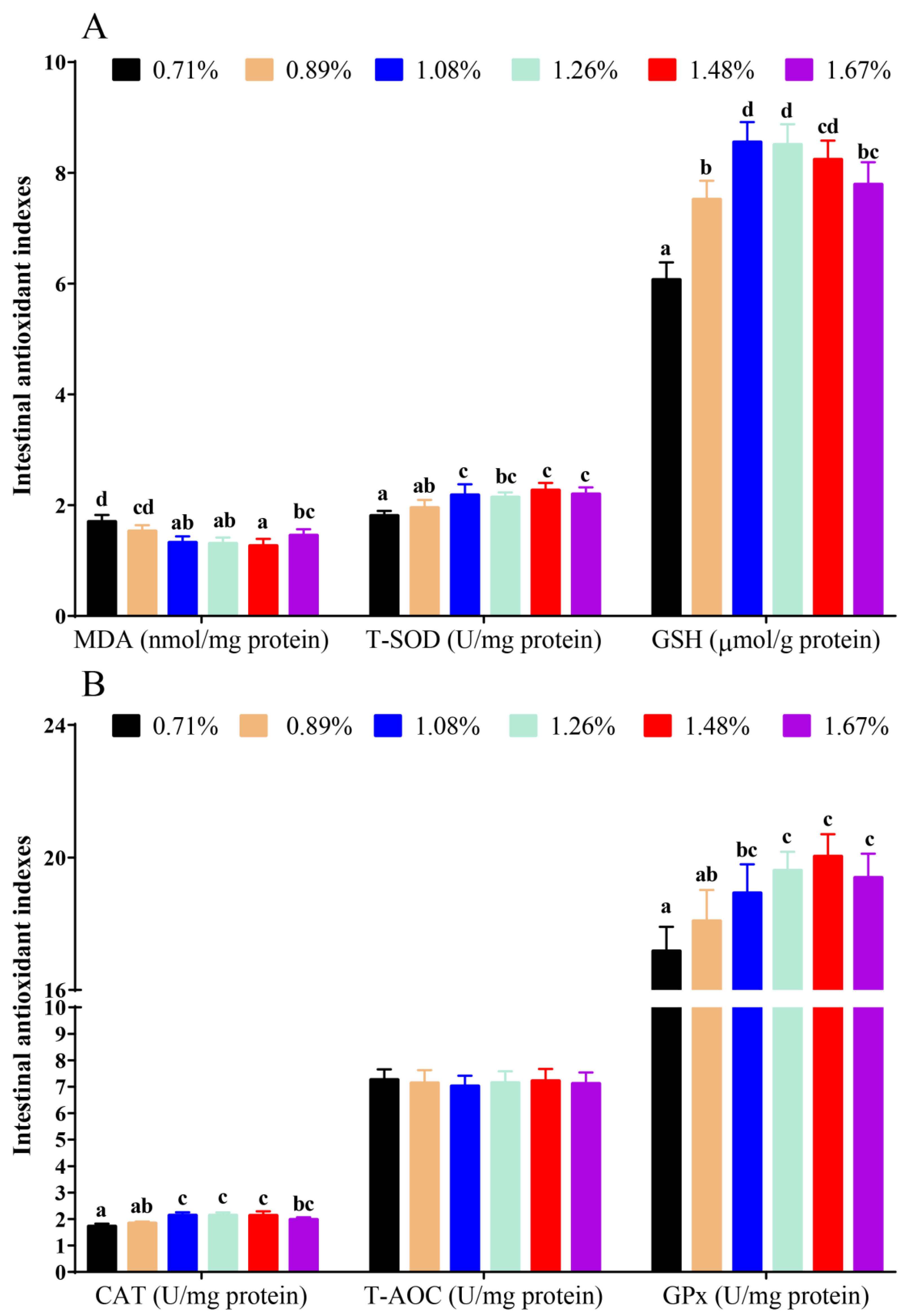
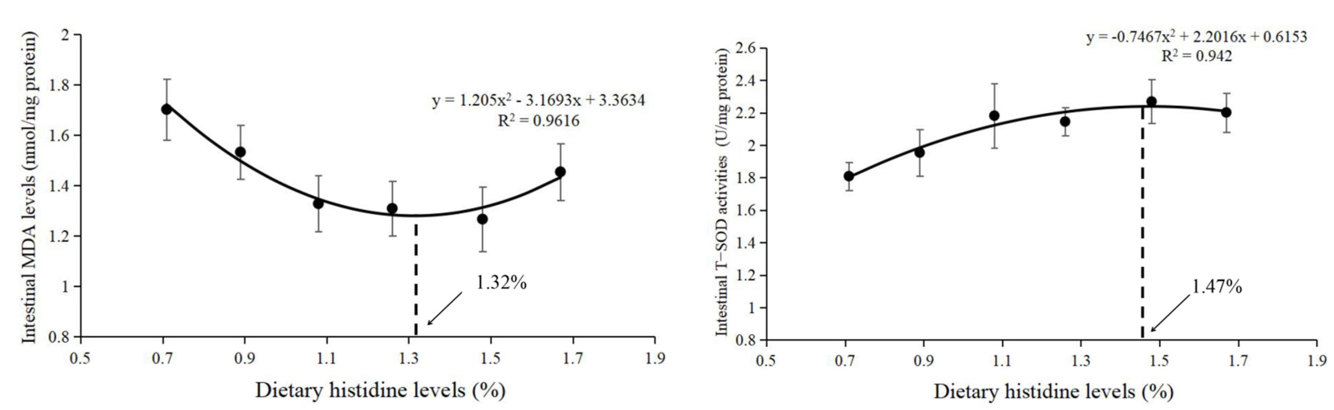
3.1. Intestinal Antioxidant Status

Compared with the 0.71% dietary histidine level (DHL), the (1.08–1.67)% DHLs significantly lowered the intestinal MDA levels and upregulated the intestinal antioxidant enzyme activities of GPx, T-SOD, and CAT, and the (0.89–1.67)% DHLs significantly upregulated the intestinal GSH levels (p < 0.05) (Figure 1A,B). However, no significant changes in T-AOC were found with different DHLs (p > 0.05) (Figure 1B). Based on the MDA and T-SOD results, the dietary histidine requirements of juvenile largemouth bass were 1.32% of the diet (2.81% dietary protein) and 1.47% of the diet (3.13% dietary protein), as determined by quadratic regression analysis (Figure 2).

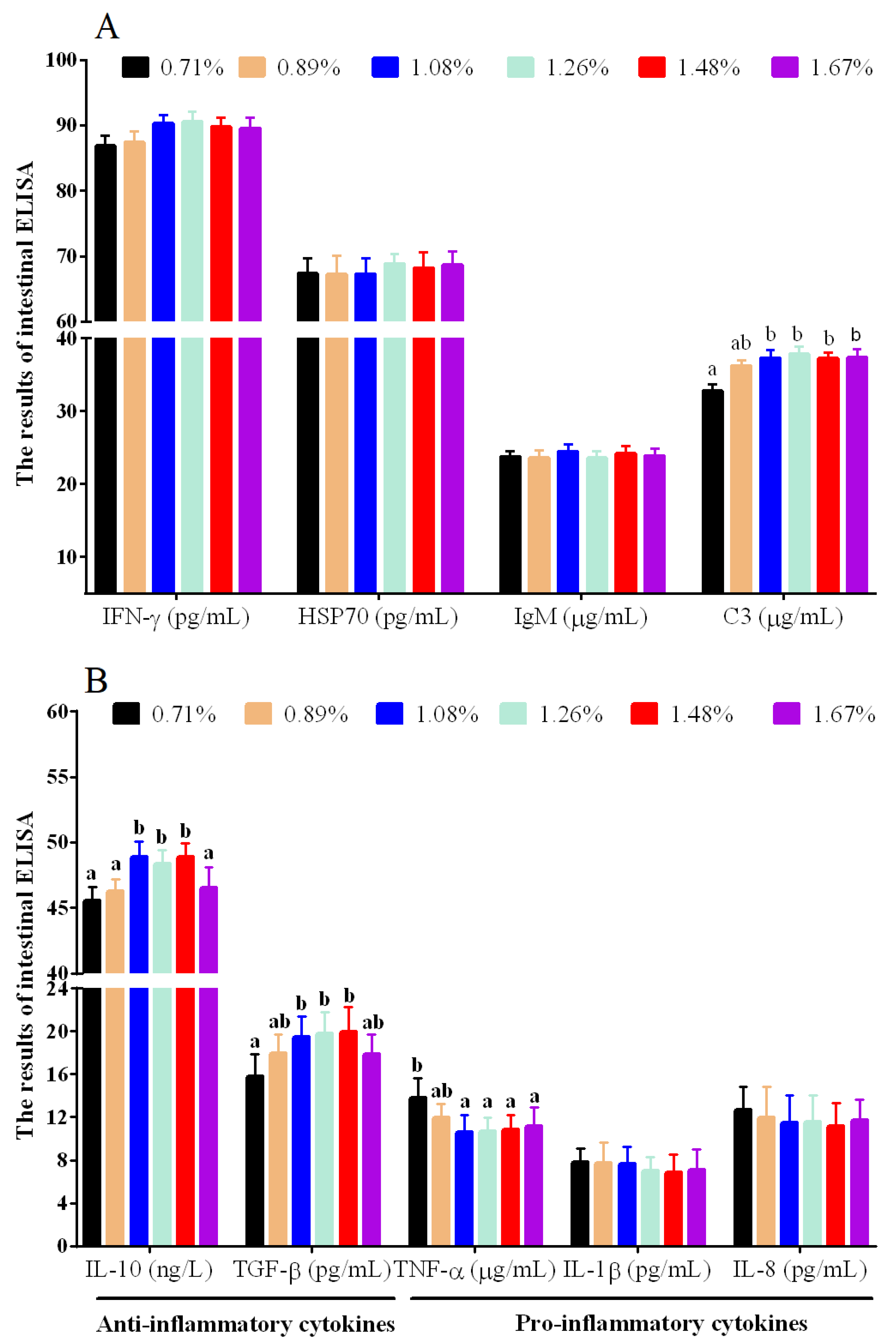
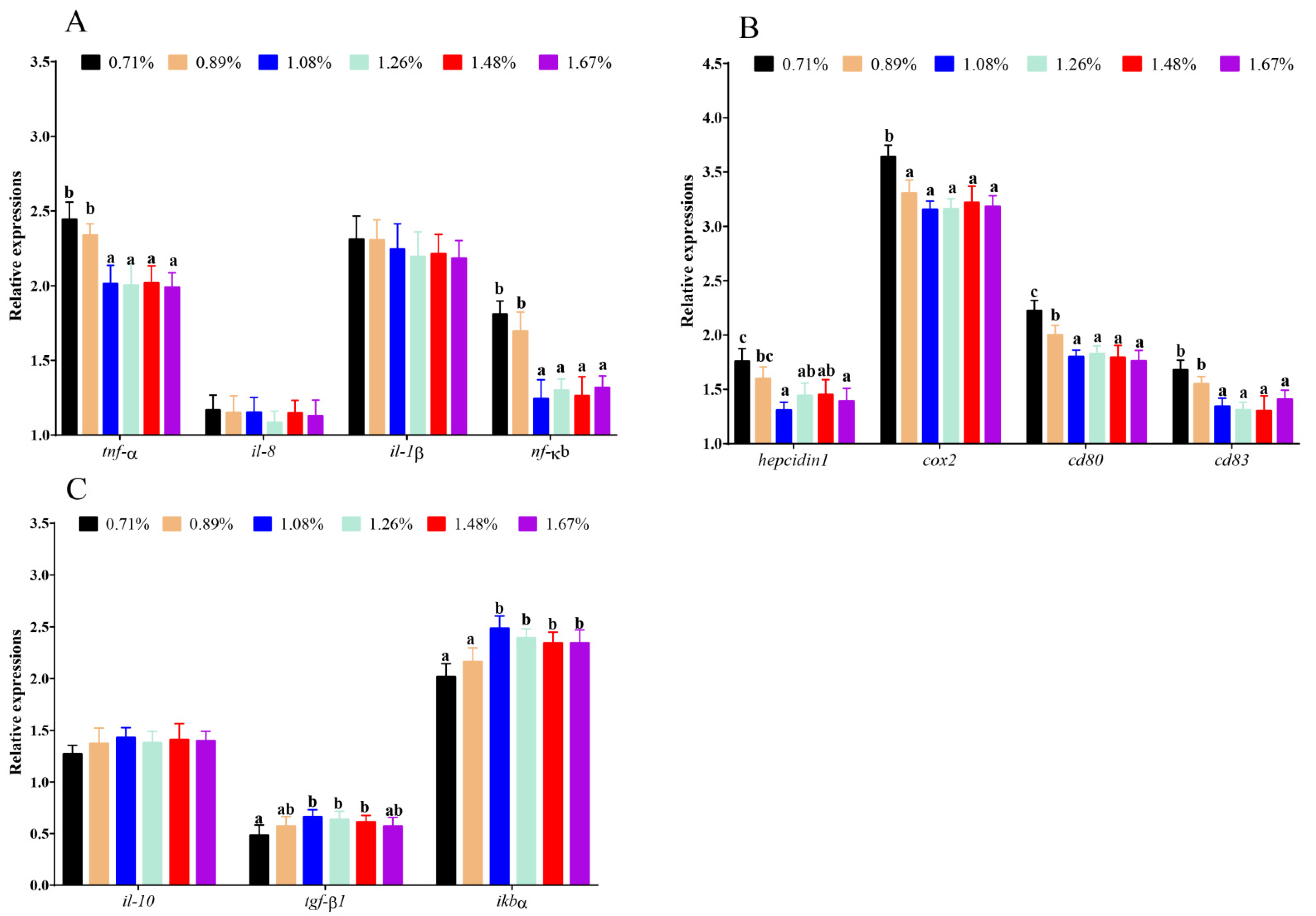
3.2. Intestinal Immune Cytokines

Compared with the 0.71% DHL, significantly higher levels of intestinal C3 were observed at the (1.08–1.67)% DHLs (p < 0.05) (Figure 3A). DHLs of (1.08–1.48)% significantly upregulated the intestinal anti-inflammatory cytokine levels of TGF-β and IL-10, and the 1.08%–1.67% DHLs significantly lowered the intestinal proinflammatory cytokine level of TNF-α (p < 0.05) (Figure 3B). However, DHL had no significant effect on the intestinal levels of the immune cytokines IL-1β, IFN-γ, IgM, HSP70, and IL-8 (p > 0.05) (Figure 3A,B).

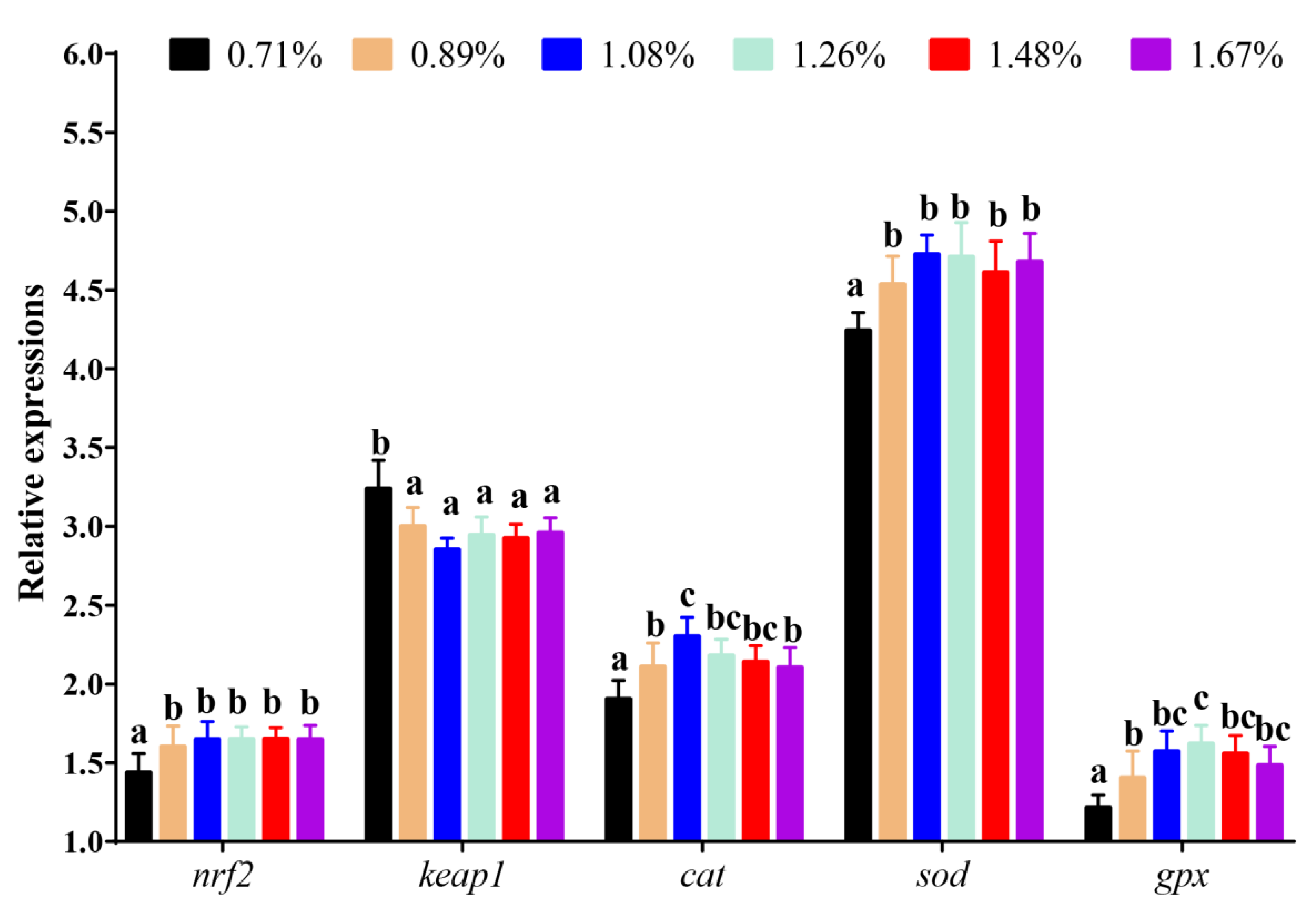
3.3. The mRNA Levels of the Key Genes in the Nrf2-Signalling Pathway

Compared with the 0.71% DHL, significantly higher mRNA levels of nrf2 were observed in the (0.89–1.67)% DHL groups (p < 0.05). However, DHLs of (0.89–1.67)% significantly lowered the mRNA levels of Kelch-like ECH-associated protein 1 (keap1) (p < 0.05). Furthermore, compared with the control group, the (0.89–1.67)% DHLs also significantly upregulated the mRNA levels of gpx, cat, and superoxide dismutase (sod) (p < 0.05) (Figure 4).

3.4. The mRNA Levels of Inflammatory-Related Genes

Compared with the 0.71% DHL, DHLs of (1.08–1.67)% significantly lowered the mRNA levels of the proinflammatory-related genes nuclear factor kappa B (nf-κb), tnf-α, hepcidin 1, and cd83 (p < 0.05) (Figure 5A,B). However, DHLs of (0.89–1.67)% also lowered the mRNA levels of the proinflammatory-related genes cyclooxygenase 2 (cox2) and cd80 (p < 0.05) (Figure 5B). Additionally, the (1.08–1.48)% DHLs significantly upregulated the mRNA levels of the anti-inflammatory-related gene transforming growth factor β1 (tgf-β1) (p < 0.05), and the (1.08–1.67)% DHLs significantly upregulated the mRNA levels of the anti-inflammatory-related gene NF-kappa-B inhibitor alpha (ikbα) (p < 0.05) (Figure 5C). However, no significant changes in il-8, il-1β, or il-10 were observed in fish fed different DHLs (p > 0.05) (Figure 5A,C).

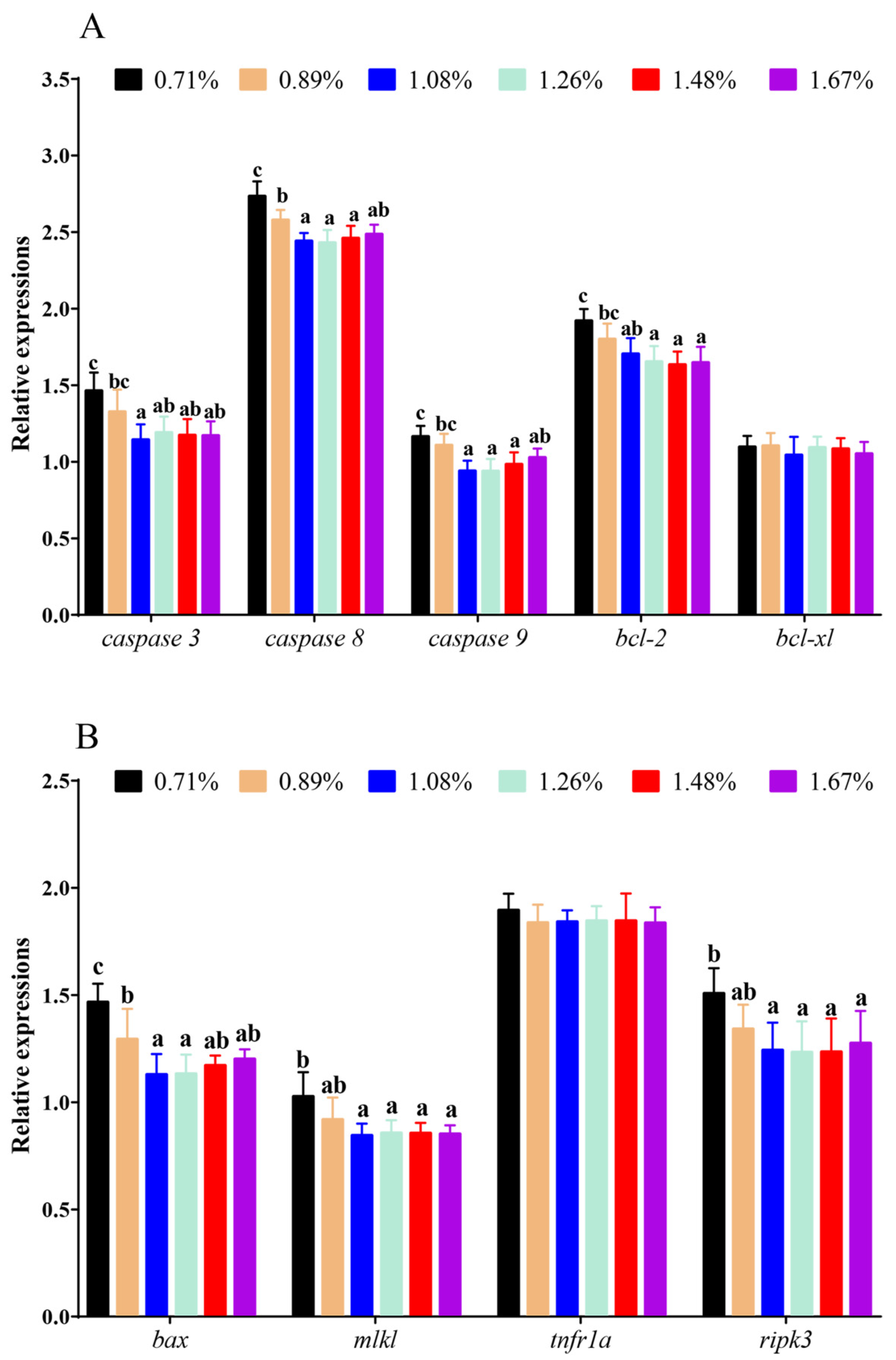
3.5. The mRNA Levels of Apoptosis and Necroptosis Genes

Compared with the 0.71% DHL, significantly lower mRNA levels of B-cell leukaemia/lymphoma 2 (bcl-2), caspase 3, mixed lineage kinase-like (mlkl), caspase 9, and receptor-interacting protein kinase-3 (ripk3) were observed with the (1.08–1.67)% DHLs (p < 0.05), and significantly lower mRNA levels of caspase 8 and bcl-2-associated x (bax) were also observed with the (0.89–1.67)% DHLs (p < 0.05) (Figure 6A,B). However, DHL had no significant effect on the mRNA levels of B-cell leukaemia/lymphoma-XL (bcl-xl) and tumour necrosis factor receptor 1A (tnfr1a) (p > 0.05) (Figure 6A,B).

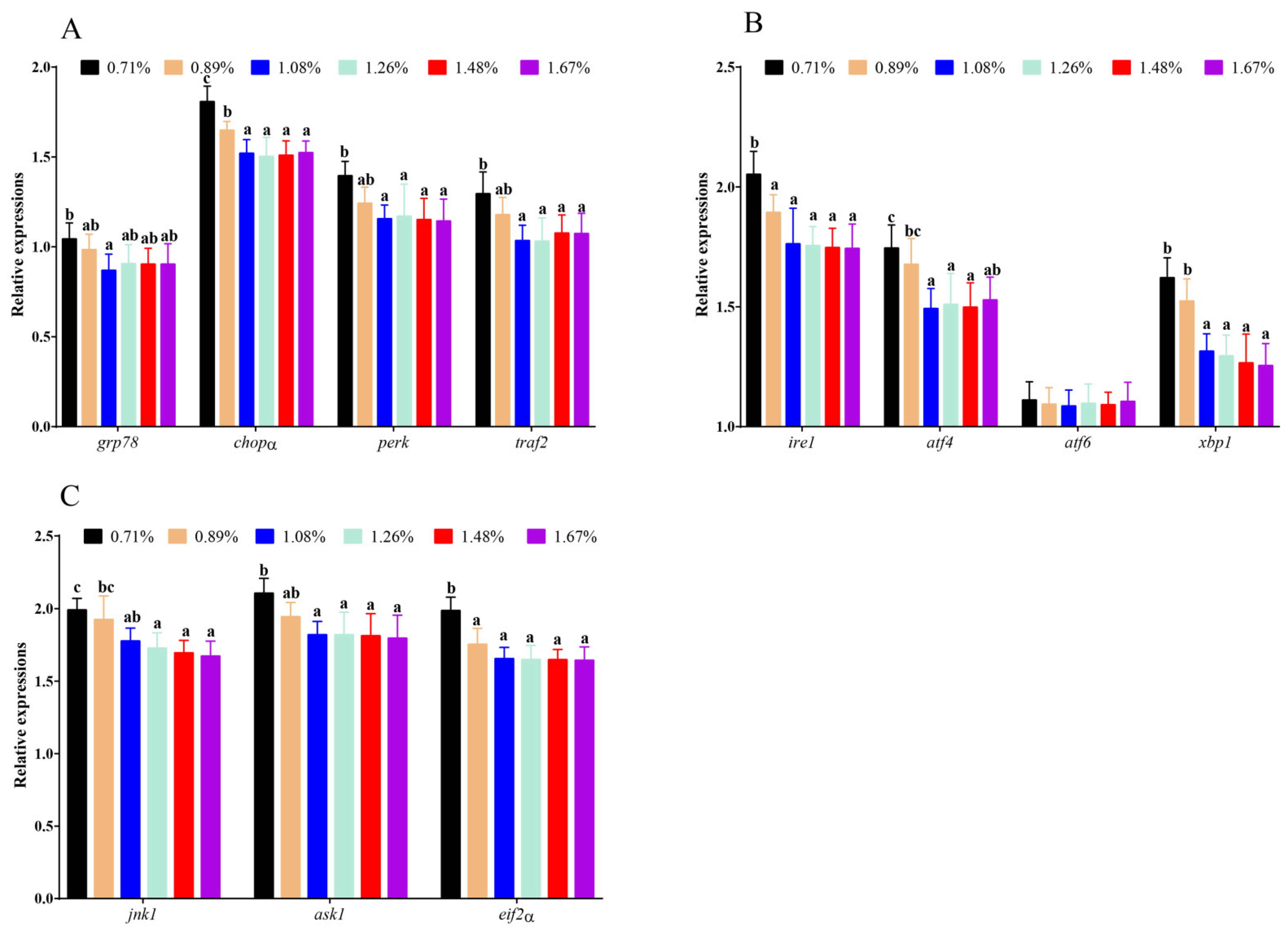
3.6. The mRNA Levels of ERS-Related Genes

Compared with the 0.71% DHL, significantly higher mRNA levels of c-Jun N-terminal kinase-1 (jnk1), activating transcription factor 4 (atf4), X-box binding protein 1 (xbp1), apoptosis signal-regulating kinase 1 (ask1), protein kinase R (PKR)-like endoplasmic-reticulum kinase (perk), and TNF receptor-associated factor 2 (traf2) were observed with the (1.08–1.67)% DHLs (p < 0.05), and significantly higher mRNA levels of eukaryotic translation initiation factor 2 (eif2α), C/EBP-homologous protein (chopα), and inositol-requiring enzyme 1 (ire1) were also observed with the (0.89–1.67)% DHLs (p < 0.05) (Figure 7A–C). Furthermore, the 1.08% DHL significantly upregulated the mRNA level of 78 kDa glucose-regulated protein (grp78) (p < 0.05) (Figure 7A). However, DHL had no significant effect on the mRNA levels of activating transcription factor 6 (atf6) (p > 0.05) (Figure 7B).

4. Discussion

4.1. Histidine Deficiency Inhibited Intestinal Antioxidant Capacity

MDA is marker for oxidative stress, which can reflect the degree of cellular oxidative stress [33]. This study found that dietary histidine deficiency (0.71% DHL) significantly upregulated the intestinal MDA level, indicating that dietary histidine deficiency could cause oxidative damage. Similar reports were also found in the serum of Jian carp (C. carpio var. Jian) [11], gills of grass carp [13], and muscles of grass carp [15]. The increase in MDA level caused by histidine deficiency may be related to a decreased ability to scavenge superoxide anions and hydroxyl radicals. The antioxidant system plays an important role in preventing oxidative stress and scavenging superoxide anions and hydroxyl radicals in fish [34]. In the present study, histidine deficiency significantly suppressed the intestinal antioxidant enzyme activities of T-SOD, CAT, and GPx and the intestinal level of GSH. Furthermore, the activity of antioxidant enzymes is regulated by the expression of antioxidant genes [35]. The Nrf2-signalling pathway regulates antioxidant genes [13,15]. In this study, dietary histidine deficiency significantly lowered the mRNA level of nrf2 and increased the mRNA level of keap1, which further lowered the mRNA levels of the downstream genes sod, cat, and gpx. Thus, dietary histidine supplementation has a positive effect on these antioxidant indices and the mRNA levels of antioxidant genes, which further lowers intestinal MDA levels. Jiang et al. [13] reported that dietary histidine deficiency suppressed the antioxidant index and gene expression in the gills of grass carp. Furthermore, similar results were reported in the muscles of grass carp [15]. The results here indicated that dietary histidine deficiency increased MDA levels, which might be partially related to the decrease of antioxidant enzymes and factors in the intestine. In our previous study, dietary histidine deficiency resulted in reduced growth performance [24]. In this study, dietary histidine deficiency also had negative effects on intestinal antioxidant capacity of largemouth bass. These results might be one of the reasons why histidine deficiency could lower the growth performance of largemouth bass. Lemire et al. [36] reported that glutamate, a product of histidine catabolism, was involved in the generation of the antioxidant α-ketoglutarate, which is also considered a crucial molecule in cellular redox regulation [36,37,38,39,40]. In aquatic animals, α-ketoglutarate supplementation in the diet enhanced the antioxidant capacity in grass carp [41,42], hybrid sturgeon (Acipenser schrenckii ♀× A. baerii ♂) [43], Songpu mirror carp [44], and common carp [45]. Hence, histidine exerts its antioxidant functions through its metabolite α-ketoglutaric acid. In this study, no significant changes in T-AOC were found with different DHLs. However, only the highest DHL (1.42%) significantly increased the activity of T-AOC in juvenile blunt snout bream [46]. In this study, the DHL in the control group was 0.71%, which was much higher than that in the juvenile blunt snout bream. Therefore, it may be related to the difference in DHL in this study. Unfortunately, there are few studies on the relationship between histidine and T-AOC in aquatic animals, and the related mechanism is still unclear, which needs to be further studied. Furthermore, the recommended histidine requirement of juvenile largemouth bass was 1.26% of the diet (2.68% of dietary protein) based on the growth performance in our previous study [24]. However, in this study, the dietary histidine requirements of juvenile largemouth bass were 1.32% of the diet (2.81% dietary protein) and 1.47% of the diet (3.13% dietary protein) based on the MDA and T-SOD results, which showed that the requirement based on antioxidant capacity was slightly higher than that based on growth performance, indicating that the level of histidine in the feed might be necessary to be further increase if a better immune effect is pursued.

4.2. Histidine Deficiency Suppressed Intestinal Immunocompetence

Immune globulin, the interferon system, and the complement system play an indispensable role in the regulation of immune capacity [47,48,49]. In this study, dietary histidine deficiency inhibited the level of C3 compared with that after appropriate histidine supplementation, indicating that dietary histidine deficiency has a negative effect on immune regulation in largemouth bass. It has been found that the active site of C3 in all animal species contains a conserved histidine residue, which can activate the thioester bond of C3 and promote covalent binding of the activated group with the hydroxyl group on the surface of target cells, leading to the occurrence of the corresponding biochemical reaction. If this key histidine residue is substituted, complement activation cannot be completed [50]. This might be a possible reason why dietary histidine deficiency decreased the production of intestinal C3. However, dietary histidine supplementation had no significant effect on IgM, HSP70, or IFN-γ. Interestingly, only a limited number of studies have reported the regulation of immune globulin, the interferon system, and the complement system in animals. Therefore, the specific regulatory mechanism needs to be further studied.

4.3. Histidine Deficiency Induces Intestinal Endoplasmic-Reticulum Stress Resulting in Inflammatory Response, Apoptosis, and Necroptosis

The ER is one of the most important organelles in eukaryotic cells and is closely related to cell homeostasis, and ER homeostasis is maintained through unfolded protein reactions involving three signalling pathways, including the PEPK-signalling pathway, IRE-1-signalling pathway, and ATF6-signalling pathway [16]. In this study, dietary histidine deficiency activated the PEPK-signalling pathway and IRE1-signalling pathway with increased core gene expression of pepk, grp78, eif2α, atf4, chopα, ire1, xbp1, traf2, ask1, and jnk1, indicating that dietary histidine deficiency induced ERS. In aquatic animals, activation of the PEPK-signalling pathway and IRE1-signalling pathway resulted in ERS under heat stress in largemouth bass [32]. Furthermore, numerous studies have reported that adverse factors can induce ERS by affecting the expression of the core genes in the IRE1- and PERK-signalling pathways in blunt snout bream [17], common carp [18,19], Gibel carp [20], and grass carp [21]. However, dietary histidine deficiency had no significant effect on the mRNA level of ATF6. In contrast to this study, ERS regulation was also involved in the ATF6-signalling pathway [32,51]. Activation of the IRE1 pathway by B-cell differentiation did not lead to the upregulation of the CHOP downstream genes in the ATF6 and PERK pathways [52,53]. Therefore, it was speculated that these three pathways could be selectively activated. However, the mechanism of selection was unclear.
In this study, dietary histidine deficiency significantly upregulated the mRNA levels of proapoptotic genes, including caspase 3, bax, caspase 8, caspase 9, and the mRNA levels of necroptosis genes, including mlkl and ripk3. Furthermore, dietary histidine deficiency also significantly lowered the mRNA levels of the antiapoptotic gene bcl-2. These results showed that chronic dietary histidine deficiency could induce apoptosis and necroptosis of the intestine. Jiang et al. [13] also reported that dietary histidine deficiency induced apoptosis in the gills of grass carp by improving the mRNA levels of caspase 3, caspase 8, and caspase 9, which supports the results of this study. When intense or persistent ERS occurs, CHOP gene expression is activated in the three above-mentioned pathways and ERS exceeds the range of cellular self-regulation, which leads to cell apoptosis and cell death [23]. Hence, chronic dietary histidine deficiency induced persistent ERS, which further led to cell apoptosis and necroptosis in largemouth bass. Furthermore, studies have reported that IRE1 binds with TRAF2 to activate ASK1 and further activate JNK, which upregulates CHOP gene expression to regulate antiapoptotic genes and caspase-mediated apoptosis pathways [54,55]. This might be another mechanism by which dietary histidine deficiency induces apoptosis. NF-κB is an important signalling pathway in inflammatory regulation [56]. In this study, dietary histidine deficiency activated the NF-κB-signalling pathway to induce an inflammatory response, which further upregulated the mRNA levels of the proinflammatory factors tnf-α, hepcidin 1, cox2, cd80, and cd83 and lowered the mRNA levels of the anti-inflammatory factors tgf-β1 and ikbα. Furthermore, dietary histidine deficiency significantly lowered the levels of the intestinal anti-inflammatory factors IL-10 and TGF-β and upregulated the level of the intestinal proinflammatory factor TNF-α, showing a trend similar to that of the gene expression of inflammatory factors. A similar study reported that dietary histidine deficiency induced an inflammatory response in the gills of grass carp [13] and intestines of blunt snout bream [46]. Furthermore, dietary amino acid deficiency can also induce inflammatory responses, such as through methionine and lysine, in blunt snout bream [5,57]. It was reported that activation of the IRE1–TRAF2–ASK1–JNK1-signalling pathway could upregulate NF-κB and mediate the inflammatory response [23]. Dietary histidine deficiency can also activate the IRE1–TRAF2–ASK1–JNK1-signalling pathway, which might be the mechanism by which histidine regulates the NF-κB-signalling pathway. However, unfortunately, there are relatively few studies on histidine at present, and the relevant mechanisms need further study.

5. Conclusions

Histidine deficiency can weaken the intestinal antioxidant capacity and induce intestinal endoplasmic-reticulum stress, which further leads to an inflammatory response, apoptosis, and necroptosis and decreases the immune capacity of largemouth bass. However, histidine supplementation could alleviate these problems. Based on the results of MDA and T-SOD, the appropriate dietary histidine requirements of juvenile largemouth bass are 1.32% of the diet (2.81% dietary protein) and 1.47% of the diet (3.13% dietary protein), respectively, as determined by quadratic regression analysis.

Author Contributions

M.R. and L.Z. (Lu Zhang) designed the study; H.L. carried out the experiments and wrote the manuscript; and P.X., L.Z. (Lin Zhang), D.H. and G.X. provided technical assistance. All authors have read and agreed to the published version of the manuscript.

Funding

This study was financially supported by the earmarked fund for CARS (CARS-46).

Institutional Review Board Statement

This experiment was based strictly on the requirements of the Institutional Animal Care and Ethics Committee of Nanjing Agricultural University, Nanjing, China [Permit number: SYXK (Su) 2011-0036].

Informed Consent Statement

Not applicable.

Data Availability Statement

All datasets generated for this study are available.

Conflicts of Interest

Author Lu Zhang is employed by Tongwei Co., Ltd. The remaining authors declare that the research was conducted in the absence of any commercial or financial relationships that could be construed as potential conflicts of interest. Lu Zhang played a major role in the design of the study, data analysis, and supervising the published results.

References

  1. Zhang, H.; Ran, C.; Teame, T.; Ding, Q.; Hoseinifar, S.H.; Xie, M.; Zhang, Z.; Yang, Y.; Olsen, R.E.; Gatlin, D.M., III; et al. Research progress on gut health of farmers teleost fish: A viewpoint concerning the intestinal mucosal barrier and the impact of its damage. Rev. Fish Biol. Fisher. 2020, 30, 569–586. [Google Scholar] [CrossRef]
  2. Dawood, M.A. Nutritional immunity of fish intestines: Important insights for sustainable aquaculture. Rev. Aquacult. 2021, 13, 642–663. [Google Scholar] [CrossRef]
  3. Takahashi, I.; Kiyono, H. Gut as the largest immunologic tissue. J. Parenter. Enter. Nutr. 1999, 23, S7–S12. [Google Scholar] [CrossRef] [PubMed]
  4. Jiang, W.; Qu, B.; Feng, L.; Jiang, J.; Kuang, S.; Wu, P.; Tang, L.; Tang, W.-N.; Zhang, Y.A.; Zhou, X.-Q.; et al. Histidine prevents cu-induced oxidative stress and the associated decreases in mRNA from encoding tight junction proteins in the intestine of grass carp (Ctenopharyngodon idella). PLoS ONE 2016, 11, e157001. [Google Scholar] [CrossRef] [PubMed] [Green Version]
  5. Huang, D.Y.; Maulu, S.; Ren, M.C.; Liang, H.L.; Ge, X.P.; Ji, K.; Yu, H. Dietary lysine levels improved antioxidant capacity and immunity via the TOR and p38 MAPK signaling pathways in grass carp, Ctenopharyngodon idellus fry. Front. Immunol. 2021, 12, 635015. [Google Scholar] [CrossRef] [PubMed]
  6. Liang, H.; Ji, K.; Ge, X.; Xi, B.; Chen, X. Tributyrin plays an important role in regulating the growth and health status of juvenile blunt snout bream (Megalobrama amblycephala), as evidenced by pathological examination. Front. Immunol. 2021, 12, 652294. [Google Scholar] [CrossRef] [PubMed]
  7. Kunisawa, J.; Kiyono, H. Vitamin-mediated regulation of intestinal immunity. Front. Immunol. 2013, 4, 189. [Google Scholar] [CrossRef] [Green Version]
  8. Zheng, X.; Feng, L.; Jiang, W.D.; Wu, P.; Liu, Y.; Kuang, S.Y.; Tang, L.; Zhou, X.-Q. The regulatory effects of pyridoxine deficiency on the grass carp (Ctenopharyngodon idella) gill barriers immunity, apoptosis, antioxidant, and tight junction challenged with flavobacterium columnar. Fish Shellfish Immunol. 2020, 105, 209–223. [Google Scholar] [CrossRef] [PubMed]
  9. Gao, Y.; Liu, Y.; Chen, X.; Yang, H.; Li, X.; Tian, L. Effects of graded levels of histidine on growth performance, digested enzymes activities, erythrocyte osmotic fragility and hypoxia-tolerance of juvenile grass carp Ctenopharyngodon idella. Aquaculture 2016, 452, 388–394. [Google Scholar] [CrossRef]
  10. Wade, A.M.; Tucker, H.N. Antioxidant characteristics of L-histidine. J. Nutr. Biochem. 1998, 9, 308–315. [Google Scholar] [CrossRef]
  11. Feng, L.; Zhao, B.; Chen, G.F.; Jiang, W.D.; Liu, Y.; Jiang, J.; Hu, K.; Li, S.; Zhou, X. Effects of dietary histidine on antioxidant capacity in juvenile Jian carp (Cyprinus carpio var. Jian). Fish Physiol. Biochem. 2013, 39, 559–571. [Google Scholar] [CrossRef] [PubMed]
  12. Lee, Y.T.; Hsu, C.C.; Lin, M.H.; Liu, K.S.; Yin, M.C. Histidine and carnosine delay diabetic deterioration in mice and protect human low density lipoprotein against oxidation and glycation. Eur. J. Pharmacol. 2005, 513, 145–150. [Google Scholar] [CrossRef] [PubMed]
  13. Jiang, W.D.; Feng, L.; Qu, B.; Wu, P.; Kuang, S.Y.; Jiang, L.; Tang, L.; Tang, W.-N.; Zhang, Y.-A.; Zhou, X.-Q.; et al. Changes in integrity of the gill during histidine deficiency or excess due to depression of cellular anti-oxidative ability, induction of apoptosis, inflammation and impair of cell-cell tight junctions related to Nrf2, TOR and NF-kB signaling in fish. Fish Shellfish Immunol. 2016, 56, 111–122. [Google Scholar] [CrossRef] [PubMed]
  14. Erickson, M.C.; Hultin, H.O. Influence of histidine on lipid peroxidation in sarcoplasmic reticulum. Arch. Biochem. Biophys. 1992, 292, 427–432. [Google Scholar] [CrossRef]
  15. Wu, P.; Qu, B.; Feng, L.; Jiang, W.D.; Kuang, S.Y.; Jiang, J.; Tang, L.; Zhou, X.-Q.; Liu, Y. Dietary histidine deficiency induced flesh quality loss associated with changes in muscle nutritive composition, antioxidant capacity, Nrf2 and TOR signaling molecules in on-growing grass carp (Ctenopharyngodon idella). Aquaculture 2020, 526, 735399. [Google Scholar] [CrossRef]
  16. Zheng, Z.; Shang, Y.; Tao, J.; Zhang, J.; Sha, B. Endoplasmic reticulum stress signaling pathways: Activation and diseases. Curr. Protein. Pept. Sci. 2019, 20, 935–943. [Google Scholar] [CrossRef]
  17. Cao, X.F.; Dai, Y.J.; Liu, M.Y.; Yuan, X.Y.; Wang, C.C.; Huang, Y.Y.; Liu, W.-B.; Jiang, G.-Z. High-fat diet induces aberrant hepatic lipid secretion in blunt snout bream by activating endoplasmic reticulum stress-associated IRE1/XBP1 pathway. Biochim. Biophys. Acta Mol. Cell Biol. Lipids 2019, 1864, 213–223. [Google Scholar] [CrossRef]
  18. Jia, R.; Du, J.L.; Cao, L.P.; Feng, W.R.; He, Q.; Xu, P.; Yin, G. Chronic exposure of hydrogen peroxide alters redox state, apoptosis and endoplasmic reticulum stress in common carp (Cyprinus carpio). Aquat. Toxicol. 2020, 229, 105657. [Google Scholar] [CrossRef]
  19. Xue, S.Q.; Lin, J.W.; Zhou, Q.; Wang, H.T.; Han, Y. Effect of ammonia stress on transcriptome and endoplasmic reticulum stress pathway for common carp (Cyprinus carpio) hepatopancreas. Aquacult. Rep. 2021, 20, 100694. [Google Scholar] [CrossRef]
  20. Li, H.Y.; Xu, W.J.; Wu, L.Y.; Dong, B.; Jin, J.Y.; Han, D.; Zhu, X.; Yang, Y.; Liu, H.; Xie, S. Differential regulation of endoplasmic reticulum stress-induced autophagy and apoptosis in two strains of gibel carp (Carassius gibelio) exposed to acute waterborne cadmium. Aquat. Toxicol. 2021, 231, 105721. [Google Scholar] [CrossRef]
  21. Xie, L.X.; Chen, S.Q.; Yao, C.R.; Li, D.P.; Li, L.; Tang, R. Nitrite induces endoplasmic reticulum stress and associates apoptosis of liver cells in grass carp (Ctenopharyngodon idella). Aquaculture 2019, 507, 275–281. [Google Scholar] [CrossRef]
  22. Hetz, C.; Saxena, S. ER stress and the unfolded protein response in neurodegeneration. Nat. Rev. Neurol. 2017, 13, 477–491. [Google Scholar] [CrossRef] [PubMed]
  23. Gómez-Sierra, T.; Bellido, B.; Reyes-Fermín, L.M.; Martínez-Klimova, E.; Pedraza-Chaverri, J. Regulation of endoplasmic reticulum stress in models of kidney disease. Adv. Redox. Res. 2021, 3, 100010. [Google Scholar] [CrossRef]
  24. Liang, H.L.; Xu, G.C.; Xu, P.; Zhu, J.; Li, S.L.; Ren, M.C. Dietary histidine supplementation maintained amino acid homeostasis and reduced hepatic fat accumulation of juvenile largemouth bass, Mieropterus salmoides. Aquacult. Nutr. 2022, 2022, 4034922. [Google Scholar] [CrossRef]
  25. AOAC (Association of Official Analytical Chemists). Official Methods of Analysis of the Association of Official Analytical Chemists, 15th ed.; Association of Official Analytical Chemists: Washington, DC, USA, 2003. [Google Scholar]
  26. Liang, H.L.; Ren, M.C.; Habte-Tsion, H.M.; Ge, X.P.; Xie, J.; Mi, H.F.; Xi, B.; Miao, L.; Liu, B.; Zhou, Q.; et al. Dietary arginine affects growth performance, plasma amino acid contents and gene expressions of the TOR signaling pathway in juvenile blunt snout bream, Megalobrama amblycephala. Aquaculture 2016, 461, 1–8. [Google Scholar] [CrossRef]
  27. Zhou, Y.L.; He, G.L.; Jin, T.; Chen, Y.J.; Dai, F.Y.; Luo, L.; Lin, S.-M. High dietary starch impairs intestinal health and microbiota of largemouth bass, Micropterus salmoides. Aquaculture 2021, 534, 736261. [Google Scholar] [CrossRef]
  28. Zhao, L.; Liang, J.; Chen, F.K.; Tang, X.H.; Liao, L.; Liu, Q.; Luo, J.; Du, Z.; Li, Z.; Luo, W.; et al. High carbohydrate diet induced endoplasmic reticulum stress and oxidative stress, promoted inflammation and apoptosis, impaired intestinal barrier of juvenile largemouth bass (Micropterus salmoides). Fish Shellfish Immunol. 2021, 119, 308–317. [Google Scholar] [CrossRef] [PubMed]
  29. Yu, L.L.; Yu, H.H.; Liang, X.F.; Li, N.; Wang, X.; Li, F.H.; Wu, X.F.; Zheng, Y.H.; Xue, M.; Liang, X.F. Dietary butylated hydroxytoluene improves lipid metabolism, antioxidant and anti-apoptotic response of largemouth bass (Micropterus salmoides). Fish Shellfish Immunol. 2018, 72, 220–229. [Google Scholar] [CrossRef]
  30. Zhao, L.L.; Liang, J.; Liu, H.; Gong, C.X.; Huang, X.L.; Hu, Y.F.; Liu, Q.; He, Z.; Zhang, X.; Yang, S.; et al. Yinchenhao Decoction ameliorates the high-carbohydrate diet induced suppression of immune response in largemouth bass (Micropterus salmoides). Fish Shellfish Immunol. 2022, 125, 141–151. [Google Scholar] [CrossRef]
  31. Yin, P.; Xie, S.W.; Hou, Y.J.; Guo, T.Y.; Fang, H.H.; Zhang, Y.M.; Liu, Y.; Tian, L.; Niu, J. Effects of dietary oxidized fish oil on growth performance, antioxidant defense system, apoptosis and mitochondrial function of juvenile largemouth bass (Micropterus salmoides). Aquaculture 2019, 500, 347–358. [Google Scholar] [CrossRef]
  32. Zhou, X.Q.; Li, L.L.; Li, C.J.; Liu, E.G.; Hao, H.; Ling, Q.F. Heat stress-induced endoplasmic reticulum stress promotes liver apoptosis in largemouth bass (Micropterus salmoides). Aquaculture 2022, 546, 737401. [Google Scholar] [CrossRef]
  33. Liang, H.; Ji, K.; Ge, X.; Ren, M.; Liu, B.; Xi, B.; Pan, L. Effects of dietary arginine on antioxidant status and immunity involved in ampk-no signaling pathway in juvenile blunt snout bream. Fish Shellfish Immunol. 2018, 78, 69–78. [Google Scholar] [CrossRef] [PubMed]
  34. Jiang, W.D.; Wen, H.L.; Liu, Y.; Jiang, J.; Wu, P.; Zhao, J.; Kuang, S.-Y.; Tang, L.; Tang, W.-N.; Zhang, Y.-A.; et al. Enhanced muscle nutrient content and flesh quality, resulting from tryptophan, is associated with anti-oxidative damage referred to the Nrf2 and TOR signalling factors in young grass carp (Ctenopharyngodon idella): Avoid tryptophan deficiency or excess. Food Chem. 2016, 199, 210–219. [Google Scholar] [CrossRef] [PubMed]
  35. Kobayashi, M.; Itoh, K.; Suzuki, T.; Osanai, H.; Nishikawa, K.; Katoh, Y.; Takagi, Y.; Yamamoto, M. Identification of the interactive interface and phylogenic conservation of the Nrf2-Keap1 system. Genes Cells 2002, 7, 807–820. [Google Scholar] [CrossRef]
  36. Lemire, J.; Milandu, Y.; Auger, C.; Bignucolo, A.; Appanna, V.P.; Appanna, V.D. Histidine is a source of the antioxidant, α-ketoglutarate, in Pseudomonas fuorescens challenged by oxidative stress. FEMS Microbiol. Lett. 2010, 309, 170–177. [Google Scholar] [CrossRef] [Green Version]
  37. Hou, Y.; Wang, L.; Ding, B.; Liu, Y.; Zhu, H.; Liu, J.; Li, Y.; Kang, P.; Yin, Y.; Wu, G. Alpha-Ketoglutarate and intestinal function. Front. Biosci. (Landmark Ed.) 2011, 16, 1186–1196. [Google Scholar] [CrossRef] [Green Version]
  38. Mailloux, R.J.; Puiseux-Dao, S.; Appanna, V.D. Alphaketoglutarate abrogates the nuclear localization of HIF-1alpha in aluminum-exposed hepatocytes. Biochimie 2009, 91, 408–415. [Google Scholar] [CrossRef]
  39. Mailloux, R.J.; Singh, R.; Brewer, G.; Auger, C.; Lemire, J.; Appanna, V.D. Alpha-ketoglutarate dehydrogenase and glutamate dehydrogenase work in tandem to modulate the antioxidant alpha-ketoglutarate during oxidative stress in Pseudomonas fluorescens. J. Bacteriol. 2009, 191, 3804–3810. [Google Scholar] [CrossRef] [Green Version]
  40. Wang, L.; Wei, Y.; Wang, C.; Li, J.; Zhao, Z.; Luo, L.; Du, X.; Qiyou, X. Effects of α-ketoglutarate on the growth performance, amino acid metabolism and related gene expression of mirror carp (Cyprinus carpio). Aquac. Nutr. 2017, 23, 926–933. [Google Scholar] [CrossRef]
  41. Wang, H.Q.; Zhao, Y.R.; Jin, B.T.; Liang, J. Effects of dietary alpha-ketoglutarate supplementation on growth and serum biochemical parameters of grass carp (Ctenopharyngodon idella) fingerlings. Isr. J. Aquacult. Bamidgeh. 2016, 68, 1–6. [Google Scholar]
  42. Lin, X.; Jin, B.T.; Wang, H.Q.; Zhao, Y.R. Effects of diet α-ketoglutarate (AKG) supplementation on the growth performance, antioxidant defense system, intestinal digestive enzymes, and immune response of grass carp (Ctenopharyngodon idellus). Aquacult. Int. 2020, 28, 511–524. [Google Scholar] [CrossRef]
  43. Wang, L.S.; Xu, Q.Y.; Wang, C.A.; Li, J.N.; Chen, D.; Zhao, Z.G.; Luo, L.; Du, X. Effects of dietary α-ketoglutarate supplementation on the antioxidant defense system and HSP 70 and HSP 90 gene expression of hybrid sturgeon Acipenser schrenckii ♀ × A. baerii ♂ exposed to ammonia-N stress. Aquac. Res. 2017, 48, 2266–2277. [Google Scholar] [CrossRef]
  44. Wu, D.; Fan, Z.; Li, J.N.; Zhang, Y.Y.; Wang, C.A.; Xu, Q.Y.; Wang, L. Evaluation of Alpha-Ketoglutarate Supplementation on the Improvement of Intestinal Antioxidant Capacity and Immune Response in Songpu Mirror Carp (Cyprinus carpio) After Infection With Aeromonas hydrophila. Front. Immunol. 2021, 12, 690234. [Google Scholar] [CrossRef] [PubMed]
  45. Wu, D.; Fan, Z.; Li, J.N.; Zhang, Y.Y.; Xu, Q.Y.; Wang, L.; Wang, L.S. Low Protein Diets Supplemented With Alpha-Ketoglutarate Enhance the Growth Performance, Immune Response, and Intestinal Health in Common Carp (Cyprinus carpio). Front. Immunol. 2022, 13, 915657. [Google Scholar] [CrossRef]
  46. Yang, Q.; Liang, H.L.; Mokrani, A.; Ji, K.; Yu, H.; Ge, X.P.; Ren, M.; Xie, J.; Pan, L.; Sun, A. Dietary histidine affects intestinal antioxidant enzyme activities, antioxidant gene expressions and inflammatory factors in juvenile blunt snout bream (Megalobrama amblycephala). Aquacult. Nutr. 2019, 25, 249–259. [Google Scholar] [CrossRef] [Green Version]
  47. Merle, N.S.; Elizabeth, C.S.; Veronique, F.B.; Roumenina, L.T. Complement system part I-molecular mechanisms of activation and regulation. Front. Immunol. 2015, 6, 262. [Google Scholar] [CrossRef] [PubMed] [Green Version]
  48. Thom, V.; Arumugam, T.V.; Tim, M.; Gelderblom, M. Therapeutic potential of intravenous immunoglobulin in acute brain injury. Front. Immunol. 2017, 8, 875. [Google Scholar] [CrossRef] [Green Version]
  49. Sekiguchi, K.; Ogawa, E.; Kurohane, K.; Konishi, H.; Mochizuki, N.; Manabe, K.; Imai, Y. Adjuvant effect of short chain triacylglycerol tributyrin on a mouse contact hypersensitivity model. Toxicol. Lett. 2018, 284, 56–62. [Google Scholar] [CrossRef]
  50. Dodds, M.W.; Law, S.K. The phylogeny and evolution of the thioester bond-containing proteins C3, C4 and alpha 2-macroglobulin. Immunol. Rev. 1998, 166, 15–26. [Google Scholar] [CrossRef]
  51. Li, M.Q.; Baumeister, P.; Roy, B.; Phan, T.; Foti, D.; Luo, S.Z.; Lee, A.S. AFT6 as a transcription activator of the endoplasmic reticulum stress element: Thapsigargin stress-induced changes and synergistic interactions with NF-Y and YY1. Mol. Cell. Biol. 2000, 20, 5096–5106. [Google Scholar] [CrossRef] [Green Version]
  52. Gass, J.N.; Gifford, N.M.; Brewer, J.W. Activation of an unfolded protein response during differentiation of antibody-secreting B cells. J. Biol. Chem. 2002, 277, 49047–49054. [Google Scholar] [CrossRef] [PubMed]
  53. Ma, Y.; Brewer, J.W.; Diehl, J.A.; Hendershot, L.M. Two distinct stress signaling pathways converge upon the chop promoter during the mammalian unfolded protein response. J. Mol. Biol. 2002, 318, 1351–1365. [Google Scholar] [CrossRef] [PubMed]
  54. Tomicic, M.T.; Meise, R.; Aasland, D.; Berte, N.; Kitzinger, R.; Krämer, O.H.; Kaina, B.; Christmann, M. Apoptosis induced by temozolomide and nimustine in glioblastoma cells is supported by JNK/c-jun-mediated induction of the BH3-only protein BIM. Oncotarget 2015, 6, 33755–33768. [Google Scholar] [CrossRef] [Green Version]
  55. Almanza, A.; Carlesso, A.; Chintha, C.; Creedican, S.; Doultsinos, D.; Leuzzi, B.; Luís, A.; McCarthy, N.; Montibeller, L.; Sanket, M.; et al. Endoplasmic reticulum stress signalling-from basic mechanisms to clinical applications. FEBS J. 2019, 286, 241–278. [Google Scholar] [CrossRef] [Green Version]
  56. Tan, L.; Li, J.; Wang, Y.; Tan, R. Anti-Neuroinflammatory effect of Alantolactone through the suppression of the NF-κB and MAPK signaling pathways. Cells 2019, 8, 739. [Google Scholar] [CrossRef] [PubMed] [Green Version]
  57. Ji, K.; Liang, H.; Ren, M.; Ge, X.; Mi, H.; Pan, L.; Yu, H. The immunoreaction and antioxidant capacity of juvenile blunt snout bream (Megalobrama amblycephala) involves the PI3K/Akt/Nrf2 and NF-κB signal pathways in response to dietary methionine levels. Fish shellfish Immun. 2020, 105, 126–134. [Google Scholar] [CrossRef] [PubMed]
Figure 1. The results of intestinal MAD and antioxidant indexes against graded different levels of dietary histidine. (A) showed the results of MDA, T-SOD and GSH, (B) showed the results of CAT, T-AOC and GPx. Data are presented as mean ± standard deviation (n = 3 × 3). Values with different alphabetical superscripts above bars are significantly different (p < 0.05).
Antioxidants 11 02399 g001
Figure 2. Quadratic regression analysis of MDA and T-SOD against graded different levels of dietary histidine.
Antioxidants 11 02399 g002
Figure 3. The results of immune cytokines of intestinal ELISA against graded different levels of dietary histidine. (A) showed the results of IFN-γ, HSP70, IgM and C3. (B) showed the results of IL-10, TGF-β, TNF-α, IL-1β and IL-8. Data are presented as mean ± standard deviation (n = 3 × 3). Values with different alphabetical superscripts above bars are significantly different (p < 0.05).
Antioxidants 11 02399 g003
Figure 4. The mRNA levels of the key genes of the Nrf2-signaling pathway against graded different levels of dietary histidine. Data are presented as mean ± standard deviation (n = 3 × 3). Values with different alphabetical superscripts above bars are significantly different (p < 0.05).
Antioxidants 11 02399 g004
Figure 5. The mRNA levels of inflammatory-related genes against graded different levels of dietary histidine. (A) showed the results of tnf-α, il-8, il-1β and nf-κb. (B) showed the results of hepcidin 1, cox2, cd80 and cd83. (C) showed the results of il-8, tgf-β1 and ikbα. Data are presented as mean ± standard deviation (n = 3 × 3). Values with different alphabetical superscripts above bars are significantly different (p < 0.05).
Antioxidants 11 02399 g005
Figure 6. The mRNA levels of apoptosis and necroptosis genes against graded different levels of dietary histidine. (A) showed the results of caspase 3, caspase 8, caspase 9, bcl-2 and bcl-xl. (B) showed the results of bax, mlkl, tnfr1a and ripk3. Data are presented as mean ± standard deviation (n = 3 × 3). Values with different alphabetical superscripts above bars are significantly different (p < 0.05).
Antioxidants 11 02399 g006
Figure 7. The mRNA levels of endoplasmic-reticulum-stress-related genes against graded different levels of dietary histidine. (A) showed the results of grp78, chopα, perk and traf2. (B) showed the results of ire1, atf4, atf6 and xbp1. (C) showed the results of jnk1, ask1 and eif2α. Data are presented as mean ± standard deviation (n = 3 × 3). Values with different alphabetical superscripts above bars are significantly different (p < 0.05).
Antioxidants 11 02399 g007
Table 1. Ingredients and proximate compositions of experimental diets (% dry matter) 1.
IngredientDiet 1 (%)Diet 2 (%)Diet 3 (%)Diet 4 (%)Diet 5 (%)Diet 6 (%)
Fish meal 2303030303030
Rapeseed meal 2888888
Soybean meal 2101010101010
Wheat meal 2161616161616
Fish oil555555
Sleeve-fish ointment222222
Amino acid mixes 313.2113.2113.2113.2113.2113.21
Choline chloride0.10.10.10.10.10.1
Vitamin premix 4111111
Mineral premix 4111111
Monocalcium phosphate2.52.52.52.52.52.5
Microcrystalline cellulose3.183.183.183.183.183.18
Rice bran777777
Ethoxylquinine0.010.010.010.010.010.01
Glycine10.80.60.40.20
L-histidine00.20.40.60.81
Compositional analysis (dry matter)
Crude protein (%)46.9347.0147.1146.8346.9147.03
Crude lipid (%)11.0210.9810.9411.0110.9710.99
Energy (MJ/kg)18.8718.8518.8318.8518.9118.88
Histidine levels (%)0.710.891.081.261.481.67
1 The experimental formulation referred to our previous study [24]; 2 Fish meal, rapeseed meal, soybean meal, wheat meal obtained from Wuxi Tongwei feedstuffs Co., Ltd. (Wuxi, China), crude protein 65.6%, 39.2%, 39.2%, and 13.1%, respectively; crude lipid 9.5%, 6.1%, 4.3%, and 4.0%, respectively; 3 Amino acid mixes: the amino acid content was balanced according to the amino acid composition of 47% protein level of the whole fish. Arginine, 0.87%; isoleucine, 0.74%; leucine, 1.21%; lysine, 1.40%; methionine, 0.37%; phenylalanine, 0.61%; threonine, 0.74%; valine, 0.74%; tryptophan, 0.14%; aspartic acid, 1.00%; serine, 0.71%; glycine, 1.16%; alanine, 0.52%; glutamic acid, 2.02%; proline, 0.98%. All amino acids obtained from Feeder Co., Ltd. (Shanghai, China); 4 Vitamins premix and mineral mix were purchased by HANOVE Biotechnology Co., Ltd. (Wuxi, China).
Table 2. Primer sequences for RT-qPCR.
Gene NameForward SequenceReverse SequenceAmplification Efficiency (%)Source
nrf2CCACACGTGACTCTGATTTCTCTCCTCCATGACCTTGAAGCAT102.5[27]
keap1GCACCTAACCGTGGAACTCAACCAGTTTTAGCCAGTCATTGTTCC99.8
catTGGTGTTCACGGATGAGATGGGGAGAAGCGGACAGCAATAGG98.6
sodCCACCAGAGGTCTCACAGCACCACTGAACCGAAGAAGGACT101.2
gpxCCCTGCAATCAGTTTGGACATTGGTTCAAAGCCATTCCCT102.5[28]
nf-κbCCACTCAGGTGTTGGAGCTTTCCAGAGCACGACACACTTC100.8XP_027136364.1
tnf-αCTTCGTCTACAGCCAGGCATCGTTTGGCACACCGACCTCACC99.9[29]
il-8CGTTGAACAGACTGGGAGAGATGAGTGGGATGGCTTCATTATCTTGT103.6
il-1βCGTGACTGACAGCAAAAAGAGGGATGCCCAGAGCCACAGTTC103.4
il-10CGGCACAGAAATCCCAGAGCCAGCAGGCTCACAAAATAAACATCT101.1
tgf-β1GCTCAAAGAGAGCGAGGATGTCCTCTACCATTCGCAATCC98.5
hepcidin1CATTCACCGGGGTGCAACCTGATGTGATTTGGCATCATC99.4[28]
cox2CACTGGGTCGTGTCACTTTTGATTCTCCTCCTTGCTGT101.3[30]
cd80TCTTCATCGTGGTAATAATAGGTGTGGTGTCTTCAGGGTCT98.9
cd83CACTGTTGTGCCTTGCTGGGAGCCTCTTTGACCTTGT99.8
ikbαCCCCAACTACAGTGGACAAAAAGGTCAAGGAGGCAACG103.1
caspase 3GAGGCGATGGACAAGAGTCACACAGACGAATGAAGCGTGG99.8XM_038713063.1
bcl-xlCATCCTCCTTGGCTCTGGGGGTCTGTTTGCCTTTGG103.5[31]
caspase 8GAGACAGACAGCAGACAACCATTCCATTTCAGCAAACACATC101.8[28]
caspase 9CTGGAATGCCTTCAGGAGACGGGGGGAGGGGCAAGACAACAGGGTG99.7
bcl-2CGCCATCCACAGAGTCCTCCGGAACAGTTCGTCTATCACC101.1[30]
baxACTTTGGATTACCTGCGGGATGCCAGAAATCAGGAGCAGA101.9
mlklCCCAAGCCTCAGTTCCTCTTTCTTCGGTCTGGTGCA102.1
tnrf1aGCATACCCAGAATGTGAGACATAACCGCCACGACTAA99.3
ripk3GTTTAGGGCAGGAGGTGATTCTGAGTTTCCCAATGTTT99.7
eif2αCCTCGTTTGTCCGTCTGTATCGCTGACTCTGTCGGCCTTG101.2[28]
traf2CTGCCAAACCTTAATCCTTACAGACTTACAGCCCACTTC99.5
xbp1ACACCCTCGACACGAAAGAAGAATGCCCAGTAGCAAATC98.9[30]
grp78TTGCCGATGACGACGAAACAATCAGACGCTCACCCT102.1
chopαGATGAGCAGCCTAAGCCACGAACAGGTCAGCCAAGAAGTCG101.5
perkCCACCGCAGAGCAGATGTAATGCTGGAGTCATCCTACCGA102.7[32]
ask1CAACTACGCCTTCATCCCGTGGTCCCAACAGCATCTCGAA99.7
ire1CTGCCAGATCCGCATACACTGGTGTCCACTCTTGAAGGCA98.5
atf6GACGCCCCGCATAAGAGTAAGCAGACTTGAGGAGAGCTGG101.6
jnk1TGCACTACCTGAGCCACTTGTGTGCTTCCTGGCTGATGTT100.3XM_038735152.1
atf4GCGGACATTTGTGTTGCACTCTGTCCTGCCAGGTGATGAA99.2XM_038712790.1
gapdhACTGTCACTCCTCCATCTTCACGGTTGCTGTATCCAA AZA04761.1
Publisher’s Note: MDPI stays neutral with regard to jurisdictional claims in published maps and institutional affiliations.

