
Circulating Malondialdehyde Concentrations in Obstructive Sleep Apnea (OSA): A Systematic Review and Meta-Analysis with Meta-Regression

 ,
,  , , , , and
Abstract
:1. Introduction
2. Methods
2.1. Search Strategy, Eligibility Criteria and Study Selection
2.2. Statistical Analysis
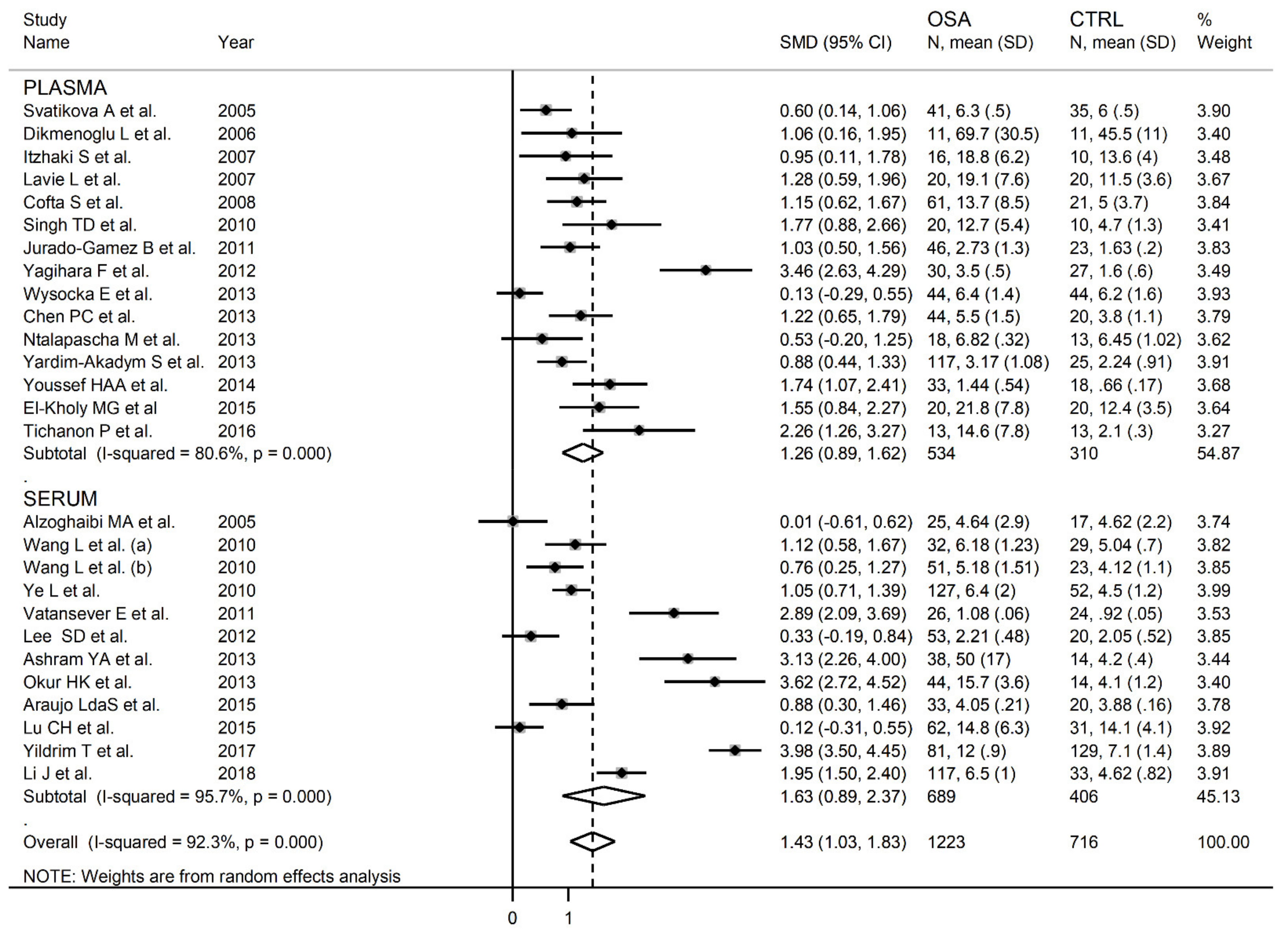
3. Results
4. Discussion
5. Conclusions
Supplementary Materials
Author Contributions
Funding
Conflicts of Interest
References
- Benjafield, A.V.; Ayas, N.T.; Eastwood, P.R.; Heinzer, R.; Ip, M.S.M.; Morrell, M.J.; Nunez, C.M.; Patel, S.R.; Penzel, T.; Pépin, J.L.D.; et al. Estimation of the global prevalence and burden of obstruc-tive sleep apnoea: A literature-based analysis. Lancet Respir. Med. 2019, 7, 687–698. [Google Scholar] [CrossRef] [Green Version]
- Garvey, J.F.; Pengo, M.F.; Drakatos, P.; Kent, B.D. Epidemiological aspects of obstructive sleep apnea. J. Thorac. Dis. 2015, 7, 920–929. [Google Scholar] [CrossRef] [PubMed]
- Azagra-Calero, E.; Espinar-Escalona, E.; Barrera-Mora, J.M.; Llamas-Carreras, J.M.; Solano-Reina, E. Obstructive sleep apnea syndrome (OSA). Review of the literature. Med. Oral. Patol. Oral. Cir. Bucal. 2012, 17, e925–e929. [Google Scholar] [CrossRef]
- Suzuki, Y.J.; Jain, V.; Park, A.-M.; Day, R.M. Oxidative stress and oxidant signaling in obstructive sleep apnea and associated cardiovascular diseases. Free Radic. Biol. Med. 2006, 40, 1683–1692. [Google Scholar] [CrossRef] [PubMed] [Green Version]
- Lavie, L. Oxidative stress in obstructive sleep apnea and intermittent hypoxia-Revisited-The bad ugly and good: Implications to the heart and brain. Sleep Med. Rev. 2015, 20, 27–45. [Google Scholar] [CrossRef]
- Peng, Y.-J.; Yuan, G.; Overholt, J.L.; Kumar, G.K.; Prabhakar, N.R. Systemic and cellular responses to intermittent hypoxia: Evidence for oxidative stress and mitochondrial dysfunction. Adv. Exp. Med. Biol. 2003, 536, 559–564. [Google Scholar] [CrossRef]
- Valko, M.; Leibfritz, D.; Moncol, J.; Cronin, M.T.D.; Mazur, M.; Telser, J. Free radicals and antioxidants in normal physiological functions and human disease. Int. J. Biochem. Cell Biol. 2007, 39, 44–84. [Google Scholar] [CrossRef] [PubMed]
- Lavie, L.; Lavie, P. Molecular mechanisms of cardiovascular disease in OSAHS: The oxidative stress link. Eur. Respir. J. 2009, 33, 1467–1484. [Google Scholar] [CrossRef] [PubMed]
- Mannarino, M.R.; di Filippo, F.; Pirro, M. Obstructive sleep apnea syndrome. Eur. J. Intern. Med. 2012, 23, 586–593. [Google Scholar] [CrossRef]
- Niki, E. Lipid peroxidation: Physiological levels and dual biological effects. Free. Radic. Biol. Med. 2009, 47, 469–484. [Google Scholar] [CrossRef]
- Fois, A.G.; Paliogiannis, P.; Sotgia, S.; Mangoni, A.A.; Zinellu, E.; Pirina, P.; Carru, C.; Zinellu, A. Evaluation of oxidative stress bi-omarkers in idiopathic pulmonary fibrosis and therapeutic applications: A systematic review. Respir. Res. 2018, 19, 51. [Google Scholar] [CrossRef] [Green Version]
- Paliogiannis, P.; Fois, A.G.; Sotgia, S.; Mangoni, A.A.; Zinellu, E.; Pirina, P.; Carru, C.; Zinellu, A. Circulating malondialdehyde concentra-tions in patients with stable COPD: A systematic review and meta-analysis. Biomark. Med. 2018, 12, 771–781. [Google Scholar] [CrossRef]
- Jelic, M.; Mandic, A.; Maricic, S.; Srdjenovic, B. Oxidative stress and its role in cancer. J. Cancer Res. Ther. 2021, 17, 22–28. [Google Scholar] [CrossRef]
- Niki, E. Lipid peroxidation products as oxidative stress biomarkers. BioFactors 2008, 34, 171–180. [Google Scholar] [CrossRef]
- Barcelo, A.; Miralles, C.; Barbe, F.; Vila, M.; Pons, S.; Agusti, A.G.N. Abnormal lipid peroxidation in patients with sleep ap-noea. Eur. Resp. J. 2000, 16, 644–647. [Google Scholar] [CrossRef] [Green Version]
- Hopps, E.; Canino, B.; Calandrino, V.; Montana, M.; Presti, R.L.; Caimi, G. Lipid peroxidation and protein oxidation are related to the severity of OSAS. Eur. Rev. Med Pharmacol. Sci. 2014, 18, 3773–3778. [Google Scholar]
- Svatikova, A.; Wolk, R.; Lerman, L.O.; Juncos, L.A.; Greene, E.L.; McConnell, J.P.; Somers, V.K. Oxidative stress in obstructive sleep apnoea. Eur. Heart J. 2005, 26, 2435–2439. [Google Scholar] [CrossRef] [Green Version]
- Wells, G.; Shea, B.; O’Connell, D.; Peterson, J.; Welch, V.; Losos, M.; Tugwell, P. The Newcastle-Ottawa Scale (NOS) for Assessing the Quality of Non Randomised Studies in Meta-Analyses. 2013. Available online: http://www.ohri.ca/programs/clinical_epidemiology/oxford.asp (accessed on 29 June 2021).
- Wan, X.; Wang, W.; Liu, J.; Tong, T. Estimating the sample mean and standard deviation from the sample size, median, range and/or interquartile range. BMC Med. Res. Methodol. 2014, 14, 1–13. [Google Scholar] [CrossRef] [PubMed] [Green Version]
- Hozo, S.P.; Djulbegovic, B.; Hozo, I. Estimating the mean and variance from the median, range, and the size of a sample. BMC Med. Res. Methodol. 2005, 5, 13. [Google Scholar] [CrossRef] [PubMed] [Green Version]
- Bowden, J.; Tierney, J.F.; Copas, A.J.; Burdett, S. Quantifying, displaying and accounting for heterogeneity in the meta-analysis of RCTs using standard and generalised Q statistics. BMC Med. Res. Methodol. 2011, 11, 41. [Google Scholar] [CrossRef] [PubMed] [Green Version]
- Higgins, J.P.T.; Thompson, S.G. Quantifying heterogeneity in a meta-analysis. Stat. Med. 2002, 21, 1539–1558. [Google Scholar] [CrossRef]
- Tobias, A. Assessing the influence of a single study in the meta-analysis estimate. Stata Tech. Bull. 1999, 47, 15–17. [Google Scholar]
- Begg, C.B.; Mazumdar, M. Operating characteristics of a rank correlation test for publication bias. Biometrics 1994, 50, 1088. [Google Scholar] [CrossRef]
- Sterne, J.A.; Egger, M. Funnel plots for detecting bias in meta-analysis: Guidelines on choice of axis. J. Clin. Epidemiol. 2001, 54, 1046–1055. [Google Scholar] [CrossRef]
- Duval, S.; Tweedie, R. Trim and fill: A simple funnel-plot-based method of testing and adjusting for publication bias in me-ta-analysis. Biomet 2000, 56, 455–463. [Google Scholar] [CrossRef] [PubMed]
- Liberati, A.; Altman, D.G.; Tetzlaff, J.; Mulrow, C.; Gøtzsche, P.C.; Ioannidis, J.P.; Clarke, M.; Devereaux, P.; Kleijnen, J.; Moher, D. The PRISMA statement for reporting systematic reviews and meta-analyses of studies that evaluate healthcare interventions: Explanation and elaboration. J. Clin. Epidemiol. 2009, 62. [Google Scholar] [CrossRef] [PubMed] [Green Version]
- Alzoghaibi, M.A.; Bahammam, A.S.O. Lipid peroxides, superoxide dismutase and circulating IL-8 and GCP-2 in patients with severe obstructive sleep apnea: A pilot study. Sleep Breath. 2005, 9, 119–126. [Google Scholar] [CrossRef] [PubMed]
- Dikmenoǧlu, N.; Çiftçi, B.; Ileri, E.; Güven, S.F.; Seringeç, N.; Aksoy, Y.; Ercil, D. Erythrocyte deformability, plasma viscosi-ty and oxidative status in patients with severe obstructive sleep apnea syndrome. Sleep Med. 2006, 7, 255–261. [Google Scholar] [CrossRef]
- Itzhaki, S.; Dorchin, H.; Clark, G.; Lavie, L.; Lavie, P.; Pillar, G. The effects of 1-year treatment with a herbst mandibular ad-vancement splint on obstructive sleep apnea, oxidative stress, and endothelial function. Chest 2007, 13, 740–749. [Google Scholar] [CrossRef]
- Lavie, L.; Vishnevsky, A.; Lavie, P. Oxidative stress and systemic inflammation in patients with sleep apnea: Role of obesity. Sleep Biol. Rhyms. 2007, 5, 100–110. [Google Scholar] [CrossRef]
- Cofta, S.; Wysocka, E.; Piorunek, T.; Rzymkowska, M.; Batura-Gabryel, H.; Torlinski, L. Oxidative stress markers in the blood of persons with different stages of obstructive sleep apnea syndrome. J. Physiol. Pharmacol. 2008, 59 (Suppl. 6), 183–190. [Google Scholar] [PubMed]
- Singh, T.D.; Patial, K.; Vijayan, V.K.; Ravi, K. Oxidative stress and obstructive sleep apnoea syndrome. Indian J. Chest Dis. Allied Sci. 2009, 51, 217–224. [Google Scholar] [PubMed]
- Wang, L.; Li, J.; Xie, Y.; Zhang, X.-G. Association between serum homocysteine and oxidative stress in elderly patients with obstructive sleep apnea/hypopnea syndrome. Biomed. Environ. Sci. 2010, 23, 42–47. [Google Scholar] [CrossRef]
- Ye, L.; Ma, G.-H.; Chen, L.; Li, M.; Liu, J.-L.; Yang, K.; Li, Q.-Y.; Li, N.; Wan, H.-Y. Quantification of circulating cell-free DNA in the serum of patients with obstructive sleep apnea–hypopnea syndrome. Lung 2010, 188, 469–474. [Google Scholar] [CrossRef]
- Jurado-Gamez, B.; Fernandez-Marin, M.C.; Gomez-Chaparro, J.L.; Munoz-Cabrera, L.; López-Barea, J.; Perez-Jimenez, F.; Lopez-Miranda, J. Relationship of oxidative stress and endothelial dysfunction in sleep apnoea. Eur. Respir. J. 2010, 37, 873–879. [Google Scholar] [CrossRef]
- Vatansever, E.; Surmen-Gur, E.; Ursavas, A.; Karadag, M. Obstructive sleep apnea causes oxidative damage to plasma lipids and proteins and decreases adiponectin levels. Sleep Breath. 2011, 15, 275–282. [Google Scholar] [CrossRef] [PubMed]
- Lee, S.D.; Ju, G.; Choi, J.-A.; Kim, J.-W.; Yoon, I.-Y. The association of oxidative stress with central obesity in obstructive sleep apnea. Sleep Breath. 2011, 16, 511–517. [Google Scholar] [CrossRef]
- Yagihara, F.; Lucchesi, L.M.; D’Almeida, V.; de Mello, M.T.; Tufik, S.; Bittencourt, L.R.A. Oxidative stress and quality of life in elderly patients with obstructive sleep apnea syndrome: Are there differences after six months of continuous positive airway pressure treatment? Clinics 2012, 67, 565–571. [Google Scholar] [CrossRef]
- Wysocka, E.; Cofta, S.; Piorunek, T.; Dziegielewska-Gesiak, S.; Bryl, W.; Torlinski, L. Blood antioxidant status, dysglycemia and obstructive sleep apnea. Adv. Exp. Med. Biol. 2012, 756, 121–129. [Google Scholar]
- Ashram, Y.A.; Wahab, N.H.A.; Diab, I.H. Non-dipping pattern of nocturnal blood pressure in obstructive sleep apnea syndrome: Possible role of oxidative stress and endothelin-1 precursor. Alex. J. Med. 2013, 49, 153–161. [Google Scholar] [CrossRef] [Green Version]
- Chen, P.-C.; Guo, C.-H.; Tseng, C.-J.; Wang, K.-C.; Liu, P.-J. Blood trace minerals concentrations and oxidative stress in patients with obstructive sleep apnea. J. Nutr. Health Aging 2013, 17, 639–644. [Google Scholar] [CrossRef] [PubMed]
- Ntalapascha, M.; Makris, D.; Kyparos, A.; Tsilioni, I.; Kostikas, K.; Gourgoulianis, K.; Kouretas, D.; Zakynthinos, E. Oxida-tive stress in patients with obstructive sleep apnea syndrome. Sleep Breath. 2013, 17, 549–555. [Google Scholar] [CrossRef]
- Okur, H.K.; Pelin, Z.; Yuksel, M.; Yosunkaya, S. Lipid peroxidation and paraoxonase activity in nocturnal cyclic and sustained intermittent hypoxia. Sleep Breath. 2013, 17, 365–371. [Google Scholar] [CrossRef] [PubMed]
- Yardim-Akaydin, S.; Caliskan-Can, E.; Gökalp, F.; Firat, H.; Ardic, S.; Simsek, B. Lipid peroxidation and DNA damage in apnea patients with or without metabolic syndrome. Sleep Biol. Rhythm. 2013, 11, 116–124. [Google Scholar] [CrossRef]
- Abu Youssef, H.A.; Elshazly, M.I.; Rashed, L.A.; Sabry, I.M.; Ibrahim, E.K. Thiobarbituric acid reactive substance (TBARS) a marker of oxidative stress in obstructive sleep apnea. Egypt. J. Chest Dis. Tuberc. 2014, 63, 119–124. [Google Scholar] [CrossRef] [Green Version]
- El-Kholy, M.G.; El-Shafey, B.I.; Hantera, M.S.; Ganna, S.A.; El-Sorogy, H.A.; Faisl, A.E.-R.F. Effect of continuous positive airway pressure on oxidative stress accompanied by obstructive sleep apnea. Egypt. J. Bronc 2015, 9, 192–197. [Google Scholar] [CrossRef]
- Araújo, L.D.S.; Fernandes, J.F.R.; Klein, M.R.S.T.; Sanjuliani, A.F. Obstructive sleep apnea is independently associated with inflammation and insulin resistance, but not with blood pressure, plasma catecholamines, and endothelial function in obese subjects. Nutriton 2015, 31, 1351–1357. [Google Scholar] [CrossRef] [PubMed]
- Lu, C.-H.; Lin, H.-C.; Huang, C.-C.; Lin, W.-C.; Chen, H.-L.; Chang, H.-W.; Friedman, M.; Chen, C.T.; Tsai, N.-W.; Wang, H.-C.; et al. Increased circulating endothelial progenitor cells and anti-oxidant capacity in obstructive sleep apnea after surgical treatment. Clin. Chim. Acta 2015, 448, 1–7. [Google Scholar] [CrossRef]
- Tichanon, P.; Wilaiwan, K.; Sopida, S.; Orapin, P.; Watchara, B.; Banjamas, I. Effect of continuous positive airway pressure on airway inflammation and oxidative stress in patients with obstructive sleep apnea. Can. Respir. J. 2016, 2016, 1–7. [Google Scholar] [CrossRef] [Green Version]
- Yildirim, T.; Alp, R. The role of oxidative stress in the relation between fibromyalgia and obstructive sleep apnea syndrome. Eur. Rev. Med. Pharm. Sci. 2017, 21, 20–29. [Google Scholar]
- Li, J.; Yu, L.Q.; Jiang, M.; Wang, L.; Fang, Q. Homocysteine level in patients with obstructive sleep apnea/hypopnea syn-drome and the impact of continuous positive airway pressure treatment. Adv. Clin. Exp. Med. 2018, 27, 1549–1554. [Google Scholar] [CrossRef] [Green Version]
- Avezov, K.; Aizenbud, D.; Lavie, L. Intermittent hypoxia induced formation of “endothelial cell-colony forming units (EC-CFUS)” is affected by ros and oxidative stress. Front. Neurol. 2018, 9. [Google Scholar] [CrossRef] [PubMed]
- Christou, K.; Kostikas, K.; Pastaka, C.; Tanou, K.; Antoniadou, I.; Gourgoulianis, K.I. Nasal continuous positive airway pressure treatment reduces systemic oxidative stress in patients with severe obstructive sleep apnea syndrome. Sleep Med. 2009, 10, 87–94. [Google Scholar] [CrossRef] [PubMed]
- Dyugovskaya, L.; Lavie, P.; Lavie, L. Increased Adhesion Molecules Expression and Production of Reactive Oxygen Species in Leukocytes of Sleep Apnea Patients. Am. J. Respir. Crit. Care Med. 2002, 165, 934–939. [Google Scholar] [CrossRef]
- Lavie, L. Oxidative stress—A unifying paradigm in obstructive sleep apnea and comorbidities. Prog. Cardiovasc. Dis. 2009, 51, 303–312. [Google Scholar] [CrossRef]
- Minoguchi, K.; Yokoe, T.; Tanaka, A.; Ohta, S.; Hirano, T.; Yoshino, G.; O’Donnell, C.P.; Adachi, M. Association between lipid peroxidation and inflammation in obstructive sleep apnoea. Eur. Respir. J. 2006, 28, 378–385. [Google Scholar] [CrossRef] [PubMed]
- Fiedorczuk, P.; Stróżyński, A.; Olszewska, E. Is the oxidative stress in obstructive sleep apnea associated with cardiovascular complications? Systematic review. J. Clin. Med. 2020, 7, 3734. [Google Scholar] [CrossRef] [PubMed]
- Wood, L.; Gibson, P.; Garg, M. Biomarkers of lipid peroxidation, airway inflammation and asthma. Eur. Respir. J. 2003, 21, 177–186. [Google Scholar] [CrossRef] [Green Version]
- Cohen, J. Statistical Power Analysis for the Behavioral Sciences, 2nd ed.; Lawrence Erlbaum Associates: Mahwah, NJ, USA, 1998. [Google Scholar]
- Ito, F.; Sono, Y.; Ito, T. Measurement and clinical significance of lipid peroxidation as a biomarker of oxidative stress: Oxidative stress in diabetes, atherosclerosis, and chronic inflammation. Antioxidants 2019, 8, 72. [Google Scholar] [CrossRef] [Green Version]
- Tug, T.; Karatas, F.; Terzi, S.M.; Ozdemir, N. Comparison of serum malondialdehyde levels determined by two different methods in patients with COPD: HPLC or TBARS methods. Lab. Med. 2005, 36, 41–44. [Google Scholar] [CrossRef]
- Fadaei, R.; Safari-Faramani, R.; Hosseini, H.; Koushki, M.; Ahmadi, R.; Rostampour, M.; Khazaie, H. Increased the circulating levels of malondialdehyde in patients with obstructive sleep apnea: A systematic review and meta-analysis. Sleep Breath. 2021, 1–8. [Google Scholar] [CrossRef]


| First Author Year. Country | Control | OSA | ||||||||||||||
|---|---|---|---|---|---|---|---|---|---|---|---|---|---|---|---|---|
| n | Age Mean ± SD Range | Gender (M/F) | BMI Mean | AHI% | ODI% | SaO2 Mean (%)/tSaO2 <90% (Min) | Comorbidities | n | Age Mean ± SD Range | Gender (M/F) | BMI Mean | AHI% | ODI% | SaO2 Mean (%)/tSaO2 <90% (Min) | Comorbities | |
| Svatikova A et al. 2005. USA | 35 | 47 ±2 - | 35/0 | 26 | NR | NR | - /1.1 | No comorbidities | 41 | 47 ±2 - | 41/0 | 29.5 | 47 | NR | - /13.2 | No comorbidities |
| Alzoghaibi MA et al. 2005. Saudi Arabia | 17 | 31 ±1.5 - | NR | 23.4 | NR | NR | NR | NR | 25 | 50 ±2.2 - | NR | 36.3 | 62 | 62.3 | 93 /- | NR |
| Dikmenoglu L et al. 2006. Turkey | 11 | 46 - - | 8/3 | 26.6 | NR | NR | NR | DIAB 36.4% HTN 45.4% | 11 | 50 - - | 8/3 | 31.1 | 55 | NR | NR | NR |
| Itzhaki S et al. 2007. Israel | 10 | 50 ±4.4 - | 8/2 | 28 | 7 | 0.9 | NR | HTN /DIAB /DLP 37.5% | 16 | 54 ±8.3 - | 11/5 | 28 | 30 | 15 | NR | HTN /DIAB /DLP 40% |
| Lavie L et al. 2007. Israel | 20 | 42 ±10 - | 16/4 | 26 | 6 | NR | - /0.5 | No comorbidities | 20 | 42 11.1 - | 16/4 | 26 | 29 | NR | - /3.1 | No comorbidities |
| Cofta S et al. 2008. Poland | 21 | 52 ±7 - | 11/10 | 33.4 | NR | NR | NR | NR | 61 | 53 ±6 - | 43/18 | 32.5 | 23 | NR | NR | NR |
| Singh TD et al. 2010. India | 10 | 31 ±1.2 - | 10/0 | 32.9 | 2 | NR | NR | No comorbidities | 20 | 44 ±2.4 - | 20/0 | 24.5 | 61 | NR | NR | HTN 15% DIAB 10% |
| Wang L et al. (a) 2010. China | 29 | 69 ±4.2 - | 27/2 | 26.8 | 3 | NR | NR | HTN 13.8% | 32 | 66 ±7.2 - | 30/2 | 23.3 | 39 | NR | NR | HTN 15.6% |
| Wang L et al. (b) 2010. China | 23 | 45 ±12.3 - | 20/3 | 25 | 3 | NR | NR | HTN 13% | 51 | 43 ±8.3 - | 46/5 | 28.3 | 45 | NR | NR | HTN 13.7 % |
| Ye L et al. 2010. China | 52 | 45 0 - | 37/15 | 26 | 2 | 3.5 | NR | No comorbidities | 127 | 45 ±11 - | 102/25 | 26.3 | 36 | 38.7 | NR | No comorbidities |
| Jurado-Gamez B et al. 2011. Spain | 23 | 48 - 44-51 | 15/8 | 30 | 3 | 7 | 94 /- | DIAB 8% | 46 | 45 - 40-47 | 34/12 | 31 | 46 | 49 | 93 /- | DIAB 4% |
| Vatansever E et al.2011. Turkey | 24 | 47 ±8 - | 24/0 | 28.4 | 2 | NR | NR | No comorbidities | 26 | 49 ±9 - | 26/0 | 28.7 | 38 | NR | NR | No comorbidities |
| Lee SD et al. 2010. South Korea | 20 | 44 ±5.7 - | 20/0 | 26.2 | 3 | 1.9 | 95.7 /0.04 | HTN 4% | 53 | 47 ±8.1 - | 53/0 | 26.6 | 32 | 26 | 94.4 /9.2 | No comorbidities |
| Yagihara F et al. 2012. Brazil | 27 | 66 ±0.7 - | 27/0 | 25.1 | 5 | NR | 94 /1.2 | DLP 40.7 DIAB 26 | 30 | 66 ±0.7 - | 30/0 | 27.9 | 38 | NR | 91 /76.7 | DLP 40% DIAB 40% |
| Wysocka E et al. 2013. Poland | 44 | 53 - 46-61 | 44/0 | 31.3 | 3 | NR | NR | DIAB 50% | 44 | 55 - 49-62 | 44/0 | 30 | 26 | NR | NR | DIAB 50% |
| Ashram YA et al. 2013. Egypt | 14 | 73 - 45-65 | 10/4 | NR | NR | NR | NR | CVD 39.5% DIAB 42% HTN. 84.2% | 38 | 75 - 33-87 | 22/16 | NR | 81 | 35 | 87 /23 | No comorbidities |
| Chen PC et al. 2013. Taiwan | 20 | 42 ±11 - | 15/5 | 26 | 3.3 | 1 | 94 /- | No comorbidities | 44 | 42 ±12 - | 33/11 | 26.7 | 15 | 10 | 94 /- | No comorbidities |
| Ntalapascha M et al. 2013. Greece | 13 | 50 ±13 - | 13/0 | 28 | 3 | 8.61 | 93 /20 | No comorbidities | 18 | 49 ±10 - | 18/0 | 31 | 58 | 61 | 90 /124 | No comorbidities |
| Okur HK et al. 2013. Turkey | 14 | 49 ±8.6 - | 11/3 | 31.8 | 2.7 | NR | 90.4 /0 | NR | 44 | 44 ±13 - | 40/4 | 30.5 | 37 | 47.2 | 76.6 /33 | NR |
| Yardim-Akadym S et al. 2013. Turkey | 25 | 43 ±8.2 - | 14/11 | 27.2 | 3 | 28 | 94.4 /0.4 | DIAB 11% HTN 23% DLP 48% | 117 | 50 ±10.7 - | 81/36 | 31.6 | 36 | 114 | 91 /54 | DLP 32% |
| Youssef HAA et al. 2014. Egypt | 18 | 45 ±12.7 - | 4/14 | 42.8 | 2 | 8 | 94.4 /6 | NR | 33 | 52 ±11.5 - | 23/10 | 44.3 | 19 | 41 | 89.6 /35.2 | NR |
| El-Kholy MG et al 2015. Egypt | 20 | 49 ±14.6 - | 10/10 | 29.4 | 2 | NR | - /3.3 | No comorbidities | 20 | 51 ±8.2 - | 9/11 | 39 | 30 | NR | - /28 | No comorbidities |
| Araujo LdaS et al. 2015. Brazil | 20 | 33 ±2 - | 5/15 | 34.5 | 2.5 | 0.9 | 96.7 /0.06 | No Comorbidities | 33 | 40 ±1.5 - | 20/13 | 34.4 | 20 | 13 | 95 /15.6 | No Comorbidities |
| Lu CH et al. 2015 Taiwan | 31 | 40 ±7.7 - | 27/4 | 24.8 | 2.4 | 0.7 | 96.8 /- | No Comorbidities | 62 | 42 ±10 - | 54/8 | 25.5 | 41 | 31.5 | 94.8 /- | No Comorbidities |
| Tichanon P et al. 2016. Thailand | 13 | 53 ±12.3 - | 10/3 | 23.3 | NR | NR | 98.2 /- | HTN 69% | 13 | 53 ±12.4 - | 10/3 | 53 | 16 | NR | 94.2 /- | No Comorbidities |
| Yildrim T et al. 2017 Turkey | 129 | 51 ±8.1 - | 78/51 | NR | NR | NR | NR | No Comorbidities | 81 | 49 ±8.4 - | 58/23 | NR | 34 | NR | NR | No Comorbidities |
| Li J et al. 2018. China | 33 | 42 ±10.1 - | 29/4 | 25.8 | 4 | 3.6 | 91.8 /- | HTN 9% | 117 | 45 ±10 - | 105/12 | 25 | 25 | 28 | 94 /- | HTN 13% |
| First Author Year. Country | NOS (stars) | Matrix Type | Assay Type | MDA Mean (µmol/l) ± SD | |
|---|---|---|---|---|---|
| Control | OSA | ||||
| Svatikova A et al. 2005. USA | 7 | P | Sp | 6.0 ± 0.5 | 6.3 ± 0.5 |
| Alzoghaibi MA et al. 2005. Saudi Arabia | 6 | S | Sp | 4.6 ± 2.2 | 4.6 ± 2.9 |
| Dikmenoglu L et al. 2006. Turkey | 8 | P | HPLC | 45.5 ± 11 # | 69.7 ± 30.7 # |
| Itzhaki S et al. 2007. Israel | 9 | P | Sp | 13.6 ± 4.0 | 18.8 ± 6.2 |
| Lavie L et al. 2007. Israel | 8 | P | Sp | 11.5 ± 3.6 | 19.1 ± 7.6 |
| Cofta S et al. 2008. Poland | 8 | P | Sp | 5.0 ± 3.7 | 13.7 ± 8.5 |
| Singh TD et al. 2010. India | 6 | P | Sp | 4.7 ± 1.3 | 12.7 ± 5.4 |
| Wang L et al. (a) 2010. China | 6 | S | Sp | 5.0 ± 0.7 | 6.2 ± 1.2 |
| Wang L et al. (b) 2010. China | 6 | S | Sp | 4.1 ± 1.1 | 5.2 ± 1.5 |
| Ye L et al. 2010. China | 8 | S | Sp | 4.5±1.2 | 6.4±2.0 |
| Jurado-Gamez B et al. 2011. Spain | 7 | P | Sp | 1.6±0.2 | 2.7±1.3 |
| Vatansever E et al.2011. Turkey | 7 | S | HPLC | 0.9 ± 0.05 | 1.1 ± 0.06 |
| Lee SD et al. 2010. South Korea | 7 | S | Sp | 2.1 ± 0.5 | 2.2 ± 0.5 |
| Yagihara F et al. 2012. Brazil | 9 | P | Sp | 1.6 ± 0.6 | 3.5 ± 0.5 |
| Wysocka E et al. 2013. Poland | 7 | P | Sp | 6.2±1.6 | 6.4±1.4 |
| Ashram YA et al. 2013. Egypt | 6 | S | Sp | 4.2 ± 0.4 | 50 ± 17 |
| Chen PC et al. 2013. Taiwan | 7 | P | Sp | 3.8 ± 1.1 | 5.5 ± 1.5 |
| Ntalapascha M et al. 2013. Greece | 7 | P | Sp | 6.5 ± 1.0 | 6.8 ± 0.3 |
| Okur HK et al. 2013. Turkey | 6 | S | Sp | 4.1 ± 1.2 | 15.7 ± 3.6 |
| Yardim-Akadym S et al. 2013. Turkey | 7 | P | HPLC | 2.2 ± 0.9 | 3.2 ± 1.1 |
| Youssef HAA et al. 2014. Egypt | 7 | P | Sp | 0.66 ± 0.17 | 1.44 ± 0.54 |
| El-Kholy MG et al 2015. Egypt | 7 | P | Sp | 12.4 ± 3.5 | 21.8 ± 7.8 |
| Araujo LdaS et al. 2015. Brazil | 7 | S | Sp | 3.88 ± 0.16 § | 4.05 ± 0.21 § |
| Lu CH et al. 2015 Taiwan | 8 | S | Sp | 14.1 ± 4.1 | 14.8 ± 6.3 |
| Tichanon P et al. 2016. Thailand | 8 | P | Sp | 2.1 ± 0.3 | 14.6 ± 7.8 |
| Yildrim T et al. 2017 Turkey | 6 | S | ELISA | 7.1 ± 1.4 | 12 ± 0.9 |
| Li J et al. 2018. China | 7 | S | Sp | 4.6 ± 0.8 | 6.5 ± 1.0 |
Publisher’s Note: MDPI stays neutral with regard to jurisdictional claims in published maps and institutional affiliations. |
© 2021 by the authors. Licensee MDPI, Basel, Switzerland. This article is an open access article distributed under the terms and conditions of the Creative Commons Attribution (CC BY) license (https://creativecommons.org/licenses/by/4.0/).
Share and Cite
Pau, M.C.; Zinellu, E.; Fois, S.S.; Piras, B.; Pintus, G.; Carru, C.; Mangoni, A.A.; Fois, A.G.; Zinellu, A.; Pirina, P. Circulating Malondialdehyde Concentrations in Obstructive Sleep Apnea (OSA): A Systematic Review and Meta-Analysis with Meta-Regression. Antioxidants 2021, 10, 1053. https://doi.org/10.3390/antiox10071053
Pau MC, Zinellu E, Fois SS, Piras B, Pintus G, Carru C, Mangoni AA, Fois AG, Zinellu A, Pirina P. Circulating Malondialdehyde Concentrations in Obstructive Sleep Apnea (OSA): A Systematic Review and Meta-Analysis with Meta-Regression. Antioxidants. 2021; 10(7):1053. https://doi.org/10.3390/antiox10071053
Chicago/Turabian StylePau, Maria Carmina, Elisabetta Zinellu, Sara S. Fois, Barbara Piras, Gianfranco Pintus, Ciriaco Carru, Arduino A. Mangoni, Alessandro G. Fois, Angelo Zinellu, and Pietro Pirina. 2021. "Circulating Malondialdehyde Concentrations in Obstructive Sleep Apnea (OSA): A Systematic Review and Meta-Analysis with Meta-Regression" Antioxidants 10, no. 7: 1053. https://doi.org/10.3390/antiox10071053