


The Effects of Cortical Reorganization and Applications of Functional Near-Infrared Spectroscopy in Deaf People and Cochlear Implant Users

Abstract
:1. Introduction
2. Role of Cross-Modal Reorganization in the Auditory Cortex
2.1. Evidence for Cross-Modal Reorganization in the Auditory Cortex
2.2. Maladaptive Plasticity Effects in the Auditory Cortex
2.3. Adaptive Plasticity Effects in the Auditory Cortex

| Manuscripts | Study Population | Stimuli | Neuroimaging Method | Main Results | Study Design |
|---|---|---|---|---|---|
| Lee D.S et al., 2001 [60] | Prelingually deaf patients (2.2–20.3 y), n = 15 | No, resting state | PET-CT | Low metabolic activity of the auditory cortex was positively correlated with CI outcomes (r = 0.81). | Longitudinal study |
| Doucet M E et al., 2006 [71] | Prelingually and postlingually deafened CI users (18–62 y), n = 13 | Visual stimuli consisted of a high-contrast sinusoidal concentric grating. | EEG | The poor CI performers exhibited broader, anteriorly distributed, high P2 amplitudes over the cortex, whereas the good CI performers showed significantly higher P2 amplitudes over visual occipital areas. | Cross-sectional |
| Lee H.J et al., 2007 [69] | Prelingually deaf patients (1.5–11.3 y), n = 22 | No, resting state | PET-CT | Decreased metabolic activity in the right Heschl’s gyrus (R = −0.45) and in the posterior superior temporal sulcus (R = −0.466) were positively correlated with CI outcomes. | Longitudinal study |
| Buckley K A et al., 2011 [70] | Prelingually and postlingually deafened CI users (14–65 y), n = 22 | Visual stimuli consisted of moving visual gradients with still pictures of cartoon characters in the center. | EEG | A clear negative association between the amplitude of the N1 VEP over the right temporal lobe and speech perception scores was observed for prelingually deafened CI users (r = −0.7703 to −0.8965) but not for postlingually deafened CI users. | Cross-sectional |
| Sandmann P et al., 2012 [58] | Postlingually deafened CI users (38–69 y), n = 11 | Visual stimuli consisted of reversing displays of chequerboard patterns. | EEG | At the P100 latency, CI users showed activation in the right auditory cortex that was inversely related to speech recognition ability in a CI recipient. | Cross-sectional |
| Campbell J et al., 2016 [73] | Prelingually and postlingually deafened CI users (4.95–15.43 y), n = 14 | Visual stimuli consisted of a high-contrast sinusoidal concentric grating. | EEG | The VEP N1 latency in the right temporal cortex was negatively related to speech perception in background noise in children with cochlear implants (r = −0.576). | Cross-sectional |
| Liang M et al., 2017 [72] | Pre-lingually deafened CI children (4.2–6.4 y), n = 20 | Visual stimuli consisted of a photograph with imaginative sound and a photograph without imaginative sound. | EEG | Good CI performers showed significant decreases in N1 amplitude in the primary auditory cortex and in the primary visual cortex, but these did not occur in the poor CI performer group. | Longitudinal study |
| Anderson et al., 2017 [62] | Prelingually, perilingually, and postlingually deaf adults (36–78 y), n = 15 | IHR number sentences (normal speech, male and female speakers) were split into visual speech (visual-only or lip-reading) and auditory speech (auditory-only) | fNIRS_ETG4000 | There was a strong positive correlation between the change in bilateral STC activation to visual speech from preimplantation to postimplantation and speech understanding with a CI (r = 0.70). | Longitudinal study |
| Anderson et al., 2019 [93] | Prelingually, perilingually, and postlingually deaf adults (36–78 y), n = 15 | IHR number sentences (normal speech, male and female speakers) were split into visual stimuli (visual-only or lip-reading) and auditory stimuli (auditory-only) | fNIRS_ETG4000 | Although stronger activation to visual speech preoperatively was predictive of poorer speech understanding outcomes postimplantation (r =−0.75), this relationship was driven by the heterogeneity in subjects. | Longitudinal study |
| Mushtaq et al., 2020 [64] | Prelingually deaf children (6–11 y), n = 19 | Visual speech, auditory speech, signal correlated noise, and steady speech shaped noise. | fNIRS_ETG4000 | Visual and auditory speech are processed synergistically in the temporal cortex of children with CIs, and they should be encouraged, rather than discouraged, to use visual speech. | Cross-sectional |
2.4. Summary of Existing Evidence
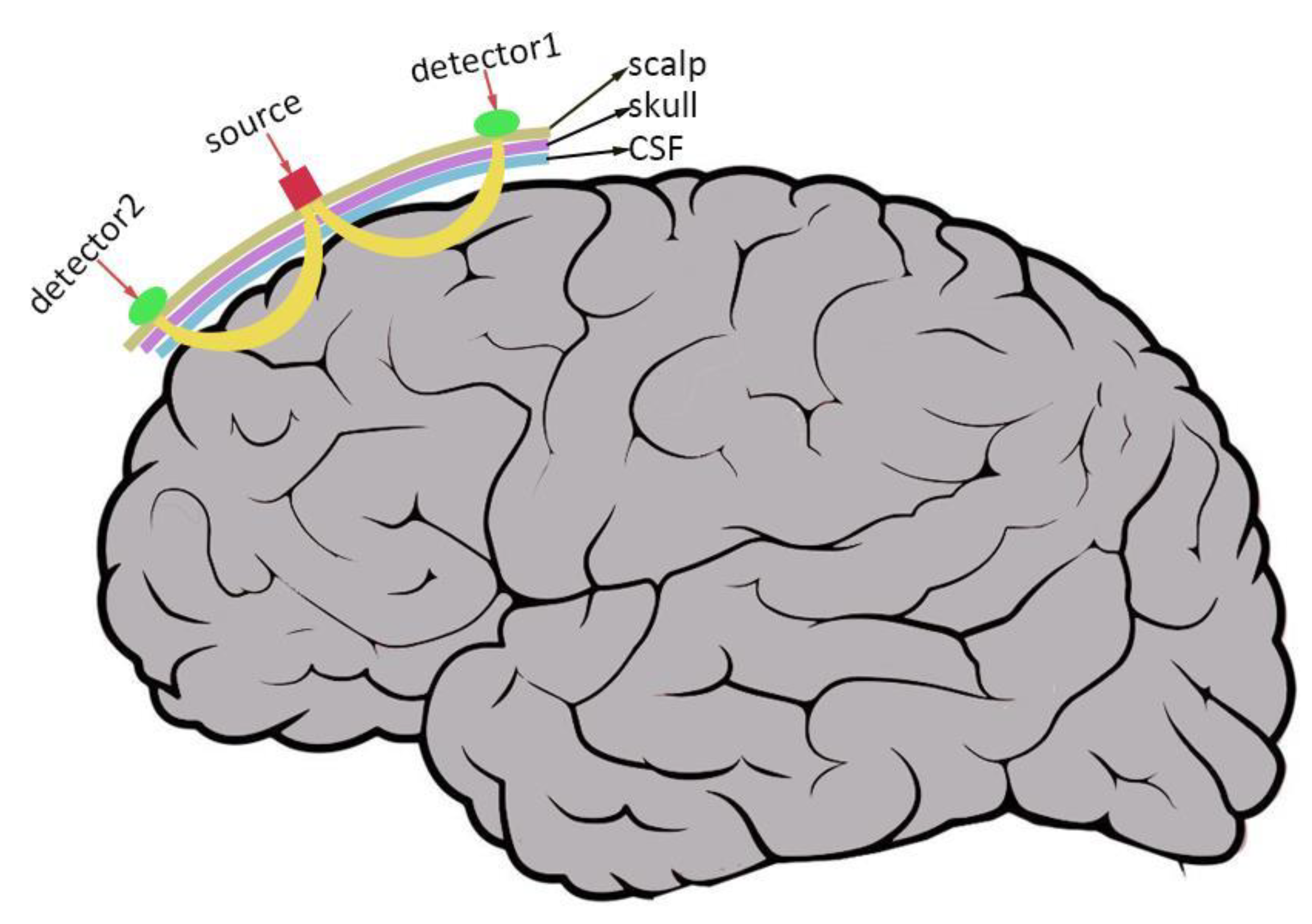
3. fNIRS in Deafness and Cochlear Implant
3.1. fNIRS as a Tool for Predicting/Assessing Speech Performance in Cochlear Implants
3.2. Future fNIRS Application Directions in CI Users
4. Conclusions
Author Contributions
Funding
Institutional Review Board Statement
Informed Consent Statement
Data Availability Statement
Conflicts of Interest
References
- Salomon, J.A.; Vos, T.; Hogan, D.R.; Gagnon, M.; Naghavi, M.; Mokdad, A.; Begum, N.; Shah, R.; Karyana, M.; Kosen, S.; et al. Common values in assessing health outcomes from disease and injury: Disability weights measurement study for the Global Burden of Disease Study 2010. Lancet 2012, 380, 2129–2143. [Google Scholar] [CrossRef]
- Blake, S.W.; Debara, L.T.; Michael, H.M.; Gerard, M.O. Global hearing health care: New findings and perspectives. Lancet 2017, 390, 2503–2515. [Google Scholar]
- Eshraghi, A.A.; Polineni, S.P.; Davies, C.; Shahal, D.; Mittal, J.; Al-Zaghal, Z.; Sinha, R.; Jindal, U.; Mittal, R. Genotype-Phenotype Correlation for Predicting Cochlear Implant Outcome: Current Challenges and Opportunities. Front. Genet. 2020, 11, 678. [Google Scholar] [CrossRef]
- Moser, T.; Dieter, A. Towards optogenetic approaches for hearing restoration. Biochem. Biophys. Res. Commun. 2020, 527, 337–342. [Google Scholar] [CrossRef]
- Eshraghi, A.A.; Nazarian, R.; Telischi, F.F.; Rajguru, S.M.; Truy, E.; Gupta, C. The cochlear implant: Historical aspects and future prospects. Anat. Rec. 2012, 295, 1967–1980. [Google Scholar] [CrossRef]
- Prochazka, A. Neurophysiology and neural engineering: A review. J. Neurophysiol. 2017, 118, 1292–1309. [Google Scholar] [CrossRef]
- Tobey, E.A.; Britt, L.; Geers, A.; Loizou, P.; Loy, B.; Roland, P.; Warner-Czyz, A.; Wright, C.G. Cochlear implantation updates: The Dallas Cochlear Implant Program. J. Am. Acad. Audiol. 2012, 23, 438–445. [Google Scholar] [CrossRef]
- Blamey, P.; Artieres, F.; Baskent, D.; Bergeron, F.; Beynon, A.; Burke, E.; Dillier, N.; Dowell, R.; Fraysse, B.; Gallego, S.; et al. Factors affecting auditory performance of postlinguistically deaf adults using cochlear implants: An update with 2251 patients. Audiol. Neurootol. 2013, 18, 36–47. [Google Scholar] [CrossRef]
- Holden, L.K.; Finley, C.C.; Firszt, J.B.; Holden, T.A.; Brenner, C.; Potts, L.G.; Gotter, B.D.; Vanderhoof, S.S.; Mispagel, K.; Heydebrand, G.; et al. Factors affecting open-set word recognition in adults with cochlear implants. Ear. Hear. 2013, 34, 342–360. [Google Scholar] [CrossRef]
- Dettman, S.J.; Dowell, R.C.; Choo, D.; Arnott, W.; Abrahams, Y.; Davis, A.; Dornan, D.; Leigh, J.; Constantinescu, G.; Cowan, R.; et al. Long-term Communication Outcomes for Children Receiving Cochlear Implants Younger Than 12 Months: A Multicenter Study. Otol. Neurotol. 2016, 37, e82–e95. [Google Scholar] [CrossRef]
- Lazard, D.S.; Vincent, C.; Venail, F.; Van de Heyning, P.; Truy, E.; Sterkers, O.; Skarzynski, P.H.; Skarzynski, H.; Schauwers, K.; O’Leary, S.; et al. Pre-, per- and postoperative factors affecting performance of postlinguistically deaf adults using cochlear implants: A new conceptual model over time. PLoS ONE 2012, 7, e48739. [Google Scholar] [CrossRef] [Green Version]
- Liang, C.; Wenstrup, L.H.; Samy, R.N.; Xiang, J.; Zhang, F. The Effect of Side of Implantation on the Cortical Processing of Frequency Changes in Adult Cochlear Implant Users. Front. Neurosci. 2020, 14, 368. [Google Scholar] [CrossRef]
- Kraaijenga, V.; Derksen, T.C.; Stegeman, I.; Smit, A.L. The effect of side of implantation on unilateral cochlear implant performance in patients with prelingual and postlingual sensorineural hearing loss: A systematic review. Clin. Otolaryngol. 2018, 43, 440–449. [Google Scholar] [CrossRef]
- Finley, C.C.; Holden, T.A.; Holden, L.K.; Whiting, B.R.; Chole, R.A.; Neely, G.J.; Hullar, T.E.; Skinner, M.W. Role of electrode placement as a contributor to variability in cochlear implant outcomes. Otol. Neurotol. 2008, 29, 920–928. [Google Scholar] [CrossRef]
- Gomaa, N.A.; Rubinstein, J.T.; Lowder, M.W.; Tyler, R.S.; Gantz, B.J. Residual speech perception and cochlear implant performance in postlingually deafened adults. Ear. Hear. 2003, 24, 539–544. [Google Scholar] [CrossRef]
- Peng, K.A.; Kuan, E.C.; Hagan, S.; Wilkinson, E.P.; Miller, M.E. Cochlear Nerve Aplasia and Hypoplasia: Predictors of Cochlear Implant Success. Otolaryngol. Head Neck Surg. 2017, 157, 392–400. [Google Scholar] [CrossRef]
- Kim, B.G.; Chung, H.J.; Park, J.J.; Park, S.; Kim, S.H.; Choi, J.Y. Correlation of cochlear nerve size and auditory performance after cochlear implantation in postlingually deaf patients. JAMA Otolaryngol. Head Neck. Surg. 2013, 139, 604–609. [Google Scholar] [CrossRef]
- O’Neill, E.R.; Kreft, H.A.; Oxenham, A.J. Cognitive factors contribute to speech perception in cochlear-implant users and age-matched normal-hearing listeners under vocoded conditions. J. Acoust. Soc. Am. 2019, 146, 195. [Google Scholar] [CrossRef]
- Olds, C.; Pollonini, L.; Abaya, H.; Larky, J.; Loy, M.; Bortfeld, H.; Beauchamp, M.S.; Oghalai, J.S. Cortical Activation Patterns Correlate with Speech Understanding After Cochlear Implantation. Ear. Hear. 2016, 37, e160–e172. [Google Scholar] [CrossRef]
- Glick, H.; Sharma, A. Cross-modal plasticity in developmental and age-related hearing loss: Clinical implications. Hear. Res. 2017, 343, 191–201. [Google Scholar] [CrossRef]
- Saliba, J.; Bortfeld, H.; Levitin, D.J.; Oghalai, J.S. Functional near-infrared spectroscopy for neuroimaging in cochlear implant recipients. Hear. Res. 2016, 338, 64–75. [Google Scholar] [CrossRef] [Green Version]
- Ibraheem, O.A.; Kolkaila, E.A.; Nada, E.H.; Gad, N.H. Auditory cortical processing in cochlear-implanted children with different language outcomes. Eur. Arch. Otorhinolaryngol. 2020, 277, 1875–1883. [Google Scholar] [CrossRef]
- Kral, A.; Kronenberger, W.G.; Pisoni, D.B.; O’Donoghue, G.M. Neurocognitive factors in sensory restoration of early deafness: A connectome model. Lancet Neurol. 2016, 15, 610–621. [Google Scholar] [CrossRef]
- Lomber, S.G.; Meredith, M.A.; Kral, A. Cross-modal plasticity in specific auditory cortices underlies visual compensations in the deaf. Nat. Neurosci. 2010, 13, 1421–1427. [Google Scholar] [CrossRef]
- Simon, M.; Lazzouni, L.; Campbell, E.; Delcenserie, A.; Muise-Hennessey, A.; Newman, A.J.; Champoux, F.; Lepore, F. Enhancement of visual biological motion recognition in early-deaf adults: Functional and behavioral correlates. PLoS ONE 2020, 15, e236800. [Google Scholar] [CrossRef]
- Capek, C.M.; Macsweeney, M.; Woll, B.; Waters, D.; McGuire, P.K.; David, A.S.; Brammer, M.J.; Campbell, R. Cortical circuits for silent speechreading in deaf and hearing people. Neuropsychologia 2008, 46, 1233–1241. [Google Scholar] [CrossRef]
- Auer, E.J.; Bernstein, L.E. Enhanced visual speech perception in individuals with early-onset hearing impairment. J. Speech Lang Hear. Res. 2007, 50, 1157–1165. [Google Scholar] [CrossRef]
- Suh, M.W.; Lee, H.J.; Kim, J.S.; Chung, C.K.; Oh, S.H. Speech experience shapes the speechreading network and subsequent deafness facilitates it. Brain 2009, 132 Pt 10, 2761–2771. [Google Scholar] [CrossRef]
- Bottari, D.; Heimler, B.; Caclin, A.; Dalmolin, A.; Giard, M.H.; Pavani, F. Visual change detection recruits auditory cortices in early deafness. Neuroimage 2014, 94, 172–184. [Google Scholar] [CrossRef]
- Fine, I.; Finney, E.M.; Boynton, G.M.; Dobkins, K.R. Comparing the effects of auditory deprivation and sign language within the auditory and visual cortex. J. Cogn. Neurosci. 2005, 17, 1621–1637. [Google Scholar] [CrossRef]
- Finney, E.M.; Fine, I.; Dobkins, K.R. Visual stimuli activate auditory cortex in the deaf. Nat. Neurosci. 2001, 4, 1171–1173. [Google Scholar] [CrossRef] [PubMed]
- Sadato, N.; Okada, T.; Honda, M.; Matsuki, K.; Yoshida, M.; Kashikura, K.; Takei, W.; Sato, T.; Kochiyama, T.; Yonekura, Y. Cross-modal integration and plastic changes revealed by lip movement, random-dot motion and sign languages in the hearing and deaf. Cereb. Cortex 2005, 15, 1113–1122. [Google Scholar] [CrossRef] [PubMed]
- Vachon, P.; Voss, P.; Lassonde, M.; Leroux, J.M.; Mensour, B.; Beaudoin, G.; Bourgouin, P.; Lepore, F. Reorganization of the auditory, visual and multimodal areas in early deaf individuals. Neuroscience 2013, 245, 50–60. [Google Scholar] [CrossRef] [PubMed]
- Shiell, M.M.; Champoux, F.; Zatorre, R.J. Reorganization of auditory cortex in early-deaf people: Functional connectivity and relationship to hearing aid use. J. Cogn. Neurosci. 2015, 27, 150–163. [Google Scholar] [CrossRef] [PubMed]
- Allison, T.; Puce, A.; Mccarthy, G. Social perception from visual cues: Role of the STS region. Trends Cogn. Sci. 2000, 4, 267–278. [Google Scholar] [CrossRef]
- Corina, D.; Chiu, Y.S.; Knapp, H.; Greenwald, R.; San, J.L.; Braun, A. Neural correlates of human action observation in hearing and deaf subjects. Brain Res. 2007, 1152, 111–129. [Google Scholar] [CrossRef]
- Macsweeney, M.; Campbell, R.; Woll, B.; Giampietro, V.; David, A.S.; McGuire, P.K.; Calvert, G.A.; Brammer, M.J. Dissociating linguistic and nonlinguistic gestural communication in the brain. Neuroimage 2004, 22, 1605–1618. [Google Scholar] [CrossRef]
- Calvert, G.A.; Bullmore, E.T.; Brammer, M.J.; Campbell, R.; Williams, S.C.; McGuire, P.K.; Woodruff, P.W.; Iversen, S.D.; David, A.S. Activation of auditory cortex during silent lipreading. Science 1997, 276, 593–596. [Google Scholar] [CrossRef]
- Calvert, G.A.; Campbell, R. Reading speech from still and moving faces: The neural substrates of visible speech. J. Cogn. Neurosci. 2003, 15, 57–70. [Google Scholar] [CrossRef]
- Macsweeney, M.; Calvert, G.A.; Campbell, R.; McGuire, P.K.; David, A.S.; Williams, S.C.; Woll, B.; Brammer, M.J. Speechreading circuits in people born deaf. Neuropsychologia 2002, 40, 801–807. [Google Scholar] [CrossRef]
- Reale, R.A.; Calvert, G.A.; Thesen, T.; Jenison, R.L.; Kawasaki, H.; Oya, H.; Howard, M.A.; Brugge, J.F. Auditory-visual processing represented in the human superior temporal gyrus. Neuroscience 2007, 145, 162–184. [Google Scholar] [CrossRef] [PubMed] [Green Version]
- Karns, C.M.; Dow, M.W.; Neville, H.J. Altered cross-modal processing in the primary auditory cortex of congenitally deaf adults: A visual-somatosensory fMRI study with a double-flash illusion. J. Neurosci. 2012, 32, 9626–9638. [Google Scholar] [CrossRef]
- Scott, G.D.; Karns, C.M.; Dow, M.W.; Stevens, C.; Neville, H.J. Enhanced peripheral visual processing in congenitally deaf humans is supported by multiple brain regions, including primary auditory cortex. Front. Hum. Neurosci. 2014, 8, 177. [Google Scholar] [CrossRef]
- Cardin, V.; Orfanidou, E.; Ronnberg, J.; Capek, C.M.; Rudner, M.; Woll, B. Dissociating cognitive and sensory neural plasticity in human superior temporal cortex. Nat. Commun. 2013, 4, 1473. [Google Scholar] [CrossRef]
- Emmorey, K.; Grabowski, T.; Mccullough, S.; Damasio, H.; Ponto, L.L.; Hichwa, R.D.; Bellugi, U. Neural systems underlying lexical retrieval for sign language. Neuropsychologia 2003, 41, 85–95. [Google Scholar] [CrossRef]
- Emmorey, K.; Mehta, S.; Grabowski, T.J. The neural correlates of sign versus word production. Neuroimage 2007, 36, 202–208. [Google Scholar] [CrossRef]
- Mayberry, R.I.; Chen, J.K.; Witcher, P.; Klein, D. Age of acquisition effects on the functional organization of language in the adult brain. Brain Lang. 2011, 119, 16–29. [Google Scholar] [CrossRef] [PubMed]
- Macsweeney, M.; Capek, C.M.; Campbell, R.; Woll, B. The signing brain: The neurobiology of sign language. Trends Cogn. Sci. 2008, 12, 432–440. [Google Scholar] [CrossRef] [PubMed]
- Emmorey, K.; Xu, J.; Braun, A. Neural responses to meaningless pseudosigns: Evidence for sign-based phonetic processing in superior temporal cortex. Brain Lang. 2011, 117, 34–38. [Google Scholar] [CrossRef]
- Moreno, A.; Limousin, F.; Dehaene, S.; Pallier, C. Brain correlates of constituent structure in sign language comprehension. Neuroimage 2018, 167, 151–161. [Google Scholar] [CrossRef]
- Auer, E.J.; Bernstein, L.E.; Sungkarat, W.; Singh, M. Vibrotactile activation of the auditory cortices in deaf versus hearing adults. Neuroreport 2007, 18, 645–648. [Google Scholar] [CrossRef] [PubMed] [Green Version]
- Levanen, S.; Hamdorf, D. Feeling vibrations: Enhanced tactile sensitivity in congenitally deaf humans. Neurosci. Lett. 2001, 301, 75–77. [Google Scholar] [CrossRef]
- Levanen, S.; Jousmaki, V.; Hari, R. Vibration-induced auditory-cortex activation in a congenitally deaf adult. Curr. Biol. 1998, 8, 869–872. [Google Scholar] [CrossRef]
- Schurmann, M.; Caetano, G.; Hlushchuk, Y.; Jousmaki, V.; Hari, R. Touch activates human auditory cortex. Neuroimage 2006, 30, 1325–1331. [Google Scholar] [CrossRef] [PubMed]
- Finney, E.M.; Clementz, B.A.; Hickok, G.; Dobkins, K.R. Visual stimuli activate auditory cortex in deaf subjects: Evidence from MEG. Neuroreport 2003, 14, 1425–1427. [Google Scholar] [CrossRef]
- Hauthal, N.; Sandmann, P.; Debener, S.; Thorne, J.D. Visual movement perception in deaf and hearing individuals. Adv. Cogn. Psychol. 2013, 9, 53–61. [Google Scholar] [CrossRef]
- Kim, M.B.; Shim, H.Y.; Jin, S.H.; Kang, S.; Woo, J.; Han, J.C.; Lee, J.Y.; Kim, M.; Cho, Y.S.; Moon, I.J.; et al. Cross-Modal and Intra-Modal Characteristics of Visual Function and Speech Perception Performance in Postlingually Deafened, Cochlear Implant Users. PLoS ONE 2016, 11, e148466. [Google Scholar] [CrossRef]
- Sandmann, P.; Dillier, N.; Eichele, T.; Meyer, M.; Kegel, A.; Pascual-Marqui, R.D.; Marcar, V.L.; Jancke, L.; Debener, S. Visual activation of auditory cortex reflects maladaptive plasticity in cochlear implant users. Brain 2012, 135 Pt 2, 555–568. [Google Scholar] [CrossRef]
- Shiell, M.M.; Champoux, F.; Zatorre, R.J. The Right Hemisphere Planum Temporale Supports Enhanced Visual Motion Detection Ability in Deaf People: Evidence from Cortical Thickness. Neural Plast. 2016, 2016, 7217630. [Google Scholar] [CrossRef]
- Lee, D.S.; Lee, J.S.; Oh, S.H.; Kim, S.K.; Kim, J.W.; Chung, J.K.; Lee, M.C.; Kim, C.S. Cross-modal plasticity and cochlear implants. Nature 2001, 409, 149–150. [Google Scholar] [CrossRef]
- Heimler, B.; Weisz, N.; Collignon, O. Revisiting the adaptive and maladaptive effects of crossmodal plasticity. Neuroscience 2014, 283, 44–63. [Google Scholar] [CrossRef] [PubMed]
- Anderson, C.A.; Wiggins, I.M.; Kitterick, P.T.; Hartley, D. Adaptive benefit of cross-modal plasticity following cochlear implantation in deaf adults. Proc. Natl. Acad. Sci. USA 2017, 114, 10256–10261. [Google Scholar] [CrossRef] [PubMed]
- Lyness, C.R.; Woll, B.; Campbell, R.; Cardin, V. How does visual language affect crossmodal plasticity and cochlear implant success? Neurosci. Biobehav. Rev. 2013, 37 Pt 2, 2621–2630. [Google Scholar] [CrossRef] [PubMed]
- Mushtaq, F.; Wiggins, I.M.; Kitterick, P.T.; Anderson, C.A.; Hartley, D. The Benefit of Cross-Modal Reorganization on Speech Perception in Pediatric Cochlear Implant Recipients Revealed Using Functional Near-Infrared Spectroscopy. Front. Hum. Neurosci. 2020, 14, 308. [Google Scholar] [CrossRef]
- Kral, A.; Sharma, A. Developmental neuroplasticity after cochlear implantation. Trends Neurosci. 2012, 35, 111–122. [Google Scholar] [CrossRef]
- Sharma, A.; Campbell, J.; Cardon, G. Developmental and cross-modal plasticity in deafness: Evidence from the P1 and N1 event related potentials in cochlear implanted children. Int. J. Psychophysiol. 2015, 95, 135–144. [Google Scholar] [CrossRef]
- Lee, H.J.; Kang, E.; Oh, S.H.; Kang, H.; Lee, D.S.; Lee, M.C.; Kim, C.S. Preoperative differences of cerebral metabolism relate to the outcome of cochlear implants in congenitally deaf children. Hear. Res. 2005, 203, 2–9. [Google Scholar]
- Seung-Ha, O.; Chong-Sun, K.; Eun, J.K.; Dong, S.L.; Hyo, J.L.; Sun, O.C.; Soon-hyun, A.; Chan, H.H.; Hong, J.P.; Ja, W.K. Speech Perception after Cochlear Implantation over a 4-Year Time Period. Acta Oto Laryngol. 2003, 123, 148–153. [Google Scholar]
- Lee, H.J.; Giraud, A.L.; Kang, E.; Oh, S.H.; Kang, H.; Kim, C.S.; Lee, D.S. Cortical activity at rest predicts cochlear implantation outcome. Cereb. Cortex 2007, 17, 909–917. [Google Scholar] [CrossRef]
- Buckley, K.A.; Tobey, E.A. Cross-modal plasticity and speech perception in pre- and postlingually deaf cochlear implant users. Ear. Hear. 2011, 32, 2–15. [Google Scholar] [CrossRef]
- Doucet, M.E.; Bergeron, F.; Lassonde, M.; Ferron, P.; Lepore, F. Cross-modal reorganization and speech perception in cochlear implant users. Brain 2006, 129 Pt 12, 3376–3383. [Google Scholar] [CrossRef] [PubMed]
- Liang, M.; Zhang, J.; Liu, J.; Chen, Y.; Cai, Y.; Wang, X.; Wang, J.; Zhang, X.; Chen, S.; Li, X.; et al. Visually Evoked Visual-Auditory Changes Associated with Auditory Performance in Children with Cochlear Implants. Front. Hum. Neurosci. 2017, 11, 510. [Google Scholar] [CrossRef] [PubMed]
- Campbell, J.; Sharma, A. Visual Cross-Modal Re-Organization in Children with Cochlear Implants. PLoS ONE 2016, 11, e147793. [Google Scholar] [CrossRef] [PubMed] [Green Version]
- Quartz, S.R.; Sejnowski, T.J. The neural basis of cognitive development: A constructivist manifesto. Behav. Brain Sci. 1997, 20, 537–556, 556–596. [Google Scholar] [CrossRef]
- Sharma, A.; Gilley, P.M.; Dorman, M.F.; Baldwin, R. Deprivation-induced cortical reorganization in children with cochlear implants. Int. J. Audiol. 2007, 46, 494–499. [Google Scholar] [CrossRef] [PubMed]
- Friston, K. The free-energy principle: A unified brain theory? Nat. Rev. Neurosci. 2010, 11, 127–138. [Google Scholar] [CrossRef] [PubMed]
- Bastos, A.M.; Usrey, W.M.; Adams, R.A.; Mangun, G.R.; Fries, P.; Friston, K.J. Canonical microcircuits for predictive coding. Neuron 2012, 76, 695–711. [Google Scholar] [CrossRef]
- Kral, A.; Yusuf, P.A.; Land, R. Higher-order auditory areas in congenital deafness: Top-down interactions and corticocortical decoupling. Hear. Res. 2017, 343, 50–63. [Google Scholar] [CrossRef]
- Harris, K.D.; Mrsic-Flogel, T.D. Cortical connectivity and sensory coding. Nature 2013, 503, 51–58. [Google Scholar] [CrossRef]
- Kral, A. Auditory critical periods: A review from system’s perspective. Neuroscience 2013, 247, 117–133. [Google Scholar] [CrossRef]
- Kral, A.; Tillein, J.; Heid, S.; Hartmann, R.; Klinke, R. Postnatal cortical development in congenital auditory deprivation. Cereb. Cortex 2005, 15, 552–562. [Google Scholar] [CrossRef] [PubMed]
- Kral, A.; Tillein, J. Brain plasticity under cochlear implant stimulation. Adv. Otorhinolaryngol. 2006, 64, 89–108. [Google Scholar] [PubMed]
- Stropahl, M.; Plotz, K.; Schonfeld, R.; Lenarz, T.; Sandmann, P.; Yovel, G.; De Vos, M.; Debener, S. Cross-modal reorganization in cochlear implant users: Auditory cortex contributes to visual face processing. Neuroimage 2015, 121, 159–170. [Google Scholar] [CrossRef] [PubMed]
- Bergeson, T.R.; Pisoni, D.B.; Davis, R.A. Development of audiovisual comprehension skills in prelingually deaf children with cochlear implants. Ear. Hear. 2005, 26, 149–164. [Google Scholar] [CrossRef] [PubMed]
- Hassanzadeh, S. Outcomes of cochlear implantation in deaf children of deaf parents: Comparative study. J. Laryngol. Otol. 2012, 126, 989–994. [Google Scholar] [CrossRef]
- Voss, P.; Gougoux, F.; Zatorre, R.J.; Lassonde, M.; Lepore, F. Differential occipital responses in early- and late-blind individuals during a sound-source discrimination task. Neuroimage 2008, 40, 746–758. [Google Scholar] [CrossRef]
- Collignon, O.; Dormal, G.; Albouy, G.; Vandewalle, G.; Voss, P.; Phillips, C.; Lepore, F. Impact of blindness onset on the functional organization and the connectivity of the occipital cortex. Brain 2013, 136 Pt 9, 2769–2783. [Google Scholar] [CrossRef]
- Bedny, M.; Konkle, T.; Pelphrey, K.; Saxe, R.; Pascual-Leone, A. Sensitive period for a multimodal response in human visual motion area MT/MST. Curr. Biol. 2010, 20, 1900–1906. [Google Scholar] [CrossRef]
- Land, R.; Baumhoff, P.; Tillein, J.; Lomber, S.G.; Hubka, P.; Kral, A. Cross-Modal Plasticity in Higher-Order Auditory Cortex of Congenitally Deaf Cats Does Not Limit Auditory Responsiveness to Cochlear Implants. J. Neurosci. Off. J. Soc. Neurosci. 2016, 36, 6175–6185. [Google Scholar] [CrossRef]
- Hall, D.A.; Fussell, C.; Summerfield, A.Q. Reading fluent speech from talking faces: Typical brain networks and individual differences. J. Cogn. Neurosci. 2005, 17, 939–953. [Google Scholar] [CrossRef]
- Rouger, J.; Lagleyre, S.; Fraysse, B.; Deneve, S.; Deguine, O.; Barone, P. Evidence that cochlear-implanted deaf patients are better multisensory integrators. Proc. Natl. Acad. Sci. USA 2007, 104, 7295–7300. [Google Scholar] [CrossRef]
- Strelnikov, K.; Rouger, J.; Lagleyre, S.; Fraysse, B.; Demonet, J.F.; Deguine, O.; Barone, P. Increased audiovisual integration in cochlear-implanted deaf patients: Independent components analysis of longitudinal positron emission tomography data. Eur. J. Neurosci. 2015, 41, 677–685. [Google Scholar] [CrossRef] [PubMed]
- Anderson, C.A.; Wiggins, I.M.; Kitterick, P.T.; Hartley, D.E. Pre-operative Brain Imaging Using Functional Near-Infrared Spectroscopy Helps Predict Cochlear Implant Outcome in Deaf Adults. J. Assoc. Res. Otolaryngol. 2019, 20, 511–528. [Google Scholar] [CrossRef] [PubMed] [Green Version]
- Macsweeney, M.; Amaro, E.; Calvert, G.A.; Campbell, R.; David, A.S.; McGuire, P.; Williams, S.C.; Woll, B.; Brammer, M.J. Silent speechreading in the absence of scanner noise: An event-related fMRI study. Neuroreport 2000, 11, 1729–1733. [Google Scholar] [CrossRef] [PubMed]
- Bernstein, L.E.; Auer, E.J.; Moore, J.K.; Ponton, C.W.; Don, M.; Singh, M. Visual speech perception without primary auditory cortex activation. Neuroreport 2002, 13, 311–315. [Google Scholar] [CrossRef]
- Sakai, K.L.; Tatsuno, Y.; Suzuki, K.; Kimura, H.; Ichida, Y. Sign and speech: Amodal commonality in left hemisphere dominance for comprehension of sentences. Brain 2005, 128 Pt 6, 1407–1417. [Google Scholar] [CrossRef]
- Petitto, L.A.; Zatorre, R.J.; Gauna, K.; Nikelski, E.J.; Dostie, D.; Evans, A.C. Speech-like cerebral activity in profoundly deaf people processing signed languages: Implications for the neural basis of human language. Proc. Natl. Acad. Sci. USA 2000, 97, 13961–13966. [Google Scholar] [CrossRef]
- Anderson, C.A.; Lazard, D.S.; Hartley, D.E. Plasticity in bilateral superior temporal cortex: Effects of deafness and cochlear implantation on auditory and visual speech processing. Hear. Res. 2017, 343, 138–149. [Google Scholar] [CrossRef]
- Lazard, D.S.; Lee, H.J.; Truy, E.; Giraud, A.L. Bilateral reorganization of posterior temporal cortices in post-lingual deafness and its relation to cochlear implant outcome. Hum. Brain Mapp. 2013, 34, 1208–1219. [Google Scholar] [CrossRef]
- Corina, D.P.; Blau, S.; Lamarr, T.; Lawyer, L.A.; Coffey-Corina, S. Auditory and Visual Electrophysiology of Deaf Children with Cochlear Implants: Implications for Cross-modal Plasticity. Front. Psychol. 2017, 8, 59. [Google Scholar] [CrossRef]
- Kim, B.G.; Kim, J.W.; Park, J.J.; Kim, S.H.; Kim, H.N.; Choi, J.Y. Adverse events and discomfort during magnetic resonance imaging in cochlear implant recipients. JAMA Otolaryngol. Head Neck Surg. 2015, 141, 45–52. [Google Scholar] [CrossRef] [PubMed]
- Bortfeld, H. Functional near-infrared spectroscopy as a tool for assessing speech and spoken language processing in pediatric and adult cochlear implant users. Dev. Psychobiol. 2019, 61, 430–443. [Google Scholar] [CrossRef] [PubMed]
- Debener, S.; Hine, J.; Bleeck, S.; Eyles, J. Source localization of auditory evoked potentials after cochlear implantation. Psychophysiology 2008, 45, 20–24. [Google Scholar] [CrossRef] [PubMed]
- Gilley, P.M.; Sharma, A.; Dorman, M.; Finley, C.C.; Panch, A.S.; Martin, K. Minimization of cochlear implant stimulus artifact in cortical auditory evoked potentials. Clin. Neurophysiol. 2006, 117, 1772–1782. [Google Scholar] [CrossRef]
- Chen, L.C.; Sandmann, P.; Thorne, J.D.; Herrmann, C.S.; Debener, S. Association of Concurrent fNIRS and EEG Signatures in Response to Auditory and Visual Stimuli. Brain Topogr. 2015, 28, 710–725. [Google Scholar] [CrossRef]
- Sevy, A.B.; Bortfeld, H.; Huppert, T.J.; Beauchamp, M.S.; Tonini, R.E.; Oghalai, J.S. Neuroimaging with near-infrared spectroscopy demonstrates speech-evoked activity in the auditory cortex of deaf children following cochlear implantation. Hear. Res. 2010, 270, 39–47. [Google Scholar] [CrossRef]
- Lawler, C.A.; Wiggins, I.M.; Dewey, R.S.; Hartley, D.E. The use of functional near-infrared spectroscopy for measuring cortical reorganisation in cochlear implant users: A possible predictor of variable speech outcomes? Cochlear Implant. Int. 2015, 16 (Suppl. S1), S30–S32. [Google Scholar] [CrossRef]
- Steinbrink, J.; Villringer, A.; Kempf, F.; Haux, D.; Boden, S.; Obrig, H. Illuminating the BOLD signal: Combined fMRI-fNIRS studies. Magn. Reason. Imaging 2006, 24, 495–505. [Google Scholar] [CrossRef]
- Toronov, V.Y.; Zhang, X.; Webb, A.G. A spatial and temporal comparison of hemodynamic signals measured using optical and functional magnetic resonance imaging during activation in the human primary visual cortex. Neuroimage 2007, 34, 1136–1148. [Google Scholar] [CrossRef]
- Tak, S.; Ye, J.C. Statistical analysis of fNIRS data: A comprehensive review. Neuroimage 2014, 85 Pt 1, 72–91. [Google Scholar] [CrossRef]
- Tsuzuki, D.; Cai, D.S.; Dan, H.; Kyutoku, Y.; Fujita, A.; Watanabe, E.; Dan, I. Stable and convenient spatial registration of stand-alone NIRS data through anchor-based probabilistic registration. Neurosci. Res. 2012, 72, 163–171. [Google Scholar] [CrossRef] [PubMed]
- Tsuzuki, D.; Dan, I. Spatial registration for functional near-infrared spectroscopy: From channel position on the scalp to cortical location in individual and group analyses. Neuroimage 2014, 85 Pt 1, 92–103. [Google Scholar] [CrossRef] [PubMed]
- Bisconti, S.; Shulkin, M.; Hu, X.; Basura, G.J.; Kileny, P.R.; Kovelman, I. Functional Near-Infrared Spectroscopy Brain Imaging Investigation of Phonological Awareness and Passage Comprehension Abilities in Adult Recipients of Cochlear Implants. J. Speech Lang. Hear. Res. 2016, 59, 239–253. [Google Scholar] [CrossRef]
- Chen, L.C.; Sandmann, P.; Thorne, J.D.; Bleichner, M.G.; Debener, S. Cross-Modal Functional Reorganization of Visual and Auditory Cortex in Adult Cochlear Implant Users Identified with fNIRS. Neural Plast. 2016, 2016, 4382656. [Google Scholar] [CrossRef] [PubMed]
- van de Rijt, L.P.; van Opstal, A.J.; Mylanus, E.A.; Straatman, L.V.; Hu, H.Y.; Snik, A.F.; van Wanrooij, M.M. Temporal Cortex Activation to Audiovisual Speech in Normal-Hearing and Cochlear Implant Users Measured with Functional Near-Infrared Spectroscopy. Front. Hum. Neurosci. 2016, 10, 48. [Google Scholar] [CrossRef]
- Wiggins, I.M.; Anderson, C.A.; Kitterick, P.T.; Hartley, D.E. Speech-evoked activation in adult temporal cortex measured using functional near-infrared spectroscopy (fNIRS): Are the measurements reliable? Hear. Res. 2016, 339, 142–154. [Google Scholar] [CrossRef]
- Lawrence, R.J.; Wiggins, I.M.; Hodgson, J.C.; Hartley, D.E. Evaluating cortical responses to speech in children: A functional near-infrared spectroscopy (fNIRS) study. Hear. Res. 2021, 401, 108155. [Google Scholar] [CrossRef]
- Lawrence, R.J.; Wiggins, I.M.; Anderson, C.A.; Davies-Thompson, J.; Hartley, D.E. Cortical correlates of speech intelligibility measured using functional near-infrared spectroscopy (fNIRS). Hear. Res. 2018, 370, 53–64. [Google Scholar] [CrossRef]
- Dewey, R.S.; Hartley, D.E. Cortical cross-modal plasticity following deafness measured using functional near-infrared spectroscopy. Hear. Res. 2015, 325, 55–63. [Google Scholar] [CrossRef]
- Gallagher, A.; Beland, R.; Lassonde, M. The contribution of functional near-infrared spectroscopy (fNIRS) to the presurgical assessment of language function in children. Brain Lang. 2012, 121, 124–129. [Google Scholar] [CrossRef]
- Arun, K.M.; Smitha, K.A.; Rajesh, P.G.; Kesavadas, C. Functional near-infrared spectroscopy is in moderate accordance with functional MRI in determining lateralisation of frontal language areas. Neuroradiol. J. 2018, 31, 133–141. [Google Scholar] [CrossRef]
- Sato, H.; Hirabayashi, Y.; Tsubokura, H.; Kanai, M.; Ashida, T.; Konishi, I.; Uchida-Ota, M.; Konishi, Y.; Maki, A. Cerebral hemodynamics in newborn infants exposed to speech sounds: A whole-head optical topography study. Hum. Brain Mapp. 2012, 33, 2092–2103. [Google Scholar] [CrossRef]
- Wijayasiri, P.; Hartley, D.; Wiggins, I.M. Brain activity underlying the recovery of meaning from degraded speech: A functional near-infrared spectroscopy (fNIRS) study. Hear. Res. 2017, 351, 55–67. [Google Scholar] [CrossRef] [PubMed]
- Strelnikov, K.; Marx, M.; Lagleyre, S.; Fraysse, B.; Deguine, O.; Barone, P. PET-imaging of brain plasticity after cochlear implantation. Hear. Res. 2015, 322, 180–187. [Google Scholar] [CrossRef] [PubMed]
- Lazard, D.S.; Bordure, P.; Lina-Granade, G.; Magnan, J.; Meller, R.; Meyer, B.; Radafy, E.; Roux, P.E.; Gnansia, D.; Pean, V.; et al. Speech perception performance for 100 post-lingually deaf adults fitted with Neurelec cochlear implants: Comparison between Digisonic(R) Convex and Digisonic(R) SP devices after a 1-year follow-up. Acta Otolaryngol. 2010, 130, 1267–1273. [Google Scholar] [CrossRef] [PubMed]
- Glennon, E.; Svirsky, M.A.; Froemke, R.C. Auditory cortical plasticity in cochlear implant users. Curr. Opin. Neurobiol. 2020, 60, 108–114. [Google Scholar] [CrossRef]
- Strelnikov, K.; Rouger, J.; Demonet, J.F.; Lagleyre, S.; Fraysse, B.; Deguine, O.; Barone, P. Visual activity predicts auditory recovery from deafness after adult cochlear implantation. Brain 2013, 136 Pt 12, 3682–3695. [Google Scholar] [CrossRef] [Green Version]

| Techniques Comparison | fNIRS | EEG | fMRI | PET |
|---|---|---|---|---|
| Spatial resolution | Medium | Low | High | High |
| Temporal resolution | Medium | High | Low | Low |
| Confounding effect of techniques | No | No | Yes, because of high levels of scanner noise | No |
| CI-compatible | Yes | Yes | No | Yes |
| Interference of CI | No | Yes, electric artefacts created by the device. | Yes, magnetic artefacts created by the device. | Yes, metal artefacts created by the device. |
| Safety for repeated testing | Yes | Yes | Yes | No, because of radionuclide exposure |
| Invasiveness | Noninvasive | Noninvasive | Noninvasive | Invasive |
| Physical constraints of subjects | Low, resistant to movement artefacts | Medium | High, susceptible to movement artefacts | High, susceptible to movement artefacts |
| Running cost | Low | Low | High | High |
| Ecological validity | High | Medium | Low | Low |
| Portability | Yes | Yes | No | No |
| Depth of detection | Surface of the cortex | cortex | Deep nuclei | Deep nuclei |
| Manuscripts | Key Finding | Advantages | Limitations |
|---|---|---|---|
| Sevy et al., 2010 [106] | NIRS has the potential to compensate for the shortcomings of behavioral assessment tools by providing an accurate measure of a CI’s ability to successfully stimulate the auditory cortex. | A seminal study that demonstrates the feasibility of NIRS neuroimaging in pediatric and adult CI recipients. | This is a preliminary study that does not look into whether there is a difference in cortical response between standardized speech testing materials in the clinic and the stimuli used in the article or what features of the acoustic stimulus are the most effective drivers of the NIRS response. |
| Dewey et al., 2015 [119] | Profoundly deaf individuals show increased activation of visual stimulation in the right auditory cortex compared with normal-hearing controls using fNIRS. There is no significant difference in activation to somatosensory stimulation between groups. | This is the first study to report cross-modal cortical responses in profoundly deaf individuals, and it demonstrated the potential of fNIRS for studying cross-modal cortical plasticity prior to and following cochlear implantation in all age groups. | Due to the heterogeneity of the deaf group and the imbalance in sample size between prelingually deaf individuals and postlingually deaf individuals, it was not possible to perform extensive subgroup analyses. |
| Olds et al., 2016 [19] | Implanted adults with good speech perception and NH controls show a similar pattern, that is, greater cortical activation for natural speech than for unintelligible speech. Poor CI users have indistinguishable cortical activation for all stimuli. Although CI participants’ cortical activation directly correlates with the CNC (R2 = 0.53 to 0.68) and AzBio (R2 = 0.55 to 0.66) scores, it does not correlate with their general auditory abilities (SRT scores). | The study reveals the neural correlates of speech processing among CI adults so that the variability in CI outcomes can be better understood. Therefore, fNIRS could be used as an objective measure of speech perception. | This study does not disclose how the general auditory abilities (SRT scores) were measured and does not quantify or control the attention of the participants. |
| Chen et al., 2016 [114] | The variability in speech rehabilitation in CI users depends on the combined effect of cross-modal reorganization in auditory cortex and visual cortex. In addition, the reorganization in auditory cortex is detrimental to CI recovery, while the reorganization in visual cortex is beneficial to CI recovery. | This is the first fNIRS study to investigate the joint influence of functional reorganization of both auditory and visual cortex on CI users’ speech recognition, and the results indicate the importance of both types of reorganization. | The author does not examine the relationship between cross-modal plasticity and CI outcome while controlling confounding factors, such as age at onset and duration of auditory deprivation. |
| Anderson et al., 2017 [62] | The increased cross-modal activation of auditory brain regions by visual speech from before to after implantation is adaptive to hearing restoration after implantation through an audiovisual mechanism. Furthermore, there is a strong positive correlation between changes in bilateral STC activation to visual speech from preimplantation to postimplantation and speech understanding with a CI (r = 0.70). | A longitudinal study that used fNIRS to examine changes in cortical function and plasticity over the period from hearing loss to hearing rehabilitation with a CI. | There was heterogeneity in these subjects, who include prelingually, perilingually, and postlingually deaf individuals, so incorporating these subjects into a unified framework of analysis requires care. |
| Anderson et al., 2019 [93] | fNIRS measures can provide additional prognostic information about future CI outcomes. Preoperative cortical imaging provides prognostic value above that of influential clinical characteristics, including the age at onset (an additional 18%) and duration of auditory deprivation (an additional 35%). | The study suggests that the use of fNIRS as an objective measure prior to cochlear implantation may enable us to deliver more accurate prognostic information to adult CI candidates. | The relationship between cross-modal plasticity and auditory outcomes was driven by the heterogeneity in adult CI-using clinical populations. |
| Mushtaq et al., 2020 [64] | Although CI users display significantly greater cortical responses to visual speech compared with NH controls, there is no significant difference between these two groups in responses to auditory speech. Visual and auditory speech are processed synergistically in the temporal cortex of children with CIs, and they should be encouraged, rather than discouraged, to use visual speech. | The first fNIRS study with pediatric CI recipients explores the relationship between speech understanding and cortical responses. | Almost all CI users score well on the speech perception test, so the authors do not compare the differences in cortical responses between CI users with good vs. poor speech perception. |
Publisher’s Note: MDPI stays neutral with regard to jurisdictional claims in published maps and institutional affiliations. |
© 2022 by the authors. Licensee MDPI, Basel, Switzerland. This article is an open access article distributed under the terms and conditions of the Creative Commons Attribution (CC BY) license (https://creativecommons.org/licenses/by/4.0/).
Share and Cite
Zhou, X.; Feng, M.; Hu, Y.; Zhang, C.; Zhang, Q.; Luo, X.; Yuan, W. The Effects of Cortical Reorganization and Applications of Functional Near-Infrared Spectroscopy in Deaf People and Cochlear Implant Users. Brain Sci. 2022, 12, 1150. https://doi.org/10.3390/brainsci12091150
Zhou X, Feng M, Hu Y, Zhang C, Zhang Q, Luo X, Yuan W. The Effects of Cortical Reorganization and Applications of Functional Near-Infrared Spectroscopy in Deaf People and Cochlear Implant Users. Brain Sciences. 2022; 12(9):1150. https://doi.org/10.3390/brainsci12091150
Chicago/Turabian StyleZhou, Xiaoqing, Menglong Feng, Yaqin Hu, Chanyuan Zhang, Qingling Zhang, Xiaoqin Luo, and Wei Yuan. 2022. "The Effects of Cortical Reorganization and Applications of Functional Near-Infrared Spectroscopy in Deaf People and Cochlear Implant Users" Brain Sciences 12, no. 9: 1150. https://doi.org/10.3390/brainsci12091150