Next Issue
Volume 12, October
Previous Issue
Volume 12, August


Brain Sci., Volume 12, Issue 9 (September 2022) – 151 articles

Cover Story (view full-size image): 
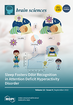
Sleep was found to enhance the consolidation of odors in adults but not in typically developing children (TDC). Previously, children with attention deficit hyperactivity disorder (ADHD) were reported to show lower perceptive thresholds for odors. Sleep-associated odor memory consolidation in children with ADHD was investigated. Children with ADHD and age-matched TDC participated in an incidental odor recognition task. For the sleep groups, encoding took place in the evening and retrieval the next morning. In the wake groups, the time schedule was reversed. Odor memory consolidation was superior in the ADHD sleep group compared to the TDC sleep and the ADHD wake groups. Abundant pre-experience due to lower perceptive thresholds is suggested as a possible explanation. View this paper
  • Issues are regarded as officially published after their release is announced to the table of contents alert mailing list.
  • You may sign up for e-mail alerts to receive table of contents of newly released issues.
  • PDF is the official format for papers published in both, html and pdf forms. To view the papers in pdf format, click on the "PDF Full-text" link, and use the free Adobe Reader to open them.
Order results
Result details
Section
Select all
Export citation of selected articles as:
10 pages, 893 KiB  
Article
Determining Whether Sex and Zygosity Modulates the Association between APOE4 and Psychosis in a Neuropathologically-Confirmed Alzheimer’s Disease Cohort
by Mila Valcic, Marc A. Khoury, Julia Kim, Luis Fornazzari, Nathan W. Churchill, Zahinoor Ismail, Vincenzo De Luca, Debby Tsuang, Tom A. Schweizer, David G. Munoz and Corinne E. Fischer
Brain Sci. 2022, 12(9), 1266; https://doi.org/10.3390/brainsci12091266 - 19 Sep 2022
Cited by 3 | Viewed by 1765 | Correction
Abstract
Background: The APOE4 allele is a genetic risk factor for developing late-onset Alzheimer’s disease (AD). Previous work by our group revealed that female APOE4 homozygotes with Lewy body (LB) pathology were more likely to experience psychosis compared to female APOE4 non-carriers, whereas in [...] Read more.
Background: The APOE4 allele is a genetic risk factor for developing late-onset Alzheimer’s disease (AD). Previous work by our group revealed that female APOE4 homozygotes with Lewy body (LB) pathology were more likely to experience psychosis compared to female APOE4 non-carriers, whereas in males there was no APOE4 dose-dependent significant effect. The objective of this study was to refine our previous findings by adjusting for covariates and determining the probability of an APOE4 sex-mediated effect on psychosis. Methods: Neuropathologically confirmed AD patients with LB pathology (n = 491) and without LB pathology (n = 716) were extracted from the National Alzheimer’s Coordinating Center (NACC). Patients were classified as psychotic if they scored positively for delusions and/or hallucinations on the Neuropsychiatric Inventory. Analysis consisted of a preliminary unadjusted binary logistic regression and a Generalized Additive binary logistic regression Model (GAM) to predict the relationship between APOE4 status and sex on the presence of psychosis in both cohorts, adjusting for age, education and MMSE. Results: In the cohort with LB pathology, female APOE4 homozygotes were significantly more likely to experience psychosis compared to female APOE4 non-carriers (OR = 4.15, 95%CI [1.21, 14.2], p = 0.023). Female heterozygotes were also more likely to experience psychosis compared to female APOE4 non-carriers, but to a lesser extent (OR = 2.37, 95%CI [1.01, 5.59], p = 0.048). There was no significant difference in males with LB pathology or in any sex in the cohort without LB pathology. Conclusions: Sex and zygosity influence the effect of APOE4 on psychosis in neuropathologically confirmed AD patients, with the effect being limited to females with LB pathology. Full article
(This article belongs to the Special Issue New Frontiers in Aging Psychiatry)
Show Figures

Graphical abstract

17 pages, 819 KiB  
Article
Does Executive Function Training Impact on Communication? A Randomized Controlled tDCS Study on Post-Stroke Aphasia
by Francesca Pisano, Alessio Manfredini, Andrea Castellano, Carlo Caltagirone and Paola Marangolo
Brain Sci. 2022, 12(9), 1265; https://doi.org/10.3390/brainsci12091265 - 19 Sep 2022
Cited by 4 | Viewed by 2843
Abstract
New approaches in aphasia rehabilitation have recently identified the crucial role of executive functions (EFs) in language recovery, especially for people with severe aphasia (PWSA). Indeed, EFs include high-order cognitive abilities such as planning and problem solving, which enable humans to adapt to [...] Read more.
New approaches in aphasia rehabilitation have recently identified the crucial role of executive functions (EFs) in language recovery, especially for people with severe aphasia (PWSA). Indeed, EFs include high-order cognitive abilities such as planning and problem solving, which enable humans to adapt to novel situations and are essential for everyday functional communication. In a randomized double-blind crossover design, twenty chronic Italian PWSA underwent ten days of transcranial direct current stimulation (tDCS) (20 min, 2 mA) over the right dorsolateral prefrontal cortex (DLPFC). Two conditions were considered, i.e., anodal and sham, while performing four types of cognitive training (alertness, selective attention, visuo-spatial working memory, and planning), all of which were related to executive functions. After anodal tDCS, a greater improvement in selective attention, visuospatial working memory and planning abilities was found compared to the sham condition; this improvement persisted one month after the intervention. Importantly, a significant improvement was also observed in functional communication, as measured through the Communication Activities of Daily Living Scale, in noun and verb naming, in auditory and written language comprehension tasks and in executive function abilities. This evidence emphasizes, for the first time, that tDCS over the right DLPFC combined with executive training enhances functional communication in severe aphasia. Full article
(This article belongs to the Special Issue New Strategies and New Rehabilitation Evaluations to Stroke)
Show Figures

Graphical abstract

9 pages, 705 KiB  
Article
Efficacy of Liquid Embolic Agent Treatment in Hemorrhagic Peripheral Intracranial Aneurysms: A Single-Center Experience
by Zong Zhuang, Qi Zhu, Xun-Zhi Liu, Hai-Ping Ling, Shi-Jie Na, Tao Liu, Yu-Hua Zhang, Chun-Hua Hang, Kai-Dong Liu and Qing-Rong Zhang
Brain Sci. 2022, 12(9), 1264; https://doi.org/10.3390/brainsci12091264 - 19 Sep 2022
Viewed by 1528
Abstract
Objective: To evaluate the efficacy of liquid embolization agents for treating various hemorrhagic peripheral intracranial aneurysms. Methods: We retrospectively analyzed 38 patients who suffered from hemorrhagic peripheral intracranial aneurysms and were treated with liquid embolization agents. We used the modified Rankin scale [...] Read more.
Objective: To evaluate the efficacy of liquid embolization agents for treating various hemorrhagic peripheral intracranial aneurysms. Methods: We retrospectively analyzed 38 patients who suffered from hemorrhagic peripheral intracranial aneurysms and were treated with liquid embolization agents. We used the modified Rankin scale for follow-up at 6 months postoperatively, and digital subtraction angiography follow-up was performed 6 months postoperatively. Results: Of the 38 patients (ten of simple peripheral intracranial aneurysms, six of Moyamoya disease (MMD), and 22 of arteriovenous malformation (AVM)), posterior circulation accounted for the most significant proportion (57.9%), followed by anterior circulation (21.1%) and intranidal aneurysms (21.1%). Intraoperative hemorrhage occurred in four cases, postoperative cerebral infarction occurred in four cases, two patients encountered microcatheter retention, and intraoperative thrombosis took place in the basilar artery of a patient with an arteriovenous malformation. A postoperative hemorrhage occurred in only one patient. At 6-month follow-up, 84.2% of patients had good prognosis outcomes, and 13.5% had poor outcomes. Conclusion: Liquid embolization agents are effective for hemorrhagic peripheral intracranial aneurysms; however, safety depends on the subtypes. For peripheral hemorrhagic aneurysms in MMD, the vessel architecture must be carefully evaluated before embolization. Full article
(This article belongs to the Section Neurosurgery and Neuroanatomy)
Show Figures

Figure 1

12 pages, 1715 KiB  
Article
Temporal Stability of Dynamic Default Mode Network Connectivity Negatively Correlates with Suicidality in Major Depressive Disorder
by Xuan Ouyang, Yicheng Long, Zhipeng Wu, Dayi Liu, Zhening Liu and Xiaojun Huang
Brain Sci. 2022, 12(9), 1263; https://doi.org/10.3390/brainsci12091263 - 17 Sep 2022
Cited by 2 | Viewed by 1643
Abstract
Previous studies have demonstrated that the suicidality in patients with major depressive disorder (MDD) is related to abnormal brain functional connectivity (FC) patterns. However, little is known about its relationship with dynamic functional connectivity (dFC) based on the assumption that brain FCs fluctuate [...] Read more.
Previous studies have demonstrated that the suicidality in patients with major depressive disorder (MDD) is related to abnormal brain functional connectivity (FC) patterns. However, little is known about its relationship with dynamic functional connectivity (dFC) based on the assumption that brain FCs fluctuate over time. Temporal stabilities of dFCs within the whole brain and nine key networks were compared between 52 MDD patients and 21 age, sex-matched healthy controls (HCs) using resting-state functional magnetic resonance imaging and temporal correlation coefficients. The alterations in MDD were further correlated with the scores of suicidality item in the Hamilton Rating Scale for Depression (HAMD). Compared with HCs, the MDD patients showed a decreased temporal stability of dFC as indicated by a significantly decreased temporal correlation coefficient at the global level, as well as within the default mode network (DMN) and subcortical network. In addition, temporal correlation coefficients of the DMN were found to be significantly negatively correlated with the HAMD suicidality item scores in MDD patients. These results suggest that MDD may be characterized by excessive temporal fluctuations of dFCs within the DMN and subcortical network, and that decreased stability of DMN connectivity may be particularly associated with the suicidality in MDD. Full article
(This article belongs to the Section Molecular and Cellular Neuroscience)
Show Figures

Figure 1

8 pages, 1299 KiB  
Article
Clinico-Radiological Phenotype of UBTF c.628G>A Pathogenic Variant-Related Neurodegeneration in Childhood: A Case Report and Literature Review
by Ching-Shiang Chi, Hsiu-Fen Lee and Chi-Ren Tsai
Brain Sci. 2022, 12(9), 1262; https://doi.org/10.3390/brainsci12091262 - 17 Sep 2022
Cited by 2 | Viewed by 1479
Abstract
Background: This work aims to describe the clinico-radiological phenotype of UBTF c.628G>A (p.Glu210Lys) pathogenic variant-related neurodegeneration in childhood. Methods: We describe the progress of clinical and neuroimaging features in a male individual who had childhood-onset neuroregression and carried the heterozygous UBTF c.628G>A (p.Glu210Lys) [...] Read more.
Background: This work aims to describe the clinico-radiological phenotype of UBTF c.628G>A (p.Glu210Lys) pathogenic variant-related neurodegeneration in childhood. Methods: We describe the progress of clinical and neuroimaging features in a male individual who had childhood-onset neuroregression and carried the heterozygous UBTF c.628G>A (p.Glu210Lys) pathogenic variant. Clinical cases reported in the literature are reviewed. Results: Fifteen individuals, from 14 reported cases and the index case, were noted. The median age at onset of neurodegeneration was 3 years. Clinical phenotype was consistent among the affected individuals, with progressive motor, speech, cognitive, and social–emotional regression together with ataxia and prominent pyramidal and extrapyramidal symptoms and signs in early to middle childhood. All individuals had the same brain MRI features in terms of symmetric and diffuse T2 high signal intensity over the bilateral subcortical, periventricular, and peritrigonal white matter and progressive cortical and subcortical supratentorial atrophy. Two individuals were reported to have bilateral thalamic involvement. All individuals had profound intellectual disability with loss of verbal and/or ambulatory functions during follow-up. Conclusions: Individuals with the heterozygous UBTF c.628G>A (p.Glu210Lys) pathogenic variant had consistent clinical progress and neuroimaging features. Familiarity with this clinico-radiological phenotype may allow earlier diagnosis of this rare disease. Full article
(This article belongs to the Topic Age-Related Neurodegenerative Diseases and Stroke)
Show Figures

Figure 1

9 pages, 1220 KiB  
Article
Comparing the Effects of Exoskeletal-Type Robot-Assisted Gait Training on Patients with Ataxic or Hemiplegic Stroke
by Sungsik Son, Kil-Byung Lim, Jiyong Kim, Changhun Lee, Sung II Cho and Jeehyun Yoo
Brain Sci. 2022, 12(9), 1261; https://doi.org/10.3390/brainsci12091261 - 17 Sep 2022
Cited by 4 | Viewed by 1801
Abstract
This study aimed to discover the effects of robotic rehabilitation utilizing an exoskeletal-type robot-assisted gait training (RAGT) device on patients with ataxic and hemiplegic stroke and to compare its effectiveness between the two groups. This was a retrospective study, and the electronic charts [...] Read more.
This study aimed to discover the effects of robotic rehabilitation utilizing an exoskeletal-type robot-assisted gait training (RAGT) device on patients with ataxic and hemiplegic stroke and to compare its effectiveness between the two groups. This was a retrospective study, and the electronic charts of 22 patients who underwent RAGT treatment from October 2019 to June 2021 were reviewed. Patients were divided into ataxic and hemiplegic groups based on their symptoms. The clinical outcome measures included the Berg balance scale (BBS), functional ambulation category (FAC), and mobility subcategories of the modified Barthel Index (MBI-m). Outcome measures were reviewed at two points within 48 h, before and after RAGT with EXOWALK®, a type of exoskeletal robot. After the RAGT sessions, total patients in both ataxic and hemiplegic groups demonstrated statistically significant improvements in BBS (p < 0.0001, p = 0.002, and p = 0.005, respectively) and MBI-m (p < 0.0001, p = 0.002, and p = 0.011, respectively). Additionally, FAC after RAGT was significantly improved (p = 0.0056). The regression coefficient of the number of RAGT treatments for BBS changes in the nine subjects was estimated to be 2.45; 3.50 in the ataxic group and 2.26 in the hemiplegic group. The regression coefficient of the number of RAGT treatments for MBI-m changes in the nine subjects was estimated to be 0.16; 4.00 in the ataxic group and −0.52 in the hemiplegic group. Our results suggest that RAGT using an exoskeletal-type robot, EXOWALK®, could be effective for improving walking capacity, balance, and daily activities of life in patients with ataxic and hemiplegic stroke. Full article
(This article belongs to the Section Sensory and Motor Neuroscience)
Show Figures

Figure 1

12 pages, 828 KiB  
Article
Enhanced Short-Term Memory Function in Older Adults with Dementia Following Music-Feedback Physical Training: A Pilot Study
by Jessica V. Strong, Maria Arnold, Lydia Schneider, Johanna Perschl, Arno Villringer and Thomas Hans Fritz
Brain Sci. 2022, 12(9), 1260; https://doi.org/10.3390/brainsci12091260 - 16 Sep 2022
Cited by 2 | Viewed by 2746
Abstract
Prior research demonstrates that music making, physical exercise, and social activity have unique, positive effects on cognition and mood. One intervention, “Jymmin®”, was developed incorporating these approaches and found effective for decreased pain perception and increased endurance, self-efficacy, mood, and muscle [...] Read more.
Prior research demonstrates that music making, physical exercise, and social activity have unique, positive effects on cognition and mood. One intervention, “Jymmin®”, was developed incorporating these approaches and found effective for decreased pain perception and increased endurance, self-efficacy, mood, and muscle efficiency. Previously, Jymmin was not piloted with older adults with dementia. The current study is a randomized pilot study of the Jymmin® with an older adult population in a long-term care facility (n = 38), evaluated across dementia levels (mild, moderate, or severe). Results found significant improvements in scores on a confrontation naming task across all conditions (p = 0.047) and a significant interaction effect for short-term memory scores (p = 0.046), suggesting higher scores at Time 2 for the experimental group and at Time 3 for the control group. There were no significant changes in mood ratings. Findings are discussed in the context of neural activity and musical agency. Full article
Show Figures

Figure 1

11 pages, 273 KiB  
Article
Emotion Recognition in Patients with Low-Grade Glioma before and after Surgery
by Anne M. Buunk, Marleen J. J. Gerritsen, Hanne-Rinck Jeltema, Michiel Wagemakers, Jan D. M. Metzemaekers, Rob J. M. Groen and Jacoba M. Spikman
Brain Sci. 2022, 12(9), 1259; https://doi.org/10.3390/brainsci12091259 - 16 Sep 2022
Cited by 4 | Viewed by 1566
Abstract
Research on patients with low-grade gliomas (LGGs) showed neurocognitive impairments in various domains. However, social cognition has barely been investigated. Facial emotion recognition is a vital aspect of social cognition, but whether emotion recognition is affected in LGG patients is unclear. Therefore, we [...] Read more.
Research on patients with low-grade gliomas (LGGs) showed neurocognitive impairments in various domains. However, social cognition has barely been investigated. Facial emotion recognition is a vital aspect of social cognition, but whether emotion recognition is affected in LGG patients is unclear. Therefore, we aimed to investigate the effect of LGG and resection by examining emotion recognition pre- and postoperatively. Additionally, the relationships among emotion recognition and general cognition and tumor location were investigated. Thirty patients with LGG who underwent resective surgery were included and matched with 63 healthy control participants (HCs). Emotion recognition was measured with the Facial Expressions of Emotion–Stimuli and Tests (FEEST) and general cognition with neuropsychological tests. Correlations and within-group and between-group comparisons were calculated. Before surgery, patients performed significantly worse than the HCs on FEEST-Total and FEEST-Anger. Paired comparisons showed no significant differences between FEEST scores before and post-surgery. No significant correlations with general cognition and tumor location were found. To conclude, the results of this study indicate that the tumor itself contributes significantly to social cognitive dysfunction and that surgery causes no additional deficit. Impairments were not related to general cognitive deficits or tumor location. Consequently, incorporating tests for emotion recognition into the neuropsychological assessment of patients with LGG is important. Full article
(This article belongs to the Special Issue Social Cognition across Healthy and Neuropsychiatric Conditions)
25 pages, 5060 KiB  
Article
COVID-19 Long-Term Effects: Is There an Impact on the Simple Reaction Time and Alternative-Forced Choice on Recovered Patients?
by Mauro Santoyo-Mora, Carlos Villaseñor-Mora, Luz M. Cardona-Torres, Juan J. Martínez-Nolasco, Alejandro I. Barranco-Gutiérrez, José A. Padilla-Medina and Micael Gerardo Bravo-Sánchez
Brain Sci. 2022, 12(9), 1258; https://doi.org/10.3390/brainsci12091258 - 16 Sep 2022
Cited by 9 | Viewed by 2218
Abstract
A comparative single-evaluation cross-sectional study was performed to evaluate cognitive damage in post-COVID-19 patients. The psychophysics tests of Two-Alternative Forced Choice (2AFC) and Simple Reaction Time (SRT), under a designed virtual environment, were used to evaluate the cognitive processes of decision-making, visual attention, [...] Read more.
A comparative single-evaluation cross-sectional study was performed to evaluate cognitive damage in post-COVID-19 patients. The psychophysics tests of Two-Alternative Forced Choice (2AFC) and Simple Reaction Time (SRT), under a designed virtual environment, were used to evaluate the cognitive processes of decision-making, visual attention, and information processing speed. The population under study consisted of 147 individuals, 38 controls, and 109 post-COVID patients. During the 2AFC test, an Emotiv EPOC+® headset was used to obtain EEG signals to evaluate their Focus, Interest, and Engagement metrics. Results indicate that compared to healthy patients or recovered patients from mild-moderate COVID-19 infection, patients who recovered from a severe-critical COVID infection showed a poor performance in different cognitive tests: decision-making tasks required higher visual sensitivity (p = 0.002), Focus (p = 0.01) and information processing speed (p < 0.001). These results signal that the damage caused by the coronavirus on the central nervous and visual systems significantly reduces the cognitive processes capabilities, resulting in a prevalent deficit of 42.42% in information processing speed for mild-moderate cases, 46.15% for decision-making based on visual sensitivity, and 62.16% in information processing speed for severe-critical cases. A psychological follow-up for patients recovering from COVID-19 is recommended based on our findings. Full article
(This article belongs to the Special Issue Impact of COVID-19 Infection on Brain Structures and Functions)
Show Figures

Figure 1

7 pages, 2089 KiB  
Case Report
Staged Surgery for Intra-Extracranial Communicating Jugular Foramen Paraganglioma: A Case Report and Systematic Review
by Qiang Li, Yanbing Yu, Li Zhang, Jiang Liu, Hongxiang Ren and Xueke Zhen
Brain Sci. 2022, 12(9), 1257; https://doi.org/10.3390/brainsci12091257 - 16 Sep 2022
Cited by 1 | Viewed by 1591
Abstract
Staged surgery strategy was preferred for patients with intra-extracranial communicating jugular foramen paraganglioma (IECJFP). A female patient who presented mild tinnitus, headache, and dizziness, together with preoperative related imaging, was diagnosed with a left intra-extracranial communicating jugular foramen lesion in November 2015 and [...] Read more.
Staged surgery strategy was preferred for patients with intra-extracranial communicating jugular foramen paraganglioma (IECJFP). A female patient who presented mild tinnitus, headache, and dizziness, together with preoperative related imaging, was diagnosed with a left intra-extracranial communicating jugular foramen lesion in November 2015 and accepted an initial operation for the intracranial tumor by retrosigmoid approach. The pathologic report was paraganglioma. In November 2021, a subtotal resection of the extracranial tumor was conducted for prominent lower cranial nerves (LCNs) deficit and middle ear involvement by infratemporal approach. In patients with IECJFP accompanied by LCNs deficit and middle ear involvement, an initial surgery for extracranial lesion and a second procedure for intracranial tumor were appropriate. However, the first operation for the intracranial lesion was preferred in IECJFP cases without LCNs deficit and middle ear involvement, as it could remove compression to the neurovascular structure and brain stem, clarify a pathological diagnosis, avoid a CSF leak, and prevent a severe neurological disorder from extracranial lesion excision. Subtotal resection of the extracranial tumor would be performed when lesion became larger combined with obvious LCNs disorder and tympanic cavity involvement. Consideration of specific staged surgical strategy for IECJFP in accordance with preoperative LCNs deficit and tympanic cavity involvement could prevent critical postoperative neurological deficit and improve quality of life in the long term. Full article
Show Figures

Figure 1

12 pages, 3911 KiB  
Opinion
Giant Non-Functioning Pituitary Adenomas: Treatment Considerations
by Domenico Solari, Luigi Maria Cavallo, Chiara Graziadio, Sergio Corvino, Ilaria Bove, Felice Esposito and Paolo Cappabianca
Brain Sci. 2022, 12(9), 1256; https://doi.org/10.3390/brainsci12091256 - 16 Sep 2022
Cited by 2 | Viewed by 3262
Abstract
Giant pituitary adenomas are a subgroup of pituitary adenomas defined by a diameter greater than 4 cm, and they account for 5–14% of adenomas in surgical series. Because of their growth patterns and locations, often involving critical neurovascular structures, they represent a true [...] Read more.
Giant pituitary adenomas are a subgroup of pituitary adenomas defined by a diameter greater than 4 cm, and they account for 5–14% of adenomas in surgical series. Because of their growth patterns and locations, often involving critical neurovascular structures, they represent a true surgical challenge, and gross total resection is difficult to achieve. There is no consensus on the optimal surgical strategy for giant pituitary adenomas, and, often, integrated multi-staged treatment strategies have been considered. Transcranial or transsphenoidal approaches, alone or combined, according to tumor and patient features are the two main routes. Each of these strategies has pros and cons. The conventional transcranial approach has for a long time been considered the first choice for the removal of giant pituitary adenomas. Currently, with endoscopic techniques, it is also possible to remove lesions that involve the intradural compartment and the adjacent neurovascular structures with the use of extended approaches. Our policy for the management of these lesions is to adopt the endoscopic endonasal approach as the first choice unless the tumor presents significant intracranial extension that results in it being outside the visibility and maneuverability of the endoscopic endonasal route. In these latter cases, we agree that the transcranial approach is more appropriate. However, accurate preoperative evaluation and refined treatment plans for each patient are mandatory to define a proper strategy in order to achieve the most effective long-term result. Full article
Show Figures

Figure 1

14 pages, 3694 KiB  
Article
Itaconate Attenuates Neuroinflammation and Exerts Dopamine Neuroprotection in Parkinson’s Disease through Inhibiting NLRP3 Inflammasome
by Guoqing Sun, Rui Zhang, Chengxiao Liu, Wenjun Meng and Qi Pang
Brain Sci. 2022, 12(9), 1255; https://doi.org/10.3390/brainsci12091255 - 16 Sep 2022
Cited by 8 | Viewed by 2332
Abstract
Parkinson’s disease (PD) is a common age-associated neurodegenerative motor disorder, which is mainly caused by dopaminergic neuron loss in the substantia nigra. This study aimed to evaluate the function and the underlying molecular mechanism of itaconate in PD. PD models were established in [...] Read more.
Parkinson’s disease (PD) is a common age-associated neurodegenerative motor disorder, which is mainly caused by dopaminergic neuron loss in the substantia nigra. This study aimed to evaluate the function and the underlying molecular mechanism of itaconate in PD. PD models were established in vivo and in vitro using 1-methyl-4-phenyl-1,2,3,6-tetrahydropyridine (MPTP) and 1-methyl-4-phenylpyridinium (MPP+), respectively. Pole and rotarod tests were applied to evaluate the motor coordination of mice. The expression of tyrosine hydroxylase (TH) in MPTP-induced mice and the MPP+ revulsive PD cell model were detected using Western blotting and immunofluorescence. The inflammatory factors level was detected by quantitative real-time polymerase chain reaction. The content of superoxide dismutase (SOD), malondialdehyde (MDA), glutathione (GSH), and reactive oxygen species (ROS) in substantia nigra, striatum, and SH-SY5Y cells were analyzed. Moreover, the apoptosis of MPP+ revulsive SH-SY5Y cells was determined using terminal deoxynucleotidyl transferase-mediated dUTP-biotin nick-end labeling (TUNEL) staining and flow cytometry. The expression of apoptosis- and Nod-like receptor family protein 3 (NLRP3) inflammasome-associated proteins was measured using Western blotting and immunofluorescence. Itaconate attenuated motor deficits of MPTP-induced PD mice. Itaconate inhibited dopamine neuronal damage, inflammatory response, oxidative stress, and neuronal apoptosis in MPTP-caused PD mice and the MPP+ revulsive PD cell model. Additionally, itaconate notably repressed the activation of NLRP3 inflammasome. This research demonstrated that itaconate could attenuate neuroinflammation and exert dopamine neuroprotection in PD through inhibiting NLRP3 inflammasome. Full article
(This article belongs to the Section Molecular and Cellular Neuroscience)
Show Figures

Figure 1

8 pages, 1518 KiB  
Case Report
Internal Carotid Injury during Skull Base Surgery—Case Report and a Review of the Literature
by Petr Matoušek, Tomáš Krejčí, Eva Misiorzová, Radim Lipina, Václav Procházka, Jakub Lubojacký, Lenka Čábalová and Pavel Komínek
Brain Sci. 2022, 12(9), 1254; https://doi.org/10.3390/brainsci12091254 - 16 Sep 2022
Cited by 1 | Viewed by 1568
Abstract
Iatrogenic injury of the internal carotid artery (ICA) is a rare, and probably underreported, complication of transnasal endoscopic skull base surgery. Although treatment algorithms have been suggested, there is no definite consensus or guideline for the management of this severe complication. We describe [...] Read more.
Iatrogenic injury of the internal carotid artery (ICA) is a rare, and probably underreported, complication of transnasal endoscopic skull base surgery. Although treatment algorithms have been suggested, there is no definite consensus or guideline for the management of this severe complication. We describe a case of ICA injury that occurred during a transsphenoidal biopsy of a tumor in the cavernous sinus and we present a treatment algorithm for managing this complication. We reviewed the articles published from 1998 to 2021, reporting on major vascular injury during transnasal endoscopic skull base surgery and endonasal endoscopic surgery, and we compare the methods and results of ICA injury management reported in the literature with the presented case. The most promising treatment for ICA injury might be packing with a muscle graft initially, then performing an endovascular intervention. Full article
Show Figures

Figure 1

10 pages, 6989 KiB  
Case Report
Poor Spontaneous Recovery of Aphemia Accompanied by Damage to the Anterior Segment of the Left Arcuate Fasciculus: A Case Report
by Qiwei Yu and Wenjun Qian
Brain Sci. 2022, 12(9), 1253; https://doi.org/10.3390/brainsci12091253 - 16 Sep 2022
Viewed by 2005
Abstract
Aphemia is a rare and special type of speech disorder, and the mechanisms underlying the occurrence and recovery remain unclear. Here, we present a clinical case of poor spontaneous recovery of aphemia, with the anterior segment of the left arcuate fasciculus server damaged [...] Read more.
Aphemia is a rare and special type of speech disorder, and the mechanisms underlying the occurrence and recovery remain unclear. Here, we present a clinical case of poor spontaneous recovery of aphemia, with the anterior segment of the left arcuate fasciculus server damaged and the posterior segment intact, as detected by diffusion tensor imaging. Aphemia could be caused by the disruption of the cortical and subcortical language circuits. In particular, our data support the view that damage to the anterior segment of the left arcuate fasciculus may result in poor spontaneous recovery from speech production deficits and that an intact posterior segment seems to be crucial for supporting residual language comprehension ability in patients with post-stroke aphasia. Collectively, these data imply the importance of the left arcuate fasciculus during recovery from the language disorder in the subacute stage of stroke. Full article
(This article belongs to the Special Issue Language and Brain: From Genes to Behavior)
Show Figures

Figure 1

10 pages, 614 KiB  
Article
Language Development in Preschool Duchenne Muscular Dystrophy Boys
by Daniela Pia Rosaria Chieffo, Federica Moriconi, Ludovica Mastrilli, Federica Lino, Claudia Brogna, Giorgia Coratti, Michela Altobelli, Valentina Massaroni, Giulia Norcia, Elisabetta Ferraroli, Simona Lucibello, Marika Pane and Eugenio Mercuri
Brain Sci. 2022, 12(9), 1252; https://doi.org/10.3390/brainsci12091252 - 16 Sep 2022
Cited by 3 | Viewed by 1780
Abstract
Background: the present study aims to assess language in preschool-aged Duchenne muscular dystrophy (DMD) boys with normal cognitive quotients, and to establish whether language difficulties are related to attentional aspects or to the involvement of brain dystrophin isoforms. Methods: 20 children aged between [...] Read more.
Background: the present study aims to assess language in preschool-aged Duchenne muscular dystrophy (DMD) boys with normal cognitive quotients, and to establish whether language difficulties are related to attentional aspects or to the involvement of brain dystrophin isoforms. Methods: 20 children aged between 48 and 72 months were assessed with language and attention assessments for preschool children. Nine had a mutation upstream of exon 44, five between 44 and 51, four between 51 and 63, and two after exon 63. A control group comprising 20 age-matched boys with a speech language disorder and normal IQ were also used. Results: lexical and syntactic comprehension and denomination were normal in 90% of the boys with Duchenne, while the articulation and repetition of long words, and sentence repetition frequently showed abnormal results (80%). Abnormal results were also found in tests assessing selective and sustained auditory attention. Language difficulties were less frequent in patients with mutations not involving isoforms Dp140 and Dp71. The profile in Duchenne boys was different form the one observed in SLI with no cognitive impairment. Conclusion: The results of our observational cross-sectional study suggest that early language abilities are frequently abnormal in preschool Duchenne boys and should be assessed regardless of their global neurodevelopmental quotient. Full article
(This article belongs to the Special Issue Language and Brain: From Genes to Behavior)
Show Figures

Figure 1

3 pages, 192 KiB  
Editorial
Underlying Mechanisms and Neurorehabilitation of Gait after Stroke
by Janis J. Daly, Svetlana Pundik and Jessica P. McCabe
Brain Sci. 2022, 12(9), 1251; https://doi.org/10.3390/brainsci12091251 - 16 Sep 2022
Viewed by 1314
Abstract
The title of this Special Issue is: “Underlying Mechanisms and Neurorehabilitation of Gait after Stroke” [...] Full article
(This article belongs to the Special Issue Underlying Mechanisms and Neurorehabilitation of Gait after Stroke)
16 pages, 18575 KiB  
Article
Brief Maternal Separation Promotes Resilience to Anxiety-like and Depressive-like Behaviors in Female C57BL/6J Offspring with Imiquimod-Induced Psoriasis
by Lin Zhou, Zuotian Wu, Yixin Li, Ling Xiao, Huiling Wang and Gaohua Wang
Brain Sci. 2022, 12(9), 1250; https://doi.org/10.3390/brainsci12091250 - 15 Sep 2022
Cited by 6 | Viewed by 1735
Abstract
Background: Psoriasis is a common chronic inflammatory skin disease that often causes depression. Early life experience affects brain development and relates to depression. Whether the effect of different MS protocols in early life on anxiety-like and depressive-like behaviors in female offspring with imiquimod [...] Read more.
Background: Psoriasis is a common chronic inflammatory skin disease that often causes depression. Early life experience affects brain development and relates to depression. Whether the effect of different MS protocols in early life on anxiety-like and depressive-like behaviors in female offspring with imiquimod (IMQ)-induced psoriasis is unknown. Methods: C57BL/6J mice were subjected to no separation (NMS), brief MS (15 min/day, MS15) or long MS (180 min/day, MS180) from postpartum days (PPD) 1 to PPD21. Then, 5% imiquimod cream was applied for 8 days in adults. Behavioral tests, skin lesions and hippocampal protein expression were also assessed. Results: We found significant psoriasis-like skin lesions in female mice following IMQ application, and mice showed anxiety-like and depressive-like behaviors. Further, increased microglial activation and decreased expression of neuroplasticity were detected in mice following IMQ application. However, after MS15 in early life, mice showed decreased anxiety-like and depressive-like behaviors, indicating resilience. Further, inhibited hippocampal neuroinflammation and increased neuroplasticity were detected. Conclusions: Collectively, this study confirms that brief MS confers resilience to the behavior deficits in female offspring with IMQ-induced psoriasis and reverses the activation of neuroinflammation and the damage of neuroplasticity injury. Full article
(This article belongs to the Section Behavioral Neuroscience)
Show Figures

Figure 1

14 pages, 1381 KiB  
Article
Effects and Adaptation of Visual-Motor Illusion Using Different Visual Stimuli on Improving Ankle Joint Paralysis of Stroke Survivors—A Randomized Crossover Controlled Trial
by Junpei Tanabe, Kazu Amimoto, Katsuya Sakai, Shinpei Osaki, Nao Yoshihiro and Tokuei Kataoka
Brain Sci. 2022, 12(9), 1249; https://doi.org/10.3390/brainsci12091249 - 15 Sep 2022
Cited by 4 | Viewed by 1754
Abstract
Visual-motor illusion (VMI) is an intervention to induce kinesthetic sensation from visual stimuli. We aimed to compare the effects of VMI of different visual stimuli on the paralyzed side ankle joint of stroke hemiplegic patients (hemiplegic patients) and to clarify their indication. We [...] Read more.
Visual-motor illusion (VMI) is an intervention to induce kinesthetic sensation from visual stimuli. We aimed to compare the effects of VMI of different visual stimuli on the paralyzed side ankle joint of stroke hemiplegic patients (hemiplegic patients) and to clarify their indication. We applied two types of VMI images of ankle dorsiflexion: ankle dorsiflexion without resistance (standard VMI (S-VMI)) and maximum effort dorsiflexion with resistance (power VMI (P-VMI)). Twenty-two hemiplegic patients were divided into two groups: Group A, which received S-VMI first and P-VMI one week later (n = 11), and Group B, which received P-VMI first and S-VMI one week later (n = 11). Immediate effects were evaluated. Outcomes were the dorsiflexion angle and angular velocity, degree of sense of agency (SoA), and sense of ownership. Patient’s characteristics of cognitive flexibility were assessed using the Trail making test-B (TMT-B). Fugl-Meyer assessment and the Composite-Spasticity-Scale were also assessed. P-VMI was significantly higher than S-VMI in SoA and dorsiflexion angular velocity. Additionally, the degree of improvement in dorsiflexion function with P-VMI was related to TMT-B and degree of muscle tone. Therefore, P-VMI improves ankle function in hemiplegic patients more than S-VMI but should be performed with cognitive flexibility and degree of muscle tone in mind. Full article
(This article belongs to the Section Neurorehabilitation)
Show Figures

Figure 1

30 pages, 1433 KiB  
Systematic Review
Imaging the Limbic System in Parkinson’s Disease—A Review of Limbic Pathology and Clinical Symptoms
by Magdalena Banwinkler, Hendrik Theis, Stéphane Prange and Thilo van Eimeren
Brain Sci. 2022, 12(9), 1248; https://doi.org/10.3390/brainsci12091248 - 15 Sep 2022
Cited by 13 | Viewed by 6289
Abstract
The limbic system describes a complex of brain structures central for memory, learning, as well as goal directed and emotional behavior. In addition to pathological studies, recent findings using in vivo structural and functional imaging of the brain pinpoint the vulnerability of limbic [...] Read more.
The limbic system describes a complex of brain structures central for memory, learning, as well as goal directed and emotional behavior. In addition to pathological studies, recent findings using in vivo structural and functional imaging of the brain pinpoint the vulnerability of limbic structures to neurodegeneration in Parkinson’s disease (PD) throughout the disease course. Accordingly, dysfunction of the limbic system is critically related to the symptom complex which characterizes PD, including neuropsychiatric, vegetative, and motor symptoms, and their heterogeneity in patients with PD. The aim of this systematic review was to put the spotlight on neuroimaging of the limbic system in PD and to give an overview of the most important structures affected by the disease, their function, disease related alterations, and corresponding clinical manifestations. PubMed was searched in order to identify the most recent studies that investigate the limbic system in PD with the help of neuroimaging methods. First, PD related neuropathological changes and corresponding clinical symptoms of each limbic system region are reviewed, and, finally, a network integration of the limbic system within the complex of PD pathology is discussed. Full article
Show Figures

Figure 1

12 pages, 1225 KiB  
Communication
Ehrlich Tumor Induces TRPV1-Dependent Evoked and Non-Evoked Pain-like Behavior in Mice
by Mariana M. Bertozzi, Telma Saraiva-Santos, Tiago H. Zaninelli, Felipe A. Pinho-Ribeiro, Victor Fattori, Larissa Staurengo-Ferrari, Camila R. Ferraz, Talita P. Domiciano, Cassia Calixto-Campos, Sergio M. Borghi, Ana C. Zarpelon, Thiago M. Cunha, Rubia Casagrande and Waldiceu A. Verri
Brain Sci. 2022, 12(9), 1247; https://doi.org/10.3390/brainsci12091247 - 15 Sep 2022
Cited by 2 | Viewed by 1933
Abstract
We standardized a model by injecting Ehrlich tumor cells into the paw to evaluate cancer pain mechanisms and pharmacological treatments. Opioid treatment, but not cyclooxygenase inhibitor or tricyclic antidepressant treatments reduces Ehrlich tumor pain. To best use this model for drug screening it [...] Read more.
We standardized a model by injecting Ehrlich tumor cells into the paw to evaluate cancer pain mechanisms and pharmacological treatments. Opioid treatment, but not cyclooxygenase inhibitor or tricyclic antidepressant treatments reduces Ehrlich tumor pain. To best use this model for drug screening it is essential to understand its pathophysiological mechanisms. Herein, we investigated the contribution of the transient receptor potential cation channel subfamily V member 1 (TRPV1) in the Ehrlich tumor-induced pain model. Dorsal root ganglia (DRG) neurons from the Ehrlich tumor mice presented higher activity (calcium levels using fluo-4 fluorescent probe) and an increased response to capsaicin (TRPV1 agonist) than the saline-injected animals (p < 0.05). We also observed diminished mechanical (electronic von Frey) and thermal (hot plate) hyperalgesia, paw flinching, and normalization of weight distribution imbalance in TRPV1 deficient mice (p < 0.05). On the other hand, TRPV1 deficiency did not alter paw volume or weight, indicating no significant alteration in tumor growth. Intrathecal injection of AMG9810 (TRPV1 antagonist) reduced ongoing Ehrlich tumor-triggered mechanical and thermal hyperalgesia (p < 0.05). Therefore, the contribution of TRPV1 to Ehrlich tumor pain behavior was revealed by genetic and pharmacological approaches, thus, supporting the use of this model to investigate TRPV1-targeting therapies for the treatment of cancer pain. Full article
(This article belongs to the Special Issue Advances in the Study of Mechanisms Underlying Touch and Pain)
Show Figures

Graphical abstract

16 pages, 8950 KiB  
Article
Infant Stimulation Induced a Rapid Increase in Maternal Salivary Oxytocin
by Kana Minami, Teruko Yuhi, Haruhiro Higashida, Shigeru Yokoyama, Takahiro Tsuji and Chiharu Tsuji
Brain Sci. 2022, 12(9), 1246; https://doi.org/10.3390/brainsci12091246 - 15 Sep 2022
Cited by 1 | Viewed by 1763
Abstract
Oxytocin (OT) is a neuropeptide involved in human social behaviors and reproduction. Non-invasive OT levels in saliva have recently roused interest as it does not require a specialized medical setting. Here, we observed one woman’s basal serum and saliva OT from pregnancy to [...] Read more.
Oxytocin (OT) is a neuropeptide involved in human social behaviors and reproduction. Non-invasive OT levels in saliva have recently roused interest as it does not require a specialized medical setting. Here, we observed one woman’s basal serum and saliva OT from pregnancy to 1 year postpartum to track OT concentration changes over this period. We examined the changes in salivary OT levels over time in response to maternal physiological and behavioral responses. The fluctuation of saliva OT levels is well correlated with serum OT during pregnancy and breastfeeding. However, while salivary OT increased rapidly during direct interaction (social interaction tests) with the infant and/or when the mother was watching her own infant’s video (video tests), no increase was observed in serum. We used social interaction and video tests on a group of mothers (nine mothers for social interaction and six for the video test) to clarify these single-subject results. In both tests, the mothers had increased OT in their saliva but not serum. Our study may suggest that salivary samples reflect not only the physical but also the emotional state and that saliva samples may be useful for monitoring women’s OT levels during pre- and postpartum periods. Further studies with larger sample numbers are necessary to confirm the rapid changes in salivary OT levels in response to maternal physiological and behavioral responses. Full article
(This article belongs to the Section Behavioral Neuroscience)
Show Figures

Figure 1

14 pages, 4765 KiB  
Article
Hydrogen Sulfide Attenuates Neuroinflammation by Inhibiting the NLRP3/Caspase-1/GSDMD Pathway in Retina or Brain Neuron following Rat Ischemia/Reperfusion
by Kun-Li Yang, Wen-Hong Li, Ya-Jie Liu, Ying-Juan Wei, Yan-Kai Ren, Chen-Di Mai, Si-Yu Zhang, Yue Zuo, Zhen-Zhou Sun, Dong-Liang Li and Chih-Huang Yang
Brain Sci. 2022, 12(9), 1245; https://doi.org/10.3390/brainsci12091245 - 15 Sep 2022
Cited by 10 | Viewed by 2301
Abstract
Gasdermin D-executing pyroptosis mediated by NLRP3 inflammasomes has been recognized as a key pathogenesis during stroke. Hydrogen sulfide (H2S) could protect CNS against ischemia/reperfusion (I/R)-induced neuroinflammation, while the underlying mechanism remains unclear. The study applied the middle cerebral artery occlusion/reperfusion (MCAO/R) [...] Read more.
Gasdermin D-executing pyroptosis mediated by NLRP3 inflammasomes has been recognized as a key pathogenesis during stroke. Hydrogen sulfide (H2S) could protect CNS against ischemia/reperfusion (I/R)-induced neuroinflammation, while the underlying mechanism remains unclear. The study applied the middle cerebral artery occlusion/reperfusion (MCAO/R) model to investigate how the brain and the retinal injuries were alleviated in sodium hydrogen sulfide (NaHS)-treated rats. The rats were assigned to four groups and received an intraperitoneal injection of 50 μmol/kg NaHS or NaCl 15 min after surgery. Neurological deficits were evaluated using the modified neurologic severity score. The quantification of pro-inflammatory cytokines, NLRP3, caspase-1, and GSDMD were determined by ELISA and Western blot. Cortical and retinal neurodegeneration and cell pyroptosis were determined by histopathologic examination. Results showed that NaHS rescued post-stroke neurological deficits and infarct progression, improved retina injury, and attenuated neuroinflammation in the brain cortexes and the retinae. NaHS administration inhibits inflammation by blocking the NLRP3/caspase-1/GSDMD pathway and further suppressing neuronal pyroptosis. This is supported by the fact that it reversed the high-level of NLRP3, caspase-1, and GSDMD following I/R. Our findings suggest that compounds with the ability to donate H2S could constitute a novel therapeutic strategy for ischemic stroke. Full article
(This article belongs to the Topic Brain Injury, Microcirculation and Tissue Perfusion)
Show Figures

Figure 1

10 pages, 1745 KiB  
Article
IL-33 Alleviates Postoperative Cognitive Impairment by Inhibiting Hippocampal Inflammation and Upregulating Excitatory Synaptic Number in Aged Mice
by Qi Li, Yuqian Zhao, Chuanchuan Shi and Xuemin Song
Brain Sci. 2022, 12(9), 1244; https://doi.org/10.3390/brainsci12091244 - 14 Sep 2022
Cited by 3 | Viewed by 1871
Abstract
Delayed neurocognitive recovery (dNCR), a postoperative complication that occurs in elderly patients, still lacks effective treatment. Interleukin-33 (IL-33) has been proved to modulate neuroinflammation and synaptic plasticity, among other effects, but the role of IL-33 in dNCR is not clear. We established a [...] Read more.
Delayed neurocognitive recovery (dNCR), a postoperative complication that occurs in elderly patients, still lacks effective treatment. Interleukin-33 (IL-33) has been proved to modulate neuroinflammation and synaptic plasticity, among other effects, but the role of IL-33 in dNCR is not clear. We established a dNCR model in aged mice by laparotomy under sevoflurane anesthesia. Cognition was evaluated by Morris water maze (MWM) and fear conditioning test (FCT). Immunofluorescence was used to detect the density of IL-33 and glial fibrillary acidic protein (GFAP) co-localization, ionized calcium-binding adapter molecule 1, vesicular glutamate transporter 1 (vGlut1) and postsynaptic density protein-95 (PSD95) co-localization in the hippocampus. IL-33, GFAP, vGlut1 and PSD95 were tested by Western blotting. Enzyme-linked immunosorbent assay was used to detect the levels of tumor necrosis factor-alpha (TNF-α), interleukin-1 beta (IL-1β) and IL-10. Surgery/anesthesia reduced the level of IL-33 in the hippocampus. Intraperitoneal injection of 200 ng IL-33 per mouse significantly decreased the latency to the platform and increased the number of platform crossings and the target quadrant dwell time in MWM, while increasing the freezing time in the context test of FCT. Furthermore, IL-33 inhibited microglial activation and the release of TNF-α and IL-1β while upregulating the markers of excitatory synapses vGlut1 and PSD95. Our findings indicated that IL-33 improved cognition by inhibiting the hippocampal inflammatory response and upregulating the number of excitatory synapses. Therefore, IL-33 is a potential drug for the treatment of dNCR. Full article
(This article belongs to the Special Issue Advances in Study on Postoperative Delirium)
Show Figures

Figure 1

11 pages, 3798 KiB  
Article
Evaluation of Rhesus Macaque Models for Cerebral Palsy
by Yong Zhu, Yanan Xiong, Jin Zhang, Haiyang Tong, Hongyi Yang, Qingjun Zhu, Xiaoyan Xu, De Wu, Jiulai Tang and Jinhua Li
Brain Sci. 2022, 12(9), 1243; https://doi.org/10.3390/brainsci12091243 - 14 Sep 2022
Cited by 1 | Viewed by 1482
Abstract
Animal models play a central role in all areas of biomedical research. The similarities in anatomical structure and physiological characteristics shared by non-human primates (NHPs) and humans make NHPs ideal models with which to study human disorders, such as cerebral palsy (CP). However, [...] Read more.
Animal models play a central role in all areas of biomedical research. The similarities in anatomical structure and physiological characteristics shared by non-human primates (NHPs) and humans make NHPs ideal models with which to study human disorders, such as cerebral palsy (CP). However, the methodologies for systematically evaluating NHP models of CP have rarely been assessed, despite the long history of using NHP models to understand CP. Such models should be evaluated using multidisciplinary approaches prior to being used to research the diagnosis and treatment of CP. In this study, we evaluated rhesus macaque CP models established by partial resection of the motor cortex and intrathecal injection of bilirubin. Abnormal posture, motor dysfunction, gross and fine motor behavior, and muscular tension were evaluated, and changes in the cerebral cortex and basal ganglia were observed using 9.4 T magnetic resonance imaging. The results clearly demonstrated the utility of the established evaluation methodology for assessing CP models. This model evaluation methodology may guide researchers through the model building process. Full article
(This article belongs to the Section Neuromuscular and Movement Disorders)
Show Figures

Figure 1

7 pages, 1534 KiB  
Case Report
Camrelizumab-Induced Isolate Abducens Neuritis: A Rare Ophthalmic Immune-Related Adverse Events
by Yanli Hou, Qiang Su, Simeng Tang and Hongyang Li
Brain Sci. 2022, 12(9), 1242; https://doi.org/10.3390/brainsci12091242 - 14 Sep 2022
Cited by 3 | Viewed by 1587
Abstract
Background: Anti-tumor immunotherapy with immune checkpoint inhibitors induces several immune-related adverse events. Camrelizumab-related isolate abducens neuritis is rare. Case presentation: We report on a 67-year-old man with esophageal cancer who presented with acute-onset isolated right abducens cranial nerve palsy after ten cycles of [...] Read more.
Background: Anti-tumor immunotherapy with immune checkpoint inhibitors induces several immune-related adverse events. Camrelizumab-related isolate abducens neuritis is rare. Case presentation: We report on a 67-year-old man with esophageal cancer who presented with acute-onset isolated right abducens cranial nerve palsy after ten cycles of Camrelizumab treatment. Magnetic resonance imaging examination revealed thickening and post-contrast enhancement at the cisternal segment of the right abducens nerve. The diagnosis was immune-related abducens neuritis caused by Camrelizumab. We put him on oral taper corticoids (methylprednisone) for neuritis treatment without Camrelizumab suspension. One month after treatment, he recovered completely. At the last follow-up, one year after the onset of diplopia, the patient was in good condition without neurological symptom recurrence. Conclusion: Abducens neuritis is a rare immune-related adverse outcome of Camrelizumab. The present case proves the efficacy and safety of using corticoids in the treatment of abducens neuritis. Full article
(This article belongs to the Special Issue Dissecting Neuroinflammation and Ocular System)
Show Figures

Figure 1

13 pages, 3806 KiB  
Article
Recognition and Processing of Visual Information after Neuronavigated Transcranial Magnetic Stimulation Session
by Wiktoria Kasprzycka, Magdalena Ligia Naurecka, Bartosz Michał Sierakowski, Paulina Putko, Zygmunt Mierczyk, Grzegorz Chabik, Stanisław Dec, Stefan Gaździński and Rafał Rola
Brain Sci. 2022, 12(9), 1241; https://doi.org/10.3390/brainsci12091241 - 14 Sep 2022
Cited by 1 | Viewed by 2172
Abstract
Background: Transcranial magnetic stimulation (TMS) is a method of noninvasive and painless stimulation of the nervous system, which is based on Faraday’s law of electromagnetic induction. Over the past twenty years, the TMS technique has been deployed as a tool for the diagnosis [...] Read more.
Background: Transcranial magnetic stimulation (TMS) is a method of noninvasive and painless stimulation of the nervous system, which is based on Faraday’s law of electromagnetic induction. Over the past twenty years, the TMS technique has been deployed as a tool for the diagnosis and therapy of neurodegenerative diseases, as well as in the treatment of mental disorders (e.g., depression). Methods: We tested the inhibitory effects of repetitive TMS (rTMS) on reaction times to militarily relevant visual stimuli amidst distractors and on accompanying blood oxygenation level dependent (BOLD) signal functional magnetic resonance imaging (fMRI) in 20 healthy people. rTMS was applied over the visual cortices, V1, on both hemispheres with the inhibitory theta burst paradigm with the intensity of 70% of the active motor threshold fMRI in 20 healthy people. Results: Analysis of the reaction time to visual stimuli after using TMS to the V1 visual cortex revealed an increase in the number of incorrect recognitions, and the reaction time was from 843 to 910 ms. In the subgroup of participants (n = 15), after the stimulation, there were significant reductions of BOLD signal in blood flow within V1 cortices. Conclusions: The studies of reaction times after the rTMS revealed the inhibitory effect of rTMS on the reaction times and recognition performance of significant (military) objects in the visual field. Full article
Show Figures

Figure 1

17 pages, 735 KiB  
Article
Continuous Neurophysiologic Data Accurately Predict Mood and Energy in the Elderly
by Sean H. Merritt, Michael Krouse, Rana S. Alogaily and Paul J. Zak
Brain Sci. 2022, 12(9), 1240; https://doi.org/10.3390/brainsci12091240 - 14 Sep 2022
Cited by 5 | Viewed by 4375
Abstract
The elderly have an elevated risk of clinical depression because of isolation from family and friends and a reticence to report their emotional states. The present study explored whether data from a commercial neuroscience platform could predict low mood and low energy in [...] Read more.
The elderly have an elevated risk of clinical depression because of isolation from family and friends and a reticence to report their emotional states. The present study explored whether data from a commercial neuroscience platform could predict low mood and low energy in members of a retirement community. Neurophysiologic data were collected continuously for three weeks at 1Hz and averaged into hourly and daily measures, while mood and energy were captured with self-reports. Two neurophysiologic measures averaged over a day predicted low mood and low energy with 68% and 75% accuracy. Principal components analysis showed that neurologic variables were statistically associated with mood and energy two days in advance. Applying machine learning to hourly data classified low mood and low energy with 99% and 98% accuracy. Two-day lagged hourly neurophysiologic data predicted low mood and low energy with 98% and 96% accuracy. This study demonstrates that continuous measurement of neurophysiologic variables may be an effective way to reduce the incidence of mood disorders in vulnerable people by identifying when interventions are needed. Full article
(This article belongs to the Section Computational Neuroscience and Neuroinformatics)
Show Figures

Figure 1

13 pages, 3164 KiB  
Article
Experimental Induction of Intracranial Aneurysms in Rats: A New Model Utilizing a Genetic Modification within the EDNRA Gene
by Tim Lampmann, Valeri Borger, Jürgen Konczalla, Suzana Gispert, Georg Auburger, Hartmut Vatter and Erdem Güresir
Brain Sci. 2022, 12(9), 1239; https://doi.org/10.3390/brainsci12091239 - 14 Sep 2022
Viewed by 1491
Abstract
The rupture of an intracranial aneurysm (IA) leads to life-threatening subarachnoid hemorrhage. Aside from well-established risk factors, recently published genome-wide association studies of IA revealed the strong association of a common variant near the endothelin receptor type A (EDNRA) gene with IA risk. [...] Read more.
The rupture of an intracranial aneurysm (IA) leads to life-threatening subarachnoid hemorrhage. Aside from well-established risk factors, recently published genome-wide association studies of IA revealed the strong association of a common variant near the endothelin receptor type A (EDNRA) gene with IA risk. However, the role of EDNRA in the pathogenesis of IA remains unclear. The aim of this study was to investigate the influence of a genetic modification within the EDNRA gene on IA pathogenesis in a novel in vivo model. Adult wild-type Sprague–Dawley rats (WT rats) and genetically modified rats (EDNRA rats) were used for the induction of IA using arterial hypertension (HT). Animals were stratified into four groups: WT rats without (WT_CTL) and with induction of HT (WT + HT), as well as EDNRA rats without (EDNRA_CTL) and with induction of HT (EDNRA + HT). Blood pressure (BP) was observed for 12 weeks. After the observation period, cerebral arteries were analyzed for morphological (i.e., aneurysmal) changes as well as histological and functional changes by immunofluorescence and functional investigation. In the groups of rats with induction of HT, BP was higher in EDNRA + HT compared with that in WT + HT. No IAs were observed in WT_CTL and EDNRA_CTL but were found in WT + HT and EDNRA + HT. There was no histological difference in the immunofluorescence of EDNRA between all groups. Contractility and potency of endothelin-1 differed between the groups in functional investigation. In summary, we created a new model that is suitable for further studies for better understanding of the role of EDNRA in IA pathogenesis. Full article
(This article belongs to the Section Neuro-oncology)
Show Figures

Graphical abstract

22 pages, 1850 KiB  
Review
Neurofeedback for the Education of Children with ADHD and Specific Learning Disorders: A Review
by Abhishek Uday Patil, Deepa Madathil, Yang-Tang Fan, Ovid J. L. Tzeng, Chih-Mao Huang and Hsu-Wen Huang
Brain Sci. 2022, 12(9), 1238; https://doi.org/10.3390/brainsci12091238 - 14 Sep 2022
Cited by 6 | Viewed by 5453
Abstract
Neurofeedback (NF) is a type of biofeedback in which an individual’s brain activity is measured and presented to them to support self-regulation of ongoing brain oscillations and achieve specific behavioral and neurophysiological outcomes. NF training induces changes in neurophysiological circuits that are associated [...] Read more.
Neurofeedback (NF) is a type of biofeedback in which an individual’s brain activity is measured and presented to them to support self-regulation of ongoing brain oscillations and achieve specific behavioral and neurophysiological outcomes. NF training induces changes in neurophysiological circuits that are associated with behavioral changes. Recent evidence suggests that the NF technique can be used to train electrical brain activity and facilitate learning among children with learning disorders. Toward this aim, this review first presents a generalized model for NF systems, and then studies involving NF training for children with disorders such as dyslexia, attention-deficit/hyperactivity disorder (ADHD), and other specific learning disorders such as dyscalculia and dysgraphia are reviewed. The discussion elaborates on the potential for translational applications of NF in educational and learning settings with details. This review also addresses some issues concerning the role of NF in education, and it concludes with some solutions and future directions. In order to provide the best learning environment for children with ADHD and other learning disorders, it is critical to better understand the role of NF in educational settings. The review provides the potential challenges of the current systems to aid in highlighting the issues undermining the efficacy of current systems and identifying solutions to address them. The review focuses on the use of NF technology in education for the development of adaptive teaching methods and the best learning environment for children with learning disabilities. Full article
(This article belongs to the Special Issue The Brain Goes to School)
Show Figures

Figure 1

24 pages, 1225 KiB  
Review
Alzheimer’s Disease and Inflammaging
by Anna Mikhailovna Kosyreva, Alexandra Vladislavovna Sentyabreva, Ivan Sergeevich Tsvetkov and Olga Vasilievna Makarova
Brain Sci. 2022, 12(9), 1237; https://doi.org/10.3390/brainsci12091237 - 13 Sep 2022
Cited by 26 | Viewed by 3679
Abstract
Alzheimer’s disease is one of the most common age-related neurodegenerative disorders. The main theory of Alzheimer’s disease progress is the amyloid-β cascade hypothesis. However, the initial mechanisms of insoluble forms of amyloid-β formation and hyperphosphorylated tau protein in neurons remain unclear. One of [...] Read more.
Alzheimer’s disease is one of the most common age-related neurodegenerative disorders. The main theory of Alzheimer’s disease progress is the amyloid-β cascade hypothesis. However, the initial mechanisms of insoluble forms of amyloid-β formation and hyperphosphorylated tau protein in neurons remain unclear. One of the factors, which might play a key role in senile plaques and tau fibrils generation due to Alzheimer’s disease, is inflammaging, i.e., systemic chronic low-grade age-related inflammation. The activation of the proinflammatory cell phenotype is observed during aging, which might be one of the pivotal mechanisms for the development of chronic inflammatory diseases, e.g., atherosclerosis, metabolic syndrome, type 2 diabetes mellitus, and Alzheimer’s disease. This review discusses the role of the inflammatory processes in developing neurodegeneration, activated during physiological aging and due to various diseases such as atherosclerosis, obesity, type 2 diabetes mellitus, and depressive disorders. Full article
(This article belongs to the Section Neuropharmacology and Neuropathology)
Show Figures

Figure 1

Previous Issue
Next Issue
Back to TopTop