
Neuropsychiatric Disorders in Pediatric Long COVID-19: A Case Series

 , and
, and
Abstract
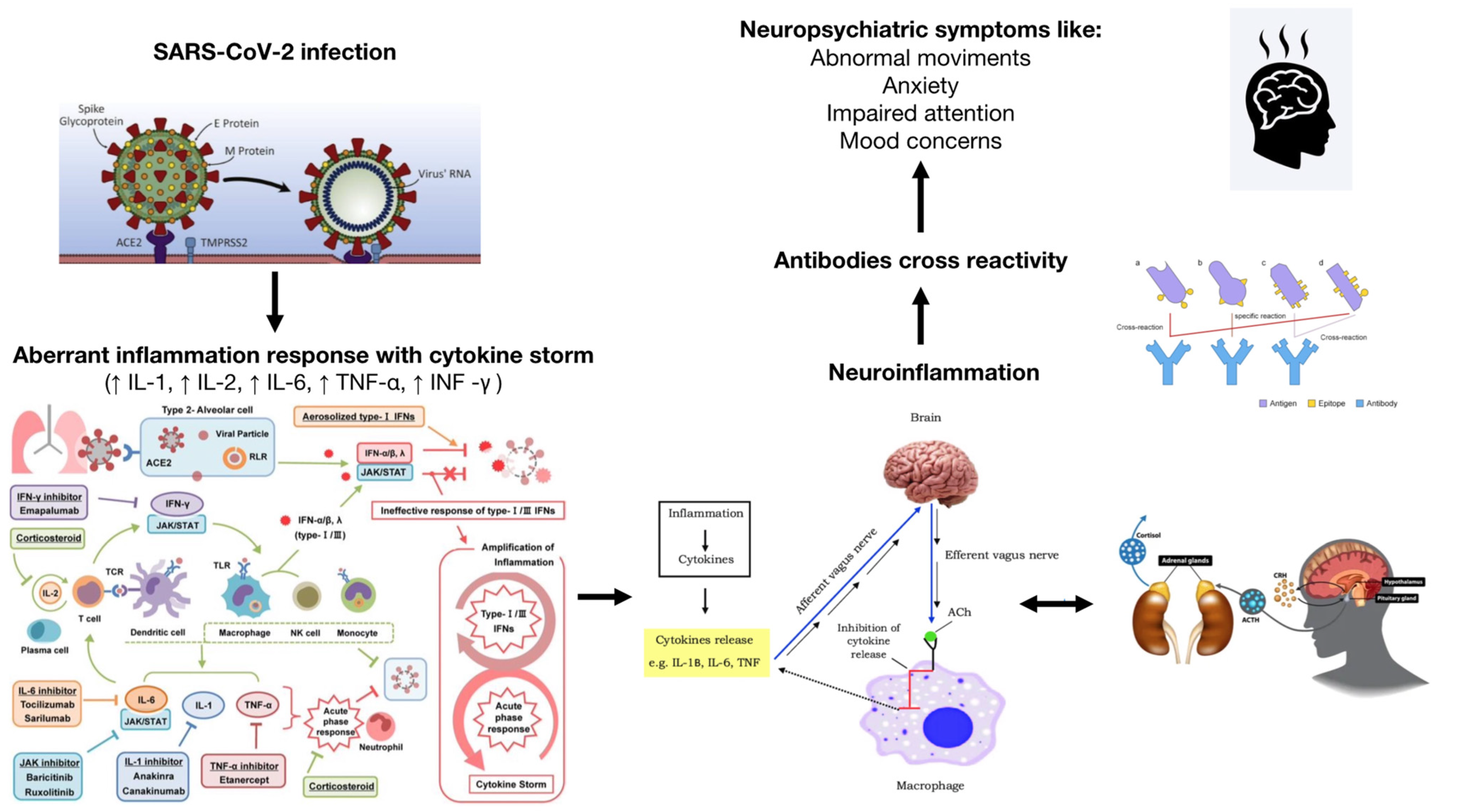
:1. Introduction
2. Methods
3. Case Description
3.1. Case 1
3.2. Case 2
3.3. Case 3
3.4. Case 4
3.5. Case 5
4. Discussion
5. Long COVID-19 and Vaccines
6. Conclusions
7. Limitations
Author Contributions
Funding
Institutional Review Board Statement
Informed Consent Statement
Conflicts of Interest
References
- Sudre, C.H.; Murray, B.; Varsavsky, T.; Graham, M.S.; Penfold, R.S.; Bowyer, R.C.; Pujol, J.C.; Klaser, K.; Antonelli, M.; Canas, L.S.; et al. Attributes and predictors of long COVID. Nat. Med. 2021, 27, 626–631. [Google Scholar] [CrossRef] [PubMed]
- Zimmermann, P.; Pittet, L.F.; Curtis, N. How Common is Long COVID in Children and Adolescents? Pediatr. Infect. Dis. J. 2021, 40, e482–e487. [Google Scholar] [CrossRef] [PubMed]
- Ortona, E.; Malorni, W. Long COVID: To investigate immunological mechanisms and sex/gender related aspects as fundamental steps for tailored therapy. Eur. Respir. J. 2022, 59, 2102245. [Google Scholar] [CrossRef] [PubMed]
- Stormorken, E.; Jason, L.A.; Kirkevold, M. Factors impacting the illness trajectory of post-infectious fatigue syndrome: A qualitative study of adults’ experiences. BMC Public Health 2017, 17, 952. [Google Scholar] [CrossRef] [PubMed] [Green Version]
- Cozzi, G.; Amaddeo, A.; Barbi, E. Post-COVID syndrome: Turning convalescence into illness? Lancet Reg. Health—Eur. 2021, 7, 100163. [Google Scholar] [CrossRef]
- Stein, M.; Ashkenazi-Hoffnung, L.; Greenberg, D.; Dalal, I.; Livni, G.; Chapnick, G.; Stein-Zamir, C.; Ashkenazi, S.; Hecht-Sagie, L.; Grossman, Z. The Burden of COVID-19 in Children and Its Prevention by Vaccination: A Joint Statement of the Israeli Pediatric Association and the Israeli Society for Pediatric Infectious Diseases. Vaccines 2022, 10, 81. [Google Scholar] [CrossRef]
- Rogers, J.P.; Chesney, E.; Oliver, D.; Pollak, T.A.; McGuire, P.; Fusar-Poli, P.; Zandi, M.S.; Lewis, G.; David, A.S. Psychiatric and neuropsychiatric presentations associated with severe coronavirus infections: A systematic review and meta-analysis with comparison to the COVID-19 pandemic. Lancet Psychiatry 2020, 7, 611–627. [Google Scholar] [CrossRef]
- Radtke, T.; Ulyte, A.; Puhan, M.A.; Kriemler, S. Long-term Symptoms After SARS-CoV-2 Infection in Children and Adolescents. JAMA 2021, 326, 869. [Google Scholar] [CrossRef]
- Buonsenso, D.; Munblit, D.; De Rose, C.; Sinatti, D.; Ricchiuto, A.; Carfi, A.; Valentini, P. Preliminary evidence on long COVID in children. Acta Paediatr. 2021, 110, 2208–2211. [Google Scholar] [CrossRef]
- Say, D.; Crawford, N.; McNab, S.; Wurzel, D.; Steer, A.; Tosif, S. Post-acute COVID-19 outcomes in children with mild and asymptomatic disease. Lancet Child Adolesc. Health 2021, 5, e22–e23. [Google Scholar] [CrossRef]
- Molteni, E.; Sudre, C.H.; Canas, L.S.; Bhopal, S.S.; Hughes, R.C.; Antonelli, M.; Murray, B.; Kläser, K.; Kerfoot, E.; Chen, L.; et al. Illness duration and symptom profile in symptomatic UK school-aged children tested for SARS-CoV-2. Lancet Child Adolesc. Health 2021, 5, 708–718. [Google Scholar] [CrossRef]
- Stephenson, T.; Pinto Pereira, S.M.; Shafran, R.; de Stavola, B.L.; Rojas, N.; McOwat, K.; Simmons, R.; Zavala, M.; O’Mahoney, L.; Chalder, T.; et al. Physical and mental health 3 months after SARS-CoV-2 infection (long COVID) among adolescents in England (CLoCk): A national matched cohort study. Lancet Child Adolesc. Health 2022, 6, 230–239. [Google Scholar] [CrossRef]
- Asadi-Pooya, A.A.; Nemati, H.; Shahisavandi, M.; Akbari, A.; Emami, A.; Lotfi, M.; Rostamihosseinkhani, M.; Barzegar, Z.; Kabiri, M.; Zeraatpisheh, Z.; et al. Long COVID in children and adolescents. World J. Pediatr. 2021, 17, 495–499. [Google Scholar] [CrossRef] [PubMed]
- Morrow, A.K.; Ng, R.; Vargas, G.; Jashar, D.T.; Henning, E.; Stinson, N.; Malone, L.A. Postacute/Long COVID in Pediatrics: Development of a Multidisciplinary Rehabilitation Clinic and Preliminary Case Series. Am. J. Phys. Med. Rehabil. 2021, 100, 1140–1147. [Google Scholar] [CrossRef] [PubMed]
- Troyer, E.A.; Kohn, J.N.; Hong, S. Are we facing a crashing wave of neuropsychiatric sequelae of COVID-19? Neuropsychiatric symptoms and potential immunologic mechanisms. Brain. Behav. Immun. 2020, 87, 34–39. [Google Scholar] [CrossRef]
- Orsini, A.; Corsi, M.; Santangelo, A.; Riva, A.; Peroni, D.; Foiadelli, T.; Savasta, S.; Striano, P. Challenges and management of neurological and psychiatric manifestations in SARS-CoV-2 (COVID-19) patients. Neurol. Sci. 2020, 41, 2353–2366. [Google Scholar] [CrossRef]
- Lin, J.E.; Asfour, A.; Sewell, T.B.; Hooe, B.; Pryce, P.; Earley, C.; Shen, M.Y.; Kerner-Rossi, M.; Thakur, K.T.; Vargas, W.S.; et al. Neurological issues in children with COVID-19. Neurosci. Lett. 2021, 743, 135567. [Google Scholar] [CrossRef]
- Desforges, M.; Le Coupanec, A.; Dubeau, P.; Bourgouin, A.; Lajoie, L.; Dubé, M.; Talbot, P.J. Human Coronaviruses and Other Respiratory Viruses: Underestimated Opportunistic Pathogens of the Central Nervous System? Viruses 2019, 12, 14. [Google Scholar] [CrossRef] [Green Version]
- He, Y.; Yu, R.; Ren, J. The correlation between psychiatric disorders and COVID-19: A narrative review. Psychiatr. Danub. 2021, 33, 76–85. [Google Scholar] [CrossRef]
- Robinson-Agramonte, M.A.; Gonçalves, C.-A.; Noris-García, E.; Préndes Rivero, N.; Brigida, A.L.; Schultz, S.; Siniscalco, D.; García García, R.J. Impact of SARS-CoV-2 on neuropsychiatric disorders. World J. Psychiatry 2021, 11, 347–354. [Google Scholar] [CrossRef]
- Dantzer, R. Neuroimmune Interactions: From the Brain to the Immune System and Vice Versa. Physiol. Rev. 2018, 98, 477–504. [Google Scholar] [CrossRef]
- Desforges, M.; Miletti, T.C.; Gagnon, M.; Talbot, P.J. Activation of human monocytes after infection by human coronavirus 229E. Virus Res. 2007, 130, 228–240. [Google Scholar] [CrossRef] [PubMed]
- Proal, A.D.; VanElzakker, M.B. Long COVID or Post-acute Sequelae of COVID-19 (PASC): An Overview of Biological Factors That May Contribute to Persistent Symptoms. Front. Microbiol. 2021, 12, 698169. [Google Scholar] [CrossRef] [PubMed]
- Osmanov, I.M.; Spiridonova, E.; Bobkova, P.; Gamirova, A.; Shikhaleva, A.; Andreeva, M.; Blyuss, O.; El-Taravi, Y.; DunnGalvin, A.; Comberiati, P.; et al. Risk factors for post-COVID-19 condition in previously hospitalised children using the ISARIC Global follow-up protocol: A prospective cohort study. Eur. Respir. J. 2022, 59, 2101341. [Google Scholar] [CrossRef] [PubMed]
- Yong, S.J. Long COVID or post-COVID-19 syndrome: Putative pathophysiology, risk factors, and treatments. Infect. Dis. 2021, 53, 737–754. [Google Scholar] [CrossRef] [PubMed]
- Morand, A.; Campion, J.-Y.; Lepine, A.; Bosdure, E.; Luciani, L.; Cammilleri, S.; Chabrol, B.; Guedj, E. Similar patterns of [18F]-FDG brain PET hypometabolism in paediatric and adult patients with long COVID: A paediatric case series. Eur. J. Nucl. Med. Mol. Imaging 2022, 49, 913–920. [Google Scholar] [CrossRef]
- De Sousa Moreira, J.L.; Barbosa, S.M.B.; Vieira, J.G.; Chaves, N.C.B.; Felix, E.B.G.; Feitosa, P.W.G.; da Cruz, I.S.; da Silva, C.G.L.; Neto, M.L.R. The psychiatric and neuropsychiatric repercussions associated with severe infections of COVID-19 and other coronaviruses. Prog. Neuropsychopharmacol. Biol. Psychiatry 2021, 106, 110159. [Google Scholar] [CrossRef]
- Antoni, M.H.; Dhabhar, F.S. The impact of psychosocial stress and stress management on immune responses in patients with cancer. Cancer 2019, 125, 1417–1431. [Google Scholar] [CrossRef]
- Eroğlu, İ.; Eroğlu, B.Ç.; Güven, G.S. Altered tryptophan absorption and metabolism could underlie long-term symptoms in survivors of coronavirus disease 2019 (COVID-19). Nutrition 2021, 90, 111308. [Google Scholar] [CrossRef]
- Anderson, G.; Maes, M. Interactions of Tryptophan and Its Catabolites With Melatonin and the Alpha 7 Nicotinic Receptor in Central Nervous System and Psychiatric Disorders: Role of the Aryl Hydrocarbon Receptor and Direct Mitochondria Regulation. Int. J. Tryptophan Res. IJTR 2017, 10, 1178646917691738. [Google Scholar] [CrossRef]
- Thomas, T.; Stefanoni, D.; Reisz, J.A.; Nemkov, T.; Bertolone, L.; Francis, R.O.; Hudson, K.E.; Zimring, J.C.; Hansen, K.C.; Hod, E.A.; et al. COVID-19 infection alters kynurenine and fatty acid metabolism, correlating with IL-6 levels and renal status. JCI Insight 2020, 5, e140327. [Google Scholar] [CrossRef] [PubMed]
- Lionetto, L.; Ulivieri, M.; Capi, M.; De Bernardini, D.; Fazio, F.; Petrucca, A.; Pomes, L.M.; De Luca, O.; Gentile, G.; Casolla, B.; et al. Increased kynurenine-to-tryptophan ratio in the serum of patients infected with SARS-CoV2: An observational cohort study. Biochim. Biophys. Acta Mol. Basis Dis. 2021, 1867, 166042. [Google Scholar] [CrossRef] [PubMed]
- Hesselmark, E.; Bejerot, S. Clinical features of paediatric acute-onset neuropsychiatric syndrome: Findings from a case– control study. BJPsych Open 2019, 5, e25. [Google Scholar] [CrossRef] [PubMed]
- Wilbur, C.; Bitnun, A.; Kronenberg, S.; Laxer, R.M.; Levy, D.M.; Logan, W.J.; Shouldice, M.; Yeh, E.A. PANDAS/PANS in childhood: Controversies and evidence. Paediatr. Child Health 2019, 24, 85–91. [Google Scholar] [CrossRef]
- Murgia, F.; Gagliano, A.; Tanca, M.G.; Or-Geva, N.; Hendren, A.; Carucci, S.; Pintor, M.; Cera, F.; Cossu, F.; Sotgiu, S.; et al. Metabolomic Characterization of Pediatric Acute-Onset Neuropsychiatric Syndrome (PANS). Front. Neurosci. 2021, 15, 645267. [Google Scholar] [CrossRef]
- Swedo, S.E.; Seidlitz, J.; Kovacevic, M.; Latimer, M.E.; Hommer, R.; Lougee, L.; Grant, P. Clinical presentation of pediatric autoimmune neuropsychiatric disorders associated with streptococcal infections in research and community settings. J. Child Adolesc. Psychopharmacol. 2015, 25, 26–30. [Google Scholar] [CrossRef] [Green Version]
- Swedo, S.E.; Frankovich, J.; Murphy, T.K. Overview of Treatment of Pediatric Acute-Onset Neuropsychiatric Syndrome. J. Child Adolesc. Psychopharmacol. 2017, 27, 562–565. [Google Scholar] [CrossRef] [Green Version]
- Pavone, P.; Ceccarelli, M.; Marino, S.; Caruso, D.; Falsaperla, R.; Berretta, M.; Rullo, E.V.; Nunnari, G. SARS-CoV-2 related paediatric acute-onset neuropsychiatric syndrome. Lancet Child Adolesc. Health 2021, 5, e19–e21. [Google Scholar] [CrossRef]
- Morabito, G.; Barbi, E.; Cozzi, G. The Unaware Physician’s Role in Perpetuating Somatic Symptom Disorder. JAMA Pediatr. 2020, 174, 9. [Google Scholar] [CrossRef]
- Murru, A.; Manchia, M.; Hajek, T.; Nielsen, R.E.; Rybakowski, J.K.; Sani, G.; Schulze, T.G.; Tondo, L.; Bauer, M.; for the International Group for The Study of Lithium Treated Patients (IGSLi). Lithium’s antiviral effects: A potential drug for CoVID-19 disease? Int. J. Bipolar Disord. 2020, 8, 21. [Google Scholar] [CrossRef]
- Parra, A.; Juanes, A.; Losada, C.P.; Álvarez-Sesmero, S.; Santana, V.D.; Martí, I.; Urricelqui, J.; Rentero, D. Psychotic symptoms in COVID-19 patients. A retrospective descriptive study. Psychiatry Res. 2020, 291, 113254. [Google Scholar] [CrossRef] [PubMed]
- Hull, M.; Parnes, M.; Jankovic, J. Increased Incidence of Functional (Psychogenic) Movement Disorders in Children and Adults Amid the COVID-19 Pandemic: A Cross-sectional Study. Neurol. Clin. Pract. 2021, 11, e686–e690. [Google Scholar] [CrossRef] [PubMed]
- Schneider, S.A.; Hennig, A.; Martino, D. Relationship between COVID-19 and movement disorders: A narrative review. Eur. J. Neurol. 2022, 29, 15217. [Google Scholar] [CrossRef] [PubMed]
- Antonelli, M.; Penfold, R.S.; Merino, J.; Sudre, C.H.; Molteni, E.; Berry, S.; Canas, L.S.; Graham, M.S.; Klaser, K.; Modat, M.; et al. Risk factors and disease profile of post-vaccination SARS-CoV-2 infection in UK users of the COVID Symptom Study app: A prospective, community-based, nested, case-control study. Lancet Infect. Dis. 2022, 22, 43–55. [Google Scholar] [CrossRef]
- Shiri, T.; Evans, M.; Talarico, C.A.; Morgan, A.R.; Mussad, M.; Buck, P.O.; McEwan, P.; Strain, W.D. Vaccinating Adolescents and Children Significantly Reduces COVID-19 Morbidity and Mortality across All Ages: A Population-Based Modeling Study Using the UK as an Example. Vaccines 2021, 9, 1180. [Google Scholar] [CrossRef] [PubMed]
- Principi, N.; Esposito, S. Reasons in favour of universal vaccination campaign against COVID-19 in the pediatric population. Ital. J. Pediatr. 2022, 48, 4. [Google Scholar] [CrossRef] [PubMed]


| Cases | Sex | Age | COVID-19 Hospitalization | COVID-19 Symptoms | NPI Symptoms | Onset after COVID-19 Disease | Follow-Up and Residual Symptoms |
|---|---|---|---|---|---|---|---|
| Case 1 (February 2021) | Male | 15 | No | Fever and anosmia | Acute psychotic state | 3 weeks | 12 months after his COVID-19 recovery: asthenia, attention deficit, poor language |
| Case 2 (March 2021) | Male | 7 | No | Mild nasal congestion | Complex motor and vocal tics | 1 week | / |
| Case 3 (May 2021) | Male | 5 | No | Only 1-day fever, nasal congestion, and asthenia | Acute-onset ocular tics, food restriction, separation anxiety, sleep disturbances, enuresis | Few weeks | 3 months after: rarely ocular tics, occasional enuresis, and anxiety |
| Case 4 (January 2022) | Female | 3 | No | No specific symptoms | Aggressive behavior, separation anxiety, ocular tics | Few months | 3-month follow-up |
| Case 5 (February 2022) | Male | 2 | No | Fever and nasal congestion, cough | Involuntary ocular movements, grimaces, upper limbs myoclonus | 4 weeks | / |
| Case 1 | Case 2 | Case 3 | Case 4 | Case 5 | |
|---|---|---|---|---|---|
| Blood count 1 with leukocyte count 2 | Neutrophils: 39.60%; Eosinophils: 5.9% | MPV: 12.7 fl; PDW: 18 fl; Monocytes: 11.5% | Normal | Hemoglobin: 10.9 mg/dL; MCH: 24.8 pg; Hematocrit 32.9% Platelets: 601 × 103/uL; PCT: 0.62% Lymphocytes 56% | RBC: 5.88 106/uL; Hemoglobin: 10.0 mg/dL; MCH: 17 pg; Hematocrit 31.60%; MCV 53.70 fl; MCH 17.00 pg. Platelets: 458 × 103/uL; PCT: 0.45% Lymphocytes 61.10% |
| Electrolytes | Normal | Normal | Normal | Normal | Normal |
| Prothrombin Time (PT) (s/INR) | Normal | Normal | Normal | Normal | Normal |
| Activated partial thromboplastin time (APTT) (s/RATIO) | Normal | Normal | Normal | Normal | Normal |
| Fibrinogen (200–450 mg/dL) | 223 mg/dL | 203 mg/dL | 217 mg/dL | 475 mg/dL | 257mg/dL |
| Thyroid function indices (ATM, ATG, TSH, FT3, FT4, Thyroglobulin) | FT3 5.68 pg/mL (n.r.2.50–5.20 pg/mL) | Normal | Normal | Normal | Normal |
| Ceruloplasmin (22–58 mg/dL) | 16 mg/dL | 19 mg/dL | 35 mg/dL | / | / |
| Copper (65–165 µg/dL) | 56 µg/dL | 66 μg/dL | 74 μg/dL | / | / |
| Transaminases (ALT, AST, GGTP) | Normal | Normal | Normal | Normal | Normal |
| Serum Creatinin +Azotemia | Normal | Normal | Normal | Normal | Normal |
| Chemical physical examination of urine and urinary sediment | Specific weight: 1028 (n.r. 1011–1025) | Normal | Normal | Specific weight: 1006 (n.r. 1011–1025) | Specific weight: 1010 (n.r. 1011–1025) |
| Serum Iron (60–160 ug/dL) | 60 ug/dL | 68 ug/dL | 76 ug/dL | 53ug/dL | 55 ug/dL |
| AST (children up to 150 U.I/mL) | 116 U.I/mL | 161 U.I/mL | 135 U.I/mL | 5 U.I/mL | / |
| CRP (0.00–2.00 mg/L) | 1.2 mg/L | 2.5 mg/L | 0.5 mg/L | 27.9 mg/L | 0.9 mg/L |
| ESR (1 h 1–16) | 9 mm/h | 17 mm/h | 2 mm/h | 56 mm/h | / |
| Cortisol (4.3–27 µg/dL h. 7–9) | 28.56 µg/dL | 7.09 µg/dL | 9 µg/dL | / | / |
| Case 1 | Case 2 | Case 3 | Case 4 | Case 5 | |
|---|---|---|---|---|---|
| EEG | Normal | Normal | Normal | Normal | Normal |
| MRI | Slight descent of the right cerebellar tonsil and thinning corpus callosum. | No abnormalities found | No abnormalities found | No abnormalities found | No abnormalities found |
| ECG | Sinus Tachycardia at 102 bpm. QT 312 ms, QTc 410 ms. | Normal sinus rhythm. QT 336 ms, QTc 425 ms. | Respiratory sinus arrhythmia at 76 bpm. | Normal sinus rhythm at 106 bpm. | Respiratory sinus arrhythmia at 75 bpm. |
| Therapy | Drugs
| CBT | Melatonin (1 mg/die) CBT | CBT + Speech therapy | CBT + Speech therapy |
Publisher’s Note: MDPI stays neutral with regard to jurisdictional claims in published maps and institutional affiliations. |
© 2022 by the authors. Licensee MDPI, Basel, Switzerland. This article is an open access article distributed under the terms and conditions of the Creative Commons Attribution (CC BY) license (https://creativecommons.org/licenses/by/4.0/).
Share and Cite
Savino, R.; Polito, A.N.; Arcidiacono, G.; Poliseno, M.; Lo Caputo, S. Neuropsychiatric Disorders in Pediatric Long COVID-19: A Case Series. Brain Sci. 2022, 12, 514. https://doi.org/10.3390/brainsci12050514
Savino R, Polito AN, Arcidiacono G, Poliseno M, Lo Caputo S. Neuropsychiatric Disorders in Pediatric Long COVID-19: A Case Series. Brain Sciences. 2022; 12(5):514. https://doi.org/10.3390/brainsci12050514
Chicago/Turabian StyleSavino, Rosa, Anna N. Polito, Giulia Arcidiacono, Mariacristina Poliseno, and Sergio Lo Caputo. 2022. "Neuropsychiatric Disorders in Pediatric Long COVID-19: A Case Series" Brain Sciences 12, no. 5: 514. https://doi.org/10.3390/brainsci12050514