Article

Particulate Matter 2.5 Level Modulates Brachial Artery Flow-Mediated Dilation Response to Aerobic Exercise in Healthy Young Men

1
Department of Applied Physiology and Kinesiology, University of Florida, Gainesville, FL 32611, USA
2
Division of Health & Kinesiology, Incheon National University, Incheon 22012, Republic of Korea
3
Department of Environmental Engineering, Incheon National University, Incheon 22012, Republic of Korea
4
Sport Science Institute, Incheon National University, Incheon 22012, Republic of Korea
5
Sports Functional Disability Institute, Incheon National University, Incheon 22012, Republic of Korea
*
Author to whom correspondence should be addressed.
Appl. Sci. 2023, 13(8), 4936; https://doi.org/10.3390/app13084936
Submission received: 13 March 2023 / Revised: 13 April 2023 / Accepted: 13 April 2023 / Published: 14 April 2023
(This article belongs to the Special Issue Exercise Interventions on Human Cardiovascular Health)

Abstract

:
Particulate matter 2.5 (PM2.5) is an environmental pollutant linked with the risk of cardiovascular disease. Aerobic exercise performed in polluted environments may have fewer benefits because of increased PM2.5 inhalation during exercise. However, the vascular responses to aerobic exercise in high PM2.5 (HPM2.5) conditions remain unknown. This study aimed to examine the acute flow-mediated dilation (FMD) response to moderate-intensity treadmill running in HPM2.5 levels compared to low PM2.5 (LPM2.5) levels in healthy young males. Treadmill running in both HPM2.5 and LPM2.5 levels was completed by nine subjects. Brachial artery FMD was measured before and after the exercise to assess vascular endothelial function. Indoor PM2.5 concentration was significantly higher in HPM2.5 than in LPM2.5 conditions (p < 0.001). Scaled FMD significantly increased after the exercise in LPM2.5 conditions but not in HPM2.5 (p = 0.03), and baseline diameter increased only in HPM2.5 conditions after the exercise (p = 0.001). Baseline diameter and peak diameter were smaller, and time to peak dilation was delayed in HPM2.5 compared to LPM2.5 in pre-exercise intervention measurements (p < 0.05). Therefore, acute PM2.5 exposure can counteract the positive effect of aerobic exercise on vascular endothelial function in young males.

1. Introduction

Cardiovascular disease (CVD) is the leading cause of mortality worldwide. Exposure to particulate matter 2.5 (PM2.5), which is an inhalable particle that is 2.5 μm or even smaller, independently increases CVD incidence and mortality [1,2,3]. Additionally, increased PM2.5 concentrations even below the United States (US) standard for PM2.5 increased the risk of CVD [2]. PM2.5 causes systemic inflammation that occurs and transfers from the respiratory system and alters the autonomic nervous system by activating alveoli sensory receptors [4,5]. Moreover, PM2.5 translocates to the bloodstream due to the size of fine particles, thereby directly damaging the cardiovascular system [4,5]. Thus, PM2.5-induced pathophysiological changes in respiratory and cardiovascular systems can negatively influence vascular structure and endothelial function.
The endothelium is an important anatomical structure that maintains vascular tone by releasing various vasoactive substances. Endothelial dysfunction is an early marker of atherosclerosis and a predictor for future CVD events [6,7,8]. Brachial artery flow-mediated dilation (FMD) is a clinical and noninvasive measure for assessing vascular endothelial function through endothelium-dependent vasodilation in response to the physiological stimulus. Exercise training is a well-known behavioral intervention to improve FMD, and even acute aerobic exercise may increase FMD [9,10,11]. However, the benefits of exercise on vascular endothelial function may differ with PM2.5 levels. Generally, increased minute ventilation during exercise increases PM2.5 inhalation compared to resting, which is a non-active condition. A higher frequency of mouth breathing during exercise circumvents the filtration in the nasal cavity [12,13,14]. Additionally, an exercise-induced increase in airflow velocity facilitates the movement of inhaled PM2.5 into the deeper region of the respiratory system [14]. Only a few studies have demonstrated the effect of exercise on endothelial function in high PM2.5 (HPM2.5) concentrations and reported inconsistent findings [15,16,17,18]. Moreover, previous studies were conducted using diesel exhaust in an indoor exposure chamber or an outdoor ambient environment. Understanding the influence of exercise in an indoor space with higher PM2.5 levels compared with lower PM2.5 conditions on vascular endothelial function is important because outdoor PM2.5 rapidly infiltrates into indoor spaces and influences indoor air quality [19]. Therefore, our study controlled the indoor PM2.5 concentrations with penetrated outdoor ambient PM2.5 to emphasize the importance of managing PM2.5 levels in indoor exercise spaces. This study aimed to examine and compare the influence of low and high PM2.5 levels during acute moderate-intensity aerobic exercise on brachial artery FMD in healthy young males. We hypothesized that the effect of exercise on brachial artery FMD would be limited in HPM2.5 levels compared to lower PM2.5 (LPM2.5) levels due to its negative influence on the cardiovascular system.

2. Materials and Methods

2.1. Subjects

This study included nine healthy young males (aged 23–27 years). They were all nonsmokers and free from cardiovascular, respiratory, and musculoskeletal diseases. The benefits, risks, and purpose of this study were fully explained to the participants, and they signed an informed consent form before participating in the study. All study procedures complied with the Declaration of Helsinki and were approved by the Incheon National University Institutional Review Board.

2.2. Study Design

Participants performed one bout of moderate-intensity aerobic exercise in both HPM2.5 and LPM2.5 conditions with a crossover study design. The exercise intervention consisted of indoor treadmill running at participants’ 70% of peak heart rate (HRpeak) for 30 min. The Polar H7 HR sensor (Polar Electro Oy, Kempele, Finland) and Polar Team App (version 1.8.8, Polar Electro Oy, Kempele, Finland) were used to continuously monitor the heart rate (HR) during the treadmill running. Vascular endothelial function was assessed by brachial artery FMD before and after the exercise intervention in HPM2.5 and LPM2.5 conditions. Participants were initially allocated to either HPM2.5 or LPM2.5 conditions based on the indoor exercise facility and outdoor ambient PM2.5 levels. A randomized crossover design could not be used because HPM2.5 and LPM2.5 condition visits had proceeded when participants’ schedules and uncontrollable PM2.5 levels were perfectly matched for each condition. Fortunately, the HPM2.5 and LPM2.5 condition visits were completed by five and four participants, respectively, as their first exercise visit. Participants were asked to fast (>6 h), avoid any intense exercise (>24 h), and refrain from caffeine and any other supplements (>12 h) that might affect the cardiovascular system before two different PM2.5 condition visits [20]. At least 7 days of washout period was established between two PM2.5 condition visits to eliminate acute confounding effects of aerobic exercise and PM2.5 exposure from the previous PM2.5 condition visit.

2.3. Body Composition

Height was measured by a conventional stadiometer, and body weight was measured with an InBody 720 Scale (Biospace, Republic of Korea). Body composition was assessed by dual-energy x-ray absorptiometry (Prodigy, GE HealthCare, Chicago, IL, USA). In HPM2.5 and LPM2.5 condition visits, the body weight and body composition were measured before the exercise intervention.

2.4. Maximal Cardiopulmonary Exercise Test

Participants completed the maximal cardiopulmonary exercise test (CPET) with a modified Bruce protocol to assess the maximal oxygen consumption (VO2max) and HR (HRmax) before the first PM2.5 condition visit (HPM2.5 or LPM2.5) [21]. The VO2max and HRmax were obtained when the participant met at least three of the following criteria: (a) a plateau in oxygen consumption with increasing exercise intensity; (b) an HR within 10 beats/min of age-predicted HRmax; (c) at least 1.15 of maximal respiratory exchange ratio; (d) a score of ≥18 on the Borg rating of perceived exertion scale. However, VO2max and HRmax are presented as peak oxygen consumption (VO2peak) and HRpeak because more than one participant did not meet at least three criteria. Measured HRpeak was used for prescribing the accurate exercise intensity. At least 72 h of washout period was established before starting the first PM2.5 condition visit to avoid the effect of maximal CPET on vascular endothelial function.

2.5. Blood Pressure and Brachial Artery Flow-Mediated Dilation

Blood pressure (BP) and brachial artery FMD were measured in a quiet and temperature-controlled laboratory with dimmed light. All measurements were performed after at least 10 min of rest in a supine position. An air purifier with a high-efficiency particulate absorbing filter in the laboratory was used to minimize the PM2.5 exposure during baseline and post-exercise measures.
A SphygmoCor Xcel device (AtCor Medical, New South Wales. Australia) was used to measure the BP. The cuff was placed on the right upper arm, and the BP was measured at least three times. We averaged two systolic BP (SBP) and diastolic BP (DBP) values within 5 mmHg between measures.
Brachial artery FMD was assessed using a doppler ultrasound system (Aloka Prosound α7, Hitachi, Japan) with an 8–15 MHz linear array transducer (UST-5412, Hitachi, Japan). The participant’s right arm was abducted in the supine position at the heart level. The pressure cuff was placed 1–2 cm distal to the antecubital fossa and connected to a rapid inflator/deflator system (E20 & AG101, D.E Hokanson, Washington, DC, USA). The brachial artery images were obtained approximately 5–10 cm proximal to the antecubital fossa. The transducer was clamped with the flexbar (Flexbar Machine Corporation, Islandia, NY, USA), after optimizing the image, to minimize the transducer’s movement during the FMD procedure. Additionally, the distance between the transducer and the antecubital fossa was measured to capture the same portion of the brachial artery in the next visit. The blood velocity was assessed with ≤60° of the doppler’s insonation angle. The baseline diameter and velocity were obtained for 1 min. Then, the pressure cuff was promptly inflated to 250 mmHg and maintained for 5 min. The diameter and velocity were recorded for 3 min after rapid cuff deflation to assess the vascular response to the reactive hyperemia. Acquired diameter and velocity were analyzed by commercially available automated wall detection software (Cardiovascular Suite, FMD Studio, Quipu, Italy). FMD was calculated as absolute (peak diameter–baseline diameter) and relative values ([peak diameter–baseline diameter]/baseline diameter). Additionally, FMD normalized by shear rate (SR) area under the curve up to the peak diameter (FMD/SRAUC) was analyzed, and allometrically scaled FMD (scaled FMD) was assessed to eliminate any potential effects of changes in SR and baseline diameter on brachial artery FMD [20].

2.6. PM2.5 Concentration

PM2.5 levels were assessed by two SidePak AM520 personal aerosol monitors (TSI, Shoreview, MN, USA). Outdoor and indoor PM2.5 concentrations were concurrently measured and recorded at each site during the exercise intervention. The recorded PM2.5 concentrations were averaged each minute, and the personal aerosol monitors were located at the same spots throughout the study.

2.7. Statistical Analyses

Alterations in body weight, body composition, BP, and vascular endothelial function were assessed at baseline between HPM2.5 and LPM2.5 condition visits with the Wilcoxon signed rank test and paired t-test. The difference in outdoor and indoor PM2.5 concentrations between HPM2.5 and LPM2.5 condition visits was confirmed with the Mann–Whitney U test. A 2 × 2 analysis of variance with repeated measures was performed to demonstrate the difference in the acute effect of moderate-intensity aerobic exercise on vascular endothelial function between HPM2.5 and LPM2.5 conditions. A generalized estimating equation (GEE) was used to control the influence of baseline diameter and SR on FMD data [20,22]. Using natural log-transforming baseline diameter (lnDbase), peak diameter (lnDpeak), and SRAUC (lnSRAUC), the GEE was employed with diameter difference (lnDpeak–lnDbase) as the dependent variable and lnDbase and lnSRAUC as covariates. Estimated means and estimated standard errors were back-transformed to scaled mean FMD and SE FMD with the following equations: scaled FMD mean = (eEM − 1) × 100; scaled FMD SE = (eESE − 1) × 100. SRAUC, FMD/SRAUC, and scaled FMD were analyzed with data from eight participants excluding one outlier. Post hoc pairwise multiple comparisons were adjusted by Bonferroni correction. Data were presented as mean ± SE, and all statistical analyses were performed using IBM Statistical Package for the Social Sciences Statistics (version 27, USA). All statistical significance was set as p < 0.05.

3. Results

3.1. Subject Characteristics and PM2.5 Concentrations

Body weight, body mass index, SBP, DBP, and HR were not different between HPM2.5 and LPM2.5 condition visits before the treadmill running (Table 1 and Table 2). Both outdoor and indoor PM2.5 concentrations in HPM2.5 condition visits were 8.6- and 7.6-fold higher than the LPM2.5 condition visits, respectively (p < 0.001; Table 1). Average indoor PM2.5 concentration during the exercise in HPM2.5 condition visits was higher than the 24 h PM2.5 standard (35 μg/m3) of the US Environmental Protection Agency (EPA) and categorized as “unhealthy for sensitive groups” or “unhealthy” by the US air quality index levels.

3.2. Responses to Treadmill Running in HPM2.5 and LPM2.5 Conditions

3.2.1. Blood Pressure

Responses of BP and HR to the acute moderate-intensity treadmill running were not different between HPM2.5 and LPM2.5 conditions. SBP, DBP, and HR immediately increased after the exercise intervention regardless of PM2.5 levels as a normal physiological response of the human body to exercise (p < 0.001 for time effect; Table 2).

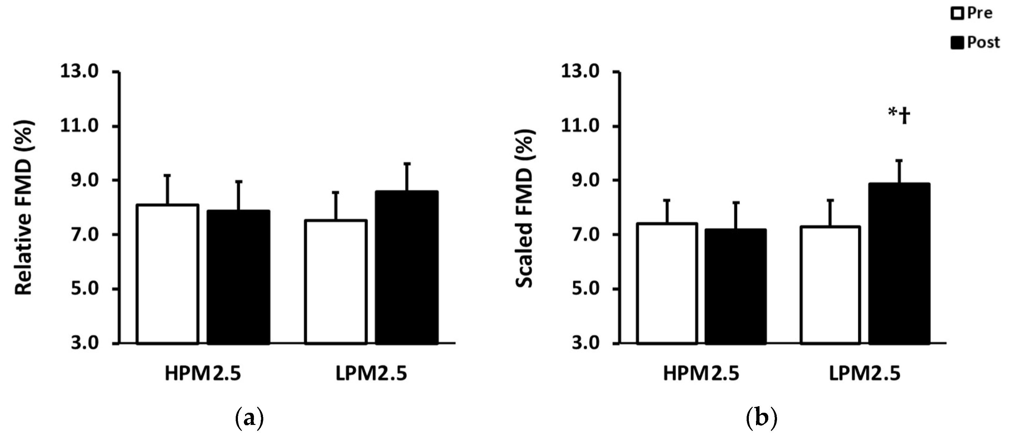
3.2.2. Brachial Artery Flow-Mediated Dilation

The absolute and relative FMD responses to aerobic exercise were not influenced by PM2.5 levels (Table 3; Figure 1). However, the scaled FMD was significantly increased after the exercise in the LPM2.5 condition (p = 0.03; Figure 1). Additionally, the post-exercise scaled FMD in the LPM2.5 condition was significantly higher than post-exercise scaled FMD in the HPM2.5 condition (p = 0.009; Figure 1). Baseline and peak diameters were smaller in the HPM2.5 condition than in the LPM2.5 condition before the exercise (p < 0.05; Table 3). The baseline diameter significantly increased after the aerobic exercise in the HPM2.5 condition (p = 0.001; Table 3). However, the baseline diameter was the same after the exercise intervention in the LPM2.5 condition. The time to peak dilation (TTP) was longer in HPM2.5 than LPM2.5 condition before exercise (p < 0.05).

4. Discussion

The current study investigated the difference in the response of vascular endothelial function to aerobic treadmill exercise between HPM2.5 and LPM2.5 levels. Consistent with our study hypothesis, the acute improvement of vascular endothelial function by aerobic exercise was demonstrated to be nullified in HPM2.5 conditions. Additionally, the baseline diameter of the brachial artery was increased only after exercising in HPM2.5 conditions. Baseline and peak diameters were smaller, and TTP was delayed in HPM2.5 conditions compared to LPM2.5 conditions before exercise.
To our knowledge, this is the first study that examined the influence of inhaled fine particulate matter on vascular endothelial function while exercising under indoor HPM2.5 and LPM2.5 conditions, naturally created by outdoor ambient PM2.5. Previous studies explored the effect of aerobic exercise in HPM2.5 concentrations on vascular function, but findings were inconsistent, and exercise interventions were conducted with different PM2.5 sources [15,16,17,18,23,24]. Studies involving outdoor cycling in a high-traffic route did not reveal any relationship between vascular function and PM2.5 concentrations [15,17]. The high-traffic route PM2.5 concentrations were lower than the US EPA 24 h PM2.5 standard, which might not negatively affect the cardiovascular system in healthy adults, although these studies were conducted with similar PM2.5 sources to our study using ambient PM2.5. Additionally, stationary cycle exercise in an exposure chamber with 300 μg/m3 of PM2.5 created by diesel exhaust did not influence brachial artery FMD in healthy young males [16]. However, our study used ambient PM2.5 to form the HPM2.5 condition, which can explain the discrepancy of findings with studies that only used diesel exhaust.
Previous studies reported FMD improvement in response to exercise [25,26]. These findings support our result in LPM2.5 conditions and suggest exercise as beneficial to enhance vascular endothelial function. However, high ambient PM environment exposure can negate the positive effect of exercise on FMD. We found that the increased scaled FMD in LPM2.5 conditions disappeared when the same exercise was performed in HPM2.5 conditions. Similarly, FMD was significantly decreased after 30 min of running in a high ambient PM1 condition in young adults [27]. PM exposure generally increases reactive oxygen species (ROS) production [28,29]. Oxidative stress induces the reduction in nitric oxide (NO) production and availability, which are key factors for vasodilation in response to reactive hyperemia [30]. Additionally, PM inhalation may facilitate leukotriene synthesis by ROS, which engenders endothelial dysfunction [31]. Furthermore, sympathetic nervous system activation by PM exposure might be associated with decreased FMD [32,33]. Collectively, increased PM inhalation during exercise may cause vascular endothelial dysfunction by oxidative stress, inflammation, and sympathetic nervous system activation.
The present study revealed lower pre-exercise baseline and peak diameters of the brachial artery in HPM2.5 conditions than in LPM2.5 conditions. We assume that vasoconstriction occurred due to PM2.5 inhalation on the way from the participants’ residence to the laboratory. Unfortunately, we were unable to accurately track the PM2.5 exposure of study participants before starting pre-exercise measures. Participants spent 30 min to 2 h on the road or on public transportation to visit our laboratory. The brachial artery could have been constricted in response to the increased PM2.5 inhalation before the pre-exercise procedures because participants were likely to be exposed to a high outdoor PM2.5 environment in the HPM2.5 visit. Interestingly, constricted pre-exercise brachial artery baseline diameter under HPM2.5 conditions in this study revealed a similar magnitude of vasoconstriction to the previous studies with a high concentration of PM2.5 exposure (150–200 μg/m3) [18,34]. We speculate that increased sympathetic nerve activity and endothelin-1 (ET-1) secretion, which is an intrinsic vasoconstrictor, constricted the brachial artery in HPM2.5 conditions. PM2.5 inhalation was found to rapidly upset the autonomic nervous system balance by activating the sympathetic nervous system [32]. Moreover, elevated systemic and local oxidative stress and inflammation by PM2.5 inhalation may increase ET-1 production [18,35].
Delayed pre-exercise TTP was found in HPM2.5 conditions compared to LPM2.5 conditions. Delayed peak dilation has been associated with the risks of CVD [36,37,38,39]. Increased arterial stiffness caused by PM2.5 exposure possibly induced the slower pre-exercise TTP in the HPM2.5 visit, although mechanisms of delayed TTP are unclear. In two previous studies, shortened TTP was found to be associated with a decrease in arterial stiffness, which makes it possible to infer the relationship between TTP and arterial stiffness that may increase with exposure to air pollution [40,41]. Additionally, PM2.5-induced oxidative stress and inflammation might impair the mechanosensor of endothelium and alter enzyme rates related to vasodilation signaling pathways. Moreover, the pre-exercise SRAUC in HPM2.5 showed a lower tendency compared to LPM2.5 conditions (p = 0.08). This might occur due to the impaired vascular shear stress sensors and cause delayed TTP. Furthermore, the diameter change rate in response to reactive hyperemia can be slowed down by increased sympathetic nervous system activation due to PM2.5 exposure [32]. Therefore, delayed vascular smooth muscle changes in response to hyperemia may occur in HPM2.5 conditions before the exercise intervention. However, future studies are necessary to explore the potential mechanisms of delayed TTP by PM2.5.
Our study demonstrated the significantly increased brachial artery baseline diameter after exercising in only the HPM2.5 condition. The different response of baseline diameter to an acute aerobic exercise between HPM2.5 and LPM2.5 conditions might be related to a smaller baseline diameter before the exercise intervention in the HPM2.5 condition than the LPM2.5 condition. Increased SR during the aerobic exercise may have redeemed the baseline diameter of the brachial artery, although the pre-exercise baseline diameter of the brachial artery can be constricted in the HPM2.5 visit, as we discussed above. Additionally, aerobic exercise can decrease plasma ET-1 concentration, which attenuates ET-1-induced brachial artery constriction [42]. The increased blood flow in the respiratory system during exercise is also likely to help reduce the circulating ET-1 by binding to ETB receptors in the lungs [43]. Thus, we assume that treadmill running helps to recover the constricted baseline diameter of the brachial artery in the HPM2.5 condition.
The strength of our study was that exercise intervention was performed in naturally formed high and low indoor PM2.5 conditions with ambient air. Additionally, it provides insight into the response of vascular endothelial function to acute aerobic exercise in HPM2.5 conditions that we often encounter in real life. Moreover, our results might convey the importance of managing PM2.5 concentration in indoor exercise facilities. However, several limitations should be mentioned. First, our sample size was small, and we did not analyze physiological mechanisms to explain our new findings. Therefore, future research with large sample sizes is required in various populations, and this research must assess possible mechanisms to explain the response of vascular endothelial function to exercising in a polluted environment. Second, we only measured brachial artery FMD to evaluate endothelium-dependent vasodilation before and approximately 30 min after the exercise in healthy young males. However, the timing of post-exercise FMD measurement is known to influence results. Additionally, the influence of inhaled PM2.5 on the cardiovascular system might be different with time. Thus, brachial artery FMD needs to be measured at multiple time points after exercise intervention to precisely analyze the effects of PM2.5 during exercise. Herein, we could not measure vascular smooth muscle responsiveness to NO donors to assess intact endothelium-independent vasodilation in our study participants. Previous studies reported inconsistent findings about the effect of PM2.5 on endothelium-independent vasodilation, and studies evaluating the effects of PM2.5 on endothelium-independent vasodilation are scarce [23,29]; thus, future studies should examine both endothelium-dependent and endothelium-independent vasodilation after exercise in HPM2.5 conditions. Third, we could not control the participants’ PM2.5 exposure before the scheduled experimental visits. The amount of inhaled PM2.5 before the pre-exercise measurements may have varied among participants and affected the vascular response to the exercise intervention. Lastly, we could not separately measure and analyze all the pollutants during exercise. Different pollutants (e.g., carbon monoxide, sulfur dioxide, ozone, nitrogen oxide, and ultrafine particles) may additionally impair the cardiovascular system [44]; thus, all pollutants have to be measured and analyzed to elucidate the influence of each pollutant on the response of vascular function to exercise.

5. Conclusions

Acute moderate-intensity treadmill running in HPM2.5 levels, which is greater than the 24 h PM2.5 standard (35 μg/m3) of the US EPA, can nullify the benefits of exercise on brachial artery FMD in healthy young males. However, aerobic exercise may help restore constricted conduit arteries that are likely induced by PM2.5 inhalation. To our best knowledge, the current study has the closest study design to a real-world situation that people can experience in daily life, but the conclusions must be interpreted carefully by considering the limitations.

Author Contributions

Conceptualization, J.-S.K. and M.-H.H.; methodology, J.-S.K., D.G.L. and M.-H.H.; software, J.-S.K., D.G.L. and M.-H.H.; formal analysis, J.-S.K. and M.-H.H.; investigation, J.-S.K. and M.-H.H.; resources, D.G.L. and M.-H.H.; data curation, J.-S.K.; writing—original draft preparation, J.-S.K. and M.-H.H.; writing—review and editing, D.G.L. and M.-H.H.; visualization, J.-S.K.; supervision, M.-H.H.; project administration, M.-H.H.; funding acquisition, M.-H.H. All authors have read and agreed to the published version of the manuscript.

Funding

This research was supported by the Incheon National University Research Grant in 2018.

Institutional Review Board Statement

The study was conducted in accordance with the Declaration of Helsinki and approved by the Institutional Review Board of Incheon National University (7007971-201907-004-01-02 and 09/05/2019).

Informed Consent Statement

Written informed consent was obtained from all participants involved in the study.

Data Availability Statement

The data presented in this study are available on request from the corresponding author.

Acknowledgments

The authors are grateful to the study participants for their time and effort.

Conflicts of Interest

The authors declare no conflict of interest. The funders had no role in the design of the study; in the collection, analyses, or interpretation of data; in the writing of the manuscript; or in the decision to publish the results.

References

  1. Alexeeff, S.E.; Liao, N.S.; Liu, X.; Van Den Eeden, S.K.; Sidney, S. Long-Term PM2.5 Exposure and Risks of Ischemic Heart Disease and Stroke Events: Review and Meta-Analysis. J. Am. Heart Assoc. 2021, 10, e016890. [Google Scholar] [CrossRef] [PubMed]
  2. Hayes, R.B.; Lim, C.; Zhang, Y.; Cromar, K.; Shao, Y.; Reynolds, H.R.; Silverman, D.T.; Jones, R.R.; Park, Y.; Jerrett, M.; et al. PM2.5 air pollution and cause-specific cardiovascular disease mortality. Int. J. Epidemiol. 2020, 49, 25–35. [Google Scholar] [CrossRef] [PubMed]
  3. Kim, O.J.; Lee, S.H.; Kang, S.H.; Kim, S.Y. Incident cardiovascular disease and particulate matter air pollution in South Korea using a population-based and nationwide cohort of 0.2 million adults. Environ. Health Glob. 2020, 19, 113. [Google Scholar] [CrossRef]
  4. Miller, M.R.; Newby, D.E. Air pollution and cardiovascular disease: Car sick. Cardiovasc. Res. 2020, 116, 279–294. [Google Scholar] [CrossRef]
  5. Brook, R.D.; Rajagopalan, S.; Pope, C.A., 3rd; Brook, J.R.; Bhatnagar, A.; Diez-Roux, A.V.; Holguin, F.; Hong, Y.; Luepker, R.V.; Mittleman, M.A.; et al. Particulate matter air pollution and cardiovascular disease: An update to the scientific statement from the American Heart Association. Circulation 2010, 121, 2331–2378. [Google Scholar] [CrossRef] [PubMed]
  6. Chan, S.Y.; Mancini, G.B.; Kuramoto, L.; Schulzer, M.; Frohlich, J.; Ignaszewski, A. The prognostic importance of endothelial dysfunction and carotid atheroma burden in patients with coronary artery disease. J. Am. Coll Cardiol. 2003, 42, 1037–1043. [Google Scholar] [CrossRef]
  7. Gokce, N.; Keaney, J.F., Jr.; Hunter, L.M.; Watkins, M.T.; Menzoian, J.O.; Vita, J.A. Risk stratification for postoperative cardiovascular events via noninvasive assessment of endothelial function: A prospective study. Circulation 2002, 105, 1567–1572. [Google Scholar] [CrossRef]
  8. Luscher, T.F.; Barton, M. Biology of the endothelium. Clin. Cardiol. 1997, 20, II-3–II-10. [Google Scholar] [CrossRef]
  9. Early, K.S.; Stewart, A.; Johannsen, N.; Lavie, C.J.; Thomas, J.R.; Welsch, M. The Effects of Exercise Training on Brachial Artery Flow-Mediated Dilation: A Meta-analysis. J. Cardiopulm. Rehabil. Prev. 2017, 37, 77–89. [Google Scholar] [CrossRef]
  10. Kapilevich, L.V.; Kologrivova, V.V.; Zakharova, A.N.; Mourot, L. Post-exercise Endothelium-Dependent Vasodilation Is Dependent on Training Status. Front. Physiol. 2020, 11, 348. [Google Scholar] [CrossRef]
  11. Siasos, G.; Athanasiou, D.; Terzis, G.; Stasinaki, A.; Oikonomou, E.; Tsitkanou, S.; Kolokytha, T.; Spengos, K.; Papavassiliou, A.G.; Tousoulis, D. Acute effects of different types of aerobic exercise on endothelial function and arterial stiffness. Eur. J. Prev. Cardiol. 2016, 23, 1565–1572. [Google Scholar] [CrossRef] [PubMed]
  12. Becquemin, M.H.; Swift, D.L.; Bouchikhi, A.; Roy, M.; Teillac, A. Particle deposition and resistance in the noses of adults and children. Eur. Respir. J. 1991, 4, 694–702. [Google Scholar] [CrossRef]
  13. Daigle, C.C.; Chalupa, D.C.; Gibb, F.R.; Morrow, P.E.; Oberdorster, G.; Utell, M.J.; Frampton, M.W. Ultrafine particle deposition in humans during rest and exercise. Inhal. Toxicol. 2003, 15, 539–552. [Google Scholar] [CrossRef]
  14. McCafferty, W.B.; Horvath, S. Air Pollution and Athletic Performance; Charles C Thomas Publisher Ltd.: Springfield, IL, USA, 1981. [Google Scholar]
  15. Cole, C.A.; Carlsten, C.; Koehle, M.; Brauer, M. Particulate matter exposure and health impacts of urban cyclists: A randomized crossover study. Environ. Health 2018, 17, 78. [Google Scholar] [CrossRef] [PubMed]
  16. Giles, L.V.; Tebbutt, S.J.; Carlsten, C.; Koehle, M.S. The effect of low and high-intensity cycling in diesel exhaust on flow-mediated dilation, circulating NOx, endothelin-1 and blood pressure. PLoS ONE 2018, 13, e0192419. [Google Scholar] [CrossRef] [PubMed]
  17. Weichenthal, S.; Hatzopoulou, M.; Goldberg, M.S. Exposure to traffic-related air pollution during physical activity and acute changes in blood pressure, autonomic and micro-vascular function in women: A cross-over study. Part Fibre Toxicol. 2014, 11, 70. [Google Scholar] [CrossRef]
  18. Peretz, A.; Sullivan, J.H.; Leotta, D.F.; Trenga, C.A.; Sands, F.N.; Allen, J.; Carlsten, C.; Wilkinson, C.W.; Gill, E.A.; Kaufman, J.D. Diesel exhaust inhalation elicits acute vasoconstriction in vivo. Environ. Health Perspect. 2008, 116, 937–942. [Google Scholar] [CrossRef]
  19. Krebs, B.; Burney, J.; Zivin, J.G.; Neidell, M. Using Crowd-Sourced Data to Assess the Temporal and Spatial Relationship between Indoor and Outdoor Particulate Matter. Environ. Sci. Technol. 2021, 55, 6107–6115. [Google Scholar] [CrossRef]
  20. Thijssen, D.H.J.; Bruno, R.M.; van Mil, A.; Holder, S.M.; Faita, F.; Greyling, A.; Zock, P.L.; Taddei, S.; Deanfield, J.E.; Luscher, T.; et al. Expert consensus and evidence-based recommendations for the assessment of flow-mediated dilation in humans. Eur. Heart J. 2019, 40, 2534–2547. [Google Scholar] [CrossRef]
  21. Stefani, L.; Mascherini, G.; Galanti, G. Aerobic threshold for exercise prescription. Int. J. Clin. Med. 2010, 1, 6–9. [Google Scholar] [CrossRef]
  22. Atkinson, G.; Batterham, A.M. Allometric scaling of diameter change in the original flow-mediated dilation protocol. Atherosclerosis 2013, 226, 425–427. [Google Scholar] [CrossRef]
  23. Barath, S.; Mills, N.L.; Lundback, M.; Tornqvist, H.; Lucking, A.J.; Langrish, J.P.; Soderberg, S.; Boman, C.; Westerholm, R.; Londahl, J.; et al. Impaired vascular function after exposure to diesel exhaust generated at urban transient running conditions. Part Fibre Toxicol. 2010, 7, 19. [Google Scholar] [CrossRef] [PubMed]
  24. Tornqvist, H.; Mills, N.L.; Gonzalez, M.; Miller, M.R.; Robinson, S.D.; Megson, I.L.; Macnee, W.; Donaldson, K.; Soderberg, S.; Newby, D.E.; et al. Persistent endothelial dysfunction in humans after diesel exhaust inhalation. Am. J. Respir. Crit. Care Med. 2007, 176, 395–400. [Google Scholar] [CrossRef]
  25. Johnson, B.D.; Padilla, J.; Wallace, J.P. The exercise dose affects oxidative stress and brachial artery flow-mediated dilation in trained men. Eur. J. Appl. Physiol. 2012, 112, 33–42. [Google Scholar] [CrossRef] [PubMed]
  26. Tinken, T.M.; Thijssen, D.H.J.; Hopkins, N.; Black, M.A.; Dawson, E.A.; Minson, C.T.; Newcomer, S.C.; Laughlin, M.H.; Cable, N.T.; Green, D.J. Impact of Shear Rate Modulation on Vascular Function in Humans. Hypertension 2009, 54, 278–285. [Google Scholar] [CrossRef] [PubMed]
  27. Rundell, K.W.; Hoffman, J.R.; Caviston, R.; Bulbulian, R.; Hollenbach, A.M. Inhalation of ultrafine and fine particulate matter disrupts systemic vascular function. Inhal. Toxicol. 2007, 19, 133–140. [Google Scholar] [CrossRef]
  28. Li, Z.W.; Hyseni, X.; Carter, J.D.; Soukup, J.M.; Dailey, L.A.; Huang, Y.C.T. Pollutant particles enhanced H2O2 production from NAD(P)H oxidase and mitochondria in human pulmonary artery endothelial cells. Am. J. Physiol.-Cell Physiol. 2006, 291, C357–C365. [Google Scholar] [CrossRef]
  29. Wauters, A.; Dreyfuss, C.; Pochet, S.; Hendrick, P.; Berkenboom, G.; van de Borne, P.; Argacha, J.F. Acute Exposure to Diesel Exhaust Impairs Nitric Oxide-Mediated Endothelial Vasomotor Function by Increasing Endothelial Oxidative Stress. Hypertension 2013, 62, 352–358. [Google Scholar] [CrossRef]
  30. Ohara, Y.; Peterson, T.E.; Harrison, D.G. Hypercholesterolemia Increases Endothelial Superoxide Anion Production. J. Clin. Investig. 1993, 91, 2546–2551. [Google Scholar] [CrossRef]
  31. Rundell, K.W.; Steigerwald, M.D.; Fisk, M.Z. Montelukast prevents vascular endothelial dysfunction from internal combustion exhaust inhalation during exercise. Inhal. Toxicol. 2010, 22, 754–759. [Google Scholar] [CrossRef]
  32. Brook, R.D.; Rajagopalan, S. Getting Sympathetic About Air Pollution Exposure. J. Am. Heart Assoc. 2021, 10, e021675. [Google Scholar] [CrossRef] [PubMed]
  33. Hijmering, M.L.; Stroes, E.S.G.; Olijhoek, J.; Hutten, B.A.; Blankestijn, P.J.; Rabelink, T.J. Sympathetic activation markedly reduces endothelium-dependent, flow-mediated vasodilation. J. Am. Coll. Cardiol. 2002, 39, 683–688. [Google Scholar] [CrossRef] [PubMed]
  34. Brook, R.D.; Brook, J.R.; Urch, B.; Vincent, R.; Rajagopalan, S.; Silverman, F. Inhalation of fine particulate air pollution and ozone causes acute arterial vasoconstriction in healthy adults. Circulation 2002, 105, 1534–1536. [Google Scholar] [CrossRef] [PubMed]
  35. Thomson, E.; Goegan, P.; Kumarathasan, P.; Vincent, R. Air pollutants increase gene expression of the vasoconstrictor endothelin-1 in the lungs. BBA Mol. Basis Dis. 2004, 1689, 75–82. [Google Scholar] [CrossRef]
  36. Black, M.A.; Cable, N.T.; Thijssen, D.H.J.; Green, D.J. Importance of measuring the time course of flow-mediated dilatation in humans. Hypertension 2008, 51, 203–210. [Google Scholar] [CrossRef]
  37. Fernandes, I.A.; Sales, A.R.K.; Rocha, N.G.; Silva, B.M.; Vianna, L.C.; da Nobrega, A.C.L. Preserved flow-mediated dilation but delayed time-to-peak diameter in individuals with metabolic syndrome. Clin. Physiol. Funct. I 2014, 34, 270–276. [Google Scholar] [CrossRef]
  38. Irace, C.; Tschakovsky, M.E.; Carallo, C.; Cortese, C.; Gnasso, A. Endothelial dysfunction or dysfunctions? Identification of three different FMD responses in males with type 2 diabetes. Atherosclerosis 2008, 200, 439–445. [Google Scholar] [CrossRef]
  39. Irace, C.; Carallo, C.; Loprete, A.; Tripolino, C.; Scavelli, F.; Gnasso, A. Delayed flow-mediated vasodilation and carotid atherosclerosis. Eur. J. Clin. Invest. 2013, 43, 49–55. [Google Scholar] [CrossRef]
  40. Zanoli, L.; Lentini, P.; Granata, A.; Gaudio, A.; Fatuzzo, P.; Serafino, L.; Rastelli, S.; Fiore, V.; D’Anca, A.; Santo Signorelli, S.; et al. A systematic review of arterial stiffness, wave reflection and air pollution. Mol. Med. Rep. 2017, 15, 3425–3429. [Google Scholar] [CrossRef]
  41. Tripolino, C.; Gnasso, A.; Carallo, C.; Scavelli, F.B.; Irace, C. Difference in carotid artery elasticity in subjects with different brachial artery kinetic of vasodilatation. J. Hum. Hypertens. 2016, 30, 493–497. [Google Scholar] [CrossRef]
  42. Ballard, K.D.; Timsina, R.; Timmerman, K.L. Influence of time of day and intermittent aerobic exercise on vascular endothelial function and plasma endothelin-1 in healthy adults. Chronobiol. Int. 2021, 38, 1064–1071. [Google Scholar] [CrossRef] [PubMed]
  43. Weitzberg, E.; Ahlborg, G.; Lundberg, J.M. Differences in Vascular Effects and Removal of Endothelin-1 in Human Lung, Brain, and Skeletal-Muscle. Clin. Physiol. 1993, 13, 653–662. [Google Scholar] [CrossRef] [PubMed]
  44. Franklin, B.A.; Brook, R.; Pope, C.A. Air Pollution and Cardiovascular Disease. Curr. Prob. Cardiol. 2015, 40, 207–238. [Google Scholar] [CrossRef] [PubMed]
Figure 1. (a) Relative flow-mediated dilation and (b) scaled flow-mediated dilation at pre- and post-exercise intervention. HPM2.5, high particulate matter 2.5; LPM2.5, low particulate matter 2.5; FMD, flow-mediated dilation. Scaled FMD showed a significant interaction (condition × time) effect (p < 0.001). * p = 0.03 vs. LPM2.5 pre is from pairwise multiple comparisons adjusted by Bonferroni correction;  p < 0.01 vs. HPM2.5 pre and HPM2.5 post are from pairwise multiple comparisons adjusted by Bonferroni correction.
Applsci 13 04936 g001
Table 1. Subject characteristics (n = 9) and PM2.5 concentrations.
HPM2.5LPM2.5
Age, years24.6 ± 0.4
Height, cm177.4 ± 1.5
Body weight, kg77.9 ± 1.677.5 ± 1.8
BMI, kg/m224.8 ± 0.624.7 ± 0.7
Outdoor PM2.5 concentration, μg/m3150.9 ± 27.1 *17.6 ± 4.7
Indoor PM2.5 concentration, μg/m359.0 ± 2.1 *7.8 ± 1.0
Values are mean ± SE. HPM2.5, high particulate matter 2.5; LPM2.5, low particulate matter 2.5; BMI, body mass index; PM, particulate matter. * p < 0.001 vs. LPM2.5.
Table 2. Blood pressure and heart rate at pre- and post-exercise intervention.
HPM2.5LPM2.5p-Value
prePostprePostCTC × T
SBP, mmHg114.2 ± 2.0120.3 ± 2.3116.0 ± 2.3119.7 ± 2.40.47<0.0010.22
DBP, mmHg70.3 ± 2.673.6 ± 2.669.8 ± 3.073.0 ± 2.70.66<0.001>0.99
PP, mmHg43.9 ± 1.546.8 ± 1.546.2 ± 1.346.7 ± 2.00.490.070.26
HR, bpm59.3 ± 2.476.9 ± 1.960.3 ± 2.876.1 ± 2.80.94<0.0010.39
Values are mean ± SE. HPM2.5, high particulate matter 2.5; LPM2.5, low particulate matter 2.5; C, condition; T, time; SBP, systolic blood pressure; DBP, diastolic blood pressure; PP, pulse pressure; HR, heart rate.
Table 3. Brachial artery diameter and flow-mediated dilation at pre- and post-exercise intervention.
HPM2.5LPM2.5p-Value
prePostprePostCTC × T
Baseline
diameter, mm
3.68 ± 0.11 #,†3.78 ± 0.10 *3.76 ± 0.113.74 ± 0.110.51<0.01<0.01
Peak
diameter, mm
3.97 ± 0.11 4.07 ± 0.104.04 ± 0.104.06 ± 0.120.420.030.15
Absolute FMD, mm0.29 ± 0.040.29 ±0.040.28 ± 0.030.32 ± 0.030.860.400.16
FMD/SRAUC,
10−3 % s
0.14 ± 0.020.11 ± 0.010.12 ± 0.010.15 ± 0.020.370.230.35
SRAUC,
103 s−1
54.13 ± 5.1463.88 ± 3.8364.75 ± 7.1165.74 ± 7.950.330.260.22
TTP, s58.8 ± 1.0 57.4 ± 3.252.1 ± 2.855.3 ± 3.50.180.760.31
Values are means ± SE. HPM2.5, high particulate matter 2.5; LPM2.5, low particulate matter 2.5; C, condition; T, time; FMD, flow-mediated dilation; SRAUC, shear rate area under the curve; TTP, time to peak diameter. * p = 0.001 vs. HPM2.5 pre is from pairwise multiple comparisons adjusted by Bonferroni correction; # p = 0.03 vs. LPM2.5 pre is from pairwise multiple comparisons adjusted by Bonferroni correction;  p < 0.05 vs. LPM2.5 pre is from paired t-test.
Disclaimer/Publisher’s Note: The statements, opinions and data contained in all publications are solely those of the individual author(s) and contributor(s) and not of MDPI and/or the editor(s). MDPI and/or the editor(s) disclaim responsibility for any injury to people or property resulting from any ideas, methods, instructions or products referred to in the content.

