Article

Advantages of Dynamic Navigation in Prosthetic Implant Treatment in Terms of the Clinical Evaluation and Salivary Pro-Inflammatory Biomarkers: A Clinical Study

1
Department of Dental Surgery, Faculty of Medical Sciences in Zabrze, Medical University of Silesia, 15 Poniatowskiego Street, 40-055 Katowice, Poland
2
Faculty of Science and Technology, University of Silesia, 75 Pułku Piechoty 1A Street, 41-500 Chorzów, Poland
3
Department of Surgical and Integrated Diagnostics Sciences, University of Genoa, Via Balbi 5, 16126 Genoa, Italy
4
Doctoral School, Department of Dental Propedeutics, Faculty of Medical Sciences in Zabrze, Medical University of Silesia in Katowice, 15 Poniatowskiego Street, 40-055 Katowice, Poland
5
Department of Microbiology and Immunology, Faculty of Medical Sciences in Zabrze, Medical University of Silesia in Katowice, 19 Jordana Str., 41-808 Zabrze, Poland
*
Author to whom correspondence should be addressed.
Appl. Sci. 2023, 13(17), 9866; https://doi.org/10.3390/app13179866
Submission received: 4 July 2023 / Revised: 30 August 2023 / Accepted: 30 August 2023 / Published: 31 August 2023
(This article belongs to the Special Issue New Techniques, Materials and Technologies in Dentistry)

Abstract

:
Successful implantation in augmented areas relies on adequate bone density and quality, along with thorough planning. The minimisation of the risks involved in the surgery and recovery phases is also of tremendous relevance. The aims of the present research were to clinically and biochemically evaluate the healing process after implant surgery (dental implants) using dynamic surgical navigation following prior bone augmentation. Thirty healthy patients who had implant treatment were analysed. The study participants (30 patients) were randomised between two groups. The 15 patients in the study group were treated with Navident dynamic navigation by using a flapless technique. The control group included 15 subjects in whom the implantation procedure was performed classically using the elevation flap full-thickness method. In all cases, the patient’s clinical condition, the patient’s subjective visual assessment of post-operative pain using the Visual Analogue Scale (VAS), and the levels of the salivary biomarkers interleukin 6 (IL 6) and C-reactive protein (CRP) immediately before surgery on the first post-operative day and on the seventh post-operative day were assessed. The healing process was shown to be faster in patients in the study group due to the low invasiveness of the treatment, which was confirmed by lower levels of pro-inflammatory cytokines in the study group versus the control group. The statistical analysis used Student’s t-test and Mann–Whitney test. The implementation of dynamic navigation and the application of the flapless technique reduced post-operative trauma, leading to a reduced risk of infection, reduced patient discomfort, and faster recovery.

1. Introduction

Conventional implant surgery involves incisions of the alveolar mucosa and elevation of the mucoperiosteal flap to visualise and access the bone. This approach ensures the identification and protection of the underlying vital anatomical structures: vessels, nerves, or the maxillary sinus [1,2]. Insufficient bone in the regions to be treated with implants requires prior bone regeneration. The materials involved in bone augmentation can be classified as autogenous (grafts taken directly from the patient’s tissues), allogenic (human bone material), xenogenic (derived from animals), or alloplastic (synthetic or natural provenance) [3]. Recently, an autogenous bone substitute extracted from the appropriately processed dentin of retained teeth has been successfully used to meet biological and physical criteria [4,5].
In the case of reduced alveolar bone, elevation of the full-thickness or partial-thickness flap results in loss of bone mass and increased osteoclast activity [6], which further aggravates the local anatomical conditions [7,8]. In extreme cases, it can lead to complications, such as injury to the inferior alveolar nerve or nasopalatine nerve and the perforation of the nasal cavity or the maxillary sinus, including accidental migration of a dental implant into the sinus [9,10].
Recently, surgical navigation techniques have been developed that provide safety, aesthetics, and comfort with a minimally invasive surgical method and are well accepted among both doctors and patients [11]. Navigation can be divided into static (using templates) and dynamic surgical navigation. The use of dynamic navigation techniques does not require the use of preprepared templates. Digital planning helps to optimise the position of the implant, taking into account the requirements for future prosthetic restoration. The accurate tracking of the position of each instrument in the surgical field by the navigation system allows for high precision in restricted anatomical conditions, shorter duration, and better outcomes regarding the procedure performed, along with less patient exposure to the procedure-related risks of infection, pain, and stress [12,13].
Researchers are attempting to objectively assess the body’s response to a range of conditions, not only systemic, such as systemic lupus erythematosus (SLE), tuberculosis, or malignant processes [14,15], but also oral conditions, including periodontitis [16], caries [17,18], lichen planus [19], or peri-implantitis [20]. To this end, the levels of various cytokines, such as IL-1β, IL-2, IL-4, IL-5, IL-6, IL-7, IL-8, IL-10, IL-12, IFN-γ, TNF-α, and CRP, are monitored for diagnostic and prognostic purposes, and their fluctuations are reflected in the patient’s clinical condition or to assess the severity of the disease [21,22].
Cytokine concentrations can be assessed in body fluids, tissues, and cells [23], and recently, human saliva has been more widely used [24]. In comparison to blood, saliva collection is a non-invasive procedure, does not involve nursing personnel, and does not evoke negative associations. Therefore, saliva sampling is especially suitable in cases where blood sampling is difficult, e.g., in very small children, the elderly, or anxious individuals [25,26]. Inflammatory mediators can affect leukocyte, osteoblast, and osteoclast activity and promote systemic and local tissue remodelling [27,28]. The frequently assessed biomarkers of inflammation are CRP and IL-6, and clinical use has been confirmed in studies [29,30].
Interleukin-6 (IL-6) is a pleiotropic cytokine involved in multiple inflammatory responses, with roles in immune regulation [31] and pathological conditions, including both acute and chronic inflammatory diseases [32]. IL-6 initiates and up-regulates inflammation, triggers the release of acute phase proteins, regulates the inflammatory response, attracts immune cells to sites of injury or infection, and stimulates coagulation [33]. IL-6 levels in saliva have different correlations with serum levels based on the conditions studied, e.g., Behçet’s disease [34] or oral lichen planus treated with photo-biomodulation [35].
C-reactive protein (CRP) operates mostly in the innate immune defence, with increased values in reaction to infection, inflammation, tissue injury, necrosis, malignant tumours, and allergic reactions [36]. CRP has a clinical diagnosis benefit as a marker of systemic inflammation and as an independent risk factor for cardiovascular disease in both adults and paediatric patients [37,38].
The main purpose of this study was to compare values of IL-6, CRP, and VAS between two surgery techniques and the evaluation of the potential benefits of dynamic navigation (compared to the classical method) by assessing the salivary levels of soluble inflammatory mediators (CRP, IL-6) and VAS pain intensity.

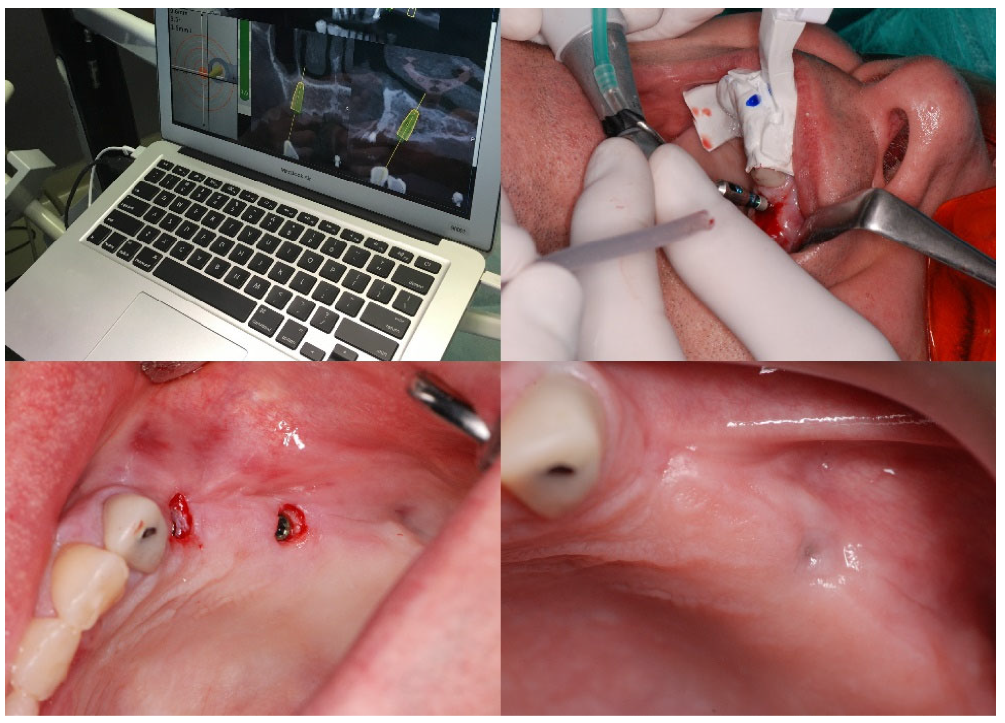
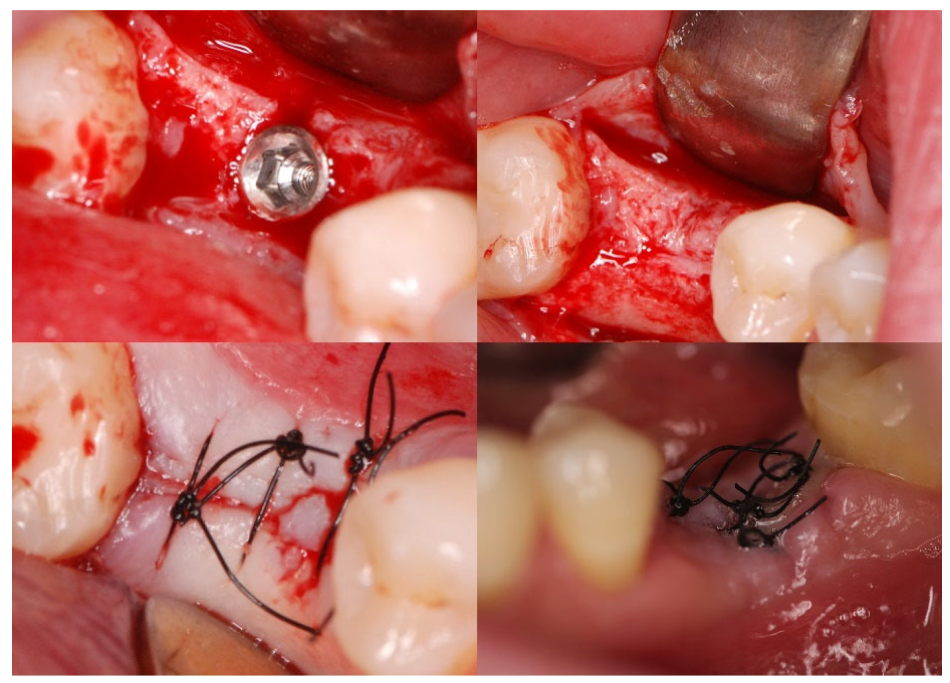
2. Materials and Methods

2.1. Patients

Implant treatment was performed on 30 patients enrolled from July 2019 to December 2020 for the rehabilitation of extracted dentition. An extraoral and intraoral examination and a CBCT scan (Carestream Dental CS 8100 3d, Carestream Dental LLC, Atlanta, GA, USA) were conducted. Patients were eligible for the study according to the criteria outlined in Table 1. Out of a total of 87 patients, 30 participants (12 men and 18 women) were finally qualified.
All participants were informed about the purpose and methodology of this study and gave written informed consent. The approval of the Bioethics Committee of the Silesian Medical Chamber in Katowice was obtained (Resolution No. 24/2019 on 25 June 2019).
The patients were then randomly divided into two groups of 15 patients: a study group and a control group. In the study group, implants were carried out using Navident dynamic navigation (Navident, ClaroNav, Toronto, ON, Canada) which allows for surgery using a flapless technique—Figure 1.
In the control group, implant treatment was carried out in a classic manner with the elevation of the mucoperiosteal flap—Figure 2. Due to the study methodology, surgery and follow-up visits were performed in the morning.

2.2. Saliva Samples

The samples of unstimulated whole saliva were collected in the morning, after a minimum hunger period of two hours (between 9 and 11 a.m.), using the Salivette® system (Sarstedt, Nümbrecht, Germany). Briefly, the patient removed the swab from Salivette® and placed the swab in the mouth. Then, the patient chewed the swab for about 1 min to stimulate salivation. Next, the patient returned the swab with the absorbed saliva to the Salivette®. The obtained saliva samples were then centrifuged for 2 min at 1000× g, and the obtained supernatants were pipetted into Eppendorf tubes which were stored at −80 °C until the day of the CRP and IL-6 assay. Then, these saliva samples were completely thawed, vortexed, and centrifuged at 1500× g for 15 min to remove mucins and other particulate matter which may interfere with antibody binding and affect the CRP or IL-6 test results.

2.3. Determination of Saliva CRP Concentration

The concentration of CRP in the saliva samples was determined by using the Salivary C-Reactive Protein ELISA Kit Generation II (Salimetrics, State College, PA, USA), which is an enzyme-linked immunoassay (ELISA) for the quantitative measurement of human CRP in oral fluid. This is an indirect sandwich ELISA kit wherein a “sandwich” is formed when the precoated captured anti-CRP antibody present on the plate binds CRP from the standard (Salimetrics’ High and Low CRP Generation II Controls) and tested samples. After each incubation, the unbound components were washed away. Bound anti-CRP antibody enzyme conjugate was then added, and the levels of detected CRP were measured by the reaction of the horseradish peroxidase enzyme to the substrate tetramethylbenzidine. The effect of this reaction is a blue colour in the solutions in the plate wells. Next, a yellow colour was formed after stopping the reaction with an acidic solution, and then the optical density was measured at 450 nm using a microplate reader EonTM Microplate Spectrophotometer (BioTek, Winooski, VT, USA). The corresponding concentration of CRP in pg/mL was determined from the standard curve (nonlinear regression curve fit) using the average optical density values of the controls and saliva samples. The functional sensitivity of the Salivary C-Reactive Protein ELISA Kit Generation II is 19.44 pg/mL.

2.4. Determination of Saliva IL-6 Concentration

The concentration of IL-6 in the saliva samples was determined using the Salivary IL-6 ELISA Kit (Salimetrics, State College, PA, USA), which is a sandwich immunoassay for the quantitative measurement of salivary IL-6. This is a sandwich ELISA kit wherein the IL-6 standard (Salimetrics’ High and Low IL-6 Controls) and tested samples bind to the antibody binding sites on a microtiter plate. After incubation, the unbound components were washed away. Next, biotin conjugated to goat antibodies and to human IL-6 was added and attached to the bound IL-6. After incubation, the unbound components were washed away, and streptavidin (conjugated to horseradish peroxidase) was added (and binds to the biotin conjugated and the goat antibodies), and the levels of detected IL-6 were measured by the reaction of the horseradish peroxidase enzyme to the substrate tetramethylbenzidine. The effect of this reaction is a blue colour in the solutions in the plate wells. Next, a yellow colour was formed after stopping the reaction with an acidic solution, and then the optical density was measured at 450 nm using a microplate reader (EonTM Microplate Spectrophotometer; BioTek, Winooski, VT, USA). The corresponding concentration of IL-6 in pg/mL was determined from a standard curve (four-parameter nonlinear regression curve fit) using the average optical density values of the controls and saliva samples. The functional sensitivity of the Salivary IL-6 ELISA Kit is 2.08 pg/mL.

2.5. Statistical Analysis
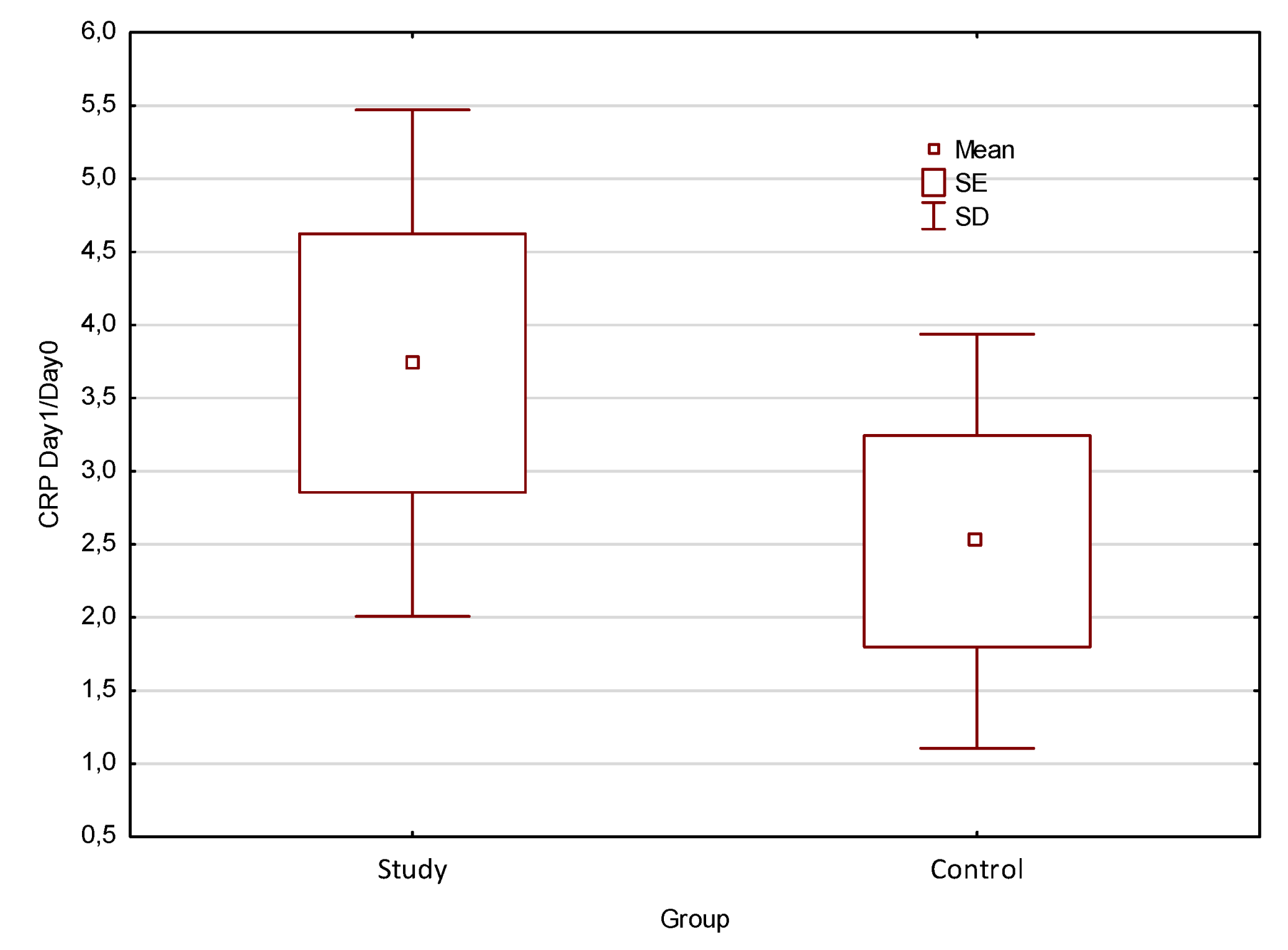
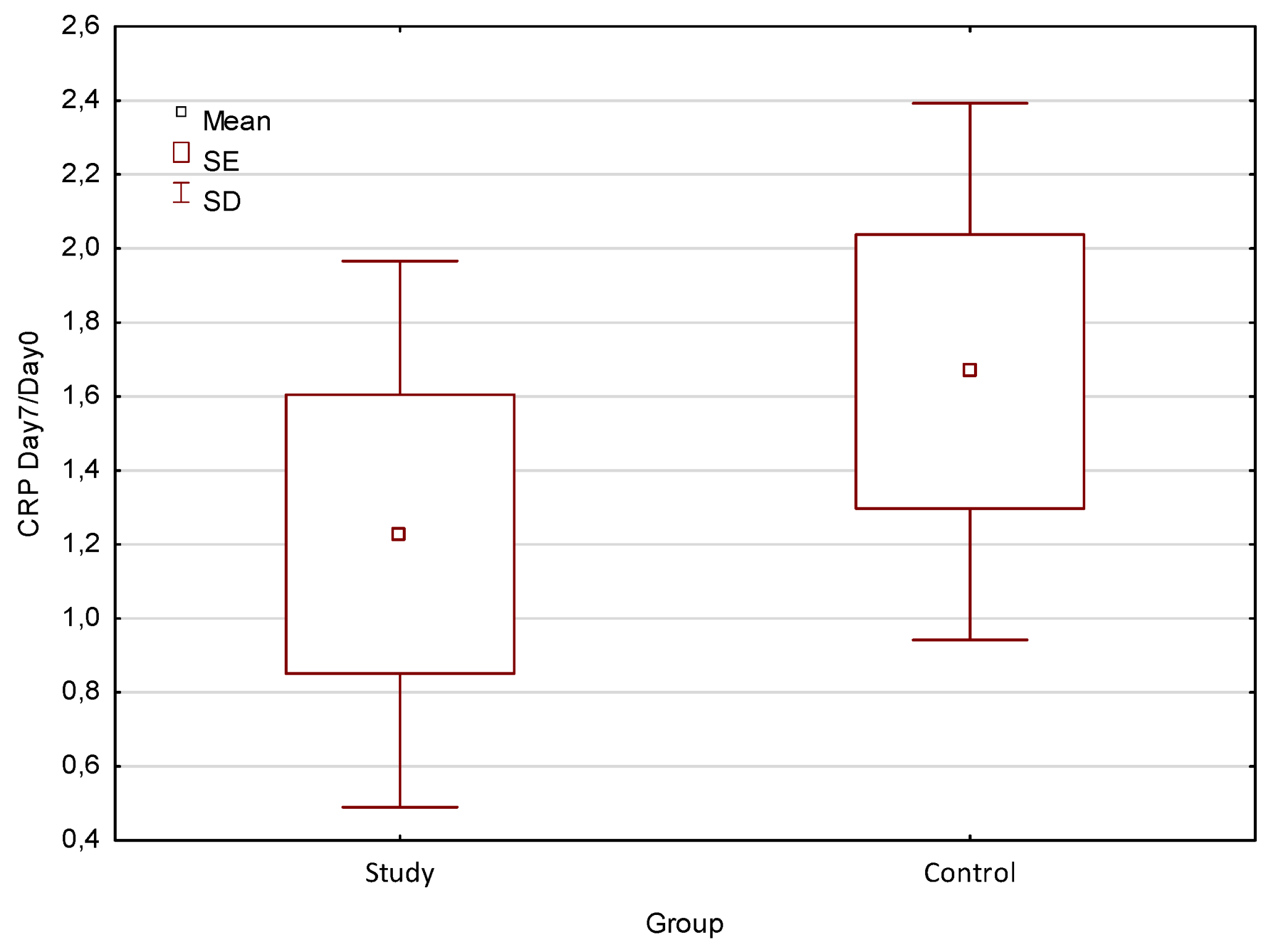
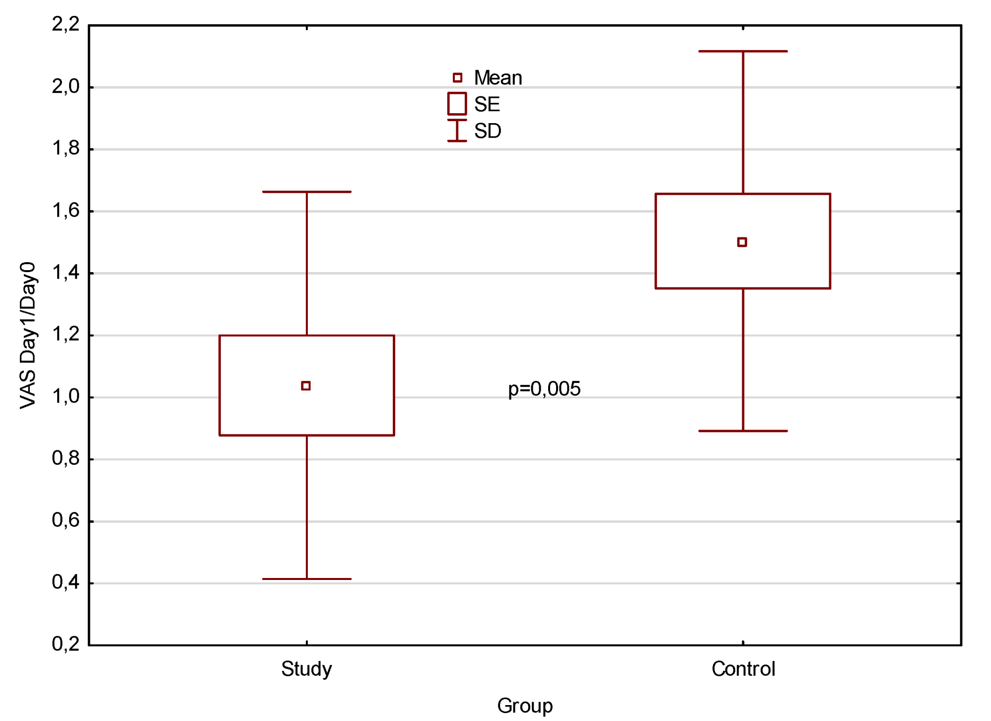
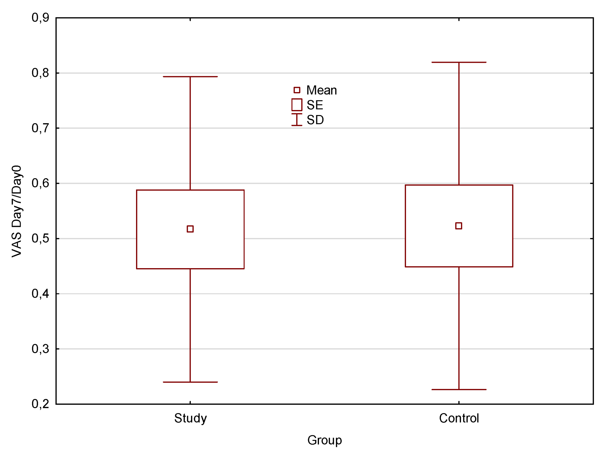
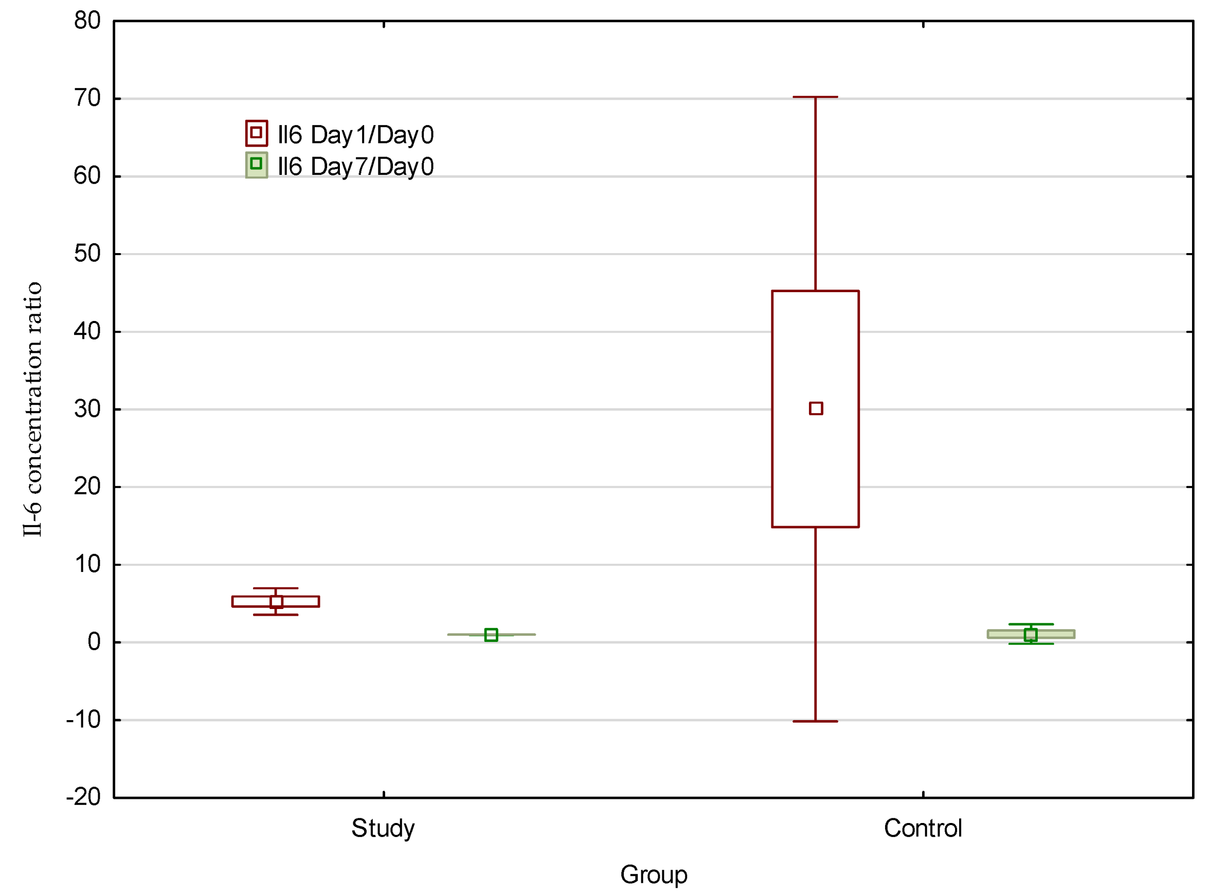
The results obtained were then collected, and statistical analyses were performed using the STATISTICA 10 program (StatSoft Polska, Kraków, Poland). For each analysis, the Shapiro–Wilk test was performed to check the type of distribution of measurable features. We checked whether the distribution of the variables was normal and if the variances were homogeneous. On the basis of those positive results, it was decided that parametric tests (including Student’s t-test for independent groups in the case of, for example, the analysis of different parameters (CRP, Il-6, and VAS) would be performed to observe changes over time between the groups of patients treated with the standard procedure and the patients free-lobe treated. When the essential assumptions for the parametric tests were not met, nonparametric tests were used (including the Mann–Whitney test). Data are expressed as the mean and median values ± standard deviation (SD) and standard error (SE). The level of significance was p < 0.05. The results are presented in box graphs.

3. Results

Comparison of CRP concentration ratio at 1st day after surgery (Day 1) and before treatment (Day 0) obtained for study group treated by flapless method was 3.74 and control group treated with standard procedure was 2.5.
Comparison of CRP concentration ratio at 7th day after surgery (Day 7) and before treatment (Day 0) obtained for study group treated by flapless method was 1.2 and control group treated with standard procedure was 1.7.
Comparison of VAS scale ratio (Day 1/Day 0) at 1st day after surgery (Day 1) and before treatment (Day 0) obtained for study group treated by flapless method was 1.04 and control group treated with standard procedure was 1.5.
Comparison of VAS scale ratio (Day 7/Day 0) at 7th day after surgery (Day 7) and before treatment (Day 0) obtained for study group treated by flapless method was 0.51 and control group treated with standard procedure was 0.52.
The presented box graphs in Figure 3 and Figure 4 show the differences between the CRP concentration ratio from day 1 after surgery and before treatment, as well as the CRP concentration ratio measured on day 7 after surgery and before treatment, obtained for the study group treated with the free-lobe method and the control group treated with the standard procedure, respectively.
Despite the fact that there were no significant differences between the mean values for the CRP concentration ratio on Day 1/Day 0 as well as on Day 7/Day 0 when comparing the study group treated with the flapless method and the control group treated with the standard procedure, there are some observable tendencies that may show completely different characters. First of all, the concentration of CRP decreases with time after surgery in both studied groups. However, the ratio coefficient calculated on day 1 after surgery seems to be higher for the study group and on day 7 after the tendency is opposed. This may suggest that the inflammatory state in the study group treated with the flapless method decreased faster over time than in the control group. Moreover, on day 7 after the procedure, the concentration drop is deeper for the study group. Such results are not statistically significant, so they may show some tendencies that can only be confirmed by using a larger group of patients.
The next analyses are presented as box graphs in Figure 5 and Figure 6, which show the differences in the VAS scale ratios on day 1 after surgery and before treatment as well as the VAS scale ratio calculated on day 7 after surgery and before treatment, obtained for the study group treated with the flapless method and the control group treated with the standard procedure, respectively.
The obtained results show statistically significant (p = 0.005) differences between the VAS scale ratio (Day1/Day0) on day 1 after surgery and before treatment, obtained for the study group treated with the flapless method and the control group treated with the standard procedure; the significantly higher value was obtained for the control group (VAS = 1.5), and for the study group, it was nearly 0.5 lower. Such results prove that the flapless method leads to less pain reported by patients just after surgery than does the standard method. Further measurements showed that the pain felt and described by patients from both groups is similar and much lower than before the surgery.
Figure 7 presents a box graph that shows the differences between the saliva Il-6 concentration ratios on day 1 after surgery and before treatment as well as the ratio calculated on day 7 after surgery and before treatment, obtained for the study group treated with the flapless method and the control group treated with the standard procedure.
It should be noted that there were no significant differences between the studied groups. However, some tendencies can be seen, such as a significantly higher mean increase in the salivary Il-6 concentration in the control group on the first post-operative day. On the other hand, the Il-6 concentration ratio on day 7 after treatment seems to be similar and significantly lower in comparison to the ratios calculated after the first day for both groups.
Generally, the saliva concentration with the pro-inflammatory factors considered (CRP, Il-6) and the VAS pain scale are lower after flapless surgery compared to the standard procedure. However, conclusions must be drawn cautiously due to the small size of the study groups and the observed trends. Only in some cases was a statistically significant (p = 0.005) higher VAS scale ratio (Day1/Day0) confirmed on day 1 after surgery when compared to the pretreatment ratio obtained for the treated control group.

4. Discussion

Saliva is one of the most important body fluids. It performs a number of vital functions, including ensuring a humid environment in the mouth, participating in water regulation, pre-digestion, protecting oral cavity structures from damage, acting as an antimicrobial agent, remineralising enamel, and participating in the perception of taste or pronunciation. Its composition and properties determine systemic homeostasis, and fluctuations in certain parameters enable it to be useful as a diagnostic marker for a number of pathologies and as a screening and treatment tool for oral squamous cell carcinoma, for instance [39].
It is very important to be able to test inflammatory markers in a non-invasive way by testing a saliva sample. Until now, the most common way to test inflammatory biomarker levels has been from a blood serum sample, which involves venipuncture and trauma, which is an invasive procedure and involves qualified personnel, laboratory equipment, and significant financial resources. In contrast, saliva collection is non-invasive, stress-free, and painless and provides an effective alternative diagnostic method [40].
A range of scientific papers that have recently been published discusses the potential use of IL-6 and CRP in the monitoring and early diagnosis of oral diseases. A study by Dineshkumar et al. [41] evaluated IL-6 among a large group of 100 patients with potentially malignant lesions (PML) in the oral cavity, 100 diagnosed oral squamous cell carcinoma (OSCC) cases, and 100 controls. Among other things, it showed a twofold to threefold higher level of inflammatory markers in saliva than in serum in those patients with diagnosed OSCC when compared with PML and a group of healthy subjects, demonstrating 96% specificity and 99% sensitivity for IL-6 in saliva in distinguishing PML from OSCC.
A study by Fonseca et al. [19] assessed the levels of a variety of pro-inflammatory cytokines in 22 edentulous patients who presented with clinical mucositis or peri-implantitis. Assessing a broad panel of markers, including, as in our study, IL-6, showed that the total IL-6 levels tended to be higher in patients with peri-implantitis in relation to a comparison group that only manifested mucositis around the implants. This demonstrated that the level of severity of the inflammatory reaction developing around the implant is reflected in IL-6 levels.
A pilot study by Draft et al. [42] on seven patients with implants, compared to three patients in the control group, assessed the levels of total antioxidant status (TAS), amounts of IL-6, IL-8, and TNF-α (tumour necrosis factor), and salivary lactate dehydrogenase (LDH) levels in correlation with bone loss around the implant over one year. Two patients in the study group had the lowest TAS levels, the highest IL-6 and IL-8 levels, and by far the highest marginal bone loss (1.0 and 0.85 mm at one year in relation to results in the range of 0.02–0.08 mm in the other implant patients). Admittedly, no strong assumptions can be drawn on such a small group; however, the authors did give consideration to screening implant patients using saliva biomarkers. Those with low TAS values or elevated levels of pro-inflammatory cytokines should undergo more frequent follow-up visits for close monitoring and the prevention of more severe complications.
A study by Cennamo et al. [43] published in May of 2023 highlights the role of point-of-care tests for the detection of salivary levels of cytokines (including IL-6) as an important tool for early diagnosis and timely treatment of inflammatory-based diseases such as periodontitis. The SPR-POF biosensor solution was presented, which allows for detecting IL-6 levels from 40 pM to 2 nM, while the GNG biosensor was designed to detect IL-6 range from 680 aM to 25 fM. The devices have applications in the diagnosis of plasma and salivary concentrations, which appears to be crucial for monitoring an individual response to a specific therapy for a particular disease.
Systemic C-reactive protein (CRP) is a sensitive marker of systemic inflammation and an independent risk factor for CVD (cardiovascular disease) [36,37].
A pilot study by Azar and Richard [43] conducted on 45 young Canadians (mean age: 18.89 years, SD = 2.62) determined the CRP levels from a saliva sample in active smokers, passive smokers, and nonsmokers. The CRP levels were highest in active smokers, lower in passive smokers, and lowest in nonsmokers. Passive smokers had significantly higher CRP levels than nonsmokers. Interestingly, the difference in CRP levels in the passive and active smokers did not reach significance. In addition, the investigators stated that salivary CRP levels appear to have a similar relationship with tobacco smoke exposure (TSE) as it is a widely used serum biomarker counterpart [44].
A cohort study [19] on 30 patients with oral lichen planus and 30 healthy patients showed that the levels of TNF-α, G-GSF, IL-1α, IL-1β, and IL-8 were statistically significantly higher in OLP patients than in the healthy group. Spearman’s rank correlation analysis showed that levels of TNF-α, GM-CSF, MIP-1α, MIP-1β, IL-1β, and IL-6 in saliva were positively correlated with the severity of the OLP lesion, meaning they have great potential as biomarkers for diagnosing and predicting the prognosis of OLP.
In implant manufacturing, several concepts have been developed for coating the implant surface and local delivery of agents regarding antimicrobial, bioactive, or therapeutic effects. For example, a coating of bioactive materials (calcium phosphates and hydroxyapatite (HA)) increases the bioactivity of the surface, improving osteointegration. Bisphosphonate coatings are designed to stimulate osteoblasts and inhibit osteoclast activity and bone resorption, and implants coated with gentamicin and polylactic acid have antimicrobial effects [45].
However, bone loss with implants progresses independently of the coatings applied to the implant surface. In the case of reduced alveolar bone, the dehiscence of the full as well as partial-thickness flap consequently leads to a loss of bone mass and increased osteoclast activity [6], which further aggravates the local anatomical conditions [7,8]. Hence, the innovative approach is complimentary implantation, which reduces the invasiveness of the procedure, improves and accelerates the healing process, and reduces the pain sensation of patients, as confirmed in our study.
The complementary technique may also lead to less bone loss compared to the traditional technique, as confirmed by a meta-analysis [46].
According to the analysis by Carosi et al. [47], computer-assisted flapless implant placement by means of mucosa-supported templates in complete arch restorations can be considered a reliable and predictable treatment choice despite the potential effects that the flapless approach can bring to the overall treatment.
Furthermore, on the basis of a comparative analysis of the planning and execution of freehand implant treatment using navigation, it has been shown that dynamic navigation may improve the quality and safety of surgical procedures and reduce the risk of complications compared to freehand implant placement [48].
Dynamic surgical navigation is also used during zygomatic implant placement procedures for the reconstruction of edentulous maxilla among patients whose osseous conditions do not allow for the placing of implants into the alveolar bone of the maxilla. A study by Bhalerao et al. confirmed that free zygomatic implant placement under dynamic navigation guidance represents a technique with minimal surgical complications [49].
Research confirms that surgery with the flapless technique provides a lower temperature for the operated area and bone bed, which contributes to improved healing and reduced likelihood of perioperative complications; this is confirmed by thermographic studies in patients operated on with flapless and classical techniques [50,51].
Equally important is the pre-operative and post-operative care of the surgical patient. Clinical studies have shown satisfactory efficacy in eliminating pathological oral flora with therapeutic preparations of natural origin, such as tea tree oil and ethanolic propolis extract [52,53], and preparation for surgery by rinsing with antiseptics before surgery, i.e., chlorhexidine, may reduce the risk of post-operative complications [54,55].
Our clinical study reveals an innovative approach to the issue of the biochemical and clinical assessment of healing in implant patients. The currently available literature has not previously explored a similar topic, indicating that there is potential space for further in-depth study of this issue. It should also be taken into account that the analysis of saliva samples and their testing requires a certain research and analytical background and an appropriate selection of patients for comparative methods. In the future, as the availability of diagnostic methods increases and their cost decreases, monitoring the clinical status of patients with objective results will allow for individual modifications to the therapy conducted.
In addition, complex surgical treatment requires the monitoring of many clinical parameters, and patient saliva may be only one of them. This also implies the limitations of the treatment method presented in this paper, which may include the patient’s general health, old age, or significant general obstructions that would disqualify the patient from surgical treatment.
In these cases, alternatives for the reconstruction of the lost dentition with implants may be proposed. In this case, “classic” prosthetic solutions such as bridges or removable denture or telescopic prosthetic devices supported by the current residual dentition may be used.

5. Conclusions

The results of this study confirm the advantages of dynamic navigation in implant treatment, supplementing previous knowledge in this aspect with observations of changes in CRP and IL-6 levels and pain intensity in the first week after surgery.
Objectively, pain is lower in the free methods than in the conventional methods and a lower level of inflammation was observed on the basis of lower biomarker parameters in the study group.
Dynamic navigation may reduce patient discomfort and facilitate faster recovery, as could be indicated by lower levels of pro-inflammatory cytokines compared to the control group.

Author Contributions

Conceptualization, K.W., T.M. and A.M.; methodology, K.W., T.M. and A.M.; software, K.W. and A.S.; validation, K.W. and T.M.; formal analysis K.W. and D.B.; investigation, K.W., A.M. and A.N.-W.; resources, K.W., T.M., A.M. and J.Ś.-W.; data curation, K.W., A.M. and J.Ś.-W.; writing—original draft preparation, K.W. and A.N.-W.; writing—review and editing, K.W., T.M. and D.B.; visualization, A.S.; supervision, K.W., T.M. and A.M.; project administration, K.W. and T.M.; funding acquisition, K.W. and T.M. All authors have read and agreed to the published version of the manuscript.

Funding

This research was funded by the Medical University of Silesia in Katowice with research numbers PCN-2-032/N/0/K and PCN-1-066/N/0/O.

Institutional Review Board Statement

This study was conducted according to the guidelines of the Declaration of Helsinki and approved by the Bioethics Committees of the Silesian Medical Chamber in Katowice (Resolution no. 24/2019, as of 25 June 2019).

Informed Consent Statement

Informed consent was obtained from all subjects involved in this study.

Data Availability Statement

The data presented in this study are available from the corresponding authors.

Conflicts of Interest

The authors declare no conflict of interest.

References

  1. Kageyama, I.; Maeda, S.; Takezawa, K. Importance of anatomy in dental implant surgery. J. Oral Biosci. 2021, 63, 142–152. [Google Scholar] [CrossRef]
  2. Greenstein, G.; Cavallaro, J.; Tarnow, D. Practical Application of Anatomy for the Dental Implant Surgeon. J. Periodontol. 2008, 79, 1833–1846. [Google Scholar] [CrossRef] [PubMed]
  3. Sheikh, Z.; Sima, C.; Glogauer, M. Bone Replacement Materials and Techniques Used for Achieving Vertical Alveolar Bone Augmentation. Materials 2015, 8, 2953–2993. [Google Scholar] [CrossRef]
  4. Gual-Vaques, P.; Polis-Yanes, C.; Estrugo-Devesa, A.; Ayuso-Montero, R.; Mari-Roig, A.; Lopez-Lopez, J. Autogenous teeth used for bone grafting: A systematic review. Med. Oral. Patol. Oral. Cir. Bucal. 2018, 23, e112–e119. [Google Scholar] [CrossRef] [PubMed]
  5. Kubaszek, B.; Morawiec, T.; Mertas, A.; Wachol, K.; Nowak-Wachol, A.; Śmieszek-Wilczewska, J.; Łopaciński, M.; Cholewka, A. Radiological and Microbiological Evaluation of the Efficacy of Alveolar Bone Repair Using Autogenous Dentin Matrix—Preliminary Study. Coatings 2022, 12, 909. [Google Scholar] [CrossRef]
  6. Fickl, S.; Kebschull, M.; Schupbach, P.; Zuhr, O.; Schlagenhauf, U.; Hürzeler, M.B. Bone loss after full-thickness and partial-thickness flap elevation. J. Clin. Periodontol. 2011, 38, 157–162. [Google Scholar] [CrossRef]
  7. Araujo, M.G.; Lindhe, J. Dimensional ridge alterations following tooth extraction. An experimental study in the dog. J. Clin. Periodontol. 2005, 32, 212–218. [Google Scholar] [CrossRef]
  8. Araújo, M.G.; Lindhe, J. Ridge alterations following tooth extraction with and without flap elevation: An experimental study in the dog. Clin. Oral Implant. Res. 2009, 20, 545–549. [Google Scholar] [CrossRef]
  9. Volberg, R.; Mordanov, O. Canalis Sinuosus Damage after Immediate Dental Implant Placement in the Esthetic Zone. Case Rep. Dent. 2019, 2019, 3462794. [Google Scholar] [CrossRef]
  10. An, J.-H.; Park, S.-H.; Han, J.J.; Jung, S.; Kook, M.-S.; Park, H.-J.; Oh, H.-K. Treatment of dental implant displacement into the maxillary sinus. Maxillofac. Plast. Reconstr. Surg. 2017, 39, 35. [Google Scholar] [CrossRef]
  11. Pellegrino, G.; Bellini, P.; Cavallini, P.F.; Ferri, A.; Zacchino, A.; Taraschi, V.; Marchetti, C.; Consolo, U. Dynamic Navigation in Dental Implantology: The Influence of Surgical Experience on Implant Placement Accuracy and Operating Time. An in Vitro Study. Int. J. Environ. Res. Public Health 2020, 17, 2153. [Google Scholar] [CrossRef] [PubMed]
  12. Vercruyssen, M.; Laleman, I.; Jacobs, R.; Quirynen, M. Computer-supported implant planning and guided surgery: A narrative review. Clin. Oral Implant. Res. 2015, 26 (Suppl. S11), 69–76. [Google Scholar] [CrossRef] [PubMed]
  13. Bover-Ramos, F.; Viña-Almunia, J.; Cervera-Ballester, J.; Peñarrocha-Diago, M.; García-Mira, B. Accuracy of Implant Placement with Computer-Guided Surgery: A Systematic Review and Meta-Analysis Comparing Cadaver, Clinical, and In Vitro Studies. Int. J. Oral Maxillofac. Implant. 2018, 33, 101–115. [Google Scholar] [CrossRef] [PubMed]
  14. Zian, Z.; Bouhoudan, A.; Mourabit, N.; Azizi, G.; Mechita, M.B. Salivary Cytokines as Potential Diagnostic Biomarkers for Systemic Lupus Erythematosus Disease. Mediat. Inflamm. 2021, 2021, 8847557. [Google Scholar] [CrossRef]
  15. Diesch, T.; Filippi, C.; Fritschi, N.; Filippi, A.; Ritz, N. Cytokines in saliva as biomarkers of oral and systemic oncological or infectious diseases: A systematic review. Cytokine 2021, 143, 155506. [Google Scholar] [CrossRef]
  16. Teles, R.P.; Likhari, V.; Socransky, S.S.; Haffajee, A.D. Salivary cytokine levels in subjects with chronic periodontitis and in periodontally healthy individuals: A cross-sectional study. J. Periodontal Res. 2009, 44, 411–417. [Google Scholar] [CrossRef]
  17. Gornowicz, A.; Bielawska, A.; Bielawski, K.; Grabowska, S.Z.; Wójcicka, A.; Zalewska, M.; Maciorkowska, E. Pro-inflammatory cytokines in saliva of adolescents with dental caries disease. Ann. Agric. Environ. Med. 2012, 19, 711–716. [Google Scholar]
  18. Ribeiro, C.C.C.; Pachêco, C.d.J.B.; Costa, E.L.; Ladeira, L.L.C.; Costa, J.F.; da Silva, R.A.; Carmo, C.D.S. Proinflammatory cytokines in early childhood caries: Salivary analysis in the mother/children pair. Cytokine 2018, 107, 113–117. [Google Scholar] [CrossRef]
  19. Zhu, Z.-D.; Ren, X.-M.; Zhou, M.-M.; Chen, Q.-M.; Hua, H.; Li, C.-L. Salivary cytokine profile in patients with oral lichen planus. J. Dent. Sci. 2022, 17, 100–105. [Google Scholar] [CrossRef]
  20. Fonseca, F.J.P.O.; Junior, M.M.; Lourenço, E.J.V.; Teles, D.d.M.; Figueredo, C.M. Cytokines expression in saliva and peri-implant crevicular fluid of patients with peri-implant disease. Clin. Oral Implant. Res. 2014, 25, e68–e72. [Google Scholar] [CrossRef]
  21. Vohra, F.; Alkhudhairy, F.; Al-Kheraif, A.A.; Akram, Z.; Javed, F. Peri-implant parameters and C-reactive protein levels among patients with different obesity levels. Clin. Implant. Dent. Relat. Res. 2018, 20, 130–136. [Google Scholar] [CrossRef] [PubMed]
  22. Melguizo-Rodríguez, L.; Costela-Ruiz, V.J.; Manzano-Moreno, F.J.; Ruiz, C.; Illescas-Montes, R. Salivary Biomarkers and Their Application in the Diagnosis and Monitoring of the Most Common Oral Pathologies. Int. J. Mol. Sci. 2020, 21, 5173. [Google Scholar] [CrossRef] [PubMed]
  23. Decker, M.-L.; Gotta, V.; Wellmann, S.; Ritz, N. Cytokine profiling in healthy children shows association of age with cytokine concentrations. Sci. Rep. 2017, 7, 17842. [Google Scholar] [CrossRef] [PubMed]
  24. Chauhan, A.; Yadav, S.S.; Dwivedi, P.; Lal, N.; Usman, K.; Khattri, S. Correlation of Serum and Salivary Cytokines Level with Clinical Parameters in Metabolic Syndrome With Periodontitis. J. Clin. Lab. Anal. 2016, 30, 649–655. [Google Scholar] [CrossRef] [PubMed]
  25. Szabo, Y.Z.; Slavish, D.C. Measuring salivary markers of inflammation in health research: A review of methodological considerations and best practices. Psychoneuroendocrinology 2021, 124, 105069. [Google Scholar] [CrossRef]
  26. Cazalis, J.; Tanabe, S.; Gagnon, G.; Sorsa, T.; Grenier, D. Tetracyclines and chemically modified tetracycline-3 (CMT-3) modulate cytokine secretion by lipopolysaccharide-stimulated whole blood. Inflammation 2009, 32, 130–137. [Google Scholar] [CrossRef]
  27. Sorsa, T.; Tjäderhane, L.; Konttinen, Y.T.; Lauhio, A.; Salo, T.; Lee, H.M.; Golub, L.M.; Brown, D.L.; Mäntylä, P. Matrix metalloproteinases: Contribution to pathogenesis, diagnosis and treatment of periodontal inflammation. Ann. Med. 2006, 38, 306–321. [Google Scholar] [CrossRef]
  28. Shahidi, M.; Jafari, S.; Barati, M.; Mahdipour, M.; Gholami, M.S. Predictive value of salivary microRNA-320a, vascular en-dothelial growth factor receptor 2, CRP and IL-6 in Oral lichen planus progression. Inflammopharmacology 2017, 25, 577–583. [Google Scholar] [CrossRef]
  29. Shiva, A.; Arab, S.; Mousavi, S.J.; Zamanian, A.; Maboudi, A. Serum and Salivary Level of Nitric Oxide (NOx) and CRP in Oral Lichen Planus (OLP) Patients. J. Dent. 2020, 21, 6–11. [Google Scholar]
  30. Rose-John, S. Interleukin-6 Family Cytokines. Cold Spring Harb. Perspect. Biol. 2017, 10, a028415. [Google Scholar] [CrossRef]
  31. Tanaka, T.; Narazaki, M.; Kishimoto, T. IL-6 in Inflammation, Immunity, and Disease. Cold Spring Harb. Perspect. Biol. 2014, 6, a016295. [Google Scholar] [CrossRef] [PubMed]
  32. Vilotić, A.; Nacka-Aleksić, M.; Pirković, A.; Bojić-Trbojević, Z.; Dekanski, D.; Krivokuća, M.J. IL-6 and IL-8: An Overview of Their Roles in Healthy and Pathological Pregnancies. Int. J. Mol. Sci. 2022, 23, 14574. [Google Scholar] [CrossRef] [PubMed]
  33. Novak, T.; Hamedi, M.; Bergmeier, L.A.; Fortune, F.; Hagi-Pavli, E. Saliva and Serum Cytokine Profiles During Oral Ulceration in Behçet’s Disease. Front. Immunol. 2021, 12, 724900. [Google Scholar] [CrossRef] [PubMed]
  34. Abboud, C.S.; Brandão, E.H.d.S.; Cunha, K.R.L.; Brito, K.d.S.; Gallo, C.d.B.; Molon, A.C.; Horliana, A.C.R.T.; Franco, A.S.L.; Thongprasom, K.; Rodrigues, M.F.S.D. Serum and salivary cytokines in patients with oral lichen planus treated with Photobiomodulation. Oral Dis. 2021, 29, 1250–1258. [Google Scholar] [CrossRef]
  35. Markanday, A. Acute Phase Reactants in Infections: Evidence-Based Review and a Guide for Clinicians. Open Forum Infect. Dis. 2015, 2, ofv098. [Google Scholar] [CrossRef]
  36. Broyles, S.T.; Staiano, A.E.; Drazba, K.T.; Gupta, A.K.; Sothern, M.; Katzmarzyk, P.T. Elevated C-Reactive Protein in Children from Risky Neighborhoods: Evidence for a Stress Pathway Linking Neighborhoods and Inflammation in Children. PLoS ONE 2012, 7, e45419. [Google Scholar] [CrossRef]
  37. Kuppa, A.; Tripathi, H.; Al-Darraji, A.; Tarhuni, W.M.; Abdel-Latif, A. C-Reactive Protein Levels and Risk of Cardiovascular Diseases: A Two-Sample Bidirectional Mendelian Randomization Study. Int. J. Mol. Sci. 2023, 24, 9129. [Google Scholar] [CrossRef]
  38. Klimek, L.; Bergmann, K.-C.; Biedermann, T.; Bousquet, J.; Hellings, P.; Jung, K.; Merk, H.; Olze, H.; Schlenter, W.; Stock, P.; et al. Visual analogue scales (VAS): Measuring instruments for the documentation of symptoms and therapy monitoring in cases of allergic rhinitis in everyday health care: Position Paper of the German Society of Allergology (AeDA) and the German Society of Allergy and Clinical Immunology (DGAKI), ENT Section, in collaboration with the working group on Clinical Immunology, Allergology and Environmental Medicine of the German Society of Otorhinolaryngology, Head and Neck Surgery (DGHNOKHC). Allergo J. Int. 2017, 26, 16–24. [Google Scholar] [CrossRef]
  39. Sahibzada, H.A.; Khurshid, Z.; Khan, R.S.; Naseem, M.; Siddique, K.M.; Mali, M.; Zafar, M.S. Salivary IL-8, IL-6 and TNF-α as Potential Diagnostic Biomarkers for Oral Cancer. Diagnostics 2017, 7, 21. [Google Scholar] [CrossRef]
  40. Ouellet-Morin, I.; Danese, A.; Williams, B.; Arseneault, L. Validation of a high-sensitivity assay for C-reactive protein in human saliva. Brain, Behav. Immun. 2011, 25, 640–646. [Google Scholar] [CrossRef]
  41. Dineshkumar, T.; Ashwini, B.K.; Rameshkumar, A.; Rajashree, P.; Ramya, R.; Rajkumar, K. Salivary and Serum Interleukin-6 Levels in Oral Premalignant Disorders and Squamous Cell Carcinoma: Diagnostic Value and Clinicopathologic Correlations. Asian Pac. J. Cancer Prev. 2016, 17, 4899–4906. [Google Scholar] [CrossRef]
  42. Azar, R.; Richard, A. Elevated salivary C-reactive protein levels are associated with active and passive smoking in healthy youth: A pilot study. J. Inflamm. 2011, 8, 37. [Google Scholar] [CrossRef] [PubMed]
  43. Cennamo, N.; Piccirillo, A.; Bencivenga, D.; Arcadio, F.; Annunziata, M.; Della Ragione, F.; Guida, L.; Zeni, L.; Borriello, A. Towards a point-of-care test to cover atto-femto and pico-nano molar concentration ranges in interleukin 6 detection exploiting PMMA-based plasmonic biosensor chips. Talanta 2023, 256, 124284. [Google Scholar] [CrossRef] [PubMed]
  44. O’Loughlin, J.; Lambert, M.; Karp, I.; McGrath, J.; Gray-Donald, K.; Barnett, T.A.; Delvin, E.E.; Levy, E.; Paradis, G. Association between cigarette smoking and C-reactive protein in a representative, population-based sample of adolescents. Nicotine Tob. Res. 2008, 10, 525–532. [Google Scholar] [CrossRef] [PubMed]
  45. Zafar, M.S.; Fareed, M.A.; Riaz, S.; Latif, M.; Habib, S.R.; Khurshid, Z. Customized Therapeutic Surface Coatings for Dental Implants. Coatings 2020, 10, 568. [Google Scholar] [CrossRef]
  46. Lahoti, K.; Dandekar, S.; Gade, J.; Agrawal, M. Comparative evaluation of crestal bone level by flapless and flap techniques for implant placement: Systematic review and meta-analysis. J. Indian Prosthodont. Soc. 2021, 21, 328–338. [Google Scholar] [CrossRef]
  47. Carosi, P.; Lorenzi, C.; Lio, F.; Cardelli, P.; Pinto, A.; Laureti, A.; Pozzi, A. Accuracy of Computer-Assisted Flapless Implant Placement by Means of Mucosa-Supported Templates in Complete-Arch Restorations: A Systematic Review. Materials 2022, 15, 1462. [Google Scholar] [CrossRef]
  48. Wachol, K.; Morawiec, T.; Nowak-Wachol, A.; Kubaszek, B.; Kasprzyk-Kucewicz, T.; Baldi, D.; Machorowska-Pieniążek, A.; Skucha-Nowak, M.; Cholewka, A. Comparative Analysis of Implant Prosthesis Treatment Planning and Execution Following Bone Repair Procedures Using Dynamic Surgical Navigation in Augmented Areas. Coatings 2022, 12, 1099. [Google Scholar] [CrossRef]
  49. Bhalerao, A.; Marimuthu, M.; Wahab, A.; Ayoub, A. Flapless placement of zygomatic implants using dynamic navigation: An innovative technical note. Br. J. Oral Maxillofac. Surg. 2022, 61, 136–140. [Google Scholar] [CrossRef]
  50. Kasprzyk-Kucewicz, T.; Cholewka, A.; Bałamut, K.; Kownacki, P.; Kaszuba, N.; Kaszuba, M.; Stanek, A.; Sieroń, K.; Stransky, J.; Pasz, A.; et al. The applications of infrared thermography in surgical removal of retained teeth effects assessment. J. Therm. Anal. Calorim. 2020, 144, 139–144. [Google Scholar] [CrossRef]
  51. Kaszuba, N.; Kasprzyk-Kucewicz, T.; Szurko, A.; Wziatek-Kuczmik, D.; Stanek, A.; Morawiec, T.; Cholewka, A. How to use thermal imaging in selected surgical dental procedures? Thermol. Int. 2021, 31, 172–181. [Google Scholar]
  52. Morawiec, T.; Dziedzic, A.; Niedzielska, I.; Mertas, A.; Tanasiewicz, M.; Skaba, D.; Kasperski, J.; Machorowska-Pieniążek, A.; Kucharzewski, M.; Szaniawska, K.; et al. The Biological Activity of Propolis-Containing Toothpaste on Oral Health Environment in Patients Who Underwent Implant-Supported Prosthodontic Rehabilitation. Evidence-Based Complement. Altern. Med. 2013, 2013, 1–12. [Google Scholar] [CrossRef] [PubMed]
  53. Wiatrak, K.; Morawiec, T.; Rój, R.; Kownacki, P.; Nitecka-Buchta, A.; Niedzielski, D.; Wychowański, P.; Machorowska-Pieniążek, A.; Cholewka, A.; Baldi, D.; et al. Evaluation of Effectiveness of a Toothpaste Containing Tea Tree Oil and Ethanolic Extract of Propolis on the Improvement of Oral Health in Patients Using Removable Partial Dentures. Molecules 2021, 26, 4071. [Google Scholar] [CrossRef] [PubMed]
  54. Hassan, S.; Dhadse, P.; Bajaj, P.; Sethiya, K.; Subhadarsanee, C. Pre-procedural Antimicrobial Mouth Rinse: A Concise Review. Cureus 2022, 14, e30629. [Google Scholar] [CrossRef]
  55. Pałka, Ł.; Nowakowska-Toporowska, A.; Dalewski, B. Is Chlorhexidine in Dentistry an Ally or a Foe? A Narrative Review. Healthcare 2022, 10, 764. [Google Scholar] [CrossRef]
Figure 1. Dynamic navigation system in use with status immediately after surgery and clinical appearance after 7 days.
Applsci 13 09866 g001
Figure 2. Implantation in the control group. Visible sutures immediately after the procedure and after 7 days. All patients were carefully examined immediately before surgery, on the first post-operative day, and on the seventh post-operative day and were asked to rate their pain on a 10-degree VAS (Visual Analogue Scale) [38].
Applsci 13 09866 g002
Figure 3. Comparison of CRP concentration ratio on day 1 after surgery (Day 1) and before treatment (Day 0), obtained for the study group treated with the flapless method and the control group treated with the standard procedure.
Applsci 13 09866 g003
Figure 4. Comparison of CRP concentration ratio on day 7 after surgery (Day 7) and before treatment (Day 0), obtained for the study group treated with the flapless method and the control group treated with the standard procedure.
Applsci 13 09866 g004
Figure 5. Comparison of VAS scale ratio (Day 1/Day 0) on day 1 after surgery (Day 1) and before treatment (Day 0), obtained for the study group treated with the flapless method and the control group treated with the standard procedure.
Applsci 13 09866 g005
Figure 6. Comparison of VAS scale ratio (Day 7/Day 0) on day 7 after surgery (Day 7) and before treatment (Day 0), obtained for the study group treated with the flapless method and the control group treated with the standard procedure.
Applsci 13 09866 g006
Figure 7. Box graph presenting the differences between saliva Il-6 concentration ratios on day 1 after surgery and before treatment (brown), as well as the ratios calculated on day 7 after surgery and before treatment (green), obtained for the study group treated with the flapless method and the control group treated with the standard procedure.
Applsci 13 09866 g007
Table 1. Patient eligibility criteria.
Inclusion CriteriaExclusion Criteria
aged 18–65 yearscritical systemic disease (ASA III-IV)
absence of systemic disease comorbiditiesgeneralised immunodeficiency
adequate oral hygiene (API < 15%)autoimmune disease
clinically and radiologically diagnosed inflammatory conditions (active caries, gingivitis and periodontitis, mucosal diseases, e.g., leukoplakia, lichen planus)
active nicotinism
use of antibiotics in the past 2 weeks
Disclaimer/Publisher’s Note: The statements, opinions and data contained in all publications are solely those of the individual author(s) and contributor(s) and not of MDPI and/or the editor(s). MDPI and/or the editor(s) disclaim responsibility for any injury to people or property resulting from any ideas, methods, instructions or products referred to in the content.

