Systematic Review

A Systematic Review of Computational Fluid Dynamics Models in the Stomach and Small Intestine

1
Auckland Bioengineering Institute, University of Auckland, Auckland 1010, New Zealand
2
Riddet Institute, Massey University, Palmerston North 4474, New Zealand
3
Department of Mechanical Engineering, University of Canterbury, Christchurch 8041, New Zealand
4
Department of Engineering Science, University of Auckland, Auckland 1010, New Zealand
*
Author to whom correspondence should be addressed.
Appl. Sci. 2023, 13(10), 6092; https://doi.org/10.3390/app13106092
Submission received: 13 April 2023 / Revised: 10 May 2023 / Accepted: 11 May 2023 / Published: 16 May 2023

Abstract

:
The use of in silico models to improve our understanding of the fluid dynamics within the gastrointestinal tract has increased over the last few decades. Computational fluid dynamics (CFD) is an in silico technique that can be used to characterize and model the fluid mechanics driving the digestion of food and absorption of nutrients. This systematic review outlines the current methodologies used to develop CFD models of the stomach and small intestine, and summarizes the flow and mixing patterns predicted from these models. A literature search was conducted on Scopus, and 15 stomach CFD studies and 15 small intestine CFD studies were included in this review after the literature selection and exclusion process. Two primary flow patterns; retropulsive flow and recirculation regions, were identified within the stomach CFD models. The flow patterns within the small intestine were depended on the type of motility pattern present. The shortcomings of the current models are discussed, and considerations for future gastric and intestinal flow modeling are provided.

1. Introduction

The gastrointestinal (GI) tract is a long continuous muscular tube responsible for ingestion, digestion, absorption and defecation. The stomach breaks down ingested foods into ‘chyme’ (partially digested food) with the aid of contractions, digestive enzymes and acid. The pyloric sphincter controls the rate at which chyme is passed into the small intestine. Nearly all of the nutrient absorption occurs within the small intestine where chyme is mixed with digestive secretions (bile and pancreatic juices) which aid in nutrient breakdown. Contractions of the intestinal wall further aid digestion and transport the nutrients to the vicinity of the intestinal walls where absorption takes place.
Our understanding of the complex processes occurring within the stomach and small intestine remains incomplete, and despite our progress in understanding the anatomy and physiology of these organs, there are many unanswered questions about the flow and mixing patterns occurring within these organs. Over the last few decades there has been a significant increase in the incidence of diet-related diseases [1]; as a result, extensive research has been conducted to improve our understanding of the complex processes involved in food digestion [2,3,4]. These studies have also sought to understand how the structure and composition of different foods affect the kinetics and nutrient absorption processes (i.e., some foods may be digested and absorbed more quickly than others due to their physical properties). Additionally, the chemical composition of foods, including the presence of different macronutrients and micronutrients can also impact how they are digested and absorbed by the body. The oral administration of pharmaceuticals is a common approach for treating many diseases, and understanding how different foods and nutrients can impact the bioavailability of these drugs is an important area of study.
GI diseases are a huge burden on the healthcare system, with annual healthcare expenditures for gastric problems comparable to that of cardiac diseases [5,6]. Approximately a quarter of the general population suffer from indigestion [7], and this can be linked to GI motility disorders [8]. GI motility disorders are often chronic or recurring in nature and can dramatically affect quality of life. Chronic intestinal pseudo-obstruction is an example of a chronic digestive disorder that is characterized by signs of intestinal obstruction without evidence of an actual mechanical obstruction in the intestinal wall [9]. Irritable bowel syndrome (IBS) is a common disorder of the small and large intestines that is characterized by abdominal pain and altered bowel habits [10]. The pathophysiology of IBS is not entirely understood and there are no effective treatments or diagnostic tests for this condition [10].
In vivo studies of intestinal motility are conducted using approaches such as advanced mapping techniques [11], magnetic resonance imaging (MRI) [12], scintigraphy [13] and endoscopy [14]. There are limited in vivo studies that measure the flow dynamics within the intestines. These range from luminal flow rate measurements [15,16], manometry pressure measurements [17] and flow patterns via combined videofluoroscopy, manometry, and multiple intraluminal impedance [18].
While advanced mapping techniques and real-time imaging tools have enabled in vivo analysis of different GI functions, the capability of these techniques to analyze and quantify the fluid dynamic behavior of the GI contents is still limited. There is also limited ability to control or manipulate experimental conditions within in vivo studies. In vitro digestion models are typically categorized into static and dynamic models. Static models have been traditionally used to study the biochemical aspect of digestion, but these systems typically are rigid, cannot model absorption and they do not accurately mimic the physiological conditions of gastric digestion [19]. In contrast, dynamic models have been developed to incorporate more realistic conditions, such as the anatomy and peristalsis of the stomach [20,21] and these systems can track the structural and physicochemical changes of foods during digestion. Numerous review papers have examined various in vitro models and emphasized their ethical advantages and reproducibility [2,22,23]. However, simulating realistic gastric emptying and mixing on the laboratory benchtop remains a significant challenge due to current limitations in experimental techniques [24].
With increased computational power, in silico models have become more sophisticated and they can provide information on the effect of gastric motility on the flow patterns, mixing, and disintegration of food particles inside the stomach. Computational Fluid Dynamics (CFD) has been identified as a promising in silico technique to model the fluid dynamics driving gastric digestion and intestinal transport. CFD is a branch of fluid mechanics that uses numerical methods to simulate and analyze the behavior of fluids and gases in complex geometries and conditions. CFD has been widely applied in engineering fields, such as aerospace, automotive, and civil engineering [25,26]. In recent years, CFD has gained attention in the biomedical engineering field for studying the fluid dynamics in the human body, including the cardiovascular system and the GI tract. However, there have been limited CFD studies on the GI tract due to its complex geometry and challenges in obtaining accurate data for boundary conditions and material properties of the GI contents for constraining and validating the numerical outputs.
There are no systematic reviews of CFD models of the GI tract. This systematic review comprehensively evaluates published CFD models used to study the dynamics of fluid flow, mixing patterns, and particle transport in the stomach and small intestine. This review identifies the key research questions that have been addressed using CFD, describes the different modeling approaches used, and evaluates the limitations of these models. Additionally, this review identifies the potential applications of CFD in the study of food digestion, drug absorption, and GI diseases. By synthesizing the current knowledge in the field, this review aims to provide insights into the future directions of CFD modeling in the stomach and small intestine.

2. Materials and Methods

The analysis of this systematic review followed the Preferred Reporting Items for Systematic Reviews and Meta-Analysis (PRISMA) statement guidelines [27].

2.1. Literature Search Strategy

A systematic literature search was conducted to identify all publications which have conducted CFD modeling of the stomach and small intestine. Several databases such as Google Scholar, Scopus and PubMed were initially considered, but due to the redundancy of the search results, the available filtering options, and the required coverage (2000–present), the literature search was limited to only Scopus. The search was conducted within the ‘article title, abstract, keywords’ option on Scopus. Two separate searches were conducted for the stomach and small intestine, and the search terms included three key criteria: (i) organ of interest, that is, stomach or small intestine, (ii) studies flow within the organ and (iii) uses numerical or computational techniques to study the flow. Common synonyms for the first and third terms were included to capture all the literature. The final search terms for the stomach CFD studies were of the form of [(“stomach” OR “gastric”) AND “flow” AND (“numerical*” OR “computational*” OR “model*”)] and for the small intestine [(“intestin*” OR “bowel”) AND “flow” AND (“numerical*” OR “computational*” OR “model*”)].

2.2. Literature Selection, Exclusion, and Data Extraction

Two of the investigators (NP and SH) conducted an extensive search of the available literature up to February 2023. The search results were filtered to only include peer-reviewed journal articles and those published in English. Two separate lists of articles were generated for the stomach and small intestine, and the article titles, abstracts and keywords were screened to identify relevant articles that conducted computational modeling of flow within the organ of interest. The list of relevant articles was screened to exclude studies that: (i) used semi-analytical approaches (such as [28,29]), (ii) used CFD models of the stomach from other animals, besides humans, (iii) used 2D or non-anatomical geometries (such as the conical one used by [30]) for the stomach, and (iv) used fixed walls with no boundary deformations.
Finally, two more stomach CFD articles [31,32] and one small intestine CFD article [33] were added from a bibliographic search cross-referenced with the other included articles. These articles did not appear in the search due to the article title, abstract and/or keywords missing the search terms outlined previously.
The investigators were not blinded by the authors, institutions, and titles of the included studies, and two investigators (NP and SH) extracted information such as the numerical technique, geometry, boundary deformation patterns, and key findings from the included articles.

3. Results

Figure 1 provides an outline of the study selection processes. A total of 5139 and 10,191 articles were initially retrieved, and these lists of articles were limited to 4414 and 8991 peer-reviewed journal articles published in English for the stomach and small intestine, respectively. Screening these lists of articles for relevant CFD studies of the stomach and small intestine identified 25 and 36 articles, respectively. Upon filtering these articles using the exclusion criteria, 15 stomach and 15 small intestine CFD studies were included for the final in-depth review.
Prior to 2010, there were no 3D CFD models of the stomach, while the first 3D small intestine model was published in 2016. There was an average of 4–5 articles published in this area (for both stomach and small intestine) in the last 2–3 years.

3.1. Governing Equations, Boundary Conditions and Initial Conditions
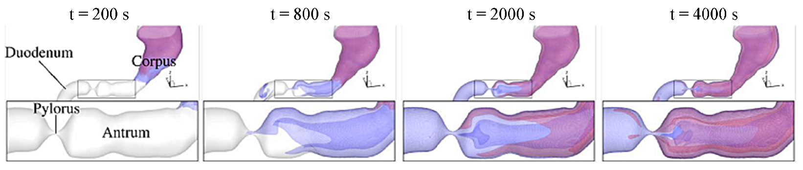
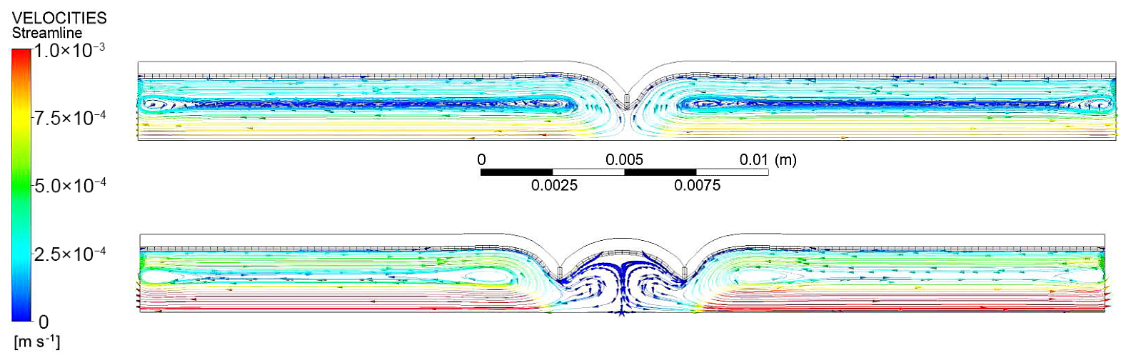
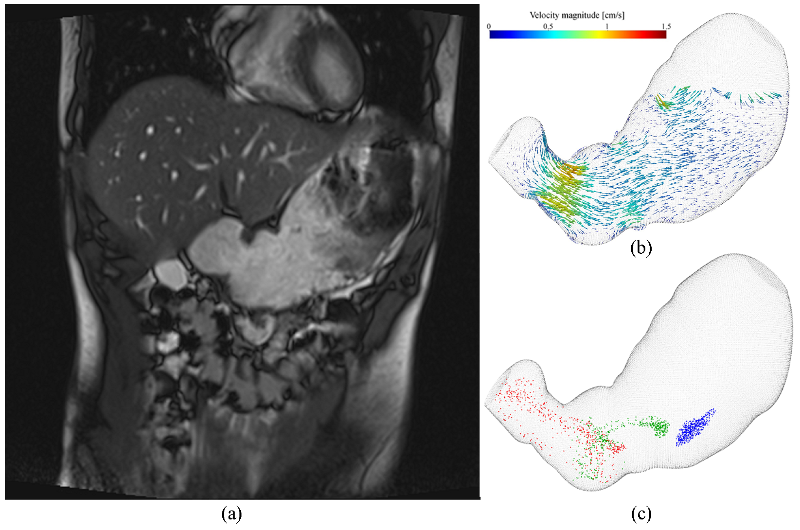
In these CFD models, the fluid is regarded as a continuum or as a continuous phase and a set of governing equations are derived based on the laws of conservation of mass, momentum, and energy. The fluid density changes within the GI are negligible, i.e., incompressible flow is present, therefore the conservation of momentum is expressed as,
u i t + u j u i x j = 1 ρ p x i + μ ρ 2 u i x j 2 + f i
where u is the flow velocity, t is the time, ρ is the fluid density, μ is the fluid viscosity, p is the fluid pressure, f is any additional body forces (e.g., gravity) and i and j denote spatial dimensions (x, y and z).
An additional equation for the conservation of mass is also required since the current system only has three equations for four unknowns (x, y and z velocities and pressure). For an incompressible fluid the conservation equation is,
u i x i = 0
Typically a no-slip boundary condition is imposed at walls i.e., the fluid velocity is equal to the wall displacement velocity,
u i = u w ,
where the wall velocity ( u w ) is determined from the prescribed boundary deformations. At an inlet, such as the entrance to the small intestine, either an inlet flow rate or a velocity profile is typically prescribed. At an outlet, such as the pylorus of the stomach, an outflow condition is typically prescribed. In these CFD models, the fluid within the domain is typically initialized with a zero velocity.

3.2. Stomach

A total of 15 articles were identified for the stomach and these are summarized in Table A1 with their relevant characteristics. The study by Pal et al. [34], which developed the first simplified 2D CFD model of the stomach incorporating contractions based on in vivo MRI data, was excluded from the analysis due to the utilization of a 2D geometry. However, the study is a significant contribution to gastric flow modeling since the motility patterns from this study have been used in 10/15 of the subsequently developed CFD models of the stomach.
CFD models of gastric motility assess different aspects of gastric motility, such as the different contraction properties [35], fluid properties [36,37], and their effects on the flow patterns and mixing. The effect of gastric motility and other factors such as posture [38] on emptying [31,39,40], mixing and disintegration of food [41,42,43], and drugs [32,44] in the human stomach were also studied.
Well-established numerical techniques such as the Finite Volume Method (FVM) were utilized in the CFD models of the stomach, with its use in both single-phase [36,45] and multi-phase [41,42] simulations. Particle-based CFD methods such as the Lattice-Boltzmann method (LBM) have been used [35,38,39], as well as the Smoothed Particle Hydrodynamics (SPH) method [40]. Due to their mesh-free formulation, SPH and LBM are well-suited for models with moving and deforming boundaries. On the other hand, models using the FVM are limited in simulating large amplitude contractions [42]. More recently, Seo and Mittal [44] and Acharya et al. [46] have used the Immersed Boundary Method to resolve fluid-structure interactions between a pill and stomach wall, respectively.
Several different anatomically realistic geometries were used to represent the stomach in these CFD models i.e., 7/15 studies generated an idealized geometry that was specific to that study, such as the one utilized by [36] (Figure 2a), some studies used a segmented geometry from the Visible Human Project (VHP) [47] (Figure 2b), and a geometry from the Virtual Population Library [48] (Figure 2c). Most studies simulated peristaltic contractions by prescribing the motion of points on the boundary, and this motion is typically prescribed mathematically with respect to the centerline. The parameters defining this motion i.e., speed (2.5 mm/s), period (20 s), and amplitude (90% radial contraction) have been primarily obtained from one MRI study of a healthy subject from Pal et al. [34]. Berry et al. [35] conducted high-resolution mapping of gastric slow waves within 10 subjects, and these recordings were averaged and used to derive peristaltic contractions. Acharya et al. [46] incorporated circular and longitudinal muscle layers to the stomach wall, and via the activation of muscle fibers embedded in these layers, gastric peristalsis was generated. Fluid flow within the stomach due to these muscle contractions was computed using fluid-structure interactions.
All simulation results showed two main flow patterns induced by gastric motility, as outlined in Figure 3. These two patterns were the ‘retropulsive jet’ located in the distal antrum and recirculating regions between pairs of antral contractions. Retropulsion is when chyme is squirted back into the stomach due to the combined effect of peristaltic contractions reaching the pylorus and the pylorus itself also being closed [49]. Thus, a ‘retropulsive jet’ refers to the resulting flow pattern due to the retropulsion motion of digesta. These fluid motions are responsible for the mixing and emulsifying of food with gastric juices and cause grinding and rubbing between food particles and/or the stomach wall [49]. Several studies have varied the viscosity of digesta, which is typically represented as a Newtonian fluid with viscosities ranging from O ( 10 3 ) to O ( 10 0 ) Pa s. An increase in the viscosity resulted in a reduction in the spatial extent of the flow structures [36,40], and a reduction in the amount of gastric emptying [39]. Negligible differences in the flow patterns were observed for a non-Newtonian fluid (shear-thinning fluid) representing tomato juice compared to a highly viscous Newtonian fluid (representing honey) [45].
Gastric mixing has been studied through several approaches, such as the evolution of particle tracers [38], evaluation of the stretching of material elements via the computation of the deformation gradient tensor [45], time integration of the rate of strain tensor [35,37], and relative vertical location changes of SPH particles [40]. Factors such as posture [38], the viscosity of digesta [40,45], velocity and acceleration of antral contractions [35] and frequency of peristaltic contractions [37] were all found to have an impact on the gastric mixing evaluated within these studies.
The content found in the stomach is a multiphase fluid consisting of gastric juices, food, and other ingested materials. The gastric juices are a complex mixture of enzymes, acids, and other components that contribute to the overall heterogeneous and dynamic nature of the gastric fluid [49]. Few studies have considered the mixing and interplay between the gastric fluid phases. Li and Jin [42] developed a CFD model of the stomach with the inclusion of gastric acid (H+ ion) secretion at the stomach wall, and this was the first study to model the folds in the mucosal surface of the stomach wall (called rugae or gastric folds) as a porous medium. Simulations conducted for three liquid foods (water, orange juice and whole milk) resulted in varying pH distributions due to density variations i.e., the H+ ions were transported from the top of the stomach to the bottom when the food is heavier than gastric juice.
Li et al. [41] also used the previously discussed model to quantify the mixing between five different food boluses and water. This study treated the individual boluses as fluids (with varying densities, diffusion coefficients, and viscosities) and solved an individual scalar transport equation per food bolus. This study assumed that large food particles were deposited in the distal stomach, and this was modeled as a porous matrix. This food matrix was found to have a larger flow resistance to food boluses than to water; therefore, rapid emptying of water was observed. This study also identified an important phenomenon related to gastric emptying known as the ‘magenstrasse’ [50], which is the fast pathway of emptying for liquids (such as water) along the lesser curvature of the stomach.
In a subsequent study, Li and Jin [43] further improved their previous model to evaluate the disintegration of small meat particles (treated as a fluid) due to the following factors; hydrogen ions (secreted from the gastric wall), enzymes and temperature effects on enzyme activity. The small meat particles were assumed to be “a collection of unlinked food particles that will separate quickly when mixed with gastric fluid”, and thus were treated as a passive scalar. This model also incorporated the breakdown of large solid particles (modeled as the porous food matrix) into small meat particles through the action of H+ ions and enzymes. Heating of meat proteins resulted in faster breakdown and emptying, while the opposite was observed with reduced contraction amplitude and reduced H+ secretion rate. The process of gastric digestion occurs over the span of 30 min to several hours, depending on the meal content [51], but only two studies have simulated digestion over O ( 1000 ) s [31,43]. Kuhar et al. [31] also modeled the chemical breakdown of a liquid meal that contains only proteins (Figure 4). Protein hydrolysis was incorporated into this model using first-order catalytic reaction kinetics, and the three species in the model (pepsin, protein and hydrolyzed protein) were modeled as passive scalars. This model included the mixing dynamics of pepsin, and consequently larger/faster contractions lead to faster hydrolysis. The delayed emptying rate observed in the stomach can be attributed to the time required for the transportation of pepsin from the secretion zones to the antrum. The interface between food and pepsin provided a direct measure of the mixing, and this study found that stronger motility led to better mixing and more widespread reaction interfaces.
Gastric emptying has been incorporated into 9/10 of the CFD models developed after 2018. However, in most of these studies the pylorus was assumed to be always open [40,41], and only a few studies have included the realistic function of the pyloric sphincter. Ishida et al. [39] included the function of the pyloric sphincter and the geometry used for the model consisted of both the stomach and duodenum from the VHP. The duodenum was static throughout the simulation and was initially completely filled with liquid, while only 80% of the stomach was filled (the rest contained air). This study concluded that impaired coordination between contractions of the antrum and pyloric closure lead to delayed gastric emptying, rapid emptying and/or bile reflux. In a subsequent study [52], using the same model, the effects of peristaltic amplitude and frequency on gastric emptying and mixing were studied. It was found that an increase in the contraction amplitude (1.2 times higher than normal) led to an increase in the emptying rate (2.7 times faster) and mixing strength becoming 5.4 times larger, with a similar effect observed for the frequency of contractions. Two other studies also included a small section of the duodenum in their CFD model of the stomach [31,32], which was used to calculate emptying rates of an active pharmaceutical ingredient and hydrolyzed protein, respectively.

3.3. Small Intestine

Table A2 summarizes the 15 articles identified for the CFD models of the small intestine. Unlike the stomach, which has relatively slow and infrequent contractions (around three contractions per minute or cpm), the small intestine exhibits more frequent contractions, ranging from 10–12 cpm [53,54]. The small intestine has a more convoluted geometry; although topologically it is still a tube, its surface area is significantly larger than the stomach.
Mesh-based methods such as the FVM [55,56,57] and Finite Element Method (FEM) [58,59,60,61] been used to model intestinal flow. Particle-based methods such as the LBM have also been utilized in [62,63]. SPH has also been utilized to model intestinal flow with Sinnott et al. [64], coupling it with the discrete element method to model the solid phase, and Alexiadis et al. [33] coupling SPH with a lattice spring model for the intestinal wall.
A small segment (3.6–25 cm in length) of the small intestine is typically modeled except for Karthikeyan et al. [60] who modeled a 120 cm jejunum and 180 cm ileum. These CFD models were based on a variety of animal species, including rats [62], rabbits [58,59], chickens [61], guinea pigs [55], pigs [56], and humans [33,63,64,65]. Most models used an axisymmetric 2D geometry to represent the intestinal segment, with others also using a 2D rectangular channel [55,62], and a 3D cylinder [33,63,64]. The validity of using an axisymmetric condition about the center of the intestine lumen should be reconsidered since this assumes that both sides of the intestinal wall are undergoing the same contraction. On the contrary, it has been experimentally observed that the contraction amplitude differs between the side of the intestinal wall which attaches to the mesentery vs. the opposite side [66]. All studies used a simplified geometry, while Trusov et al. [65] used an idealized 3D geometry with aspects of realistic anatomy, which consisted of a ‘C’ shaped cylinder to represent the duodenum. Palmada et al. [67] was the first study to incorporate an anatomically realistic geometry of the duodenum derived from the VHP (Figure 5).
Three distinct motility patterns are observed within the small intestine that facilitate food digestion and absorption. Peristalsis is responsible for the propulsion of digesta along the intestine, and these contractions were studied by 10/15 studies. The majority of these studies only simulate a single peristaltic contraction moving in an antegrade direction, while Oyama et al. [63] modeled the effect of multiple contractions and [61,67] also investigated the fluid mechanics of colliding peristaltic waves. Segmentation contractions involve localized contractions and relaxations with no overall propulsion along the intestine, and their role in mixing of digesta with surrounding fluids (enzymes and chemicals secreted into the intestinal lumen) has been studied by [56,57,59,62]. Pendular contractions consist of a ‘to-and-fro’ motion of the intestinal wall as identified experimentally by Lammers [68], and the fluid mechanical consequences of these contraction patterns have been analyzed in a few studies [58,59,62].
Fullard et al. [59] considered the effect of combining pendular and segmentation motions. These motility patterns are prescribed into the CFD model as boundary deformations, which range from sinusoidal waves [63,64,65], Gaussian distribution function [60], fluid-structure interactions [33], and more realistic deformation patterns obtained from experimental recordings of ex-vivo intestinal samples [56,58,61,62]. A model of slow wave propagation was utilized by Palmada et al. [67] to obtain anatomically realistic boundary deformations to be applied in the model of duodenum motility (Figure 5a).
Laminar flow was present across all CFD models of the small intestine with specific flow patterns being dependent on the motility prescribed in the model. Similar to the stomach, in contracting regions, flow is opposite to the direction of peristaltic wave propagation, and vortices/vortical flow is present in regions upstream and/or downstream of the contraction [55,56,60,63,64]. These studies all highlight the importance of vortical flow in enhancing digestion and absorption of nutrients within the intestines. Vortices were also observed with segmentation contractions (Figure 6), with no net movement of fluid along the length of the intestine and the authors concluded that these contractions may serve to transport nutrients from the lumen to the intestinal wall [56,57,59,62]. The resulting flow patterns from pendular/longitudinal contractions were dependent on the specific type of intestinal wall motion i.e., orad (intestinal wave moving in the oral direction), stretching or compression [58]. In their paper, de Loubens et al. [62] observed non-steady vortices within contractile regions while more ordered axial flow was present in [58]. These differences are likely due to the specific ex-vivo recordings of longitudinal motion used to define the contraction patterns. Colliding peristaltic waves resulted in swirling flow regions [67] and high-velocity fluid jets exiting the colliding region [61].
Several studies have investigated the effect of fluid viscosity on the flow behavior within the intestine. Chyme within the small intestine is a complex suspension of undigested particulate matter, solubilized nutrients, secreted enzyme, bile and mucin [69]. Most studies have used arbitrary viscosities of fluids such as water (≈1 mPa s) and honey (≈1 Pa s), with only a few studies using experimentally recorded viscosities of the liquid phase of digesta (after filtering out particulate matter), represented as a Newtonian fluid [60,67]. Whole digesta has been experimentally observed to be more viscous and non-Newtonian due to the particulate matter and exhibits pseudoplastic behavior where the apparent viscosity decreases with increased shear rate [70]. Two CFD models have considered the effect of representing digesta as a non-Newtonian fluid [56,67]. The modeling framework of [71] used particle-based techniques, SPH and Discrete Element Method (DEM), to predict collisional and hydrodynamic interactions between the fluid and solid phases of digesta. This model allows for simulating the non-Newtonian behavior based on system mechanics rather than a rheology model, and this also separates the simpler Newtonian behavior of the fluid phase from the more complex behavior of the solid phase.
Mixing within the intestine occurs at several length scales, and most CFD models studied macroscale mixing using several approaches. The passive transport of a scalar/species was computed, the variance in concentration [55] and mixing of a glucose bolus [57,67] (Figure 5b) provided a metric of intestinal mixing. The displacement of massless particles [62,72], area of vortices [56], and stretch of fluid elements (deformation gradient tensor) [58] were also used to assess the macroscale mixing occurring within the small intestine. Sinnott et al. [71] visualized the dispersion of solid DEM particles colored according to their starting axial position in Figure 7. An increase in the contraction amplitude [63,67], reduction in the viscosity of digesta [56], and colliding peristaltic contractions [61] were all found to increase the amount of intestinal mixing.

4. Discussion and Future Directions

This systematic review has identified 15 stomach and 15 small intestine articles that have used CFD modeling to assess fluid flow patterns, mixing, and digestion processes within the respective organs. Characteristics of these studies including the numerical techniques, the type of geometry used, and the approaches used to simulate motility were compared. Overall, there has been significant progress in the development of CFD models of the stomach and small intestine. These models provide a unique insight into the complex fluid dynamics and transport phenomena occurring in the GI tract, which cannot be easily studied in vivo or in vitro. However, despite the progress made, there are still numerous challenges that need to be tackled.
The lack of subject-specific stomach geometries and deformation patterns has been a major limitation in previous CFD models of stomach motility. Nearly all studies have used the MRI data from Pal et al. [34] as a basis for their models, which may not accurately represent the anatomical and physiological variability between different individuals. However, recent developments in MR imaging has led to the creation of subject-specific CFD models of gastric motility [73], such as the one presented in Figure 8. These models allow for a more accurate representation of individual variation and could lead to more personalized diagnoses and treatments for GI disorders.
To accurately simulate the digestive processes within the stomach, it is also essential to model the breakdown of solid foods. Most current models only focus on the fluid phase, or even treat the boluses as a fluid, which can lead to inaccurate results. Accurate modeling of solid food digestion will require coupling different numerical techniques to simulate the complex interactions between solid and fluid phases. Additional experiments are required to gather data on the mechanical and chemical properties of different solid foods that have undergone mastication. Such developments will likely lead to more realistic simulations of the digestive process and enable researchers to better understand the effects of different food properties and compositions on the digestive process.
The ability to compute gastric flow and mixing patterns over longer periods of time ( O ( 1000 ) s ) is crucial for predicting gastric breakdown and digestion. Digestive processes in the stomach takes several hours and it is essential to capture the temporal evolution of the flow and mixing patterns to derive meaningful results. However, current limitations in computational power make it challenging to perform such simulations over longer periods of time. Kuhar et al. [31] highlights the computational expenses associated with their model which required ∼6000 h of CPU time to simulate a cycle of 20 s. They were able to significantly reduce the computational cost by taking advantage of the periodicity of the flow patterns i.e., the flow fields from the third cycle (after 40 s) were repeated, and only the transport equations for the chemical species were solved. This enabled simulation of digestion over 5000 s, therefore future studies with periodic deformation patterns should consider a similar approach. This would not work for the simulation of solid food digestion due to the fluid–structure interactions required to compute the disintegration of particulate matter. The progress made in utilizing GPU acceleration of particle-based methods [74] also appears to be a viable route for developing future models of gastric motility and digestion.
Future CFD models of the small intestine should incorporate the complex physical properties of chyme, which can be achieved through multiphase modeling approaches. The rheology of chyme also undergoes changes as it traverses the length of the small intestine, owing to both mechanical and chemical processes. More precise experimental measurements of digesta rheology, especially in human subjects, are necessary to improve the accuracy of in silico models in capturing the fluid properties of chyme. Small intestine models incorporating nutrient absorption should model the enzymatic digestion occurring within the bulk digesta and the kinetics of transport proteins in the intestinal epithelium.
Validation of CFD models is crucial for their successful application in predicting the gastric flow and mixing patterns. While significant progress has been made in developing and refining these models, validation remains a challenge, with only 6/30 studies presenting evidence of validation. Experimental validation is necessary to establish the accuracy and reliability of the numerical results, especially in complex physiological systems like the GI tract. However, obtaining experimental data for validation purposes can be difficult, as it requires invasive techniques or non-invasive imaging techniques that are limited by resolution and accessibility. Flow visualization experiments conducted within a phantom model [75,76] could provide the necessary data for numerical validation.

5. Conclusions

The use of CFD modeling to study the fluid dynamics, mixing, and digestion processes within the stomach and small intestine has made significant advances in recent years. This review has aimed to provide a comprehensive evaluation of these models, i.e., summarizing their methodologies, findings and shortcomings. Despite the progress in recent years, there are numerous challenges that need to be addressed, such as the use of subject-specific geometries, modeling of solid food digestion, and validation of the models. One possible way to address some of these challenges is to take inspiration from other engineering fields or other bioengineering research where CFD is heavily used. For instance, the modeling of fluid flow in blood vessels has led to significant improvements in the understanding and treatment of cardiovascular diseases. As computational power and techniques continue to improve, we anticipate that these CFD models will become even more valuable in the study of GI physiology and disease. Ultimately, the continued development and refinement of CFD models will help to improve our understanding of digestion in both health and disease, leading to better diagnosis and treatment options for patients.

Author Contributions

Conceptualization, N.P., S.H., R.A., J.E.C., V.S. and L.K.C.; data curation, N.P. and S.H.; methodology, N.P., S.H. and L.K.C.; writing—original draft, N.P.; writing—review & editing, N.P., S.H., R.A., J.E.C., V.S. and L.K.C. All authors have read and agreed to the published version of the manuscript.

Funding

This research was funded by Riddet Institute Centre of Research Excellence, and Marsden Council Funding administered by Royal Society Te Apārangi. S.H. was supported by a University of Auckland Doctoral Scholarship and the Auckland Bioengineering Institute.

Institutional Review Board Statement

Not applicable.

Informed Consent Statement

Not applicable.

Data Availability Statement

The data presented in this study are available on request from the corresponding author.

Conflicts of Interest

The authors declare no conflict of interest.

Appendix A

Table A1 and Table A2 present the key characteristics extracted from the selected literature relevant to the stomach and small Intestine CFD models respectively.
Table A1. Characteristics of stomach CFD literature included within this review. All studies were unsteady simulations with laminar flow.
ArticleNumerical TechniqueCFD PackageTime StepGrid/Particle ResolutionGeometryBoundary DeformationsEmptying
Ferrua and Singh (2010) [36]FVMAnsys FLUENT0.005–0.1 s1–1.4 mmIdealizedPrescribed boundary motion with respect to the centerline with parameters derived from Pal et al. [34]No
Imai et al. (2013) [38]MPSVHPAs per [36]No
Ferrua et al. (2014) [45]FVMAnsys FLUENT0.05 s1.5 mmIdealizedAs per [36]No
Berry et al. (2016) [35]LBM1.5 mmVHPSlow-wave recordingsNo
Miyagawa et al. (2016) [37]LBM1.5 mmVHPAs per [36]No
Harrison et al. (2018) [40]SPH3 mmIdealizedAs per [36]Yes
Ishida et al. (2019) [39]LBM1.5 mmVHPAs per [36]Yes
Li et al. (2021) [41]FVMOpenFOAM0.6 mmIdealizedPrescribed boundary motion with respect to the centerline with own parametersYes
Li and Jin (2021) [42]FVMOpenFOAM0.6 mmIdealizedAs per [41]Yes
Seo and Mittal (2022) [44]IBM0.002 s0.5 mmVPLAs per [36]No
Lee et al. (2022) [32]IBM0.002 s0.5 mmVPLAs per [36]Yes
Acharya et al. (2022) [46]IBMIBAMR 1.5 × 10 5 s1.5 mmIdealizedPrescribed muscle contractions and FSIYes
Kuhar et al. (2022) [31]IBM0.05 s0.5 mmVPLAs per [36]Yes
Ebara et al. (2023) [52]LBMVHPAs per [36]Yes
Li and Jin (2023) [43]FVMOpenFOAM0.6 mmIdealizedAs per [41]Yes
Abbreviations: FVM, Finite Volume Method; MPS, Moving Particle Semi-implicit; LBM, Lattice-Boltzmann Method SPH, Smoothed Particle Hydrodynamics IBM, Immersed Boundary Method; VHP, Visible Human Project; VPL, Virtual Population Library; FSI, Fluid-Structure Interaction.
Table A2. Characteristics of Small Intestine CFD literature included within this review. All studies were unsteady simulations with laminar flow.
ArticleAnimal OrganNumerical TechniqueCFD PackageTime StepGrid/Particle ResolutionGeometryContraction Type/sBoundary Deformations
Jeffrey et al. (2003) [55]Guinea pig ileumFVM0.015 mm2D rectangular channelPeristalsisEx-vivo intestinal sample
Love et al. (2013) [56]Pig duodenumFVMFEMLAB0.7 mmAxisymmetric cylinderPeristalsis and segmentationEx-vivo intestinal sample
de Loubens et al. (2013) [62]Rat and guinea pig duodenumLBM2D rectangular channelSegmentation and pendularEx-vivo intestinal sample
de Loubens et al. (2014) [77]Rat duodenumLBM2D rectangular channelPendularEx-vivo intestinal sample
Fullard et al. (2014) [58]Rabbit ileumFEMANSYS Polyflow0.08 mmAxisymmetric cylinderPendularEx-vivo intestinal sample
Fullard et al. (2015) [59]Rabbit ileumFEMANSYS Polyflow0.04 s0.08 mmAxisymmetric cylinderSegmentation and pendularEx-vivo intestinal sample
Trusov et al. (2016) [65]Human Antrum, Pylorus and DuodenumFVMANSYS Fluent0.027–5.5 mmC shaped 3D cylinderPeristalsisSinusoidal waves
Sinnott et al. (2017) [71]Human duodenumSPH-DEM4.3–4.7 × 10 5 s1.25 mm3D cylinderPeristalsisSinusoidal waves
Yang et al. (2017) [72]Zebrafish larvaeFVMAnsys CFX3D anatomically realistic geometryPeristalsisMathematical function based on in-vivo recordings
Karthikeyan et al. (2021) [60]Human jejunum and ileumFEMCOMSOL Multiphysics0.005 s0.3 μ m to 0.4 mmAxisymmetric cylinderPeristalsisGaussian distribution function with parameters derived from MRI
Oyama et al. (2021) [63]Human ‘Section of small intestine’LBM3D cylinderPeristalsisSinusoidal waves
Zha et al. (2021) [57]Human duodenumFVMANSYS Fluent0.005 s0.19 mmAxisymmetric cylinderSegmentationEx-vivo intestinal sample
Alexiadis et al. (2021) [33]Human intestineSPH-LSMLAMMPS0.002 s6 mm3D cylinderPeristalsisArtificial neural network governing the deformation of LSM particles on the wall
Amedzrovi Agbesi and Chevalier (2022) [61]ChickenFEMCOMSOL MultiphysicsAxisymmetric cylinderPeristalsisTraveling Gaussian force density function with one-way fluid-structure interaction
Palmada et al. (2023) [67]Human duodenumFVMOpenFOAM0.018 s0.4 mm3D VHPPeristalsisElectrophysiological model of slow wave propagation
Abbreviations: FVM, Finite Volume Method; FEM, Finite Element Method; LBM, Lattice-Boltzman Method; SPH, Smoothed Particle Hydrodynamics; DEM, Discrete Element Method; LSM, Lattice Spring Model; VHP, Visible Human Project.

References

  1. Afshin, A.; Sur, P.J.; Fay, K.A.; Cornaby, L.; Ferrara, G.; Salama, J.S.; Mullany, E.C.; Abate, K.H.; Abbafati, C.; Abebe, Z.; et al. Health effects of dietary risks in 195 countries, 1990–2017: A systematic analysis for the Global Burden of Disease Study 2017. Lancet 2019, 393, 1958–1972. [Google Scholar] [CrossRef] [PubMed]
  2. Sensoy, I. A review on the food digestion in the digestive tract and the used in vitro models. Curr. Res. Food Sci. 2021, 4, 308–319. [Google Scholar] [CrossRef] [PubMed]
  3. Le Feunteun, S.; Mackie, A.R.; Dupont, D. In silico trials of food digestion and absorption: How far are we? Curr. Opin. Food Sci. 2020, 31, 121–125. [Google Scholar] [CrossRef]
  4. Dupont, D.; Le Feunteun, S.; Marze, S.; Souchon, I. Structuring food to control its disintegration in the gastrointestinal tract and optimize nutrient bioavailability. Innov. Food Sci. Emerg. Technol. 2018, 46, 83–90. [Google Scholar] [CrossRef]
  5. Camilleri, M.; Dubois, D.; Coulie, B.; Jones, M.; Kahrilas, P.J.; Rentz, A.M.; Sonnenberg, A.; Stanghellini, V.; Stewart, W.F.; Tack, J.; et al. Prevalence and socioeconomic impact of upper gastrointestinal disorders in the United States: Results of the US Upper Gastrointestinal Study. Clin. Gastroenterol. Hepatol. 2005, 3, 543–552. [Google Scholar] [CrossRef]
  6. Peery, A.F.; Crockett, S.D.; Murphy, C.C.; Jensen, E.T.; Kim, H.P.; Egberg, M.D.; Lund, J.L.; Moon, A.M.; Pate, V.; Barnes, E.L.; et al. Burden and Cost of Gastrointestinal, Liver, and Pancreatic Diseases in the United States: Update 2021. Gastroenterology 2022, 162, 621–644. [Google Scholar] [CrossRef]
  7. Oustamanolakis, P.; Tack, J. Dyspepsia: Organic versus functional. J. Clin. Gastroenterol. 2012, 46, 175–190. [Google Scholar] [CrossRef] [PubMed]
  8. Brandstaeter, S.; Fuchs, S.L.; Aydin, R.C.; Cyron, C.J. Mechanics of the stomach: A review of an emerging field of biomechanics. GAMM-Mitteilungen 2019, 42, e201900001. [Google Scholar] [CrossRef]
  9. Faulk, D.L.; Anuras, S.; Christensen, J. Chronic intestinal pseudoobstruction. Gastroenterology 1978, 74, 922–931. [Google Scholar] [CrossRef]
  10. El-Salhy, M. Recent developments in the pathophysiology of irritable bowel syndrome. World J. Gastroenterol. 2015, 21, 7621. [Google Scholar] [CrossRef]
  11. Janssen, P.W.; Lentle, R.G. Spatiotemporal Mapping Techniques for Quantifying Gut Motility. In New Advances in Gastrointestinal Motility Research; Springer: Dordrecht, The Netherlands, 2013; pp. 219–241. [Google Scholar] [CrossRef]
  12. Froehlich, J.M.; Patak, M.A.; von Weymarn, C.; Juli, C.F.; Zollikofer, C.L.; Wentz, K.U. Small bowel motility assessment with magnetic resonance imaging. J. Magn. Reson. Imaging 2005, 21, 370–375. [Google Scholar] [CrossRef] [PubMed]
  13. Christensen, F.N.; Davis, S.S.; Hardy, J.G.; Taylor, M.J.; Whalley, D.R.; Wilson, C.G. The use of gamma scintigraphy to follow the gastrointestinal transit of pharmaceutical formulations. J. Pharm. Pharmacol. 2011, 37, 91–95. [Google Scholar] [CrossRef]
  14. Yamamoto, H.; Kita, H.; Sunada, K.; Hayashi, Y.; Sato, H.; Yano, T.; Iwamoto, M.; Sekine, Y.; Miyata, T.; Kuno, A.; et al. Clinical outcomes of double-balloon endoscopy for the diagnosis and treatment of small-intestinal diseases. Clin. Gastroenterol. Hepatol. 2004, 2, 1010–1016. [Google Scholar] [CrossRef] [PubMed]
  15. Kerlin, P.; Zinsmeister, A.; Phillips, S. Relationship of motility to flow of contents in the human small intestine. Gastroenterology 1982, 82, 701–706. [Google Scholar] [CrossRef] [PubMed]
  16. Gutzeit, A.; Patak, M.A.; von Weymarn, C.; Graf, N.; Doert, A.; Willemse, E.; Binkert, C.A.; Froehlich, J.M. Feasibility of small bowel flow rate measurement with MRI. J. Magn. Reson. Imaging 2010, 32, 345–351. [Google Scholar] [CrossRef]
  17. Camilleri, M. Novel diet, drugs, and gastric interventions for gastroparesis. Clin. Gastroenterol. Hepatol. 2016, 14, 1072–1080. [Google Scholar] [CrossRef] [PubMed]
  18. Imam, H.; Sanmiguel, C.; Larive, B.; Bhat, Y.; Soffer, E. Study of intestinal flow by combined videofluoroscopy, manometry, and multiple intraluminal impedance. Am. J. Physiol.-Gastrointest. Liver Physiol. 2004, 286, G263–G270. [Google Scholar] [CrossRef]
  19. Li, Y.; Fortner, L.; Kong, F. Development of a Gastric Simulation Model (GSM) incorporating gastric geometry and peristalsis for food digestion study. Food Res. Int. 2019, 125, 108598. [Google Scholar] [CrossRef]
  20. Hashem, R.; Kazemi, S.; Stommel, M.; Cheng, L.K.; Xu, W. SoRSS: A Soft Robot for Bio-Mimicking Stomach Anatomy and Motility. Soft Robot. 2022. [Google Scholar] [CrossRef]
  21. Dang, Y.; Liu, Y.; Hashem, R.; Bhattacharya, D.; Allen, J.; Stommel, M.; Cheng, L.K.; Xu, W. SoGut: A Soft Robotic Gastric Simulator. Soft Robot. 2021, 8, 273–283. [Google Scholar] [CrossRef]
  22. Zhong, C.; Langrish, T. A comparison of different physical stomach models and an analysis of shear stresses and strains in these system. Food Res. Int. 2020, 135, 109296. [Google Scholar] [CrossRef]
  23. Dupont, D.; Alric, M.; Blanquet-Diot, S.; Bornhorst, G.; Cueva, C.; Deglaire, A.; Denis, S.; Ferrua, M.; Havenaar, R.; Lelieveld, J.; et al. Can dynamic in vitro digestion systems mimic the physiological reality? Crit. Rev. Food Sci. Nutr. 2019, 59, 1546–1562. [Google Scholar] [CrossRef] [PubMed]
  24. Li, C.; Yu, W.; Wu, P.; Chen, X.D. Current in vitro digestion systems for understanding food digestion in human upper 486 gastrointestinal tract. Trends Food Sci. Technol. 2020, 96, 114–126. [Google Scholar] [CrossRef]
  25. Mani, M.; Dorgan, A.J. A perspective on the state of aerospace computational fluid dynamics technology. Annu. Rev. Fluid Mech. 2023, 55, 431–457. [Google Scholar] [CrossRef]
  26. Thordal, M.S.; Bennetsen, J.C.; Koss, H.H.H. Review for practical application of CFD for the determination of wind load on high-rise buildings. J. Wind Eng. Ind. Aerodyn. 2019, 186, 155–168. [Google Scholar] [CrossRef]
  27. Page, M.J.; McKenzie, J.E.; Bossuyt, P.M.; Boutron, I.; Hoffmann, T.C.; Mulrow, C.D.; Shamseer, L.; Tetzlaff, J.M.; Akl, E.A.; Brennan, S.E.; et al. The PRISMA 2020 statement: An updated guideline for reporting systematic reviews. BMJ 2021, 372. [Google Scholar] [CrossRef]
  28. Tripathi, D. A mathematical model for the peristaltic flow of chyme movement in small intestine. Math. Biosci. 2011, 233, 90–97. [Google Scholar] [CrossRef]
  29. Ibanez, R.; Shokrian, M.; Nam, J.H.; Kelley, D.H. Simple analytic model for peristaltic flow and mixing. Phys. Rev. Fluids 2021, 6, 103101. [Google Scholar] [CrossRef]
  30. Alokaily, S.; Feigl, K.; Tanner, F.X. Characterization of peristaltic flow during the mixing process in a model human stomach. Phys. Fluids 2019, 31, 103105. [Google Scholar] [CrossRef]
  31. Kuhar, S.; Lee, J.H.; Seo, J.H.; Pasricha, P.J.; Mittal, R. Effect of stomach motility on food hydrolysis and gastric emptying: Insight from computational models. Phys. Fluids 2022, 34, 111909. [Google Scholar] [CrossRef]
  32. Lee, J.H.; Kuhar, S.; Seo, J.H.; Pasricha, P.J.; Mittal, R. Computational modeling of drug dissolution in the human stomach: Effects of posture and gastroparesis on drug bioavailability. Phys. Fluids 2022, 34, 081904. [Google Scholar] [CrossRef]
  33. Alexiadis, A.; Simmons, M.J.H.; Stamatopoulos, K.; Batchelor, H.K.; Moulitsas, I. The virtual physiological human gets nerves! How to account for the action of the nervous system in multiphysics simulations of human organs. J. R. Soc. Interface 2021, 18, 20201024. [Google Scholar] [CrossRef] [PubMed]
  34. Pal, A.; Indireshkumar, K.; Schwizer, W.; Abrahamsson, B.; Fried, M.; Brasseur, J.G. Gastric flow and mixing studied using computer simulation. Proc. R. Soc. Lond. Ser. B Biol. Sci. 2004, 271, 2587–2594. [Google Scholar] [CrossRef] [PubMed]
  35. Berry, R.; Miyagawa, T.; Paskaranandavadivel, N.; Du, P.; Angeli, T.R.; Trew, M.L.; Windsor, J.A.; Imai, Y.; O’Grady, G.; Cheng, L.K. Functional physiology of the human terminal antrum defined by high-resolution electrical mapping and computational modeling. Am. J. Physiol.-Gastrointest. Liver Physiol. 2016, 311, G895–G902. [Google Scholar] [CrossRef] [PubMed]
  36. Ferrua, M.; Singh, R. Modeling the Fluid Dynamics in a Human Stomach to Gain Insight of Food Digestion. J. Food Sci. 2010, 75, R151–R162. [Google Scholar] [CrossRef] [PubMed]
  37. Miyagawa, T.; Imai, Y.; Ishida, S.; Ishikawa, T. Relationship between gastric motility and liquid mixing in the stomach. Am. J. Physiol.-Gastrointest. Liver Physiol. 2016, 311, G1114–G1121. [Google Scholar] [CrossRef]
  38. Imai, Y.; Kobayashi, I.; Ishida, S.; Ishikawa, T.; Buist, M.; Yamaguchi, T. Antral recirculation in the stomach during gastric mixing. Am. J. Physiol.-Gastrointest. Liver Physiol. 2013, 304, G536–G542. [Google Scholar] [CrossRef]
  39. Ishida, S.; Miyagawa, T.; O’Grady, G.; Cheng, L.K.; Imai, Y. Quantification of gastric emptying caused by impaired coordination of pyloric closure with antral contraction: A simulation study. J. R. Soc. Interface 2019, 16, 20190266. [Google Scholar] [CrossRef]
  40. Harrison, S.M.; Cleary, P.W.; Sinnott, M.D. Investigating mixing and emptying for aqueous liquid content from the stomach using a coupled biomechanical-SPH model. Food Funct. 2018, 9, 3202–3219. [Google Scholar] [CrossRef]
  41. Li, C.; Xiao, J.; Chen, X.D.; Jin, Y. Mixing and emptying of gastric contents in human-stomach: A numerical study. J. Biomech. 2021, 118, 110293. [Google Scholar] [CrossRef]
  42. Li, C.; Jin, Y. A CFD model for investigating the dynamics of liquid gastric contents in human-stomach induced by gastric motility. J. Food Eng. 2021, 296, 110461. [Google Scholar] [CrossRef]
  43. Li, C.; Jin, Y. Digestion of meat proteins in a human-stomach: A CFD simulation study. Innov. Food Sci. Emerg. Technol. 2023, 83, 103252. [Google Scholar] [CrossRef]
  44. Seo, J.H.; Mittal, R. Computational Modeling of Drug Dissolution in the Human Stomach. Front. Physiol. 2022, 12, 755997. [Google Scholar] [CrossRef]
  45. Ferrua, M.J.; Xue, Z.; Paul Singh, R. On the kinematics and efficiency of advective mixing during gastric digestion—A numerical analysis. J. Biomech. 2014, 47, 3664–3673. [Google Scholar] [CrossRef]
  46. Acharya, S.; Halder, S.; Kou, W.; Kahrilas, P.J.; Pandolfino, J.E.; Patankar, N.A. A fully resolved multiphysics model of gastric peristalsis and bolus emptying in the upper gastrointestinal tract. Comput. Biol. Med. 2022, 143, 104948. [Google Scholar] [CrossRef]
  47. Spitzer, V.; Ackerman, M.J.; Scherzinger, A.L.; Whitlock, D. The visible human male: A technical report. J. Am. Med Inform. Assoc. 1996, 3, 118–130. [Google Scholar] [CrossRef]
  48. Gosselin, M.C.; Neufeld, E.; Moser, H.; Huber, E.; Farcito, S.; Gerber, L.; Jedensjö, M.; Hilber, I.; Di Gennaro, F.; Lloyd, B.; et al. Development of a new generation of high-resolution anatomical models for medical device evaluation: The Virtual Population 3.0. Phys. Med. Biol. 2014, 59, 5287. [Google Scholar] [CrossRef]
  49. Kong, F.; Singh, R. Disintegration of Solid Foods in Human Stomach. J. Food Sci. 2008, 73, R67–R80. [Google Scholar] [CrossRef]
  50. Pal, A.; Brasseur, J.G.; Abrahamsson, B. A stomach road or “Magenstrasse” for gastric emptying. J. Biomech. 2007, 40, 1202–1210. [Google Scholar] [CrossRef]
  51. Koziolek, M.; Garbacz, G.; Neumann, M.; Weitschies, W. Simulating the postprandial stomach: Physiological considerations for dissolution and release testing. Mol. Pharm. 2013, 10, 1610–1622. [Google Scholar] [CrossRef]
  52. Ebara, R.; Ishida, S.; Miyagawa, T.; Imai, Y. Effects of peristaltic amplitude and frequency on gastric emptying and mixing: A simulation study. J. R. Soc. Interface 2023, 20, 20220780. [Google Scholar] [CrossRef]
  53. Angeli, T.R.; O’Grady, G.; Vather, R.; Bissett, I.P.; Cheng, L.K. Intra-operative high-resolution mapping of slow wave propagation in the human jejunum: Feasibility and initial results. Neurogastroenterol. Motil. 2018, 30, e13310. [Google Scholar] [CrossRef]
  54. Clifton, J.A.; Christensen, J.; Schedl, H. The Human Small Intestinal Slow Wave. Trans. Am. Clin. Climatol. Assoc. 1966, 77, 217. [Google Scholar]
  55. Jeffrey, B.; Udaykumar, H.S.; Schulze, K.S. Flow fields generated by peristaltic reflex in isolated guinea pig ileum: Impact of contraction depth and shoulders. Am. J. Physiol.-Gastrointest. Liver Physiol. 2003, 285, G907–G918. [Google Scholar] [CrossRef]
  56. Love, R.J.; Lentle, R.G.; Asvarujanon, P.; Hemar, Y.; Stafford, K.J. An Expanded Finite Element Model of the Intestinal Mixing of Digesta. Food Dig. 2013, 4, 26–35. [Google Scholar] [CrossRef]
  57. Zha, J.; Zou, S.; Hao, J.; Liu, X.; Delaplace, G.; Jeantet, R.; Dupont, D.; Wu, P.; Dong Chen, X.; Xiao, J. The role of circular folds in mixing intensification in the small intestine: A numerical study. Chem. Eng. Sci. 2021, 229, 116079. [Google Scholar] [CrossRef]
  58. Fullard, L.; Lammers, W.; Wake, G.C.; Ferrua, M.J. Propagating longitudinal contractions in the ileum of the rabbit—Efficiency of advective mixing. Food Funct. 2014, 5, 2731–2742. [Google Scholar] [CrossRef]
  59. Fullard, L.A.; Lammers, W.J.; Ferrua, M.J. Advective mixing due to longitudinal and segmental contractions in the ileum of the rabbit. J. Food Eng. 2015, 160, 1–10. [Google Scholar] [CrossRef]
  60. Karthikeyan, J.; Salvi, D.; Karwe, M.V. Modeling of fluid flow, carbohydrate digestion, and glucose absorption in human small intestine. J. Food Eng. 2021, 292, 110339. [Google Scholar] [CrossRef]
  61. Amedzrovi Agbesi, R.J.; Chevalier, N.R. Flow and mixing induced by single, colinear, and colliding contractile waves in the intestine. Phys. Rev. Fluids 2022, 7, 043101. [Google Scholar] [CrossRef]
  62. de Loubens, C.; Lentle, R.G.; Love, R.J.; Hulls, C.; Janssen, P.W.M. Fluid mechanical consequences of pendular activity, segmentation and pyloric outflow in the proximal duodenum of the rat and the guinea pig. J. R. Soc. Interface 2013, 10, 20130027. [Google Scholar] [CrossRef]
  63. Oyama, T.; Ishida, S.; Maeyama, K.; Miyagawa, T.; Imai, Y. Liquid transport produced by a cluster of peristaltic contractions in a circular channel. Phys. Rev. Fluids 2021, 6, 093102. [Google Scholar] [CrossRef]
  64. Sinnott, M.; Cleary, P.; Arkwright, J.; Dinning, P. Investigating the relationships between peristaltic contraction and fluid transport in the human colon using Smoothed Particle Hydrodynamics. Comput. Biol. Med. 2012, 42, 492–503. [Google Scholar] [CrossRef]
  65. Trusov, P.V.; Zaitseva, N.V.; Kamaltdinov, M.R. A Multiphase Flow in the Antroduodenal Portion of the Gastrointestinal Tract: A Mathematical Model. Comput. Math. Methods Med. 2016, 2016, 5164029. [Google Scholar] [CrossRef]
  66. Schulze-Delrieu, K. Visual parameters define the phase and the load of contractions in isolated guinea pig ileum. Am. J. Physiol.-Gastrointest. Liver Physiol. 1999, 276, G1417–G1424. [Google Scholar] [CrossRef] [PubMed]
  67. Palmada, N.; Cater, J.E.; Cheng, L.K.; Suresh, V. Anatomically realistic computational model of flow and mixing in the human duodenum. Phys. Fluids 2023, 35, 011907. [Google Scholar] [CrossRef]
  68. Lammers, W.J. Spatial and temporal coupling between slow waves and pendular contractions. Am. J. Physiol.-Gastrointest. Liver Physiol. 2005, 289, G898–G903. [Google Scholar] [CrossRef]
  69. Shelat, K.J.; Nicholson, T.; Flanagan, B.M.; Zhang, D.; Williams, B.A.; Gidley, M.J. Rheology and microstructure characterisation of small intestinal digesta from pigs fed a red meat-containing Western-style diet. Food Hydrocoll. 2015, 44, 300–308. [Google Scholar] [CrossRef]
  70. Lentle, R.; Janssen, P. Physical characteristics of digesta and their influence on flow and mixing in the mammalian intestine: A review. J. Comp. Physiol. Biol. 2008, 178, 673–690. [Google Scholar] [CrossRef] [PubMed]
  71. Sinnott, M.D.; Cleary, P.W.; Harrison, S.M. Peristaltic transport of a particulate suspension in the small intestine. Appl. Math. Model. 2017, 44, 143–159. [Google Scholar] [CrossRef]
  72. Yang, J.; Shimogonya, Y.; Ishikawa, T. Mixing and pumping functions of the intestine of zebrafish larvae. J. Theor. Biol. 2017, 419, 152–158. [Google Scholar] [CrossRef] [PubMed]
  73. Hosseini, S.; Avci, R.; Paskaranandavadivel, N.; Suresh, V.; Cheng, L.K. Quantification of the Regional Properties of Gastric Motility using Dynamic Magnetic Resonance Images. IEEE Open J. Eng. Med. Biol. 2023, 4, 38–44. [Google Scholar] [CrossRef]
  74. Zhan, L.; Peng, C.; Zhang, B.; Wu, W. A stabilized TL–WC SPH approach with GPU acceleration for three-dimensional fluid–structure interaction. J. Fluids Struct. 2019, 86, 329–353. [Google Scholar] [CrossRef]
  75. Palmada, N.; Cater, J.E.; Cheng, L.K.; Suresh, V. Experimental and Computational Studies of Peristaltic Flow in a Duodenal Model. Fluids 2022, 7, 40. [Google Scholar] [CrossRef]
  76. Kozu, H.; Kobayashi, I.; Nakajima, M.; Uemura, K.; Sato, S.; Ichikawa, S. Analysis of flow phenomena in gastric contents induced by human gastric peristalsis using CFD. Food Biophys. 2010, 5, 330–336. [Google Scholar] [CrossRef]
  77. de Loubens, C.; Lentle, R.G.; Hulls, C.; Janssen, P.W.M.; Love, R.J.; Chambers, J.P. Characterisation of Mixing in the Proximal Duodenum of the Rat during Longitudinal Contractions and Comparison with a Fluid Mechanical Model Based on Spatiotemporal Motility Data. PLoS ONE 2014, 9, e95000. [Google Scholar] [CrossRef] [PubMed]
Figure 1. PRISMA study flow diagram. The number of articles related to the stomach and small intestine are identified by n and m, respectively.
Applsci 13 06092 g001
Figure 2. Comparison of three main stomach geometries used for CFD simulations: (a) Idealized 3D stomach geometry constructed by Ferrua and Singh [36], (b) gastroduodenal geometry segmented from the Visible Human Project by Ishida et al. [39] and (c) stomach geometry segmented from the Virtual Population Library by Seo and Mittal [44]; (a) has been reproduced with permission from Ferrua and Singh, Journal of Food Science; published by Wiley, 2010; (b,c) were published under a Creative Commons Attribution 4.0 license [39,44].
Applsci 13 06092 g002
Figure 3. Fluid flow patterns predicted by the CFD model developed by Ferrua and Singh [36], using (a) low-viscosity, and (b) high-viscosity Newtonian fluid. Reproduced with permission from Ferrua and Singh, Journal of Food Science; published by Wiley, 2010.
Applsci 13 06092 g003
Figure 4. Enzymatic hydrolysis, via pepsin (red), of a protein-rich meal (blue) predicted by Kuhar et al. [31]. Reproduced with permission from Kuhar et al., Physics of Fluids; published by AIP Publishing, 2022.
Applsci 13 06092 g004
Figure 5. Anatomically realistic model of duodenum motility developed by Palmada et al. [67], (a) Model of slow-wave propagation used to derive peristaltic contractions within the duodenum, and (b) computed mixing of a glucose bolus (red sphere) with digesta visualized using an isosurface. Reproduced with permission from Palmada et al., Physics of Fluids; published by AIP Publishing, 2023.
Applsci 13 06092 g005
Figure 6. Streamlines generated due to a single (top) and double (bottom) segmental contractions within a segment of the small intestine simulated by Fullard et al. [59]. Adapted with permission from Fullard et al., Journal of Food Engineering; published by Elsevier, 2015.
Applsci 13 06092 g006
Figure 7. CFD model of the small intestine developed by Sinnott et al. [71] using a coupled SPH-DEM modeling framework. Velocity contours of the liquid phase (left) and motion of the solid phase (right) at three different points in time. Negative values of Vz indicate reversed flow present under contracting regions. Adapted with permission from Sinnott et al., Applied Mathematical Modelling; published by Elsevier, 2017.
Applsci 13 06092 g007
Figure 8. Subject-specific CFD model of gastric motility developed by Hosseini et al. [73]: (a) MR image of gastric peristalsis, (b) resulting flow patterns visualized with velocity vectors and (c) spatial variation in gastric mixing (after 50 s) visualized using particle tracers. Three sets of 500 particles were initially randomly distributed inside spheres (red, glue and blue) located respectively in the distal antrum, proximal antrum and corpus.
Applsci 13 06092 g008
Disclaimer/Publisher’s Note: The statements, opinions and data contained in all publications are solely those of the individual author(s) and contributor(s) and not of MDPI and/or the editor(s). MDPI and/or the editor(s) disclaim responsibility for any injury to people or property resulting from any ideas, methods, instructions or products referred to in the content.

