
A Comparative Study on Bioleaching Properties of Various Sulfide Minerals Using Acidiphilium cryptum


1
Research Institute of Agriculture and Life Sciences, Seoul National University, Seoul 08826, Republic of Korea
2
Department of Energy and Resource Engineering, Chosun University, Gwangju 61452, Republic of Korea
3
Department of Environmental and Safety Engineering, Ajou University, Suwon 16499, Republic of Korea
4
Department of Bioresources and Rural System Engineering, Hankyong National University, Anseong 17579, Republic of Korea
*
Authors to whom correspondence should be addressed.
Appl. Sci. 2023, 13(10), 5997; https://doi.org/10.3390/app13105997
Submission received: 23 March 2023
/
Revised: 10 May 2023
/
Accepted: 11 May 2023
/
Published: 13 May 2023
(This article belongs to the Special Issue New Perspectives in Water Treatment and Soil Remediation)
Abstract
:Bioleaching has been regarded as a green alternative to chemical leaching in metal extraction processes. In this study, the bioleaching properties of indigenous acidophilic bacteria for various sulfide minerals were compared and evaluated in terms of pH reduction and metal leaching. The primary minerals in the samples were sphalerite (ZnS) (SP), galena (PbS) (GN1 and GN2), pyrite (FeS2) (PY), and chalcopyrite (CuFeS2) (CCP), and an indigenous acidophilic bacterium, Acidiphilium cryptum (99.56%), was applied for bioleaching experiments. The metal extraction in bioleaching differed according to the mineral content. The leached metal concentration of Zn was higher than that of Pb for the SP sample with a high ZnS content, whereas the concentration of Pb was higher than that of Zn for the GN1 and GN2 samples with a high PbS content. Meanwhile, the leaching rate of Zn was faster than that of Pb for all samples. Corrosion action was observed on the surface of bacterial residues in SP and GN1 samples. These results show that the bioleaching mechanism based on sulfide minerals proceeds through indirect biological oxidation, chemical oxidation, and direct bacterial oxidation. The results of this study can provide basic research data for process optimization when employing bioleaching to extract valuable metals.
1. Introduction
Sulfide minerals in ores containing useful metals have been a major source of environmental pollution in mineral and smelting processes [1,2,3]. Bioleaching is considered a green alternative to chemical leaching processes as it is a method of extracting metals through biologically produced metabolites [4]. If valuable metals in sulfide minerals are recovered using bioleaching, ore transportation costs can be reduced, and environmental pollution can be mitigated by recirculating the leaching solution [5,6,7]. The understanding of bioleaching, the application of microbial processes to extract metals from near-insoluble ores, has been rapidly established over the past few decades [8]. The oxidation of sulfide minerals using bioleaching can be classified into direct and indirect routes. Depending on the pH conditions in the leachate, direct and indirect oxidation occur simultaneously, in a phenomenon termed “cooperative leaching” [9,10].
When ores undergo chemical or biological oxidation, the minerals present on the surface dissolve. During the dissolution process, the diameter of mineral particles is reduced and the relative surface area increases. This diameter reduction through dissolution was numerically analyzed using the shrinkage model and the shrinkage particle model [11,12]. However, when the mineral surface is dissolved in solution, all crystal phases of the mineral are not uniformly dissolved, i.e., dissolution occurs selectively. For example, dislocations or stacking faults may exist because of the weak S and Fe bonds in the crystal plane of pyrite (FeS2) [13]. Therefore, a crystal plane that is weakly crystallized (i.e., contains mineral imperfections) dissolves better than a crystal plane that is highly crystallized [14,15]. Meanwhile, direct oxidation by bacteria serves to increase the leaching concentration due to the selective attachment of bacteria to mineral imperfections. In particular, bacteria preferentially attach to weakly crystallized crystal planes; hence, the crystal plane is more soluble [16,17]. In indirect oxidation, bacteria do not attach to the surface of sulfide minerals, whereas in direct oxidation, bacteria directly contact and effectively dissolve the surface of sulfide minerals. However, comparative studies on the effect of the crystal/chemical structure of sulfide minerals and the direct attachment of bacteria to the surface of sulfide minerals on bioleaching properties are still insufficient and need to be further studied.
Therefore, in this study, we tried to evaluate and compare the bioleaching properties of different sulfide minerals. Sphalerite (ZnS), galena (PbS), pyrite (FeS2), and chalcopyrite (CuFeS2) collected from local mines were used as sulfide minerals for comparative evaluation. A cultivated indigenous acidophilic bacterium, Acidiphilium cryptum (99.56%), was applied to the bioleaching experiments. The microscopical analyses were conducted to characterize the sulfide minerals before and after bacterial application. The change of pH and mineral leaching properties according to the reaction time were quantitatively evaluated, and bioleaching mechanisms for sulfide minerals were also proposed.
2. Materials and Methods
2.1. Sample Preparation and Analyses
The mineral samples used in this study were collected from mineral sample loading sites in two abandoned mines. The primary minerals in the samples from the Yeonhwa mine in Korea were sphalerite (in samples labeled SP) and galena (samples GN1, GN2), while the samples from the Gwangyang mine in Korea were pyrite (PY) and chalcopyrite (CCP). The collected crushed samples were pulverized to a size of 200 mesh (<75 μm) using a disk mill to avoid the ambiguous results obtained due to the inhomogeneity of crushed samples prior to characterization [18,19]. X-ray diffraction (XRD, X’Pert Pro MRD, PANalytical, Almelo, The Netherlands) and a reflecting microscope (Eclipse LV100POL, Nikon, Tokyo, Japan) were used to determine the mineralogical characteristics of the mineral samples. A scanning electron microscope-energy dispersive spectrometer (SEM-EDS, S4800, Hitachi, Tokyo, Japan) was used to analyze the sample surface.
2.2. Bacterial Sampling and Culture
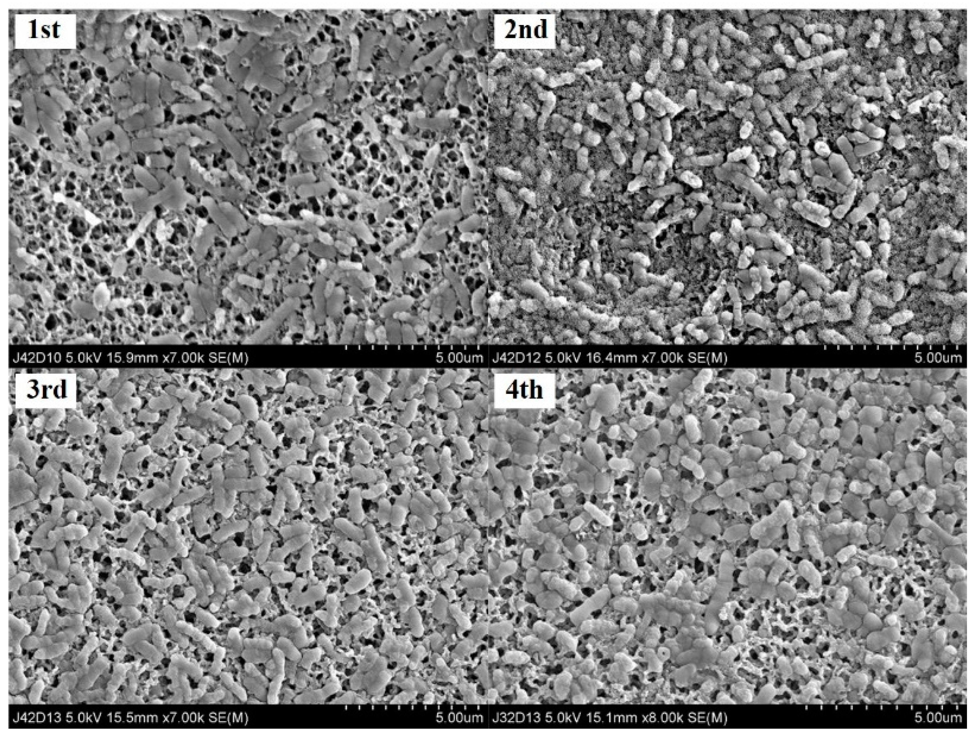
The indigenous acidophilic bacteria used in this study were collected from hot spring water (pH 3.36) near the Hatchōbaru Geothermal Power Plant in Japan. The collected bacteria were inoculated and grown in an incubator (HST-101M, Hanst, Incheon, Korea) at 32 °C to develop a growth culture medium containing inorganic components and electron donors. To prepare the growth culture medium, 0.2 g of (NH4)2SO4, 0.5 g of MgSO4·2H2O, 0.25 g of CaCl2, and 3.0 g of KH2PO4 were mixed with the inorganic components in 1.0 L of sterile triple distilled water (pH 5.12). As electron donors, 1.0 g/L of elemental sulfur powder and 5.0 mg/L of FeSO4·7H2O were used [20]. Bacterial adaptation experiments were performed to isolate indigenous acidophilic bacteria, adapt bacteria in growth culture medium, and secure biomass used for bioleaching experiments. In the bacterial adaptation experiment, 10 mL of the culture medium was collected from the growth culture medium cultured for 3 weeks (21 days) and inoculated into a new growth culture medium of the same chemical composition. The bacterial adaptation experiment was performed a total of 4 times. In addition, in order to investigate the shape of the adapted bacteria, 10 mL of the culture solution was collected after the adaptation experiment. The collected culture was filtered using GF/C with a pore size of 0.2 μm, and the filtered bacteria were observed using SEM-EDS (Figure 1).
In order to identify the biological characteristics of indigenous acidophilic bacteria, microbial identification and isolation and pure culture were commissioned by a specialized company (Cosmo Genetech, Seoul, Korea). For preactivation, 10 μL of indigenous acidophilic bacteria samples were collected from adaptation experiments and then inoculated into 3 mL of broth medium and cultured (aerobic, 32 °C, 6 days). An amount of 100 μL of the preactivated culture medium was smeared on a solid medium using a sterile spreader, and colonies were identified through culture (Aerobic, 32 °C, 6 days). Pure culture was performed while picking the colony of the target strain, followed by pure culture (repeated 3 times) and strain reconfirmation in the same medium and culture conditions. Microbial identification was performed by macroscopic and microscopic analysis, microbial identification sequencing (27F, 1492R), and align and blast. As a result, the similarity to the standard strain Acidiphilium cryptum (ATCC 33463) was 99.56%.
2.3. Bioleaching Experiments
In the bioleaching experiment, 3 g of the mineral sample was mixed as an electron donor in a 500 mL Erlenmeyer flask, following which 200 mL of a growth medium, including 10 mL of indigenous acidophilic bacterial solution, was injected and stirred at 32°C and 300 rpm. To compare the chemical leaching efficiencies, an abiotic control experiment (using a sample without bacteria) was performed under the same conditions as the bioleaching experiment. The initial pH of the test solution was adjusted to 5.12 using HNO3 and NaOH. 10 mL of the elution solution was collected, and the metal concentration eluted during the experiment was measured. After collection, 10 mL of the new growth culture solution were added. The collected solution sample was filtered with 0.45 μm filter paper to remove suspended matter in the sample. After measuring the pH of the filtered sample using a pH meter (Eijkelkamp 18.28 Multiparameter Analyzer, Giesbeek, The Netherlands), acid treatment was performed using nitric acid prior to metal concentration analysis. The concentration of the eluted metal in the acid-treated sample was measured using an atomic absorption spectrophotometer (AA-7000, Shimadzu, Tokyo, Japan).
The metal leaching properties in the leachate according to time-dependent biological and chemical reactions were quantified using the following equation:
where E (mg/L) is the leaching concentration at time t (day), EI (mg/L) is the maximum leaching concentration, and k (day−1) is the leaching rate constant.
E = EI × (1 − e−kt)
3. Results and Discussion
3.1. Composition Analysis of Various Sulfide Mineral Samples
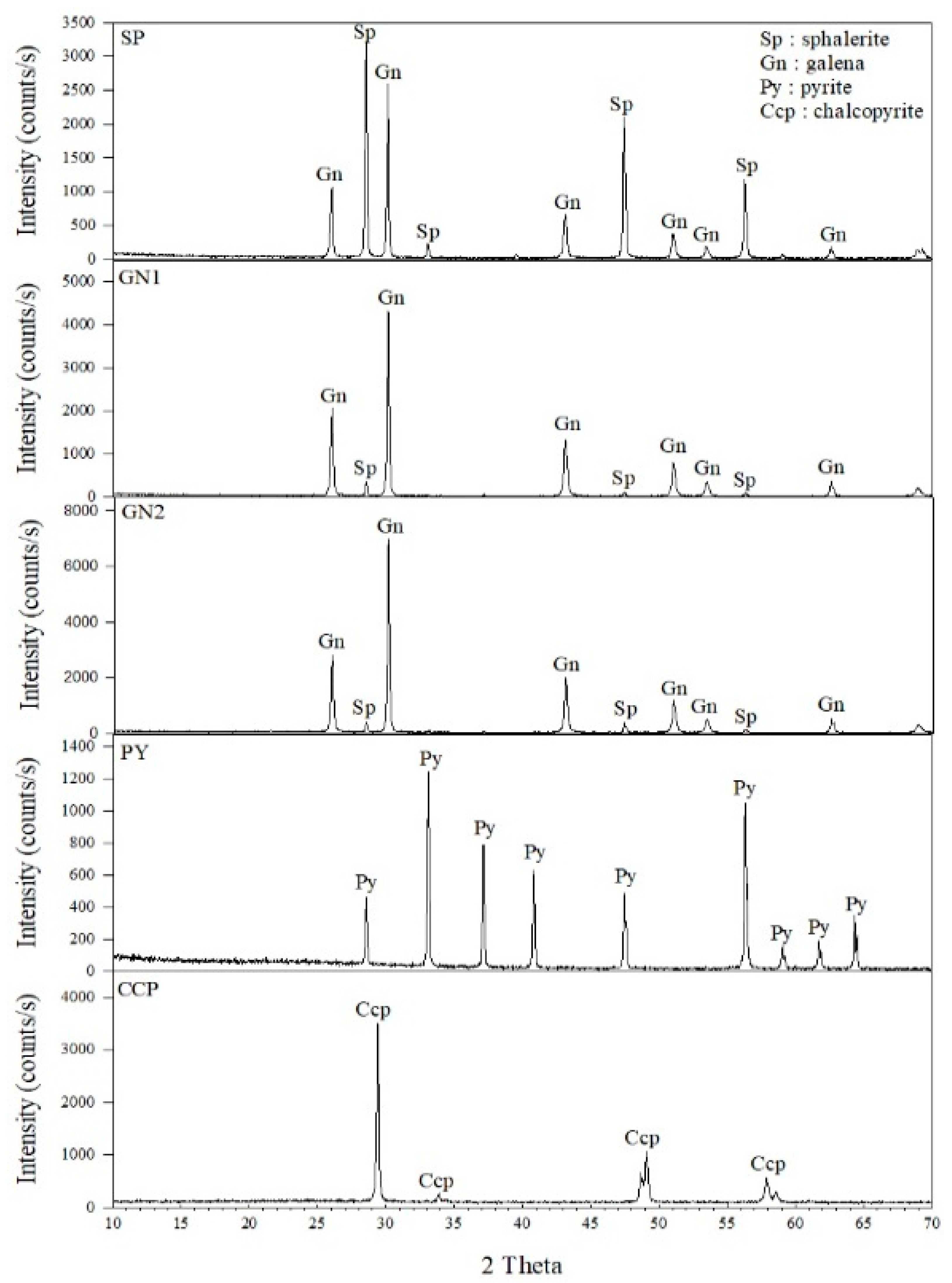
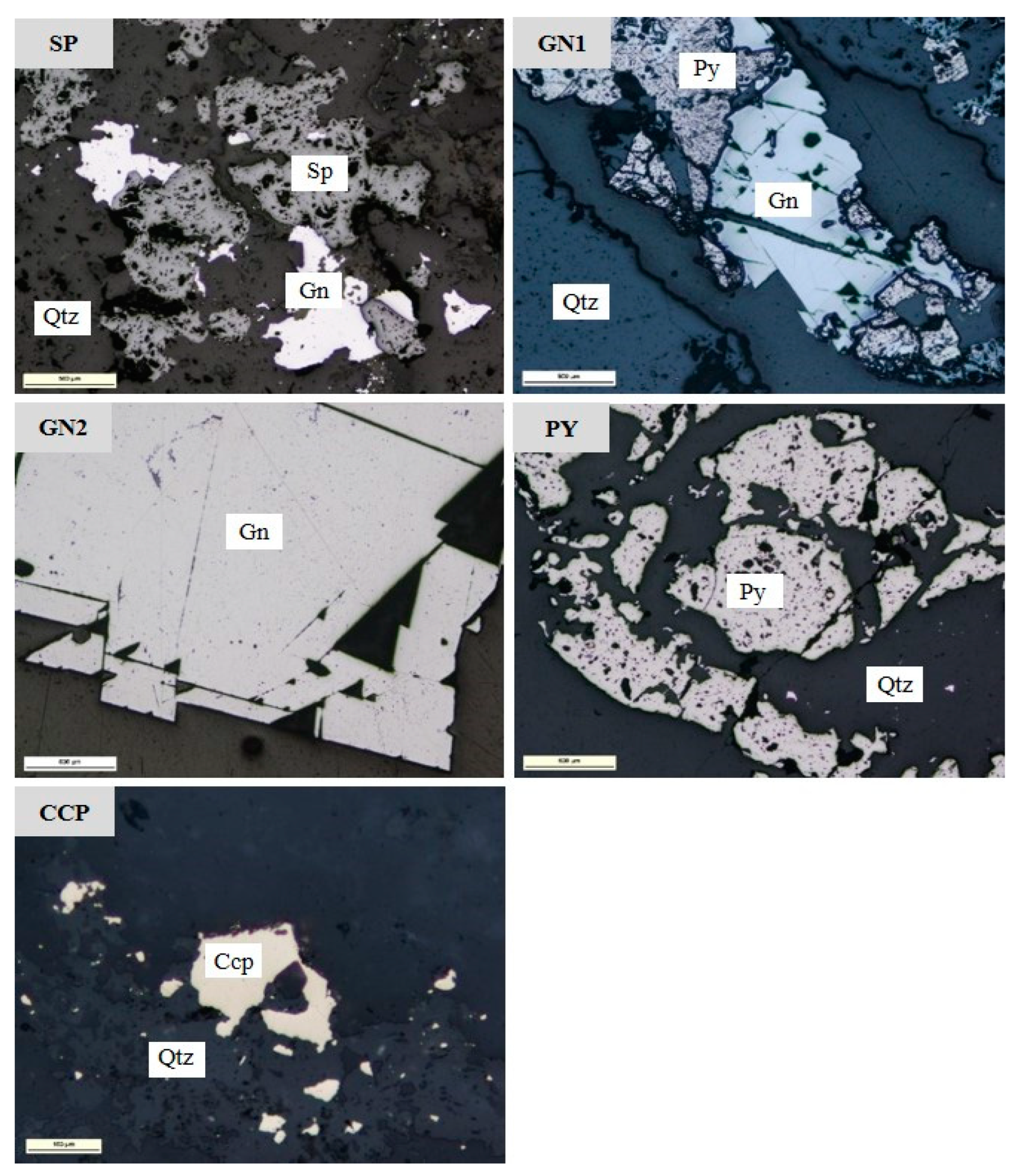
The XRD and reflecting microscope results for each sample are shown in Figure 2 and Figure 3, respectively. Based on the XRD results, the content of major metal minerals in the sample was analyzed using the Match program (Match!, Crystal Impact, Bonn, Germany) (Table 1). The SP samples were composed of 78% sphalerite and 21% galena. In the GN1 and GN2 samples, galena was found to be present in 57% and 67%, respectively, while sphalerite was present in 42% and 32%, respectively. The primary mineral of the PY sample was pyrite (>95%), and that of the CCP sample was chalcopyrite (>95%). These results were also confirmed by the reflecting microscope image (Figure 3). Sphalerite, galena, and quartz were observed on the SP surface, while only pyrite and chalcopyrite were identified on the PY and CCP surfaces, except for quartz. In the GN2 sample, it was confirmed that the distribution of galena significantly increased compared to the GN1 sample. As such, since this study aimed to compare the bioleaching characteristics of various sulfide minerals based on their composition, it has a limitation in that the bulk contents of each element could not be measured.
3.2. Comparison of pH Change during Bioleaching of Mineral Samples
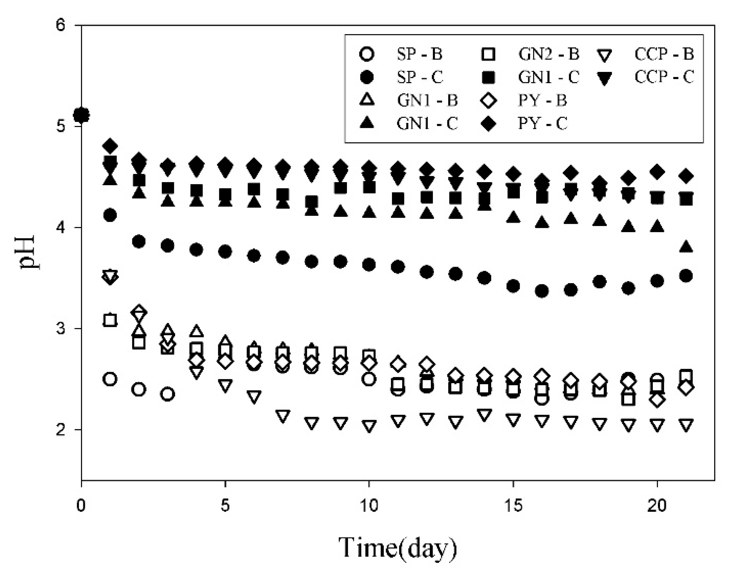
Figure 4 shows the change in the pH of the leachate during the chemical and biological oxidation of the mineral samples. During the 21 d leaching experiment, the pH decreased for the abiotic control case because of the consumption of H+ ions in the growth culture medium by the inorganic oxidation of sulfide minerals (shown as -C in Figure 4). For the bacterial samples (shown as -B in Figure 4), the change in the pH of the leachate was lower than that in the abiotic control samples for all sample conditions. The pH decreased rapidly at the beginning of the experiment and then decreased slowly after 7 d. The pH was lower than 3 in all samples after 3 d of the bioleaching experiment. This is because metal sulfides oxidatively solubilize sulfur and metals due to bioleaching processes, which reduce the pH and enhance the solubilization of additional metal compounds [21]. In addition, as the pH in the leachate decreased from 5.11 to less than 3, the bacterial samples exhibited a pH below the point of zero charge (PZC) [22,23,24]. Therefore, the surface charge of the mineral sample changed from negative to positive, and the negatively charged bacteria in the leachate came into contact with the surface of the mineral sample due to electrical attraction. This suggests the possibility of direct biological oxidation. The PZC of the mineral samples used in this study was confirmed in other studies [25]. The PZCs of sphalerite, galena, and pyrite were 4–5 in a study on sulfide mineral flotation using recycled water. Liang et al. [26] showed that the PZC of chalcopyrite in a bioleaching experiment using Sulfolobus metallicus YN24 was 3.80, and the PZC of pyrite was 2.7 [27].
The decrease in pH (ΔpH) for the bacterial samples compared to abiotic samples is summarized in Table 2. This estimation was based on the measured data for the change in the leachate pH through the chemical and biological oxidation of various mineral samples. The maximum decreasing pH (ΔpH) was estimated to be approximately 2.62–3.01, and there was no difference in ΔpH that correlated to the characteristics of the mineral samples. However, the pH decrease rate was highest in the SP sample (sphalerite content: 78%) and lowest in the CCP sample (chalcopyrite content: >95%). Similarly, Watling [28] reported that the efficiency of heap/dump leaching is highly dependent on the constituent minerals of the ore. The leaching of ores takes several months for chalcocite (Cu2S) and covellite (CuS), whereas it takes years for CCP.
3.3. Metal Extraction in Bioleaching and the Bioleaching Mechanism Based on Sulfide Minerals
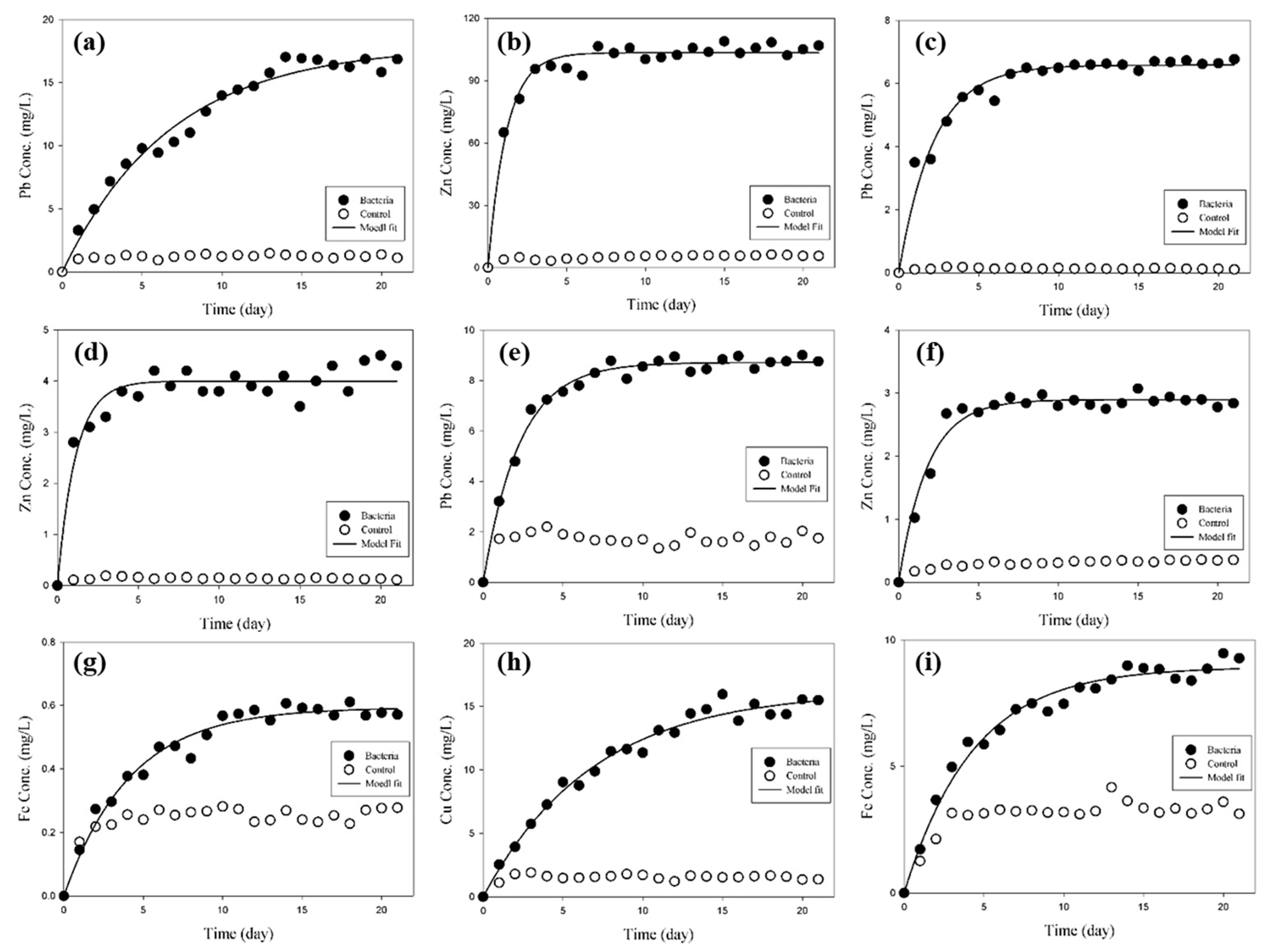
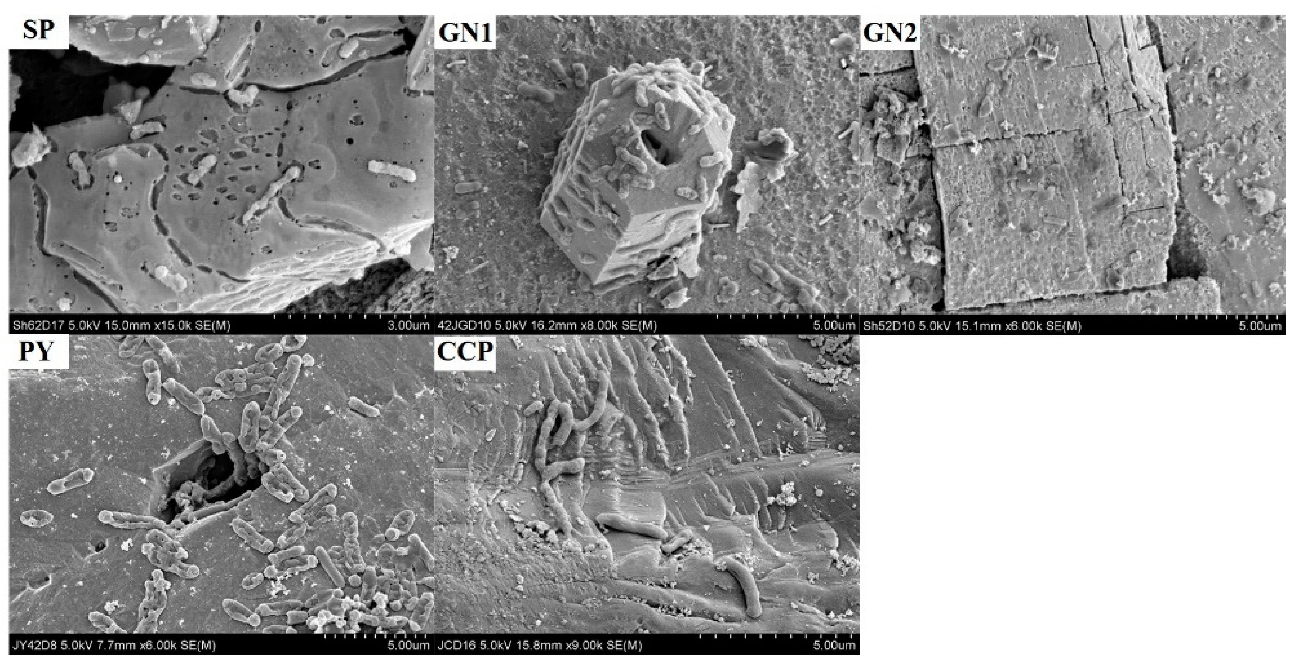
Figure 5 shows the results for the metal concentration leached over time in the leaching experiment. Although the metal extraction in bioleaching acts on the mineral surface and results in low metal concentrations in the leachate, those with bacteria showed a higher leaching concentration than the abiotic control case for all mineral samples. This is due to biological leaching by biological oxidation, including biological direct contact oxidation and chemical oxidation of the mineral samples. In order to confirm the direct oxidation in the bioleaching experiment, SEM-EDS analysis was performed on the leaching residue after the experiment was completed (Figure 6). As a result, bacteria were attached to the surface of the residue under all mineral sample conditions. Corrosion action (direct oxidation) was observed on the surface of bacterial residues in SP and GN1 samples. In the case of the SP and PY samples, a large number of bacteria were attached to the imperfections, crevasses, and pores of the residue surface. Meanwhile, trace elements that may be present in the mineral samples may inhibit microbial biogeochemical activity [29], but their effects were not considered in this study.
The metal extraction in bioleaching differed depending on the mineral content of the samples (Table 2). The leaching concentration was higher for Zn (103.40 mg/L) than for Pb (17.90 mg/L) in SP with a high ZnS content (78%). In GN1 and GN2 with high PbS content (57% and 67%), Pb (6.58 mg/L and 8.72 mg/L) showed higher concentrations than Zn (4.00 mg/L and 2.90 mg/L). These results confirm that as the composition ratio of PbS increased in GN2, the metal extraction in bioleaching of Pb also increased compared to GN1, and the opposite result was shown in the case of Zn. Nonetheless, the leaching rate of Zn was faster than that of Pb (Table 3), and this trend was also observed in previous studies. According to literature studies of heavy metal leaching on mine tailings using indigenous sulfur-oxidizing bacteria, the leaching rate of Zn was about 2.2 times higher than that of Pb during the same leaching time [30,31]. The leaching concentration of Fe in PY was minimal (0.59 mg/L) because the surface accumulation of Fe2+ could result in inhibition of the oxidizing capacity of microorganisms [32]. In the results of biological leaching experiments on low grade sulfide minerals by Ilyas et al. [33], the leaching concentrations over the same leaching time were in the order of Ni > Zn > Fe > Cu = Co. In a biological leaching study on lead zinc mine tailings consisting of complex minerals such as sphalerite, pyrite, calcite, and quartz, Zn leaching rates were 2.74 times (10% pulp density) and 9.25 times (20% pulp density) higher than the Fe leaching rate [34]. Meanwhile, some precipitates, such as anglesite, may be formed during the bioleaching process [35,36], but they were not considered in this study.
Based on the above results, the bioleaching mechanism based on sulfide minerals was able to be categorized into the following reactions: indirect biological oxidation (Equation (8)) by increased bacterial activity, chemical oxidation derived from reduced solution pH (Figure 4 and Figure 5), and consequently direct bacterial oxidation (Equations (2)–(7)) under low PZC (Figure 6) [37,38,39]. Therefore, the rate of pH decrease is faster than that of metal leaching because of the reaction time required to increase the bacterial activity in the leachate and the direct oxidation by bacteria at low PZC. In a comparative study on the bioleaching pyrrhotite at PZC 3.0 for direct bacterial oxidation (pH 3.2) and direct/indirect bacterial oxidation (pH 2.8), the maximum leaching concentration of Fe was 2.08 times (44.2 mg/L) higher in direct/indirect bacterial oxidation than in direct oxidation (21.3 mg/L) [40].
Sphalerite:
ZnS + 0.5O2 + H2SO4 → ZnSO4 + H2O + S
Galena:
PbS + 0.5O2 + H2SO4 → PbSO4 + H2O + S
Pyrite:
FeS2 + 3.5O2 + H2O → FeSO4 + H2SO4
Chalcopyrite:
CuFeS2 + O2 + 2H2SO4 → CuSO4 + FeSO4 + 2H2O + 2S
2S + 3O2 + 2H2O → 2H2SO4
4FeSO4 + O2 + 2H2SO4 → 2Fe2(SO4)3 + 2H2O
(Zn Pb Fe Cu)S + Fe2(SO4)3 → (Zn Pb Fe Cu)SO4 + 2FeSO4 + S
4. Conclusions
In this study, indigenous acidophilic bacteria were used for bioleaching experiments on various sulfide mineral samples, including sphalerite, galena, pyrite, and chalcopyrite. For the bacterial samples, the pH change in the leachate was lower than that in the abiotic control case for all samples. After 3 d of the experiment, the pH of the leachate in all samples was below the PZC, which was an experimental condition for direct biological oxidation. Obviously, the leached metal concentration of Zn was higher than that of Pb for the SP sample with a high ZnS content, whereas the concentration of Pb was higher than that of Zn for the GN1 and GN2 samples with a high PbS content. Meanwhile, the leaching rate of Zn was faster than that of Pb for all samples. Bacteria used in the bioleaching experiment were able to attach to the residue surface under all mineral sample conditions, and corrosive action was also observed. These results suggest that the bioleaching mechanism based on sulfide minerals proceeds through indirect biological oxidation, chemical oxidation, and direct bacterial oxidation. This study provides valuable insights into the direct and indirect oxidation mechanisms involved in the biological leaching of various sulfide minerals, which can guide further research and practical applications in this field.
Author Contributions
K.-H.C.: Formal analysis, Methodology; H.-S.K.: Validation; C.-G.L.: writing—review and editing; S.-J.P.: Supervision; N.-C.C.: Conceptualization, writing—review and editing. All authors have read and agreed to the published version of the manuscript.
Funding
This work was supported by R&D Project of the Korea Mine Rehabilitation and Mineral Resources Corporation in 2023.
Institutional Review Board Statement
Not applicable.
Informed Consent Statement
Not applicable.
Data Availability Statement
Not applicable.
Conflicts of Interest
The authors declare no conflict of interest.
References
- Brierley, J.; Brierley, C. Present and future commercial applications of biohydrometallurgy. Hydrometallurgy 2001, 59, 233–239. [Google Scholar] [CrossRef]
- Marsden, J.; House, I. The Chemistry of Gold Extraction; SME: Littleton, CO, USA, 2006. [Google Scholar]
- Nkuna, R.; Ijoma, G.N.; Matambo, T.S.; Chimwani, N. Accessing Metals from Low-Grade Ores and the Environmental Impact Considerations: A Review of the Perspectives of Conventional versus Bioleaching Strategies. Minerals 2022, 12, 506. [Google Scholar] [CrossRef]
- Vyas, S.; Ting, Y.-P. Sequential biological process for molybdenum extraction from hydrodesulphurization spent catalyst. Chemosphere 2016, 160, 7–12. [Google Scholar] [CrossRef] [PubMed]
- Brierley, J.A. Response of microbial systems to thermal stress in biooxidation-heap pretreatment of refractory gold ores. Hydrometallurgy 2003, 71, 13–19. [Google Scholar] [CrossRef]
- Groudev, S.; Groudeva, V. Microbial communities in four industrial copper dump leaching operations in Bulgaria. FEMS Microbiol. Rev. 1993, 11, 261–267. [Google Scholar] [CrossRef]
- Jia, Y.; Sun, H.; Tan, Q.; Xu, J.; Feng, X.; Ruan, R. Industrial Heap Bioleaching of Copper Sulfide Ore Started with Only Water Irrigation. Minerals 2021, 11, 1299. [Google Scholar] [CrossRef]
- Kang, J.-K.; Cho, K.-H.; Kim, S.-B.; Choi, N.-C. Artificial Neural Network Modeling for Prediction of Dynamic Changes in Solution from Bioleaching by Indigenous Acidophilic Bacteria. Appl. Sci. 2020, 10, 7569. [Google Scholar] [CrossRef]
- Tributsch, H. Direct versus indirect bioleaching. Hydrometallurgy 2001, 59, 177–185. [Google Scholar] [CrossRef]
- Sand, W.; Schippers, A.; Hedrich, S.; Vera, M. Progress in bioleaching: Fundamentals and mechanisms of microbial metal sulfide oxidation–Part A. Appl. Microbiol. Biotechnol. 2022, 106, 6933–6952. [Google Scholar]
- Chaudhury, G.R.; Sukla, L.; Das, R. Kinetics of biochemical leaching of sphalerite concentrate. Metall. Trans. B 1985, 16, 667–670. [Google Scholar] [CrossRef]
- Blancarte-Zurita, M. Application of a Shrinkage Particle Model to the Kinetics of Microbiological Leaching. In Fundamental and Applied Biohydrometallurgy; Lawrence, R.W., Branion, R.M.R., Ebner, H.G., Eds.; Elsevier Science Publishers: Amsterdam, The Netherlands, 1985; pp. 243–253. [Google Scholar]
- Martello, D.; Vecchio, K.; Diehl, J.; Graham, R.; Tamilia, J.; Pollack, S. Do dislocations and stacking faults increase the oxidation rate of pyrites? Geochim. Cosmochim. Acta 1994, 58, 4657–4665. [Google Scholar] [CrossRef]
- Rojas-Chapana, J.A.; Tributsch, H. Interfacial activity and leaching patterns of Leptospirillum ferrooxidans on pyrite. FEMS Microbiol. Ecol. 2004, 47, 19–29. [Google Scholar] [CrossRef]
- Deng, S.; Gu, G. Insight into the Influence of Mineralogical Properties of Pristine Pyrite on Its Bioleaching with Thermophiles. Miner. Process. Extr. Metall. Rev. 2022, 43, 733–738. [Google Scholar] [CrossRef]
- Murr, L.; Berry, V. Direct observations of selective attachment of bacteria on low-grade sulfide ores and other mineral surfaces. Hydrometallurgy 1976, 2, 11–24. [Google Scholar] [CrossRef]
- Ohmura, N.; Kitamura, K.; Saiki, H. Selective adhesion of Thiobacillus ferrooxidans to pyrite. Appl. Environ. Microbiol. 1993, 59, 4044–4050. [Google Scholar] [CrossRef]
- Joshi, V.; Shah, N.; Wakte, P.; Dhakephalkar, P.; Dhakephalkar, A.; Khobragade, R.; Naphade, B.; Shaikh, S.; Deshmukh, A.; Adhapure, N. Comparative bioleaching of metals from pulverized and non-pulverized PCBs of cell phone charger: Advantages of non-pulverized PCBs. Environ. Sci. Pollut. Res. Int. 2017, 24, 28277–28286. [Google Scholar] [CrossRef]
- Lee, G.; Kim, K. Evaluation of Feasibility of Chemical Washing Treatment of Nickel in Deep-sea Mining Tailings by Aluminum Sulfate as Extractant. J. Korean Soc. Environ. Eng. 2022, 44, 77–85. [Google Scholar] [CrossRef]
- Mahmood, M.; Turner, A. The selective leaching of zinc from chalcopyrites-phalerite concentrates using slurry electrodes. Hydrometallurgy 1985, 14, 317–329. [Google Scholar]
- Fang, D.; Zhao, L.; Yang, Z.; Shan, H.; Gao, Y.; Yang, Q. Effect of sulphur concentration on bioleaching of heavy metals from contaminated dredged sediments. Environ. Technol. 2009, 30, 1241–1248. [Google Scholar] [CrossRef]
- Kim, S.-B.; Park, S.-J.; Lee, C.-G.; Choi, N.-C.; Kim, D.-J. Bacteria transport through goethite-coated sand: Effects of solution pH and coated sand content. Colloids Surf. B Biointerfaces 2008, 63, 236–242. [Google Scholar]
- Kim, S.B.; Park, S.J.; Lee, C.G.; Kim, H.C. Transport and retention of Escherichia coli in a mixture of quartz, Al-coated and Fe-coated sands. Hydrol. Process. Int. J. 2008, 22, 3856–3863. [Google Scholar] [CrossRef]
- Kang, J.-K.; Lee, C.-G.; Park, J.-A.; Kim, S.-B.; Choi, N.-C.; Park, S.-J. Adhesion of bacteria to pyrophyllite clay in aqueous solution. Environ. Technol. 2013, 34, 703–710. [Google Scholar] [CrossRef] [PubMed]
- Bulut, G.; Yenial, Ü. Effects of major ions in recycled water on sulfide minerals flotation. Miner. Metall. Process. 2016, 33, 137–143. [Google Scholar] [CrossRef]
- Liang, Y.-t.; Zhu, S.; Wang, J.; Ai, C.-b.; Qin, W.-q. Adsorption and leaching of chalcopyrite by Sulfolobus metallicus YN24 cultured in the distinct energy sources. Int. J. Miner. Metall. Mater. 2015, 22, 549–552. [Google Scholar] [CrossRef]
- Widler, A.; Seward, T. The adsorption of gold (I) hydrosulphide complexes by iron sulphide surfaces. Geochim. Cosmochim. Acta 2002, 66, 383–402. [Google Scholar] [CrossRef]
- Watling, H. The bioleaching of sulphide minerals with emphasis on copper sulphides—A review. Hydrometallurgy 2006, 84, 81–108. [Google Scholar] [CrossRef]
- Mahowald, N.M.; Hamilton, D.S.; Mackey, K.R.M.; Moore, J.K.; Baker, A.R.; Scanza, R.A.; Zhang, Y. Aerosol trace metal leaching and impacts on marine microorganisms. Nat. Commun. 2018, 9, 2614. [Google Scholar] [CrossRef]
- Liu, Y.-G.; Zhou, M.; Zeng, G.-M.; Li, X.; Xu, W.-H.; Fan, T. Effect of solids concentration on removal of heavy metals from mine tailings via bioleaching. J. Hazard. Mater. 2007, 141, 202–208. [Google Scholar] [CrossRef]
- Liu, Y.-G.; Zhou, M.; Zeng, G.-M.; Wang, X.; Li, X.; Fan, T.; Xu, W.-H. Bioleaching of heavy metals from mine tailings by indigenous sulfur-oxidizing bacteria: Effects of substrate concentration. Bioresour. Technol. 2008, 99, 4124–4129. [Google Scholar] [CrossRef]
- Rodrıguez, Y.; Ballester, A.; Blazquez, M.; González, F.; Munoz, J. New information on the pyrite bioleaching mechanism at low and high temperature. Hydrometallurgy 2003, 71, 37–46. [Google Scholar] [CrossRef]
- Ilyas, S.; Bhatti, H.N.; Bhatti, I.A.; Sheikh, M.A.; Ghauri, M.A. Bioleaching of metal ions from low grade sulphide ore: Process optimization by using orthogonal experimental array design. Afr. J. Biotechnol. 2010, 9, 2801–2810. [Google Scholar]
- Ye, M.; Li, G.; Yan, P.; Ren, J.; Zheng, L.; Han, D.; Sun, S.; Huang, S.; Zhong, Y. Removal of metals from lead-zinc mine tailings using bioleaching and followed by sulfide precipitation. Chemosphere 2017, 185, 1189–1196. [Google Scholar] [CrossRef]
- Mejía, E.; Ospina, J.; Márquez, M.; Morales, A. Bioleaching of Galena (PbS). In Fourier Transform-Materials Analysis; Salih, S.M., Ed.; BoD—Books on Demand: Norderstedt, Germany, 2012; pp. 185–206. [Google Scholar]
- Chaerun, S.K.; Putri, E.A.; Mubarok, M.Z. Bioleaching of Indonesian galena concentrate with an iron-and sulfur-oxidizing mixotrophic bacterium at room temperature. Front. Microbiol. 2020, 11, 557548. [Google Scholar] [CrossRef]
- Bosecker, K. Bioleaching: Metal solubilization by microorganisms. FEMS Microbiol. Rev. 1997, 20, 591–604. [Google Scholar] [CrossRef]
- Rouchalova, D.; Rouchalova, K.; Janakova, I.; Cablik, V.; Janstova, S. Bioleaching of iron, copper, lead, and zinc from the sludge mining sediment at different particle sizes, pH, and pulp density using Acidithiobacillus ferrooxidans. Minerals 2020, 10, 1013. [Google Scholar] [CrossRef]
- Kim, S.; Eom, H.; Kang, W.; Oh, S.-E. Evaluation of Individual and Mixture Toxicity of Heavy Metals using Semi-continuous Type Sulfur Oxidizing Bacteria (SOB) Bioreactor. J. Korean Soc. Environ. Eng. 2022, 44, 225–234. [Google Scholar] [CrossRef]
- Kim, B.-J.; Koh, Y.-K.; Kwon, J.-S. Bioleaching of pyrrhotite with bacterial adaptation and biological oxidation for iron recovery. Metals 2021, 11, 295. [Google Scholar] [CrossRef]
Figure 1.
SEM image of the adapted bacteria (4 times).

Figure 2.
XRD analysis results of mineral samples used in bioleaching experiments.

Figure 3.
Reflecting microscope image of mineral samples used in bioleaching experiments (Sp: sphalerite, Gn: galena, Py: pyrite, Ccp: chalcopyrite, Qtz: quartz).
Figure 3.
Reflecting microscope image of mineral samples used in bioleaching experiments (Sp: sphalerite, Gn: galena, Py: pyrite, Ccp: chalcopyrite, Qtz: quartz).

Figure 4.
pH change in leachate according to reaction time under various mineral sample conditions (bacterial samples denoted “-B” and control samples denoted “-C”).
Figure 4.
pH change in leachate according to reaction time under various mineral sample conditions (bacterial samples denoted “-B” and control samples denoted “-C”).

Figure 5.
Changes in metal concentration in leachate under various mineral sample conditions ((a,b) SP, (c,d) GN1, (e,f) GN2, (g) PY, and (h,i) CCP).
Figure 5.
Changes in metal concentration in leachate under various mineral sample conditions ((a,b) SP, (c,d) GN1, (e,f) GN2, (g) PY, and (h,i) CCP).

Figure 6.
SEM image of the leaching residue surface with attached bacteria after the bioleaching experiment.
Figure 6.
SEM image of the leaching residue surface with attached bacteria after the bioleaching experiment.


Table 1.
Match! phase analysis results of mineral samples used in bioleaching experiments.
| Samples | Matched Phases | Quantity (wt %) |
|---|---|---|
| SP | PbS | 21 |
| ZnS | 78 | |
| GN1 | PbS | 57 |
| ZnS | 42 | |
| GN2 | PbS | 67 |
| ZnS | 32 | |
| PY | FeS2 | >95 |
| CCP | CuFeS2 | >95 |
Table 2.
pH reduction and leaching metal concentrations derived from bioleaching experiments using an indigenous acidophilic bacterium (Acidiphilium cryptum).
Table 2.
pH reduction and leaching metal concentrations derived from bioleaching experiments using an indigenous acidophilic bacterium (Acidiphilium cryptum).
| Sample | ΔpH | Elements | Leaching Concentration (mg/L) |
|---|---|---|---|
| SP | 2.62 | Pb | 17.90 |
| Zn | 103.40 | ||
| GN1 | 2.49 | Pb | 6.58 |
| Zn | 4.00 | ||
| GN2 | 2.55 | Pb | 8.72 |
| Zn | 2.90 | ||
| PY | 2.55 | Fe | 0.59 |
| CCP | 3.01 | Cu | 16.23 |
| Fe | 8.93 |
Table 3.
Estimation of leaching parameters by bioleaching using Equation (1).
| Sample | Elements | EI (mg/L) | K (1/day) | R2 |
|---|---|---|---|---|
| SP | Pb | 17.90 | 0.15 | 0.98 |
| Zn | 103.40 | 0.86 | 0.97 | |
| GN1 | Pb | 6.58 | 0.46 | 0.96 |
| Zn | 4.00 | 0.89 | 0.91 | |
| GN2 | Pb | 8.72 | 0.44 | 0.99 |
| Zn | 2.90 | 0.56 | 0.97 | |
| PY | Fe | 0.59 | 0.24 | 0.97 |
| CCP | Cu | 16.23 | 0.15 | 0.98 |
| Fe | 8.93 | 0.23 | 0.98 |
Disclaimer/Publisher’s Note: The statements, opinions and data contained in all publications are solely those of the individual author(s) and contributor(s) and not of MDPI and/or the editor(s). MDPI and/or the editor(s) disclaim responsibility for any injury to people or property resulting from any ideas, methods, instructions or products referred to in the content. |
© 2023 by the authors. Licensee MDPI, Basel, Switzerland. This article is an open access article distributed under the terms and conditions of the Creative Commons Attribution (CC BY) license (https://creativecommons.org/licenses/by/4.0/).
Share and Cite
MDPI and ACS Style
Cho, K.-H.; Kim, H.-S.; Lee, C.-G.; Park, S.-J.; Choi, N.-C. A Comparative Study on Bioleaching Properties of Various Sulfide Minerals Using Acidiphilium cryptum. Appl. Sci. 2023, 13, 5997. https://doi.org/10.3390/app13105997
AMA Style
Cho K-H, Kim H-S, Lee C-G, Park S-J, Choi N-C. A Comparative Study on Bioleaching Properties of Various Sulfide Minerals Using Acidiphilium cryptum. Applied Sciences. 2023; 13(10):5997. https://doi.org/10.3390/app13105997
Chicago/Turabian StyleCho, Kang-Hee, Hyun-Soo Kim, Chang-Gu Lee, Seong-Jik Park, and Nag-Choul Choi. 2023. "A Comparative Study on Bioleaching Properties of Various Sulfide Minerals Using Acidiphilium cryptum" Applied Sciences 13, no. 10: 5997. https://doi.org/10.3390/app13105997
Note that from the first issue of 2016, this journal uses article numbers instead of page numbers. See further details here.