Effects of Cadmium Exposure on Lactating Mice and Rats: A Systematic Review of Breastfeeding Experiments

 , and
, and
Abstract
:Featured Application
Abstract
1. Introduction
2. Materials and Methods
2.1. Study Design
- Population: Mice and/or rats in lactating period
- Exposure: Cd administered to dams and lactating animals exposed by breastfeeding
- Control: Animal not exposed to Cd
- Outcomes: Effects on litters exposed to Cd by breastfeeding
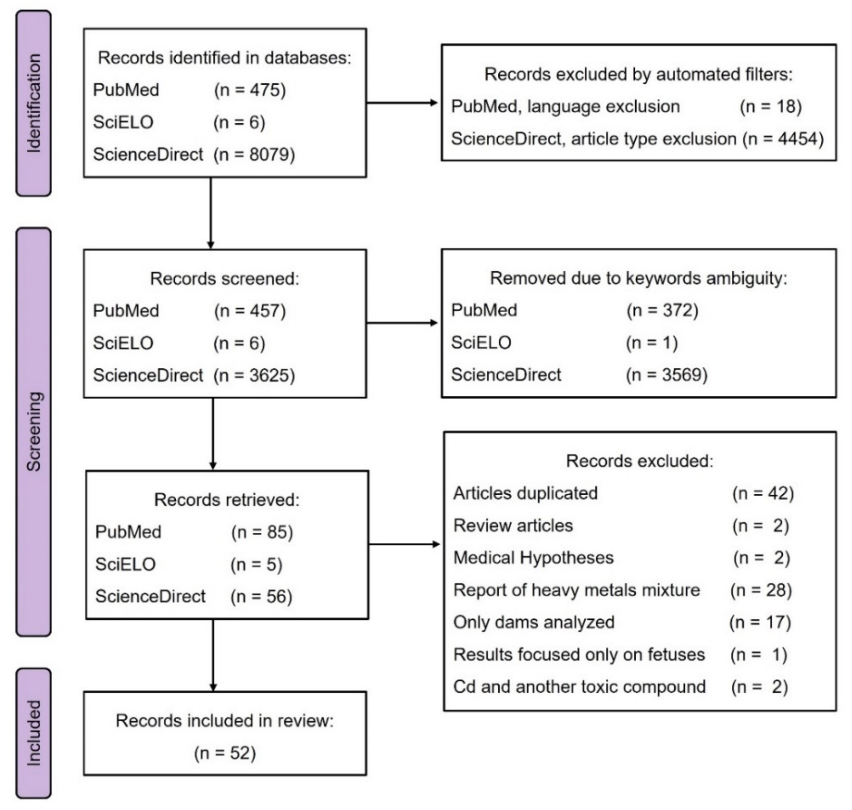
2.2. Search Strategy and Selection Process
Inclusion and Exclusion Criteria
2.3. Data Extraction and Results Synthesis
3. Results and Discussion
3.1. Selected Studies
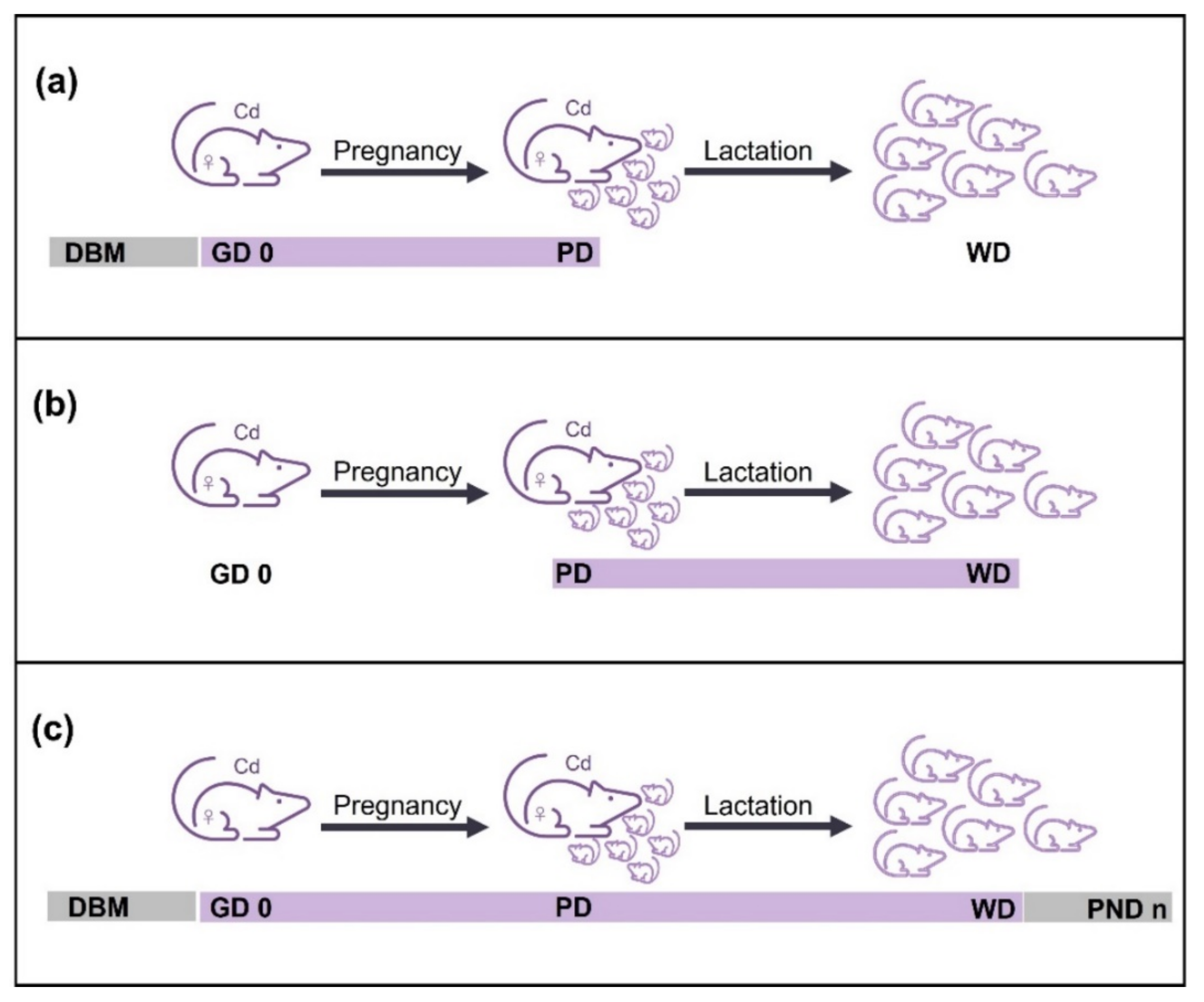
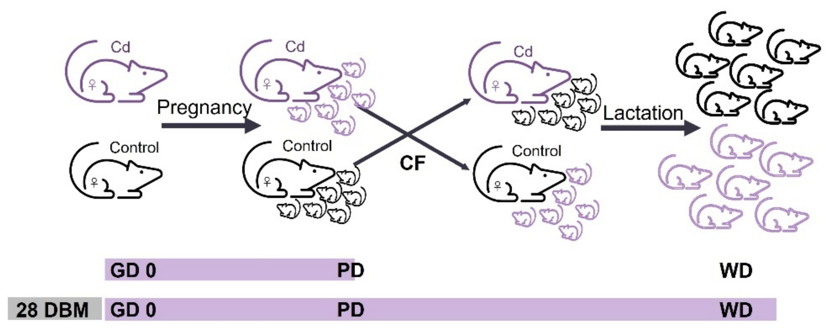
3.2. Cd Exposure Schemes
3.2.1. Cd Exposure of Dams during Pregnancy Period
Effects of Cd Exposure during Pregnancy Period

| Strain (n) Dose/Concentration, Admin Route Exposure Period Litter (n), Lactation Days | Collection Time Organ Analyzed | Results | Year Author |
|---|---|---|---|
| Wistar rats (n = 6) 50 mg CdCl2/L, d.w. gestation (n = 6), 21 | PD and PND 21 Brain | Increased acetylcholinesterase and Na+, K+-ATPase activities in the newborn rat brain that were ameliorated through a Cd-free lactation. | 2013 Liapi et al. ‡ [14] |
| Wistar rats (n = 12) 20 mg CdCl2/kg bw, i.g GD 6 to GD 14 (n = 8), 21 | GD 20 Fetuses PND 1 to 180 Physical and behavioral parameters | External malformations in fetuses at GD 20 Improved testes descent, delayed vaginal opening, and modified male and female sexual behavior | 2004 Salvatori et al. [17] |
| Wistar rats (---) 14, 7, and 3.5 mg CdCl2/kg bw, i.g. GD 5 to GD 15 (n = 8), 28 | PND 84 Behavioral and neurotoxicological analysis | Cd dose dependently altered the spontaneous and evoked electrophysiological functions | 1998 Dési et al. ‡ [15] |
| Sprague-Dawley rats (n = 6–7) 2.5 mg 109CdCl2/kg bw, i.p. GD 8, 10, 12 and 14 (n = 10), 12, CF | PND 3, and 12 Kidney, liver, and gastrointestinal tract | Decreases renal alkaline phosphatase activity determined in pups fed by their biological mothers exposed to Cd. 109Cd in liver at birth and in the gastrointestinal tract at PND 12 of newborns fed by their mothers. Prenatal Cd exposure cannot induce alone kidney damage | 1992 Saillenfait et al. [12] |
| Rats (---) 2 mg Cd(NO3)2/kg bw, i.g. GD 6 to GD 15 (n = 10), --- | PND 120 Blood | At PND 120 increased plasma and erythrocyte levels of MDA, reduced glutathione, activities of glutathione-S-transferase, glutathione reductase, and γ-glutamyl transferase. Effects less pronounced than lactation exposure. | 2008 Slyuzova et al. ‡ [13] |
| Wistar rats (---) 1 mg CdCl2/kg bw, i.v. GD 18 (n = 8), 24 | PND 2, 9, and 24 Blood, liver, brain, kidneys, spleen, pancreas, stomach, duodenum, and femur | Decreased Zn in liver and delayed Zn deposition in bone. Probably Cd increases the demand of Zn in the newborn. | 1981 Bakka et al. [18] |
| Wistar rats (---) 0.16 and 0.02 mg CdO/m3, 5 h daily, i.c. 140 DBM to GD 20 (---), 21 | PND 90 Behavioral tests | Reduction of exploratory motor activity. Central nervous system dysfunction. | 1984 Barański [16] |
3.2.2. Cd Exposure of Dams during Lactation Period
Effects of Cd Exposure during Lactation Period
3.2.3. Cd Exposure of Dams during Pregnancy–Lactation Period
| Strain (n) Dose/Concentration, Admin Route Exposure Period Litter (n), Lactation Days | Collection Time Organ Analyzed | Results | Year Author |
|---|---|---|---|
| Albino rats (n = 12) 100 mg CdCl2/L, d.w. GD 0 to PND 21 (n = 6), 21 | PND 7 and 126 Blood, liver, kidneys, and gut | Pups retained higher Cd-levels in blood, carcass, and gut at PND 7 than at PND 126. | 1979 Kostial et al. [39] |
| ICR mice (n = 10) 100 and 10 mg CdCl2/L, d.w. (23.7, 26.9, and 68.2 mg Cd/kg bw for 100 mg/L at DBM, GDs, and PNDs) ** (3.2, 3.4, and 9.3 mg Cd/kg bw for 10 mg/L at DBM, GDs, and PNDs) ** 28 DBM to PND 21 (n = 10), 21 | PND 21, 35 and 56 Blood and ovaries | Vaginal opening delay, irregular estrous cycle, inhibition of follicular development. The level of serum estradiol was reduced, and the mRNA levels of genes related to steroidogenesis were downregulated. | 2022 Li et al. [1] |
| Wistar rats (n = 6) 50 mg CdCl2/L, d.w. GD 0 to PND 21 (n = 6), 21 | PD and PND 21 Brain | Decrease in acetylcholinesterase activity. | 2013 Liapi et al. ‡ [14] |
| PND 21 Brain | Cd exposure modified the activities of acetylcholinesterase and Na+, K+-ATPase in the frontal cortex and cerebellum, and increased Mg2+ -ATPase activity in the hippocampus. | 2013 Stolakis et al. [40] | |
| Wistar rats (n = 64) 50 mg CdCl2/L, d.w. 28 DBM to PND 21 (n = 6), 21, CF | PD, PND 21, and PND 49 Blood, liver, kidney, and brain | Gestational plus lactational Cd exposure decreased Fe and Zn levels and caused hematotoxic effects more pronouncedly than exposure during either gestational or lactational period alone. | 2016 Mikolic et al. [41] |
| Rats (---) 50 mg CdCl2/L, d.w. (4.8, and 5.7 mg Cd/kg bw for DBM and pregnancy) ** 28 DBM to PND 21 (n = 6), 21 | PND 11, 21, 49, and 55 Liver and kidney | Between PND 11 and 21 Cd deposition increased in liver and kidney. Cd exposure during lactation was more important for pups than prenatal exposure. | 1993 Kostial et al. [35] |
| Sprague-Dawley rats (n = 8) 50 mg CdCl2/kg diet, f.i. 30 DBM to PND 15 (n = 10), 21 | PND 21 Blood, kidney, liver, and brain | Cocaine sensitization was attenuated in rats perinatally exposed to Cd. | 2003 Smith and Nation [51] |
| Sprague-Dawley rats (---) 50 and 10 mg CdCl2/L, d.w. (5 and 1 mg Cd/kg bw) * GD 0 to PND 21 (---), 21 | PND 21, 35, and 56 Brain | The synapses and neurites in the hippocampus were destroyed by high Cd exposure. Cognitive behavior deficit lasting from childhood to adulthood. Cd affects Cornin-1a pathway. | 2019 Feng et al. [29] |
| Sprague-Dawley rats (---) 50 and 25 mg CdCl2/kg diet, f.i. 30 DBM to PND 15 (n = 10), 21 | PND 1 and 21 Blood | Developmental Cd exposure attenuated the development/expression of morphine sensitization. Suppressive effect of an antagonist of the dopamine D2 receptor was decreased. | 2002 Smith et al. [53] |
| Wistar rats (n = 8) 30 mg CdCl2/L, d.w. (2.2 mg Cd/kg bw) ** GD 0 to PND 21 (n = 10), 21 | PND 21 Blood, liver, and kidney PND 60 Aortas | Echocardiography showed altered heart morphology. In aortic rings (PND 60) reduced endothelium-dependent reactivity and increased HO-1 (greater in females than males). VCAM-1 was lower in adult females than males | 2011 Ronco et al. [36] |
| Druckrey rats (n = 20) 20 mg Cd(CH3COO)2/L, d.w. (2.8 mg Cd/kg bw) ** GD 0 to PND 21 (n = 8), 21 | PND 1 7, 14 and 21 Brain | Cd increased thiobarbituric acid reactive substances and evoked a marked variation in reduced glutathione levels. Brain total sulphydryls were increased at PND 1. The activities of SOD and GPx were increased at PND 1 but inhibited at PND 7, 14 and 21. Brain catalase and glutathione reductase were elevated. | 1995 Gupta and Shukla [37] |
| Wistar rats (n = 16) 20 and 10 mg CdCl2/kg bw, i.g. GD 18 and PD to PND 7 (n = 8), 21 | PND 100 Blood, testes, epididymis, seminal vesicle, and ventral prostate | Cd exposure in utero and through lactation affected sperm quality and increased rate of cell death in testis. | 2012 Couto-Moraes et al. [56] |
| GD 18 to PND 7 or PND 21 | Physical and reflex development test | Cd increased bw, reduced the anogenital index and delayed physical and reflexes development. Perinatal Cd exposure promoted changes in the development of male rat offspring, reprogramming the pup’s development. | 2010 Couto-Moraes et al. [58] |
| Swiss albino rats (n = 8) 15 mg CdCl2/L, d.w. GD 0 to PND 22 (n = 15), 22 | PND 60 Blood, kidney, and brain | Pre- and post-natal Cd exposure caused a significant increase of lipid peroxidation in the brain. | 1997 Yargiçoğlu et al. [42] |
| Wistar rats (---) 14, 7, and 3.5 mg CdCl2/kg bw, i.g. GD 5 to GD 15 and PND2 to PND 28 (n = 8), 28 Males treated until PND 84 | PND 84 Behavioral and neurotoxicological analysis | Alterations in spontaneous and evoked electrophysiological functions, decreased horizontal and vertical exploratory activity and diminished exploration frequency of the open-field center. Low-level pre- and post-natal Cd exposure affected the bioelectrical and higher order functions of the nervous system. | 1998 Dési et al. ‡ [15] |
| Wistar rats (n = 10) 14, 7, and 3.5 mg CdCl2/kg bw, i.g 49 DBM to PND 28 (n = 8), 28 | PND 84 Behavioral investigations | Changes in vertical exploration activity and increased exploration of an open field center. Spontaneous and evoked electrophysiological variables showed dose- and generation-dependent variations, signaling a change in neural functions. | 1997 Nagymajtényi, et al. [50] |
| C57BL/6 mice (n = 10) 10 mg CdCl2/L, d.w. GD 0 to PND 21 (---), 21, CF | PND 21, 35, 49, and 84 Blood and brain | At birth, Cd serum levels were increased in exposed group. Serum estradiol of female offspring was decreased at PND 49. Histopathological results showed a sparse arrangement of cells in hippocampal area. Prolonged scape latency and exploring time were shown at PND 35 and 49. Learning and memory were affected, especially in female offspring, through changed structure in the hippocampal area and protein expression of γ-aminobutyric acid receptor subtype subunits. | 2018 Zhao et al. [43] |
| C57BL/6J mice (---) 10 mg CdCl2/L, d.w. GD 0 to PND 10 (---), 10 | PND 10 Brain | Transferrin receptor was upregulated in the neonatal brain | 2013 Honda et al. [44] |
| Wistar rats (n = 4–10) 10 mg Cd(CH3CO2)2/L, d.w. GD 0 to PND 21 (n = 8–12), 21 (1.12 and 2.41 mg Cd/kg bw for GDs and PNDs, respectively) * | PND 90 Blood | Male offspring at PND 90 presented no differences in testosterone levels, cell proliferation and apoptosis indexes compared with control group, but stromal inflammatory foci and multifocal inflammation increased in ventral prostates of treated group. | 2016 Santana et al. [45] |
| PND 90 Blood, testes, epididymis, vas deferens, ventral prostate, and seminal vesicle | Cd exposure affected sperm quality (morphology and motility) and increased apoptosis in testis. | 2012 Petrochelli et al. [46] | |
| PND 21 Blood and brain | Cd showed neurochemical disturbances on serotoninergic and aminoacidergic systems during development. Hippocampal levels of serotonin and 5-hydroxyindolacetic acid were significantly reduced but dopamine content was maintained. Glutamate concentration decreased in hypothalamus and increased in hippocampus, while γ-aminobutiric acid decreased only in cerebral cortex | 2010 Antonio et al. [30] | |
| Wistar rats (n = 4) 10 mg Cd(CH3CO2)2/L, d.w. (1.13 mg Cd/kg bw) * GD 0 to PND 5 (n = 10–15), 5 | PD or PND 5 Brain | Cd increased the 5-hydroxytryptamine and 5-hydroxyindoleacetic acid content in all areas of the brain and decreased the dopamine and 3,4-dihydroxyphenylacetic acid levels in mesencephalon. Decrease in brain nuclei acids was observed at PND 5. | 1998 Antonio et al. [31] |
| Druckrey rats (---) 10 mg Cd(CH3COO)2·2H2O/L, d.w. (0.97–1.44 mg/kg bw) * GD 0 to PND 21 (n = 8), 21 Pups received 10 ppm, d.w. PND 21 to PND 45 | PND 15, 21, 30, and 45 Brain | Brain lipids and cholesterol were reduced at PND 15, 21, 30 and 45. Zn and Cu levels were reduced in brains. Cd may produce central nervous system dysfunctions. | 1996 Gupta and Shukla [32] |
| C57BL/6J Jcl mice (n = 6) 10, and 1 mg CdCl2/L, d.w. GD 0 to PND 10 (---), 21 | PND 10 and 21 Brain, kidney, and liver PND 50 Vaginal smears | Cd levels in brain were higher at birth than in control group, while Cd in kidney and liver was increased at PND 10. Zn was elevated in kidney and liver at birth, while hepatic Cu was diminished at birth and PND 10. Female offspring presented delay in the timing of vaginal opening and had perturbed estrous cycles. | 2005 Ishitobi and Watanabe [47] |
| Sprague-Dawley rats (n = 10) 10, 5, and 2 mg CdCl2/L, d.w. (0.96, 0.44, and 0.089 mg Cd/kg bw) * GD 0 to PND 21 (---), 21 | PND 63 and 77 Vaginal smears PND 84 Blood, brain, thyroid, heart, liver, spleen, lung, kidney adrenals, seminal vesicle, prostate, testes, epididymis, and ovaries | There were no adverse effects on the physical and sexual development in the pups, except to delay the development of offspring. The relative weights of livers and kidneys in the adult female offspring were decreased after exposure to 10 ppm Cd. | 2015 Luo et al. [33] |
| Sprague-Dawley rats (n = 10) 8, 2, and 0.5 mg CdCl2/kg bw/day, i.g. GD 0 to PND 21, F1-F2 (---), 21 | PND 21 and 56 Blood and testes | Testicular development disorder and decrease in serum testosterone was determined in F1, but testosterone increase was observed in F2. Cd caused male reproductive problems in a multigenerational manner. The protein expression for testicular steroidogenic factor 1 and steroidogenic enzymes at PND 21 and 56 had different patterns in F1 and F2 rats. | 2020 Huang et al. [54] |
| Sprague-Dawley rats (---) 5 and 1 mg CdCl2/kg bw, i.g. GD 0 to PND 21 (n = 12), 21 | PND 21, 35, and 56 Blood and testes | CORO1A and cofilin 1 were up-regulated, while profilin 1 was down-regulated in the testis of maternal Cd-exposed male offspring. | 2022 Wang et al. [57] |
| PND 21, 35, and 56 Ovarian granulosa cells and serum | In the ovarian granulosa cells of female offspring exposed to Cd the lipid droplets were smaller than normal; ADRP was down-regulated, accompanied by decrease in PLCβ2 and PKCα. The HSL phosphorylation was increased and StAR and CYP11A1 were up-regulated. This series of events resulted in a high level of progesterone in serum. | 2020 Liu et al. [59] | |
| PND 21, 35, and 56 Blood and testes | Decreased relative testis weight and steroid hormone levels, disrupted Leydig cell development, increased SRD5α expression, inhibited activation of the cAMP/PKA signaling pathway and down-regulated steroidogenic enzymes. | 2018 Tian et al. [55] | |
| PND 21, 35, and 56 Blood, ovaries, and uterus F1-F2 | Increased biosynthesis of steroid hormones by activation of cAMP/PKA pathway, and up-regulated steroidogenesis related proteins, such as StAR, CYP11A1, 3β-HSD and CYP19A1. Elevated levels of steroid hormones contributed to early puberty onset and promoted differentiation and maturation of follicles in female offspring (F1). In the ovaries of F2 female rats the levels of CYP11A1 and CYP19A1 were also high, accompanied by increased serum progesterone. Hormonal changes induced by Cd exposure in utero might have a lasting effect beyond the first generation. | 2018 Li et al. [60] | |
| C57BL/J mice 5 and 0.5 mg CdCl2/L, d.w. (1.23 and 0.1 mg Cd/kg bw) ** 196 DBM to PND 21 21 days | PND 189 Blood and liver | Cd exacerbated liver injury and lipid deposition associated with a high fat diet, contributing to nonalcoholic fatty liver disease development. | 2022 Young et al. [38] |
| Wistar rats (n = 5) 5, 2, and 1 mg CdCl2/kg bw/day, i.g. 21 DBM to PND 28 (---), 28 | PND 1, and 28 Blood, liver, and kidney | At PND 28 Cd in kidney was higher than in liver. Uterine MT increased with Cd accumulation. Probably, MT in placenta and uterus did not play a significant role preventing Cd transport to the fetus. | 2012 Nakamura et al. [63] |
| Sprague-Dawley rats (---) 5 mg CdCl2, i.g. 30 DBM to PND 21 (n = 8), 21 | PND 3 Blood and ingested milk | Cd exposure during early life affect cocaine sensitivity. | 2004 Cardon et al. [52] |
| CF1 mice (n = 10) 5 mg CdCl2/kg diet, f.i. 139 DBM to PND 21 (---), 21 | PD, PND 7, 14, and 21 Whole body | At birth, 109Cd in pups was less than 1% of the total 109Cd transferred during the full reproductive period. During lactation, 109Cd levels tripled with each seven days interval. Approximately 94% of the total 109Cd in pups’ bodies was sequestered in the gastrointestinal tract in PND 21. 109Cd transfer to pups was about 30% increased for multiparous versus uniparous females. Transfer was not significantly affected by nutrient quality of the dams’ diet. | 1993 Whelton et al. [62] |
| C57BL/6J mice (n = 10) 0.1, 0.01, 0.001 mg CdCl2/L, d.w. (100.12, 10.03, and 1.089 µg Cd/kg bw) * 30, 90, or 150 DBM to PND 21 (---), 21 Pups received Cd until PND 70 | PND 70 Blood, brain, liver, kidney, seminal vesicle, prostate, testis, epididymis, uterus, and ovary | Cd induced alteration of spermatogenic epithelial staging in testis (reproductive toxicity) and anxiety (neurotoxicity) in male offspring. The levels of total protein, globulins, total bile acid and direct bilirubin were altered. | 2019 Zhang et al. [34] |
| Wistar rats (n = 15–17) 0.5, and 0.05 mg CdCl2/kg bw/day, d.w. 21 DBM to MD and GD 0 to PND 35 (n = 8), 21 | PND 21, 26, and 60 Blood and kidney PND 60 Urine PND 14 milk * | Maternal Cd exposure led to significant amounts of Cd in the liver and kidney of pups. In pups, insulin secretion was transiently affected by Cd exposure. | 2019 Jacquet et al. [61] |
| 129/SvJ MT1,2KO mice (n = 9) 0.15 µg Cd/L (74 µCi), 109CdCl2, d.w. GD 0 to PND 11 (n = 5), 11 | PND 11 Stomach, intestine, and feces | Placental 109Cd were higher in MT1,2KO dams than normal control. MT had no effect on the amount of 109Cd transferred to pups via milk, 85–90% of total pup 109Cd was recovered in gastrointestinal tracts. Specific sequestration of 109Cd by both maternal and neonatal intestinal tract does not require MT. | 2003 Brako et al. [48] |
3.3. Experimental Features
Supplementary Materials
Author Contributions
Funding
Data Availability Statement
Acknowledgments
Conflicts of Interest
References
- Li, C.; Wang, B.; Lu, X.; Huang, Y.; Wang, H.; Xu, D.; Zhang, J. Maternal exposure to cadmium from puberty through lactation induces abnormal reproductive development in female offspring. Ecotoxicol. Environ. Saf. 2022, 242, 113927. [Google Scholar] [CrossRef] [PubMed]
- World Health Organization; International Atomic Energy Agency. Minor and Trace Elements in Breast Milk: Report of a Joint WHO/IAEA Collaborative Study; World Health Organization: Geneva, Switzerland, 1989. [Google Scholar]
- Khanjani, N.; Jafari, M.; Ahmadi Mousavi, E. Breast milk contamination with lead and cadmium and its related factors in Kerman, Iran. J. Environ. Health Sci. Eng. 2018, 16, 323–335. [Google Scholar] [CrossRef] [PubMed]
- Herrera, S.R.; Vincent, K.L.; Poole, A.; Olson, G.; Patrikeev, I.; Saada, J.; Gamble, P.; Motamedi, M.; Saade, G.R.; Stuebe, A.M.; et al. Long-Term Effect of Lactation on Maternal Cardiovascular Function and Adiposity in a Murine Model. Am. J. Perinatol. 2019, 36, 490–497. [Google Scholar] [CrossRef] [PubMed]
- Tinia Hasianna, S.; Gunadi, J.W.; Rohmawaty, E.; Lesmana, R. Potential role of β-carotene-modulated autophagy in puerperal breast inflammation (Review). Biomed. Rep. 2022, 17, 75. [Google Scholar] [CrossRef] [PubMed]
- Motas, M.; Jiménez, S.; Oliva, J.; Cámara, M.; Pérez-Cárceles, M.D. Heavy Metals and Trace Elements in Human Breast Milk from Industrial/Mining and Agricultural Zones of Southeastern Spain. Int. J. Environ. Res. Public Health 2021, 18, 9289. [Google Scholar] [CrossRef]
- World Health Organization; International Programme on Chemical Safety. Cadmium: Environmental Aspects/Published under the Joint Sponsorship of the United Nations Environment Programme, the International Labour Organisation, and the World Health Organization; World Health Organization: Geneva, Switzerland, 1992. [Google Scholar]
- Scheuplein, R.; Charnley, G.; Dourson, M. Differential Sensitivity of Children and Adults to Chemical Toxicity: I. Biological Basis. Regul. Toxicol. Pharmacol. 2002, 35, 429–447. [Google Scholar] [CrossRef] [Green Version]
- Chandravanshi, L.; Shiv, K.; Kumar, S. Developmental toxicity of cadmium in infants and children: A review. Env. Anal. Health Toxicol. 2021, 36, e2021003. [Google Scholar] [CrossRef]
- Page, M.J.; Moher, D.; Bossuyt, P.M.; Boutron, I.; Hoffmann, T.C.; Mulrow, C.D.; Shamseer, L.; Tetzlaff, J.M.; Akl, E.A.; Brennan, S.E.; et al. PRISMA 2020 explanation and elaboration: Updated guidance and exemplars for reporting systematic reviews. BMJ 2021, 372, n160. [Google Scholar] [CrossRef]
- Morgan, R.L.; Whaley, P.; Thayer, K.A.; Schünemann, H.J. Identifying the PECO: A framework for formulating good questions to explore the association of environmental and other exposures with health outcomes. Environ. Int. 2018, 121, 1027–1031. [Google Scholar] [CrossRef]
- Saillenfait, A.M.; Payan, J.P.; Ban, M.; de Ceaurriz, J. Indirect and lactation-associated changes in renal alkaline phosphatase of newborn rats prenatally exposed to cadmium chloride. J. Appl. Toxicol. 1992, 12, 205–210. [Google Scholar] [CrossRef]
- Slyuzova, O.V.; Stepanova, E.V.; Temraleeva, A.D.; Kireev, R.A.; Ignatov, V.V. Effects of prenatal and neonatal cadmium intoxication on the intensity of lipid peroxidation and activity of glutathione system in progeny of albino rats. Bull. Exp. Biol. Med. 2008, 146, 41–44. [Google Scholar] [CrossRef] [PubMed]
- Liapi, C.; Stolakis, V.; Zarros, A.; Zissis, K.M.; Botis, J.; Al-Humadi, H.; Tsakiris, S. Gestational exposure to cadmium alters crucial offspring rat brain enzyme activities: The role of cadmium-free lactation. Environ. Toxicol. Pharm. 2013, 36, 835–839. [Google Scholar] [CrossRef] [PubMed]
- Dési, I.; Nagymajtényi, L.; Schulz, H. Behavioural and neurotoxicological changes caused by cadmium treatment of rats during development. J. Appl. Toxicol. 1998, 18, 63–70. [Google Scholar] [CrossRef]
- Barański, B. Behavioral alterations in offspring of female rats repeatedly exposed to cadmium oxide by inhalation. Toxicol. Lett. 1984, 22, 53–61. [Google Scholar] [CrossRef]
- Salvatori, F.; Talassi, C.B.; Salzgeber, S.A.; Spinosa, H.S.; Bernardi, M.M. Embryotoxic and long-term effects of cadmium exposure during embryogenesis in rats. Neurotoxicol. Teratol. 2004, 26, 673–680. [Google Scholar] [CrossRef]
- Bakka, A.; Samarawickrama, G.P.; Webb, M. Metabolism of zinc and copper in the neonate: Effect of cadmium administration during late gestation in the rat on the zinc and copper metabolism of the newborn. Chem. Biol. Interact. 1981, 34, 161–171. [Google Scholar] [CrossRef]
- Grawé, K.P.; Teiling-Gårdlund, A.; Jalkesten, E.; Oskarsson, A. Increased spontaneous motor activity in offspring after maternal cadmium exposure during lactation. Environ. Toxicol. Pharmacol. 2004, 17, 35–43. [Google Scholar] [CrossRef]
- Grawé, K.P.; Pickova, J.; Dutta, P.C.; Oskarsson, A. Fatty acid alterations in liver and milk of cadmium exposed rats and in brain of their suckling offspring. Toxicol. Lett. 2004, 148, 73–82. [Google Scholar] [CrossRef]
- Andersson, H.; Petersson-Grawé, K.; Lindqvist, E.; Luthman, J.; Oskarsson, A.; Olson, L. Low-level cadmium exposure of lactating rats causes alterations in brain serotonin levels in the offspring. Neurotoxicol. Teratol. 1997, 19, 105–115. [Google Scholar] [CrossRef]
- Friedrichi, C.; Lopes, R.A.; Sala, M.A.; Felippini, A.L.d.C.; Issa, J.P.M.; Watanabe, I.-S.; Lopes, T.R.V.P. Efectos del Cadmio Sobre las Glándulas Salivales de Rata, Durante la Lactancia. Int. J. Morphol. 2009, 27, 1129–1137. [Google Scholar] [CrossRef]
- Ribas, P.; Lopes, R.A.; Sala, M.A.; Ribas, L.M.R.; de Mattos, M.d.G.C.; Semprini, M.; Watanabe, I.-S.; Regalo, S.C.H. Effect of Cadmium on Rat Maxillary Molar Junctional Epithelium During Lactation: Efecto Del Cadmio Sobre El Epitelio De La Zona De Unión Maxilo-Molar De Ratas Durante La Lactancia. Int. J. Morphol. 2004, 22, 257–262. [Google Scholar] [CrossRef] [Green Version]
- Picoli, L.C.; Watanabe, I.S.; Lopes, R.A.; Sala, M.A.; Picoli, F. Effect of cadmium on the floor of the mouth on rats during lactation. Braz. Oral. Res. 2004, 18, 105–109. [Google Scholar] [CrossRef] [PubMed] [Green Version]
- Picoli, L.C.; Watanabe, I.-S.; Lopes, R.A.; Sala, M.A.; Picoli, F. Efectos del cadmio en la mucosa yugal de la rata durante la lactancia. Estudio morfológico e histométrico. Int. J. Morphol. 2003, 21, 191–198. [Google Scholar] [CrossRef]
- Pillet, S.; Rooney, A.A.; Bouquegneau, J.M.; Cyr, D.G.; Fournier, M. Sex-specific effects of neonatal exposures to low levels of cadmium through maternal milk on development and immune functions of juvenile and adult rats. Toxicology 2005, 209, 289–301. [Google Scholar] [CrossRef] [PubMed]
- Halder, S.; Kar, R.; Galav, V.; Mehta, A.K.; Bhattacharya, S.K.; Mediratta, P.K.; Banerjee, B.D. Cadmium exposure during lactation causes learning and memory-impairment in F1 generation mice: Amelioration by quercetin. Drug Chem. Toxicol. 2016, 39, 272–278. [Google Scholar] [CrossRef] [PubMed]
- Grawé, K.P.; Oskarsson, A. Cadmium in milk and mammary gland in rats and mice. Arch. Toxicol. 2000, 73, 519–527. [Google Scholar] [CrossRef]
- Feng, J.; Chen, S.; Wang, Y.; Liu, Q.; Yang, M.; Li, X.; Nie, C.; Qin, J.; Chen, H.; Yuan, X.; et al. Maternal exposure to cadmium impairs cognitive development of male offspring by targeting the Coronin-1a signaling pathway. Chemosphere 2019, 225, 765–774. [Google Scholar] [CrossRef]
- Antonio, M.T.; Peinado, V.; González, J.C.; Leret, M.L. Effects of maternal cadmium administration on development of monoaminergic, GABAergic and glutamatergic systems. Environ. Toxicol Pharm. 2010, 29, 87–90. [Google Scholar] [CrossRef]
- Antonio, M.T.; Benito, M.J.; Leret, M.L.; Corpas, I. Gestational administration of cadmium alters the neurotransmitter levels in newborn rat brains. J. Appl. Toxicol. 1998, 18, 83–88. [Google Scholar] [CrossRef]
- Gupta, A.; Shukla, G.S. Ontogenic profile of brain lipids following perinatal exposure to cadmium. J. Appl. Toxicol. 1996, 16, 227–233. [Google Scholar] [CrossRef]
- Luo, X.; Li, L.; Ma, M.; Li, R. Effects of low-dose cadmium exposure during gestation and lactation on development and reproduction in rats. Environ. Sci. Pollut. Res. Int. 2015, 22, 10569–10579. [Google Scholar] [CrossRef] [PubMed]
- Zhang, T.; Gao, X.; Luo, X.; Li, L.; Ma, M.; Zhu, Y.; Zhao, L.; Li, R. The effects of long-term exposure to low doses of cadmium on the health of the next generation of mice. Chem. Biol. Interact. 2019, 312, 108792. [Google Scholar] [CrossRef] [PubMed]
- Kostial, K.; Blanusa, M.; Schönwald, N.; Arezina, R.; Piasek, M.; Jones, M.M.; Singh, P.K. Organ cadmium deposits in orally exposed female rats and their pups and the depleting efficiency of sodium N-(4-methoxybenzyl)-D-glucamine-N-carbodithioate monohydrate (MeOBDCG). J. Appl. Toxicol. 1993, 13, 203–207. [Google Scholar] [CrossRef] [PubMed]
- Ronco, A.M.; Montenegro, M.; Castillo, P.; Urrutia, M.; Saez, D.; Hirsch, S.; Zepeda, R.; Llanos, M.N. Maternal exposure to cadmium during gestation perturbs the vascular system of the adult rat offspring. Toxicol. Appl. Pharmacol. 2011, 251, 137–145. [Google Scholar] [CrossRef]
- Gupta, A.; Shukla, G.S. Development of brain free radical scavenging system and lipid peroxidation under the influence of gestational and lactational cadmium exposure. Hum. Exp. Toxicol. 1995, 14, 428–433. [Google Scholar] [CrossRef]
- Young, J.L.; Cave, M.C.; Xu, Q.; Kong, M.; Xu, J.; Lin, Q.; Tan, Y.; Cai, L. Whole life exposure to low dose cadmium alters diet-induced NAFLD. Toxicol. Appl. Pharmacol. 2022, 436, 115855. [Google Scholar] [CrossRef]
- Kostial, K.; Kello, D.; Blanusa, M.; Maljković, T.; Rabar, I. Influence of some factors on cadmium pharmacokinetics and toxicity. Environ. Health Perspect 1979, 28, 89–95. [Google Scholar] [CrossRef]
- Stolakis, V.; Tsakiris, S.; Kalafatakis, K.; Zarros, A.; Skandali, N.; Gkanti, V.; Kyriakaki, A.; Liapi, C. Developmental neurotoxicity of cadmium on enzyme activities of crucial offspring rat brain regions. Biometals 2013, 26, 1013–1021. [Google Scholar] [CrossRef]
- Mikolić, A.; Schönwald, N.; Piasek, M. Cadmium, iron and zinc interaction and hematological parameters in rat dams and their offspring. J. Trace Elem. Med. Biol. 2016, 38, 108–116. [Google Scholar] [CrossRef]
- Yargiçoğlu, P.R.; Ağar, A.; Oğuz, Y.; izgüt-Uysal, V.N.; Sentürk, Ü.K.; Öner, G. The effect of developmental exposure to cadmium (Cd) on visual evoked potentials (VEPs) and lipid peroxidation. NeuroToxicol. Teratol. 1997, 19, 213–219. [Google Scholar] [CrossRef]
- Zhao, Q.; Gao, L.; Liu, Q.; Cao, Y.; He, Y.; Hu, A.; Chen, W.; Cao, J.; Hu, C.; Li, L.; et al. Impairment of learning and memory of mice offspring at puberty, young adulthood, and adulthood by low-dose Cd exposure during pregnancy and lactation via GABA(A)R α5 and δ subunits. Ecotoxicol Env. Saf. 2018, 166, 336–344. [Google Scholar] [CrossRef] [PubMed]
- Honda, A.; Watanabe, C.; Yoshida, M.; Nagase, H.; Satoh, M. Microarray analysis of neonatal brain exposed to cadmium during gestation and lactation. J. Toxicol. Sci. 2013, 38, 151–153. [Google Scholar] [CrossRef] [PubMed] [Green Version]
- Santana, V.P.; Salles, É.S.; Correa, D.E.; Gonçalves, B.F.; Campos, S.G.; Justulin, L.A.; Godinho, A.F.; Scarano, W.R. Long-term effects of perinatal exposure to low doses of cadmium on the prostate of adult male rats. Int. J. Exp. Pathol. 2016, 97, 310–316. [Google Scholar] [CrossRef] [PubMed]
- Petrochelli Banzato, T.; Godinho, A.F.; da Silva Zacarin, E.C.; Perobelli, J.E.; Dal Bianco Fernandez, C.; Favareto, A.P.; De Grava Kempinas, W. Sperm quality in adult male rats exposed to cadmium in utero and lactation. J. Toxicol. Env. Health A 2012, 75, 1047–1058. [Google Scholar] [CrossRef] [PubMed]
- Ishitobi, H.; Watanabe, C. Effects of low-dose perinatal cadmium exposure on tissue zinc and copper concentrations in neonatal mice and on the reproductive development of female offspring. Toxicol. Lett. 2005, 159, 38–46. [Google Scholar] [CrossRef]
- Brako, E.E.; Wilson, A.K.; Jonah, M.M.; Blum, C.A.; Cerny, E.A.; Williams, K.L.; Bhattacharyya, M.H. Cadmium pathways during gestation and lactation in control versus metallothoinein 1,2-knockout mice. Toxicol. Sci. 2003, 71, 154–163. [Google Scholar] [CrossRef] [Green Version]
- Fatima, G.; Raza, A.M.; Hadi, N.; Nigam, N.; Mahdi, A.A. Cadmium in Human Diseases: It’s More than Just a Mere Metal. Indian J. Clin. Biochem. 2019, 34, 371–378. [Google Scholar] [CrossRef]
- Nagymajtényi, L.; Schulz, H.; Dési, I. Behavioural and functional neurotoxicological changes caused by cadmium in a three-generational study in rats. Hum. Exp. Toxicol. 1997, 16, 691–699. [Google Scholar] [CrossRef]
- Smith, K.R.; Nation, J.R. Developmental exposure to cadmium alters responsiveness to cocaine in the rat. Drug Alcohol Depend 2003, 72, 1–11. [Google Scholar] [CrossRef]
- Cardon, A.L.; Rocha, A.; Valles, R.; Bratton, G.R.; Nation, J.R. Exposure to Cadmium During Gestation and Lactation Decreases Cocaine Self-Administration in Rats. Neuro Toxicol. 2004, 25, 869–875. [Google Scholar] [CrossRef]
- Smith, K.R.; Nation, J.R.; Bratton, G.R. The effects of developmental cadmium exposure on morphine sensitization and challenge with selective D1 and D2 antagonists. Pharmacol. Biochem. Behav. 2002, 72, 581–590. [Google Scholar] [CrossRef]
- Huang, Y.; Zhu, J.; Li, H.; Wang, W.; Li, Y.; Yang, X.; Zheng, N.; Liu, Q.; Zhang, Q.; Zhang, W.; et al. Cadmium exposure during prenatal development causes testosterone disruption in multigeneration via SF-1 signaling in rats. Food Chem. Toxicol. 2020, 135, 110897. [Google Scholar] [CrossRef] [PubMed]
- Tian, H.; Chen, S.; Leng, Y.; Li, T.; Li, Z.; Chen, H.; Zhang, Q. Exposure to cadmium during gestation and lactation affects development and function of Leydig cells in male offspring. Env. Toxicol. 2018, 33, 351–360. [Google Scholar] [CrossRef]
- Couto-Moraes, R.; Felício, L.F.; de Oliveira, C.A.; Bernardi, M.M. Post-partum testosterone administration partially reverses the effects of perinatal cadmium exposure on sexual behavior in rats. Psychol. Neurosci. 2012, 5, 221–229. [Google Scholar] [CrossRef] [Green Version]
- Wang, Y.; Li, T.; Li, H.; Liang, Y.; Mai, W.; Liu, C.; Chen, H.; Huang, Y.; Zhang, Q. CORO1A regulates lipoprotein uptake in Leydig cells exposed to cadmium. Ecotoxicol. Environ. Saf. 2022, 232, 113255. [Google Scholar] [CrossRef] [PubMed]
- Couto-Moraes, R.; Felicio, L.F.; Bernardi, M.M. Post-partum testosterone administration does not reverse the effects of perinatal exposure to cadmium on rat offspring development. J. Appl. Toxicol. 2010, 30, 233–241. [Google Scholar] [CrossRef]
- Liu, Q.; Liang, Y.; Gao, N.; Gao, J.; Wang, Y.; Li, X.; Qin, J.; Xiang, Q.; Wu, X.; Chen, H.; et al. Regulation of lipid droplets via the PLCβ2-PKCα-ADRP pathway in granulosa cells exposed to cadmium. Env. Pollut. 2020, 267, 115541. [Google Scholar] [CrossRef]
- Li, Z.; Li, T.; Leng, Y.; Chen, S.; Liu, Q.; Feng, J.; Chen, H.; Huang, Y.; Zhang, Q. Hormonal changes and folliculogenesis in female offspring of rats exposed to cadmium during gestation and lactation. Env. Pollut. 2018, 238, 336–347. [Google Scholar] [CrossRef]
- Jacquet, A.; Barbeau, D.; Arnaud, J.; Hijazi, S.; Hazane-Puch, F.; Lamarche, F.; Quiclet, C.; Couturier, K.; Fontaine, E.; Moulis, J.-M.; et al. Impact of maternal low-level cadmium exposure on glucose and lipid metabolism of the litter at different ages after weaning. Chemosphere 2019, 219, 109–121. [Google Scholar] [CrossRef]
- Whelton, B.D.; Toomey, J.M.; Bhattacharyya, M.H. Cadmium-109 metabolism in mice. IV. Diet versus maternal stores as a source of cadmium transfer to mouse fetuses and pups during gestation and lactation. J. Toxicol. Env. Health 1993, 40, 531–546. [Google Scholar] [CrossRef]
- Nakamura, Y.; Ohba, K.; Suzuki, K.; Ohta, H. Health effects of low-level cadmium intake and the role of metallothionein on cadmium transport from mother rats to fetus. J. Toxicol. Sci. 2012, 37, 149–156. [Google Scholar] [CrossRef] [PubMed]


| Strain (n) Dose/Concentration, Admin Route Exposure Period Litter (n), Lactation Days | Collection Time Organ Analyzed | Results | Year Author |
|---|---|---|---|
| Wistar rats (---) 300 mg CdCl2/L, d.w. PD to PND 21 (n = 5–8), 21 | PND 21 Salivary glands | Decrease in body weight, adenomere histology modified and development of salivary glands delayed. | 2009 Friedrichi et al. [22] |
| PND 21 Head | Decrease in body weight and epithelial hypotrophy. The epithelium was thinner with more numerous and smaller cells. | 2004 Ribas et al. [23] | |
| PND 21 Head | Decrease in body weight and epithelial hypotrophy. Epithelium from the floor of the mouth was thinner. | 2004 Picoli et al. [24] | |
| PND 21 Head | Hypertrophy of oral mucosal epithelial cells. | 2003 Picoli et al. [25] | |
| Sprague-Dawley rats (---) 25 and 5 mg CdCl2·2H2O/L, d.w. (4.8 and 1.1 mg Cd/kg bw) * PD to PND 17 (n = 8), 24–27 | PND 20 and 42 Brain PND 26–29 Kidney | At PND 29 Cd levels in the kidney were 17.4 and 30 μg/kg in pups breastfed by dams exposed to 5 and 25 mg Cd/L, respectively. Spontaneous motor activity increased at the highest Cd-dose. | 2004 Grawé et al. [19] |
| PND 19 Brain | Fatty acid metabolism in lactating rats was altered. | 2004 Grawé et al. [20] | |
| Sprague-Dawley rats (---) 10 and 5 mg CdCl2/L, d.w. PD to PND 24 (---), 24 | PND 28, and 63 Spleen, thymus, liver, and kidney | Decrease in body, kidney, and spleen weights of female offspring, and reduced levels of hepatic MT mRNA. Immunotoxic effects, such as modification of the cytotoxic activity of splenic NK cells, and inhibition of the proliferative response of thymocytes. | 2005 Pillet et al. [26] |
| Sprague-Dawley rats (n = 8) 5 mg CdCl2/L, d.w. (0.9 mg Cd/kg bw) * PD to PND 19 (---), 19 | PND 45–51 Blood, kidney, liver, and brain | Neurochemical disturbances of the serotonergic system, such as reduced cortical levels of serotonin and decreased 5-hydroxyindoleacetic in cortex and hippocampus. | 1997 Andersson et al. [21] |
| Rats 2 mg Cd(NO3)2/kg bw, i.g. PND 1 to PND 10 (n = 10), --- | PND 120 Blood | Plasma and erythrocytes showed MDA increased, glutathione reduced, and enzyme activities of GST, glutathione reductase, and γ-glutamyl transferase were decreased. Effects were pronounced compared with only prenatal exposure. | 2008 Slyuzova et al. ‡ [13] |
| Swiss albino mice 1.2 mg CdCl2/kg bw/day, i.p. PND 1 to PND 7 (---), 7 | PND 100 Brain | Impairment of memory and increased escape latency. Elevated MDA levels in brain tissue. | 2016 Halder et al. [27] |
| Sprague-Dawley rats (n = 5) 300, 68, and 8.8 µg 109Cd/kg bw, i.v. PND 3 to PND 16 (n = 8), 16 | PND 10 and 16 Blood and kidney | Milk was analyzed and Cd levels in kidneys of suckling pups correlated with Cd in milk. Low transfer of Cd to litters was observed; from Cd dose < 0.05% was retained in pups on PND 16. | 2000 Grawé and Oskarsson [28] |
Publisher’s Note: MDPI stays neutral with regard to jurisdictional claims in published maps and institutional affiliations. |
© 2022 by the authors. Licensee MDPI, Basel, Switzerland. This article is an open access article distributed under the terms and conditions of the Creative Commons Attribution (CC BY) license (https://creativecommons.org/licenses/by/4.0/).
Share and Cite
Araujo-Padilla, X.; Briseño-Bugarín, J.; López-Luna, A.; Flores de la Torre, J.A. Effects of Cadmium Exposure on Lactating Mice and Rats: A Systematic Review of Breastfeeding Experiments. Appl. Sci. 2022, 12, 11412. https://doi.org/10.3390/app122211412
Araujo-Padilla X, Briseño-Bugarín J, López-Luna A, Flores de la Torre JA. Effects of Cadmium Exposure on Lactating Mice and Rats: A Systematic Review of Breastfeeding Experiments. Applied Sciences. 2022; 12(22):11412. https://doi.org/10.3390/app122211412
Chicago/Turabian StyleAraujo-Padilla, Xelha, Jorge Briseño-Bugarín, Argelia López-Luna, and Juan Armando Flores de la Torre. 2022. "Effects of Cadmium Exposure on Lactating Mice and Rats: A Systematic Review of Breastfeeding Experiments" Applied Sciences 12, no. 22: 11412. https://doi.org/10.3390/app122211412