Article

Antifungal Efficacy of Sodium Perborate and Microwave Irradiation for Surface Disinfection of Polymethyl Methacrylate Polymer

1
Prosthodontics Department, Dr Ishrat-ul-Ebad Khan Institute of Oral Health Sciences, Dow University of Health Sciences, Karachi 74200, Pakistan
2
Department of Science of Dental Material, Dr Ishrat-ul-Ebad Khan Institute of Oral Health Sciences, Dow University of Health Sciences, Karachi 74200, Pakistan
3
Microbiology Section, Pakistan Council for Scientific and Industrial Research (PCSIR), Laboratories Complex Karachi, Karachi 75270, Pakistan
4
Prosthodontics Department, Liaquat College of Medicine and Dentistry, Darul Sehat Hospital, Karachi 75290, Pakistan
5
Department of Surgery, Section of Dentistry, Aga Khan University, Karachi 74800, Pakistan
6
School of Public Health, Dow University of Health Sciences, Karachi 74200, Pakistan
7
Department of Restorative Dental Sciences, College of Dentistry, King Faisal University, Al Ahsa 31982, Saudi Arabia
8
Department of Clinical Dental Sciences, College of Dentistry, Princess Nourah Bint Abdulrahman University, Riyadh 11564, Saudi Arabia
9
Prosthetic Dental Science Department, College of Dentistry, King Saud University, Riyadh 11451, Saudi Arabia
10
Department of Restorative Dental Science, College of Dentistry, University of Ha’il, Ha’il 55476, Saudi Arabia
*
Authors to whom correspondence should be addressed.
Appl. Sci. 2022, 12(14), 7004; https://doi.org/10.3390/app12147004
Submission received: 6 June 2022 / Revised: 1 July 2022 / Accepted: 4 July 2022 / Published: 11 July 2022
(This article belongs to the Special Issue New Techniques, Materials and Technologies in Dentistry)

Abstract

:
Various disinfecting agents showing variable success in disinfecting polymethyl methacrylate (PMMA) are available. The aim of our study was to evaluate the antifungal efficacy of sodium perborate (denture cleaning tablet-DC), microwave irradiation, and their combination for eradicating candida albicans (C. albicans) from polymethyl methacrylate (PMMA) denture base polymer. One hundred and sixty-eight PMMA resin specimens (30 × 30 × 15 mm) were divided into four groups, including control (no disinfection), microwave disinfection in distilled water (MW-DW), sodium perborate with distilled water (DC-DW), and a combination of MW-DC-DW (n = 10). Biofilms of C. albicans were cultured on the PMMA resin denture base specimens for 96 h. The samples were exposed to three different antifungal regimes, i.e., MW, denture cleaning agent-sodium perborate (DC) and DW, and a combination of MW-DC-DW for 1 to 5 min. Scanning electron microscopy (SEM) was performed to evaluate colony formation. The colony-forming units (CFU) among the experimental groups were assessed using ANOVA, a Kruskal–Wallis test, and a Mann–Whitney test. The mean CFU values were compared with the control for each disinfecting regime at 96 h growth time. For MW-DC-DW, the CFU were significantly low at 2 and 3 min of exposure when compared with the control (DW) (p < 0.05). For the MW-DW treated group, the CFU were significantly low at 3 min of exposure when compared with the control (DW) (p < 0.05). It was also found that for DC-DW, the CFU were significantly low at 5 minutes when compared with the control specimens (DW) (p < 0.05). Microwave disinfection in combination with sodium perborate is a more effective disinfecting regime against C. albicans than that of microwave disinfection and sodium perborate alone.

1. Introduction

Denture-related stomatitis (DRS) is an inflammatory reaction of the oral mucosa under different dental appliances [1]. Previous studies have revealed a 50% prevalence of DRS under removable dental prosthesis [2]. Among various etiological factors related to DRS, fungi are most often implicated in the pathogenesis of the disease [3]. Among fungi, Candida albicans (C. albicans) is considered the most common oral microbiota found in humans [4]. It presents all the properties of opportunistic pathogen, however, indicating that it can cause any pathologic infection when it finds the favorable environment. An infection caused by a Candida species can arise due to any local factors, such as poor hygiene, rough denture surface, and systemic conditions including diabetes mellitus, acquired immunodeficiency syndrome, and an immunocompromised state [5].
Moreover, the microorganisms present on a polymethyl methacrylate (PMMA) denture surface may transmit an infection from patients to the dental and laboratory staff [6]. A study by Powell et al. revealed that around 67% of the dentures sent to the dental laboratories from dental clinics have different types of opportunistic organisms on the surface [7]. Therefore, in order to reduce the incidence of DRS infections, denture disinfection is necessary. Various chemical agents are used for disinfection of PMMA dentures, including sodium perborate, sodium hypochlorite, glutaraldehyde and chlorine dioxide, 0.12% Chlorhexidine Gluconate, polymeric nanomaterials, and alkaline peroxide [8,9]. However, soaking dentures in chemical disinfectants may cause damage on the acrylic resin denture surface, alteration of material properties, and cytotoxicity [10,11]. Among several disinfectants used, chemical disinfecting agents in the form of denture cleaning tablets (containing sodium perborate) have gained widespread acceptance and are commonly used by denture patients [8]. Sodium-perborate decomposes into an alkaline peroxide solution, forming hydrogen peroxide, sodium metaborate, and oxygen in the presence of water [12]. The resulting peroxide releases oxygen bubbles, which help in mechanical and chemical cleaning [12,13]. In addition, the oxygen free radicals produced can cause effective antimicrobial activity through cell wall destruction [14]. However, sodium perborate was reported to show cytotoxic effects on human cells, including fibroblasts [15].
In order to overcome such disadvantages, alternatives for disinfecting PMMA denture bases have been researched. Among recent techniques, microwave oven sterilization has shown potential due to its antimicrobial efficacy and low cost [16]. It was also suggested that fungi and bacteria do not develop resistance against it. The literature has revealed that microwave sterilization for PMMA dentures is as potent as sodium hypochlorite alone [17,18]. While the mechanism of action for microwave irradiation is not yet clear, Companha et al. revealed in their study that microwave irradiation causes thermal alterations on bacterial and fungal cells’ membrane permeability, thus causing cell death [19]. However, data related to its effectiveness as an antifungal denture disinfectant have not yet been determined and require further research.
According to the available indexed literature, microwave irradiation was used to disinfect PMMA dentures at higher watts, along with increased exposure times [20]. However, high temperature and exposure time negatively impacted the properties of the resin denture bases. Hence, the aim of our study was to evaluate the antifungal efficacy of sodium perborate (denture cleaning tablet-DC), microwave irradiation, and their combination for eradicating candida albicans (C. albicans) from a polymethyl methacrylate (PMMA) denture base polymer.

2. Materials and Methods

In our study, the sample preparation was performed at the Dr. Ishrat-ul-Ebad Khan Institute of Oral Health Sciences. (Department of Prosthodontics). The microbiological testing, along with disinfection interventions, were undertaken at the Pakistan Council of Scientific & Industrial Research Laboratories (PCSIR). The study protocol was reviewed by the ethics and review committee of the Dr. Ishrat-ul-Ebad Khan Institute of Oral Health Sciences and the Dow University of Health Sciences, with No. IRB-1101/DUHS/Approval/2020. The study was performed within the ethical guidelines of the declaration of Helsinki (2013). The study assessed the antifungal disinfection of PMMA denture resin using distilled water (DW), microwave irradiation (MW), and a denture cleaning agent (DC-sodium perborate), both individually and combined, at variable disinfecting durations (1 min, 2 min, and 3 min) on a candida culture incubated for 96 h.

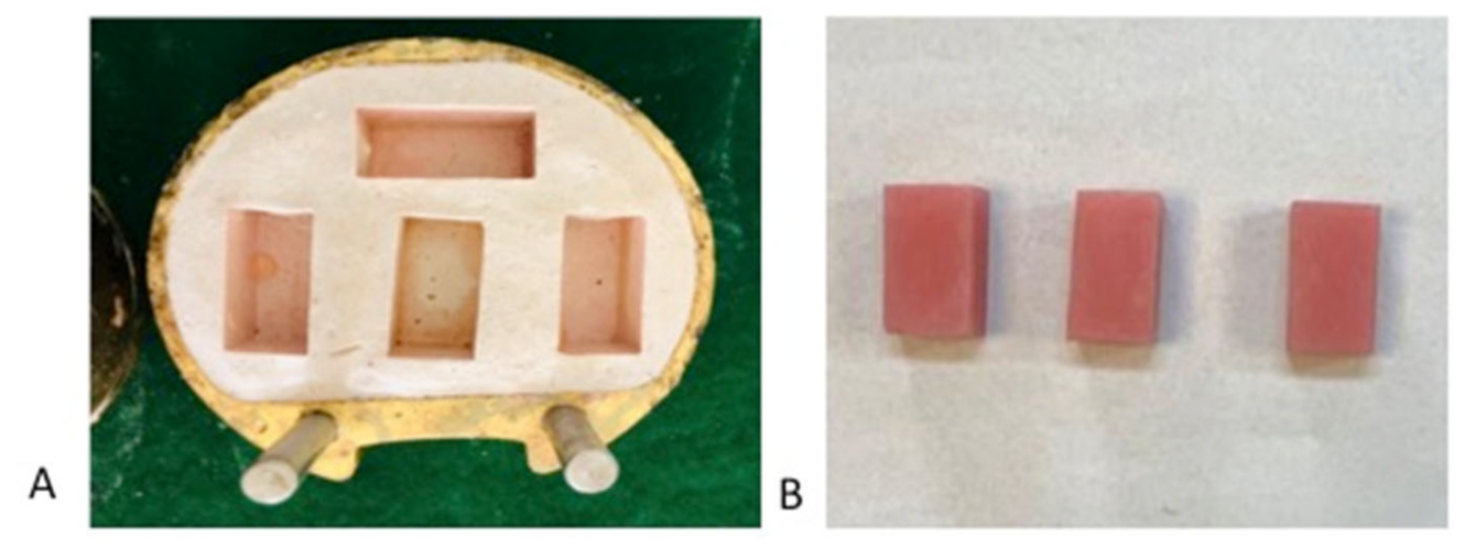
2.1. Specimen Preparation

The sample size was obtained using Pass version 11 (NCSS Statistical software, Kaysville, UT, USA), employing one-way ANOVA with 99% confidence interval at 90% power, and with the means and standard deviation of viable cells in groups (DW = 7.47 ± 0.68, DC = 4.82 ± 0.73, DW + MW 1 min = 4.49 ± 0.45, DC + MW 1 min = 2.64 ± 0.59) from a previous study [21]. A sample size of ten per group was obtained using calculations (total 12 subgroups). A total of 128 specimens were fabricated (Table 1).
In order to prepare the test specimens from PMMA acrylic resin, modelling wax (Yeti Dental, Berlin, Germany) was melted with help of a wax pot (Manfredi, Salerno, Italy) and poured into a three-part preformed metal mold with dimensions of 30 × 30 × 15 mm. Wax patterns were invested in a metallic denture flask filled with type III dental stone (Garrico Lab Stone, Rochester, NY, USA) to produce PMMA samples. De-waxing was performed using boiling water for 6 min. Heat-polymerized PMMA acrylic resin was mixed and packed at dough stage according to the manufacturer’s recommendations at a powder-to-liquid ratio of 2.3 (grams of polymer powder) to 1 mL of liquid (monomer) (heat-cured acrylic provided by MR Dental, Plymouth, UK). A hydraulic press was used for packing the denture base resin, with a sheet of plastic separating the two halves. Heat-cured PMMA was polymerized in a thermostatically controlled water bath (Manfredi—Acrydig 12, Salerno, Italy) and processed at 74 °C for two hours, followed by 100 °C for one hour (Figure 1). The amount of residual monomer for heat-cured PMMA (2 h cycle) ranged from 16 to 40 × 10−3 v/v% [22]. However, the curing cycle protocol and storage conditions were standardized among all study specimens in order to standardize the residual monomer among samples.
All samples were cooled at room temperature before being deflasked and immersed in distilled water at room temperature for 48 h in order to eliminate residual monomer. A metal bur (Denfac acrylic trimming burs) was then used to trim excess resin, and the finishing was performed using abrasive paper in a hand-held micromotor. All the specimens were autoclaved at 121 °C for 15 min. For reliability, a single operator prepared all the samples. All specimens’ dimensions were checked repeatedly. Intra-operator reliability was assessed at a kappa score of 0.85.

2.2. Study Groups

Based on the disinfection protocol, all the samples were randomly divided into the following groups.
Group MW-DW: The specimens were placed in a glass container filled with distilled water and disinfected with microwave (MW) radiation at 450 W. Depending on the MW radiation duration, the specimens were divided into MW-DW1 (1 min), MW-DW2 (2 min), and MW-DW3 (3 min). The protocol for MW disinfection was adopted from a previous study [23]. Different specimens in each subgroup were assessed at 96 h in a C. Albicans culture.
Group DC-DW: The specimens were dipped in a mixture of distilled water and denture cleaning tablet (DC) (Fittydent, Wien, Austria). Depending on the duration of immersion in the DC, the specimens were divided into DC-DW1 (1 min), DC-DW2 (2 min), and DC-DW3 (3 min). Different specimens in each subgroup were assessed at 96 h in a C. Albicans culture.
Group MW-DC-DW: In this group, the specimens were immersed in a glass beaker containing a mixture of distilled water and denture-cleaning tablet for five minutes. The glass beaker was placed in a microwave oven and irradiated at 650 W. Depending on the MW radiation’s duration, the specimens were divided into MW-DC-DW1 (1 min), MW-DC-DW2 (2 min), and MW-DC-DW3 (3 min). The temperature of the solution was kept between 65 °C to 71 °C, with ±2 °C. The different specimens in each subgroup were assessed at 96 h in the culture.
Positive Control Group: In this group, specimens were dipped in a glass container filled with 200 mL of distilled water kept at room temperature. The container was placed in the microwave oven (Samsung 2450 MHz, 800 W) without irradiation.
Negative Control Group: The purpose of this group was to establish the sterilization of the specimens and the accuracy of the test. For C. Albicans, the sterilized specimens were placed in a container with sterilized water.

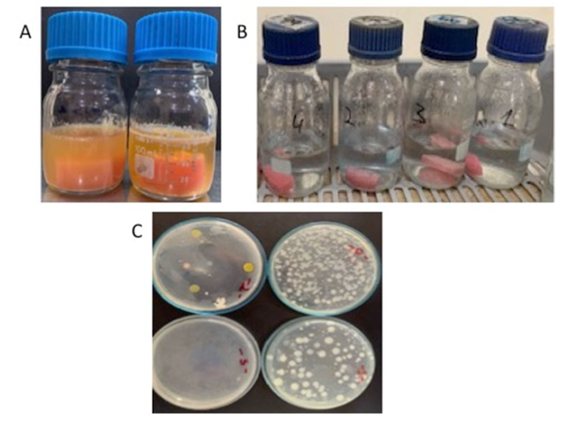
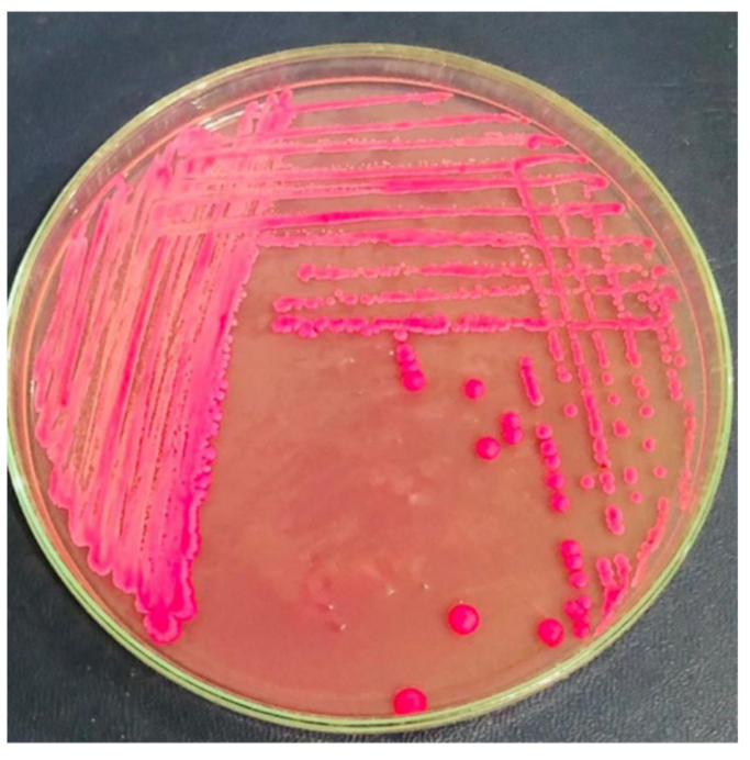
2.3. Biofilm Formation Assay

Tryptone Soya Broth (TSB) was used to enrich C. albicans (ATCC #90028) for 4 days. We then transferred 0.1 mL of this to 100 mL TSB that contained acrylic specimens (n = 163) and incubated it at 25 °C for 4 days. After 4 days’ incubation, the growth was monitored, and acrylic specimens were collected from the flask, washed with sterile distilled water, placed in 100 mL PBS (pH-7.0), and vortexed [24] (Figure 2). This flask was exposed to three treatment regimes (DW+MW, DW+DC, & DC) at different exposure times, after which 1 mL of PBS was transferred to a sterile Petri plate, poured with Dichloran Rose-Bengal Chloramphenicol Agar (Merck, Germany) (Figure 3), and incubated at 25 °C for 48 h. The growth was monitored, and the CFU were counted.

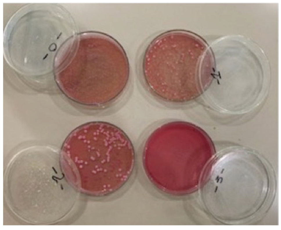
2.4. Quantification of Biofilm

All samples were incubated for 96 h for the biofilm assessment. The acrylic slides were collected between 24 h to 96 h, and the adhesion was monitored using a crystal violet binding assay as described earlier [25]. Briefly, the growth was fixed with acetic acid for 15 min, stained with 3% crystal violet (Ezzy Stain), washed with PBS at 7.0 pH, fixed gently by heating for 30 s, and finally washed with acetone. Each experiment was repeated four times to check the accuracy and precision of results. The colony-forming units (CFU) were evaluated by a single experienced microbiologist, and intra-operator reliability was observed (k = 0.84).

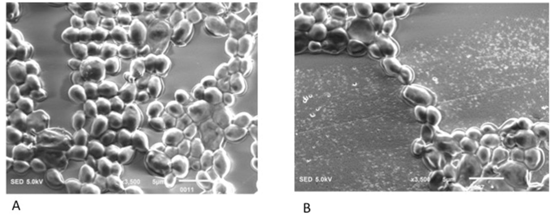
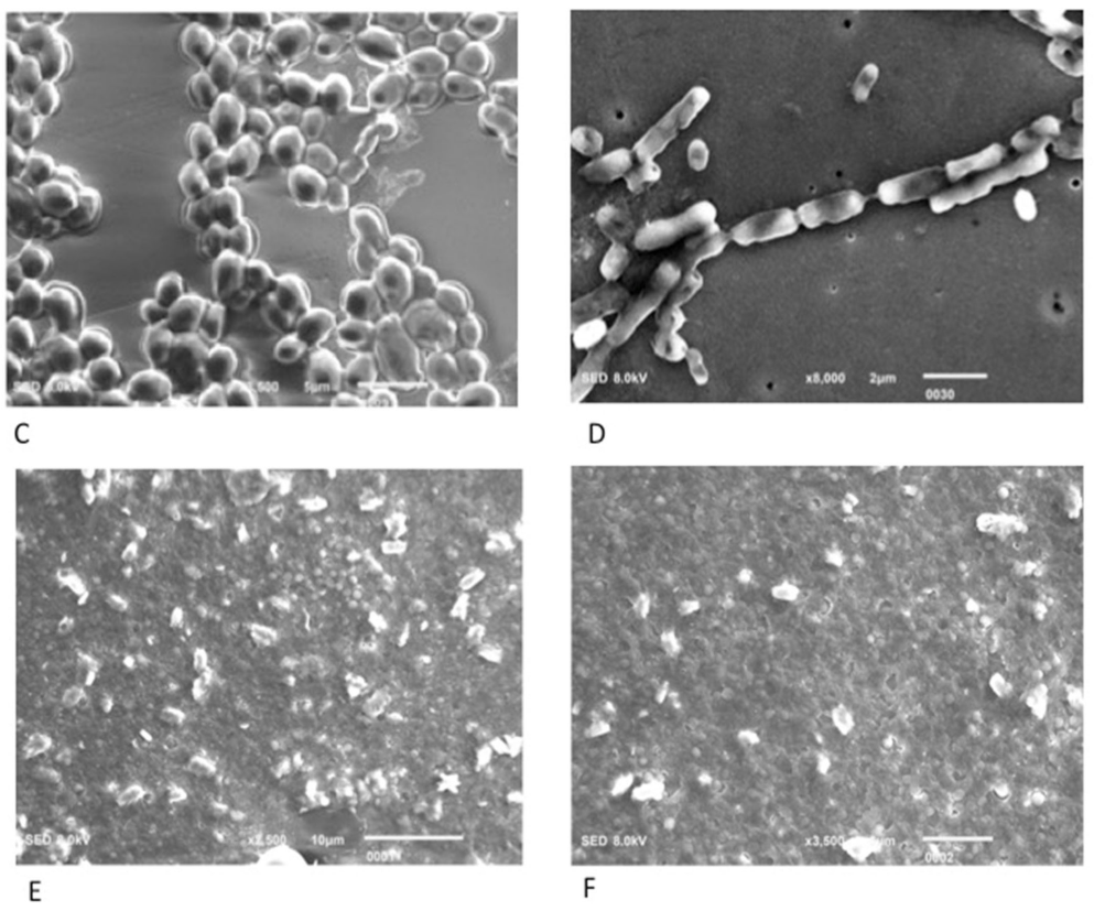
2.5. Scanning Electron Microscopy (SEM) Analysis

SEM was performed to analyze the production of the extracellular matrix material (ECM), as described earlier. All slides with biofilm were divided into 4 mm sections and then rinsed with distilled water, followed by staining with 0.02% Uranyl acetate for about 30 s. These 4 mm slide were coated with platinum in a coating machine (JEOL 3000 FC, Tokyo, Japan). All sections then displayed the existence of biofilm material under the examination of a scanning electron microscope (JEOL, Japan).

2.6. Statistical Analysis

The data were analyzed using IBM SPSS Statistics software version 21 (IBM Inc., Armonk, NY, USA). For disinfectant exposure time comparison, the Kruskal–Wallis test was applied for the C. albicans by treatment and growth time, as the CFU count was not normally distributed (Shapiro–Wilk test). For pairwise comparison of each exposure time with the control, the Mann–Whitney test was applied by treatment and growth time. A p-value of 0.05 or less was considered as statistically significant.

3. Results

The mean CFU comparison for C. albicans, with the controls for each disinfection technique at 96 h of growth, is presented in Table 1. We observed that specimens in DC-DW, MW-DW, and MW-DC-DW showed significant differences in the mean CFU of C. Albicans in comparison with the control without disinfection (p < 0.05). The MW-DC-DW disinfection regime showed near-complete disinfection (99.14%) of C. Albicans, compared with the controls (p < 0.05). The specimens treated with MW-DW showed 96% disinfection at 3 min, which was significantly lower than the controls (p < 0.05). The treatment with denture cleaning tablets with sodium perborate (DC-DW) showed only 53.57% of disinfection, compared with the control CFU after 5 min of treatment.
In the treatment with MW without using a denture cleaning tablet (MW-DW), the CFU count was reduced to 14% at 1 min of exposure and 21% at 2 min, whereas 3 min of microwave irradiation reduced 96% of the CFU count. In the disinfection treatment with DC (sodium perborate-cleaning tablet), the CFU count for C. Albicans was reduced to 6% at 1 min of exposure and to 7% at 2 and 3 min of exposure (Figure 4). However, DC use showed a maximum of 96% CFU reduction at 5 min of treatment. The combination of MW with DC showed the maximum disinfection, with the lowest CFU levels at 3 min among all three groups. MW alone showed significantly lower CFU levels (better disinfection) compared with MW-DC-DW combined; however, it exhibited higher disinfection than sodium perborate (disinfection tablet) alone (DC-DW) at 3 min of treatment. For all three disinfection regimes, the duration of disinfection showed significant influence on CFU levels (p < 0.05), with 3 min showing higher disinfection than the disinfection at 1 and 2 min (p < 0.05) (Figure 5).
A SEM analysis of the samples showed oval yeast-budding forms of C. Albicans cells, along with single-celled C. Albicans. Using SEM, the live cells of C. Albicans adhered to the PMMA polymer specimens were observed (Figure 6). After the disinfection procedure, the dead cells attached to PMMA surface were observed in the form of a cluster of irregular organelles.

4. Discussion

This study was performed to analyze the cumulative disinfection efficacy of a denture cleaning tablet (sodium perborate) and microwave irradiation on the eradication of C. albicans cultured on a PMMA denture base polymer. Our study was based on the hypothesis that there would be no significant difference in the disinfection efficacy of denture cleaning tablet, microwave irradiation, and their combination on the disinfection of C. albicans cultured on denture base. However, the postulated hypothesis was rejected as microwave irradiation combined with cleaning tablets (sodium perborate) (MW-DC-DW) showed better disinfection efficacy than that of microwave (MW) and cleaning tablets (DC) alone.
The outcomes of our study show that the CFU count of C. albicans was significantly reduced after 2 min of exposure to the MW-DW-DC group. However, 3 min of exposure to MW-DW-DC displayed a nearly zero CFU count of C. albicans. These results are in line with the findings of the study conducted by Sesma et al. [26]. Their study revealed that a combination of microwave and denture cleaning tablets demonstrated bactericidal as well as fungicidal effects against C. albicans. Moreover, they also identified that this combination possesses the ability to remove dead organisms from the PMMA denture bases. Denture cleaning tablets containing sodium perborate dissolve into peroxides, which results in the physical cleaning of the PMMA due to the bubbling and chemical destruction of the microbial cell walls [12,13,14]. Similar findings were also observed by Senna et al. [21]. They concluded that a denture cleaning tablet along with microwave irradiation caused the elimination of yeast cells from the PMMA resin base. Our study demonstrated almost 99% reduction in the CFU count of C. albicans. The slight difference observed in the previous studies may be due to the difference in the chemical composition of sodium perborate in the cleaning tablets and the different parameters of MW radiation [27,28].
Microwave disinfection is considered to be an effective and time-saving denture disinfection approach in clinical dentistry [29]. Microwaves disinfect by using thermal and non-thermal methods [30,31]. A thermal effect requires the presence of water or ionic molecules to produce a bactericidal influence at low energy levels [30], whereas high-energy, high-frequency microwaves cause cell wall and organelle destruction of microorganisms [31]. Our study found that microwave-disinfected samples displayed a significantly decreased CFU count of C. albicans at 3 min of exposure. Previous studies revealed that microwave disinfection initiates a change in the structure and permeability of the fungal cells, resulting in alterations in cell metabolism, causing cell death [32,33,34]. These results agree with the outcomes of the study conducted by Al-Saadi et al. [28]. In contrast, a study by Webb et al. showed a 1.3% survival rate of C. albicans when exposed to microwave irradiation at 350 W for 4 min [35]. In our study, MW irradiation disinfection was applied at higher power (450 Watts) for less time (3 min); therefore, a change in power and exposure time parameters may have ensured better disinfection. Moreover, in our study, the denture bases were immersed in distilled water prior to the exposure to MW irradiation. This technique may have provided the samples with a sterilized environment to prevent re-infection and a cleaner sample surface, compared with the dry sample’s MW exposure. It was proposed that PMMA denture disinfection achieved by microwave irradiation is due to the presence of distilled water, which forms bubbles upon boiling, thus removing microorganisms from the sample surface [36,37]. A study by Najdovski et al. revealed that microwave irradiation at reduced power can be used for a high level of disinfection, but not for sterilization [22], which is in line with our findings.
In our study, fungal cell adhesion was analyzed by a crystal violet binding assay, and cell count was analyzed by an aerobic plate count assay. The plate count showed that the control acrylic slide carried 1400 cfu/mL of C. albicans. The results show that the subject isolate of C. albicans showed a high level of antimicrobial resistance against denture cleaning tablets (sodium perborate). This was attributed to biofilm formation, as C. albicans nullified the toxic effect of sodium perborate and survived even after 5 min of exposure. This agreed with the findings of the previous studies by Dills et al. and Drake et al. [32,38]. They both identified that alkaline peroxide denture cleansers are effective against the streptococcus species and do not show much activity against C. albicans. This also agrees with the study by Ferreira et al., who studied the effect of a denture cleaning tablet (Bonyplus) against Candida species and S. mutans and found that the denture cleansing solution did not show significant re-educating of Candida species [39]. In addition, the alkaline peroxide tablets were not effective against S. mutans.
Interestingly, samples were fabricated in our study by mixing powder and liquid. However, CAD-CAM dentures are also available, fabricated from prepolymerized PMMA resin blocks [40]. The prepolymerized resin dentures allow for fewer surface defects and irregularities, and the incidence of fungal infection among these dentures and their disinfection protocol may differ from conventional PMMA dentures. Therefore, studies assessing the disinfection of CAD-CAM PMMA dentures are warranted. Our study showed higher disinfection efficacy of MW with sodium perborate-disinfection tablets for the use of Candida in comparison with microwave (MW) and cleaning tablets (DC) alone. It is pertinent to mention that MW disinfection of PMMA denture bases can also result in distortion and discrepancy in their fit and adaptation on stone casts [17]. However, Pavan et al. showed that at high MW energy, distortion was observed for the PMMA denture, while at 500 W or below, the dimensional accuracy was comparable to the control [17]. As MW disinfection in our study was performed at 450 W power, the PMMA resin specimens when exposed to our study’s protocol for disinfection were dimensionally accurate and therefore could be clinically utilized. In addition, our study displayed some inherent limitations. Primarily, the study was based on in vitro design, assessing a single species of Candida, which may have limited the clinical implications. Moreover, only one type of denture disinfecting tablet was used; however, cleansing agents with hydrogen peroxide, sodium hypochlorite, and Chlorhexidine are also available. Therefore, further studies assessing the different candida species with contemporary denture cleansing agents are warranted to investigate the antimicrobial efficacy of microwave irradiation.

5. Conclusions

The microwave disinfection of PMMA for 3 min at 450 W in combination with a denture cleaning tablet (sodium perborate) is a more effective disinfecting regime against C. albicans than that of microwave and sodium perborate alone. Further studies assessing the efficacy of low-level MW irradiation on the disinfection of various bacterial species is recommended.

Author Contributions

Conceptualization, Z.C., S.A.K., S.M., S.M.R.K., W.A.F., M.A.A. and Z.A.M.; methodology, K.A.A., S.A.K., M.A.A., W.A.F., S.M.R.K., A.H.A. and A.A. software, K.A.A., W.A.F., S.M.R.K., A.A., A.H.A., M.A. and T.A.; validation, Z.C., A.A., W.A.F. and S.M.; formal analysis, S.A.K., Z.A.M., A.A., S.M.R.K., M.A. and K.A.A.; resources, A.H.A., T.A., M.A.A. and A.A.; data curation, S.A.K., Z.A.M., Z.C., M.A.A., S.M.R.K., W.A.F. and A.A.; writing—original draft preparation, K.A.A., T.A., M.A. and Z.C.; writing—review and editing, S.M.R.K., S.A.K., A.A., T.A., A.H.A. and K.A.A. All authors have read and agreed to the published version of the manuscript.

Funding

This research was funded by Princess Nourah bint Abdulrahman University researchers, supporting project number PNURSP2022R6, Princess Nourah bint Abdulrahman University, Riyadh, Saudi Arabia.

Institutional Review Board Statement

The study protocol was reviewed by the ethics and review committee of Dr. Ishrat-ul-Ebad Khan Institute of Oral Health Sciences and the Dow University of Health Sciences, with No. IRB-1101/DUHS/Approval/2020.

Informed Consent Statement

No human patients or human tissue was included in the study.

Data Availability Statement

The data are available on contact from the corresponding author.

Acknowledgments

The authors wish to thank the Princess Nourah bint Abdulrahman University researchers, supporting project number PNURSP2022R6, Princess Nourah bint Abdulrahman University, Riyadh, Saudi Arabia.

Conflicts of Interest

The author declares no conflict of interest.

References

  1. Hayran, Y.; Sarikaya, I.; Aydin, A.; Tekin, Y.H. Determination of the effective anticandidal concentration of denture cleanser tablets on some denture base resins. J. Appl. Oral Sci. 2018, 26, e20170077. [Google Scholar] [CrossRef] [PubMed] [Green Version]
  2. Gendreau, L.; Loewy, Z.G. Epidemiology and etiology of denture stomatitis. J. Prosthodont. 2011, 20, 251–260. [Google Scholar] [CrossRef] [PubMed]
  3. Marsh, P.D.; Percival, R.S.; Challacombe, S.J. The influence of denture-wearing and age on the oral microflora. J. Dent. Res. 1992, 71, 1374–1381. [Google Scholar] [CrossRef] [PubMed]
  4. Berger, J.C.; Driscoll, C.F.; Romberg, E.; Luo, Q.; Thompson, G. Surface roughness of denture base acrylic resins after processing and after polishing. J. Prosthodont. Implant Esthet. Reconstr. Dent. 2006, 15, 180–186. [Google Scholar] [CrossRef]
  5. Busscher, H.J.; Weerkamp, A.H.; Van Der Mei, H.C.; Van Pelt, A.W.; De Jong, H.P.; Arends, J. Measurement of the surface free energy of bacterial cell surfaces and its relevance for adhesion. Appl. Environ. Microbiol. 1984, 48, 980–983. [Google Scholar] [CrossRef] [Green Version]
  6. De Freitas Fernandes, F.S.; Pereira-Cenci, T.; Da Silva, W.J.; Ricomini Filho, A.P.; Straioto, F.G.; Cury, A.A. Efficacy of denture cleansers on Candida spp. biofilm formed on polyamide and polymethyl methacrylate resins. J. Prosthet. Dent. 2011, 105, 51–58. [Google Scholar] [CrossRef]
  7. Powell, G.L.; Runnells, R.D.; Saxon, B.A.; Whisenant, B.K. The presence and identification of organisms transmitted to dental laboratories. J. Prosthet. Dent. 1990, 64, 235–237. [Google Scholar] [CrossRef]
  8. Cakan, U.; Kara, O.; Kara, H.B. Effects of various denture cleansers on surface roughness of hard permanent reline resins. Dent. Mater. J. 2015, 34, 246–251. [Google Scholar] [CrossRef] [Green Version]
  9. Yudaev, P.; Chuev, V.; Klyukin, B.; Kuskov, A.; Mezhuev, Y.; Chistyakov, E. Polymeric Dental Nanomaterials: Antimicrobial Action. Polymers 2022, 14, 864. [Google Scholar] [CrossRef]
  10. Coenye, T.; De Prijck, K.; Nailis, H.; Nelis, H.J. Prevention of Candida albicans Biofilm Formation. Open Mycol. J. 2011, 5, 9–20. [Google Scholar] [CrossRef]
  11. Yasui, M.; Ryu, M.; Sakurai, K.; Ishihara, K. Colonisation of the oral cavity by periodontopathic bacteria in complete denture wearers. Gerodontology 2012, 29, e494–e502. [Google Scholar] [CrossRef] [PubMed] [Green Version]
  12. Yatabe, M.; Seki, H.; Shirasu, N.; Sone, M. Effect of the reducing agent on the oxygen-inhibited layer of the cross-linked reline material. J. Oral Rehabil. 2001, 28, 180–185. [Google Scholar] [CrossRef] [PubMed]
  13. Budtz-Jørgensen, E. Materials and methods for cleaning dentures. J. Prosthet. Dent. 1979, 42, 619–623. [Google Scholar] [CrossRef]
  14. Shakouie, S.; Milani, A.S.; Eskandarnejad, M.; Rahimi, S.; Froughreyhani, M.; Galedar, S.; Ranjbar, E. Antimicrobial activity of tetraacetylethylenediamine-sodium perborate versus sodium hypochlorite against Enterococcus faecalis. J. Dent. Res. Dent. Clin. Dent. Prospect. 2016, 10, 43–47. [Google Scholar] [CrossRef]
  15. Fernandes, A.M.; Marques, M.M.; Camargo, S.E.A.; Cardoso, P.E.; Camargo, C.H.R.; Valera, M.C. Cytotoxicity of non-vital dental bleaching agents in human gingival fibroblasts. Braz. Dent. Sci. 2013, 16, 59–65. [Google Scholar] [CrossRef] [Green Version]
  16. Arendorf, T.; Walker, D. Denture stomatitis: A review. J. Oral Rehabil. 1987, 14, 217–227. [Google Scholar] [CrossRef]
  17. Pavan, S.; Arioli Filho, J.N.; Santos, P.H.d.; Mollo, F.d.A., Jr. Effect of microwave treatments on dimensional accuracy of maxillary acrylic resin denture base. Braz. Dent. J. 2005, 16, 119–123. [Google Scholar] [CrossRef] [Green Version]
  18. Sanita, P.V.; Vergani, C.E.; Giampaolo, E.T.; Pavarina, A.C.; Machado, A.L. Growth of Candida species on complete dentures: Effect of microwave disinfection. Mycoses 2009, 52, 154–160. [Google Scholar] [CrossRef]
  19. Campanha, N.H.; Pavarina, A.C.; Brunetti, I.L.; Vergani, C.E.; Machado, A.L.; Spolidorio, D.M.P. Candida albicans inactivation and cell membrane integrity damage by microwave irradiation. Mycoses 2007, 50, 140–147. [Google Scholar] [CrossRef]
  20. Silva, M.M.; Vergani, C.E.; Giampaolo, E.T.; Neppelenbroek, K.H.; Spolidorio, D.M.; Machado, A.L. Effectiveness of microwave irradiation on the disinfection of complete dentures. Int. J. Prosthodont. 2006, 19, 272–278. [Google Scholar]
  21. Senna, P.M.; Sotto-Maior, B.S.; da Silva, W.J.; Cury, A.A.D.B. Adding denture cleanser to microwave disinfection regimen to reduce the irradiation time and the exposure of dentures to high temperatures. Gerodontology 2013, 30, 26–31. [Google Scholar] [CrossRef] [PubMed]
  22. Huda, I.; Nabi, A.T.; Turner, P.S.; Hegde, C. Study to determine and estimate residual monomer leached out in heat cure polymethylmethacrylate Resins of commonly used brands using different polymerization cycles: (An invitro study). IP Ann. Od Prosthodont. Restor. Dentistry 2019, 5, 28–36. [Google Scholar] [CrossRef]
  23. Najdovski, L.; Dragaš, A.; Kotnik, V. The killing activity of microwaves on some non-sporogenic and sporogenic medically important bacterial strains. J. Hosp. Infect. 1991, 19, 239–247. [Google Scholar] [CrossRef]
  24. Mirani, Z.; Urooj, S.; Khan, M.; Khan, A.; Shaikh, I.; Siddiqui, A. An effective weapon against biofilm consortia and small colony variants of MRSA. Iran. J. Basic Med. Sci. 2020, 23, 1494–1498. [Google Scholar] [PubMed]
  25. Haney, E.; Trimble, M.; Cheng, J.; Vallé, Q.; Hancock, R. Critical assessment of methods to quantify biofilm growth and evaluate antibiofilm activity of host defence peptides. Biomolecules 2018, 8, 29. [Google Scholar] [CrossRef] [Green Version]
  26. Sesma, N.; Rocha, A.L.; Laganá, D.C.; Costa, B.; Morimoto, S. Effectiveness of denture cleanser associated with microwave disinfection and brushing of complete dentures: In vivo study. Braz. Dent. J. 2013, 24, 357–361. [Google Scholar] [CrossRef]
  27. Dantas, A.; Consani, R.L.X.; Sardi, J.; Mesquita, M.; Silva, M.; Sinhoreti, M. Biofilm formation in denture base acrylic resins and disinfection method using microwave. J. Res. Pract. Dent. 2014, 2014, 112424. [Google Scholar] [CrossRef] [Green Version]
  28. Al-Saadi, M.H. Effectiveness of chemical and microwave disinfection on denture biofilm fungi and the influence of disinfection on denture base adaptation. J. Indian Prosthodont. Soc. 2014, 14, 24–30. [Google Scholar] [CrossRef]
  29. Arita, M.; Nagayoshi, M.; Fukuizumi, T.; Okinaga, T.; Masumi, S.; Morikawa, M.; Kakinoki, Y.; Nishihara, T. Microbicidal efficacy of ozonated water against Candida albicans adhering to acrylic denture plates. Oral Microbiol. Immunol. 2005, 20, 206–210. [Google Scholar] [CrossRef]
  30. Anaraki, M.R.; Mahboubi, S.; Pirzadeh, T.; Lotfipour, F.; Torkamanzad, N. Disinfection effect of microwave radiation on Bacillus subtilis as indicator organism on contaminated dental stone casts under dry and wet conditions. GMS Hyg. Infect. Control 2017, 12, 294. [Google Scholar]
  31. Banik, S.; Bandyopadhyay, S.; Ganguly, S. Bioeffects of microwave—A brief review. Bioresour. Technol. 2003, 87, 155–159. [Google Scholar] [CrossRef]
  32. Mese, A.; Mese, S. Effect of Microwave Energy on Fungal Growth of Resilient Denture Liner Material. Biotechnol. Biotechnol. Equip. 2007, 21, 91–93. [Google Scholar] [CrossRef] [Green Version]
  33. Tarbet, W.J. Denture plaque: Quiet destroyer. J. Prosthet. Dent. 1982, 48, 647–652. [Google Scholar] [CrossRef]
  34. Dills, S.; Olshan, A.; Goldner, S.; Brogdon, C. Comparison of the antimicrobial capability of an abrasive paste and chemical-soak denture cleaners. J. Prosthet. Dent. 1988, 60, 467–470. [Google Scholar] [CrossRef]
  35. Webb, B.C.; Thomas, C.J.; Harty, D.W.; Willcox, M. Effectiveness of two methods of denture sterilization. J. Oral Rehabil. 1998, 25, 416–423. [Google Scholar] [CrossRef] [PubMed]
  36. Polyzois, G.L.; Zissis, A.J.A.; Yannikakis, S. The effect of glutaraldehyde and microwave disinfection on some properties of acrylic denture resin. Int. J. Prosthodont. 1995, 8, 150–154. [Google Scholar] [PubMed]
  37. Neppelenbroek, K.; Pavarina, A.C.; Spolidorio, D.M.P.; Vergani, C.E.; Mima, E.G.D.O.; Machado, A.L. Effectiveness of microwave sterilization on three hard chairside reline resins. Int. J. Prosthodont. 2003, 16, 264–270. [Google Scholar]
  38. Drake, D.; Wells, J.; Ettinger, R. Efficacy of denture cleansing agents in an in vitro bacteria-yeast colonization model. Int. J. Prosthodont. 1992, 5, 300–307. [Google Scholar]
  39. Ferreira, M.F.; Pereira-Cenci, T.; De Vasconcelos, L.M.R.; Rodrigues-Garcia, R.C.M.; Cury, A.A.D.B. Efficacy of denture cleansers on denture liners contaminated with Candida species. Clin. Oral Investig. 2009, 13, 237–242. [Google Scholar] [CrossRef]
  40. Heikal, M.M.A.; Nabi, N.A.; Elkerdawy, M.W. A study comparing patient satisfaction and retention of CAD/CAM milled complete dentures and 3D printed CAD/CAM complete dentures versus conventional complete dentures: A randomized clinical trial. Braz. Dent. Sci. 2022, 3, 25. [Google Scholar] [CrossRef]
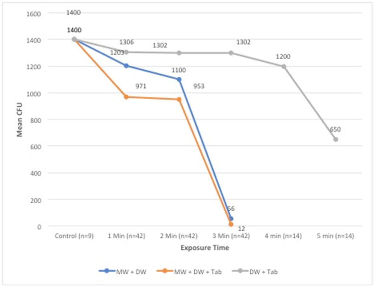
Figure 1. Fabrication of PMMA samples. (A) Dental stone mold. (B) Finished PMMA samples.
Applsci 12 07004 g001
Figure 2. Procedure for biofilm formation assay. (A) TSB containing PMMA specimens. (B) Acquisition of PMMA specimens. (C) Candida incubation.
Applsci 12 07004 g002
Figure 3. Pure culture of C. albicans on Dichloran Rose-Bengal Chloramphenicol Agar.
Applsci 12 07004 g003
Figure 4. The effect of a denture cleaning tablet and MW 450 watt on the growth of C. albicans. Plate 0 was the untreated control, plate 1 was exposed for 1 min, plate 2 was exposed for 2 min, and plate 3 was exposed for 3 min.
Applsci 12 07004 g004
Figure 5. Colony-forming units of C. Albicans for different antifungal protocols at 96 h of growth.
Applsci 12 07004 g005
Figure 6. SEM micrographs presenting the attachment of C. albicans to PMMA samples. (A) Control sample with C. albicans growth. (B) MC-DC-DW sample at 1 min treatment. (C) MW-DW sample at 1 min treatment. (D) DC-DW sample at 5 min. (E) MW-DC-DW disinfection sample at 3 min. (F) MW-DW sample at 3 min.
Applsci 12 07004 g006aApplsci 12 07004 g006b
Table 1. Pairwise mean CFU comparison for C. Albicans.
Growth Time & Exposure Time vs. ControlControl CFUMW-DC-DWMW + DWDC + DW
96 h1400MD (p-value)MD (p-value)MD (p-value)
1 min −429 (0.008 **)−197 (0.008 **)−94 (0.008 **)
2 min −447 (0.008 **)−300 (0.008 **)−98 (0.008 **)
3 min −1388 (0.007 **)−1344 (0.008 **)−98 (0.007 **)
4 min −1388 (0.008 **)−1344 (0.008 **)−200 (0.008 **)
5 min −1388 (0.008 **)−1344 (0.008 **)−750 (0.008 **)
** Significant at 1% using the Mann–Whitney test; MW: microwave; DW: distilled water; DC: sodium perborate tablet. Values are represented as the mean difference (p-value).
Publisher’s Note: MDPI stays neutral with regard to jurisdictional claims in published maps and institutional affiliations.

