Article

Plywood Jig—A New Technique for Root Canal Curvature Measurement

by
Saleem D. Makandar
1,*,
Mohammed Imran Khaiser
2,
Sneha R. Mali
3,
Mohmed Isaqali Karobari
1,
Anand Marya
4,
Pietro Messina
5 and
Giuseppe Alessandro Scardina
5,*
1
Conservative Dentistry Unit, School of Dental Sciences, Health Campus, Universiti Sains Malaysia, Kubang Kerian 16150, Kelantan, Malaysia
2
Department of Endodontics, King Fahad Hospital Madinah Munawwarah, Medina 42351, Saudi Arabia
3
Y.M.T. Dental College and Hospital, Institutional Area, Sector 4, Kharghar, Navi Mumbai 410210, India
4
Department of Orthodontics, Faculty of Dentistry, University of Puthisastra, Phnom Penh 12211, Cambodia
5
Department of Surgical, Oncological and Stomatological Disciplines, University of Palermo, 90133 Palermo, Italy
*
Authors to whom correspondence should be addressed.
Appl. Sci. 2021, 11(9), 3999; https://doi.org/10.3390/app11093999
Submission received: 16 April 2021 / Revised: 23 April 2021 / Accepted: 26 April 2021 / Published: 28 April 2021
(This article belongs to the Special Issue Innovative Techniques in Endodontics)

Abstract

:
The successful outcome of endodontic treatment is dependent on complete cleaning, shaping as well as three dimensional obturation of the root canal system. A conventional radiograph is a two-dimensional replication of a three-dimensional object and does not provide any conclusive evidence for canal curvatures. An accurate knowledge of the tooth anatomy and curvature is essential to avoid procedural errors. 100 freshly extracted human teeth were used in this study. Digital images were obtained using the plywood JIG and Schneider’s technique. These images were analyzed using the VixWin Pro digital image analyzing software (Gendex system). Statistical analysis was done using paired t test. The canal curvature average values measured using Jig method and Schneider method for mandibular teeth are 28.23° (±9.96) and 22.07° (±9.46) respectively. The smallest/largest curvature angles measured using Jig method and Schneider technique are 12/52° and 8/44° respectively. Canal curvature average values measured using Jig method and Schneider method for maxillary teeth were 23.40° (±11.36) and 19.23° (±11.94) respectively. The smallest/largest average curvature angles measured using Jig method, Schneider technique were 9/70° and 5/72° respectively. The values of the canal curvature angle obtained during routine radiographs in clinics have lower curvature angle and higher radius values as compared to the values obtained by this innovative JIG technique. Therefore, a clinician should always keep in mind the difference while measuring the curvature angle on radiographs during root canal treatment.

1. Introduction

Successful endodontic therapy is dependent on the efficiency of removing micro-organisms by means of detailed chemo-mechanical instrumentation of the root canal system. This includes the removal of the infected dentine as well as the organic tissue by cleaning and shaping procedures. The success rate of non-surgical root canal treatment ranges from 31% to 96% [1]. Precise knowledge of the complex internal canal anatomy and detailed planning of root canal treatment are essential to reduce failure rates. Apart from the problems encountered during endodontic therapy, root canal curvatures pose extreme difficulties. Determination of canal curvature will help to identify the appropriate instruments and earmark the accurate technique, which are of crucial importance for treatment success.
The root canal anatomy differs among different ethnic groups and sub-populations [2]. So, it is essential for the dentist to know about the anatomic variabilities in his practicing population. Few authors have previously tried to analyze and measure the degree of curvature present in root canals of various teeth. Schneider was one of the first researchers to provide a detailed analysis and report on determination of canal curvatures from clinical radiographs [3,4]. The drawback was that he did not investigate curvatures seen from the proximal view. But in truth, curvatures are present not only in a mesio-distal direction, as seen in a clinical radiograph, but also in a bucco-lingual direction. Pineda and Kuttler used a roentgenographic method to analyze and evaluate root canals from both the proximal as well as the clinical views [5]. Three-dimensional imaging techniques can give the exact picture of root canal anatomy like MRI and Computed Tomography, but which are expensive and tedious and will not be applicable to clinical practice [6]. Research has been conducted using different techniques to measure the canal curvatures of the mesio-buccal root of the maxillary first molar and mesial root of the mandibular first molar because of the increase in the frequency of performing endodontic treatment in these teeth and the presence of innumerable number of anomalies coupled with variable amount of canal curvatures [7,8]. Canal curvature is known to be influenced during shaping by the different file system or by the pressure on the canal walls, a research conducted evaluated the pressure distribution against the root canal wall by using finite element analysis (FEA), wherein authors concluded that the instrument shaping ability can be predicted by using FEA as a virtual tool [9]. Here we come with a new, simple, inexpensive and accurate method to measure the canal curvature angle, radius and peak height of curvature in an in vitro situation and the accuracy was compared with the standard Schneider’s canal curvature measurement.

2. Materials and Methods

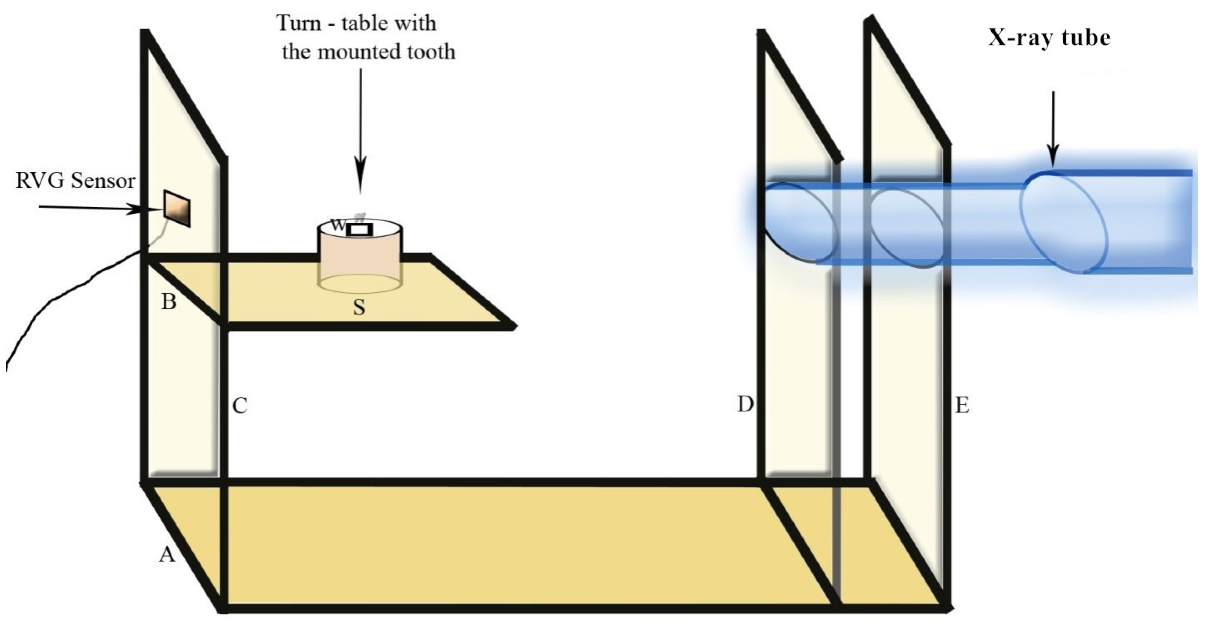
2.1. Construction of Plywoog Jig

A standard plywood jig (Figure 1) was designed in the following manner. It consists of two flat horizontal, parallel stacks (A and B) and three vertical stakes (C, D and E) made of plywood projecting from the horizontal one. The upper horizontal stake was short and fitted with a turn table which was eight inches from the X-ray tube and can be rotated to 360 degrees. The angle of rotation can be checked with an angle measuring scale (S) fitted on the surface of the turn table. The tooth for which the measurement to be carried out was mounted over the turn table. In the vertical stakes on one side, vents were made, and X-ray tube was fitted so that the tube can pass through the stakes and firm in position. Digital sensor from Radio Visio Graphy equipment was fixed on the backside of the stake in an ideal manner so that the X-ray can pass through the tooth mounted over the turn table and reach the sensor in a parallel direction.

2.2. Procedure

Freshly extracted 50 human mandibular and 50 maxillary first molars were used for analysis in this study. Any teeth with an open apex, showing external resorption, visible carious lesions, presence of very narrow or obstructed canals, were eliminated. Standard access cavities were prepared, and orifices were enlarged using GG drills in order to avoid acute bending of the files that may interfere with the measurement of the curvature. Sodium hypochlorite (2.5%) was used as an irrigant and EDTA (RC Prep) was used for lubrication of the canal [10]. A size 15 K-file was inserted into the mesio-buccal canal of the maxillary and mandibular first molars extending till the root apex and digital images were recorded. The teeth were held in position on the turntable with the help of soft modeling wax (W). Each tooth was positioned so that their long axis was parallel to the X-ray tube as well as the digital sensor. Exposure time and the distance of the tooth from the X-ray tube was kept constant.

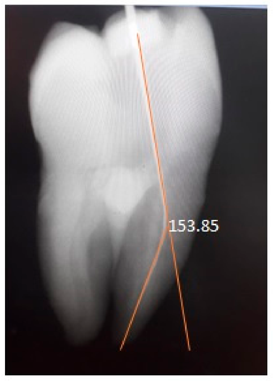
2.3. Determination of Canal Curvature Using Plywood Jig Technique

Digital images are obtained using the above-mentioned technique. These images were analyzed using the VixWin Pro digital image analyzing software (Gendex system) to obtain a straight line passing through the middle of the root. In case this is not obtained, the turn table was rotated, and another image was taken until a straight line was obtained (mimicking the straight instrument in the canal). Once a straight line was obtained, the turn table was rotated to 90 degrees and another image was taken. The instrument in the canal now was in its maximum curvature. The image indicates that the curved canal is made up of two segments, one was extending from the pulpal chamber floor down the long axis of the root as much as the coronal two thirds of the root and the second segment from the apex of the root extending back to the cervical to intercept the first line. These two lines intersect to form four angles. The interior angle is the estimate of the canal curvature. The angle of curvature, the radius, the height of curvature and the direction of curvature were calculated using VixWin Pro digital image analyzing software (using the angle measurement tool) (Figure 2).

2.4. Determination of Canal Curvature Using Schneider Technique
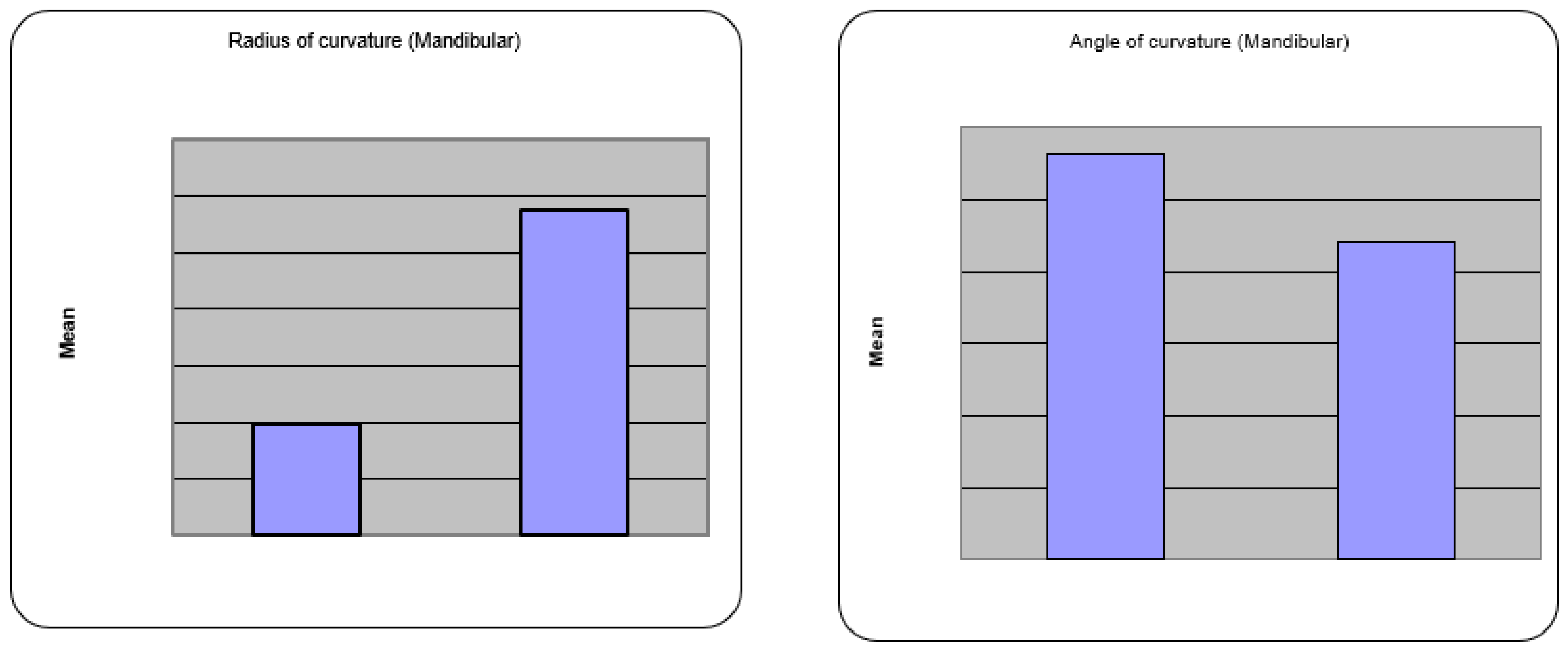
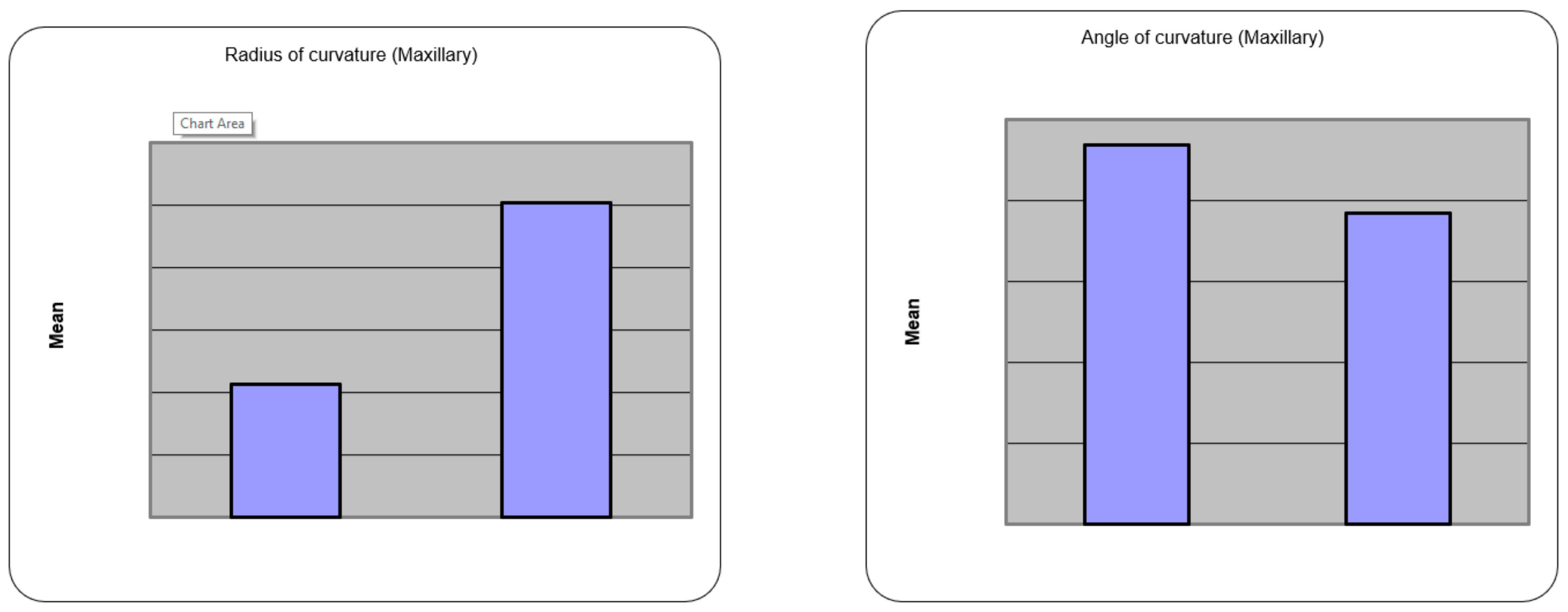
Teeth were imaged in a buccal direction and using the same technique the angle was measured in the digitalized image. A radiographic image indicated that the curved canal had two segments, one extending from the pulpal chamber floor down the long axis of much of the coronal two thirds of the root and the second segment from the apex of the root extending back to the occlusal through the apical third of the root. Radiovisiographic software was utilized to calculate the radius, the angle of curvature, the height of the curvature as well as the direction of the curvature. The curvature values were evaluated statistically using paired t test, percentage difference as compared to Schneider method (Figure 3).

3. Results

The canal curvature angle average values measured using Jig method and Schneider method for mandibular teeth are 28.23° (±9.96) and 22.07° (±9.46) respectively. (Table 1) The smallest/largest average curvature angles measured using Jig method and Schneider technique are 12°/52° and 8°/44° respectively. Paired t-test showed that there was highly significant difference between the curvature angle measured using each technique (p < 0.001) (Figure 4 and Table 2).
Canal curvature angle average values measured using Jig method and Schneider method for maxillary teeth are 23.40° (±11.36) and 19.23° (±11.94) respectively. (Table 1) The smallest/largest average curvature angles measured using Jig method, Schneider technique are 9°/70° and 5°/72° respectively. Paired t-test showed that there was highly significant difference between the curvature angle measured using each technique (p < 0.001). (Table 2)
The results of radius of curvature measured using Jig method and Schneider method for mandibular teeth are 9.79 mm (±3.65) and 10.55 mm (±3.34) respectively. The smallest/largest average curvature radius measured using Jig method, Schneider technique are 2.4 mm/18.4 mm and 2.8 mm/18.4 mm respectively. Paired t test showed that there was highly significant difference between the curvature angle measured using each technique (p < 0.001). For maxillary teeth using Jig method 16.06 mm (±8.12) compared to Schneider method 17.51 mm (±7.88). The smallest/largest average curvature radius measured using Jig method, Schneider technique are 4.2/36.6 mm and 3.4/37.8 mm respectively. Paired t test showed that there was significant difference between the curvature angle measured using each technique (p < 0.05). (Figure 5 and Table 2). The mean peak height of curvature for maxillary and mandibular teeth was 3.86 mm (±1.159) and 3.9 mm (±1.426) respectively (Table 2).

4. Discussion

The Schneider technique and the plywood jig methods were used to determine angle of root curvature, radius, height, and direction of curvature. The benefits of using this method are that it is reproducible, easy to use as well as efficient, particularly when using radiovisiographic (RVG) images. Precise knowledge of root curvature radius is required to ensure accurate planning of the instrumentation and reduces the impact of the anatomic limitations of various endodontic instruments. By modifying the instrumentation techniques, we can eliminate the disastrous consequences that may occur during root canal preparation, such as loss of working length, apical transportation, creation of elbows, zips, ledges, perforations, and even instrument fracture.
Schneider proposed a method to determine curvature based on the angle that is obtained by two straight lines [3,4]. The first line runs parallel along the length of the root canal, while the second one runs through the apical foramen till it intersects with the first line at the start of the curvature. The formed angle (a) was named according to the degree of root canal curvature: straight: 5°; moderate: 10–20°; and severe: 25–70°.
There are major problems encountered during routine endodontic treatment such as deformation or fracture of instruments. Mechanical preparation errors may come about because of an increase in the canal curvature [8]. Additionally, the file may fracture due to stress arising from contact with the canal surface. Despite their super-elasticity recently developed nickel titanium canal instruments can suffer from cyclic fatigue effect in curved canals, a sharp canal curvature increases the stress on instrument [11,12,13]. Recent literature evaluated the importance of stress at 360 degree in extracted teeth, assesses the recently developed NiTi rotary instrumentations, and new dynamic stress assessment technique, which revealed the excellent resistant to cyclic effort [14,15].
The type of canal exhibiting secondary curvature in plane opposite to that of direction of primary curvature as in mesio-buccal canal. The highest mean of curvature in mesio-buccal canal. Slowey described the mesio-lingual canal of mandibular molar usually straighter than the mesio-buccal canal [16]. Previous studies reported that most fractures occurred in the apical portion of the canal, because of the presence of a curvature with a small radius [10]. Pruett et al. introduced a new parameter described as the curvature radius that could be used to measure the root canal curvature. Schneider performed pioneer work on measuring canal curvature [17]. Most studies have used the Schneider methods for determination of the root canal curvature.
Using the plywood jig method described in the present study, it is possible to analyze a specific curvature of root canal. This is an important finding since more than one curvature can be located within the same root canal, that would not allow for accuracy in calculating the radius across the entire length of the root. Here the study was done to compare the Schneider’s technique and plywood jig to compare the canal curvature. Root canals with increased angle of curvatures and the small radii can be associated with a negative impact on the instrument or the instrumentation technique. Utilizing conventional radiographic images, the Schneider’s technique for detection of root canal curvature should be done with care because of the two-dimensional view with a high possibility of false recording of curvature. One principal advantage of using the Plywood jig in endodontics is its usefulness in aiding in the identification of maximum curvature dimensions, and in the selection of instruments for cleaning and shaping with a highly accurate non-invasive technique. Plywood jig with multiple radiography is proved to be an accurate method for measuring root canal curvature angle, radius, height, and direction of curvature.
The aim of endodontic therapy is to remove the necrotic tissue, disinfect the canal, shape the canal, and these goals can be achieved by cleaning and shaping, the successful root canal preparation depends on the angle of curvature and radius of root canal. The canal curvature finding in clinical situation is how it is measured in intra oral periapical X-ray, but the hidden curve and the maximum curve of canal is very difficult to gauge [18]. Weine [19] reported that the case became more complex, and complications were more likely occur during canal preparation if the angle of canal curvature exceeded 30°. In this study it has been calculated the difference in the values of angle and radius of curvature between routine radiographs taken and with jig technique. The mean differences obtain are mentioned in Table 2 which always can be added to the values of angle of curvature measured during clinical regular periapical radiographs. The mean curvature values can be deducted from the values measured during regular clinical periapical X-rays. The clinical techniques of canal preparation can be decided according to the severity of curvature and with more cautiously the canals can be cleaned and shaped. The mishaps during endodontics treatment like ledging, transportation, zipping, perforation can be avoided [20].

5. Conclusions

The Plywood Jig with multi-radiographic technique for determination of root curvature angle, radius, height and direction of curvature detailed in this article is easy to use, measures maximum curvature, is reproducible as well as allows for effective, efficient and reliable treatment planning. The values of canal curvature angle obtained during routine radiographs in clinics are actually has lower curvature angle value and higher radius values as compared to the values obtained by this innovative JIG technique, clinician should always keep in mind the difference while measuring the curvature angle on radiographs during root canal treatment.

Author Contributions

Conceptualization, S.D.M., M.I.K. (Mohammed Imran Khaiser), S.R.M. and M.I.K. (Mohmed Isaqali Karobari); methodology S.D.M., M.I.K. (Mohammed Imran Khaiser), S.R.M. and M.I.K. (Mohmed Isaqali Karobari); validation, M.I.K. (Mohmed Isaqali Karobari), A.M. and G.A.S.; formal analysis, M.I.K. (Mohmed Isaqali Karobari), A.M., P.M. and G.A.S.; investigation, S.D.M., M.I.K. (Mohammed Imran Khaiser), S.R.M.; construction of jig, S.D.M., M.I.K. (Mohammed Imran Khaiser) and M.I.K. (Mohmed Isaqali Karobari); writing—original draft preparation, S.D.M., and M.I.K. (Mohmed Isaqali Karobari); writing—review and editing, A.M., P.M. and G.A.S.; visualization, M.I.K. (Mohmed Isaqali Karobari) and A.M.; supervision, S.D.M. and G.A.S. All authors have read and agreed to the published version of the manuscript. Authorship is limited to those who have contributed substantially to the work reported.

Funding

This research received no external funding.

Data Availability Statement

The data used to support the findings of this study are available from the corresponding authors upon request.

Conflicts of Interest

The authors deny any conflict of interest.

References

  1. Ng, Y.L.; Mann, V.; Rahbaran, S.; Lewsey, J.; Gulabivala, K. Outcome of primary root canal treatment: Systematic review of the literature—Part 1. Effects of study characteristics on probability of success. Int. Endod. J. 2007, 40, 921–939. [Google Scholar] [CrossRef] [PubMed]
  2. Karobari, M.I.; Noorani, T.Y.; Halim, M.S.; Ahmed, H.M.A. Root and canal morphology of the anterior permanent dentition in Malaysian population using two classification systems: A CBCT clinical study. Aust. Endod. J. 2020. [Google Scholar] [CrossRef]
  3. Schneider, S.W. A comparison of canal preparations in straight and curved root canals. Oral Surg. Oral Med. Oral Pathol. 1971, 32, 271–275. [Google Scholar] [CrossRef]
  4. Pineda, F.; Kuttler, Y. Mesiodistal and buccolingual roentgenographic investigation of 7,275 root canals. Oral Surg. Oral Med. Oral Pathol. 1972, 33, 101–110. [Google Scholar] [CrossRef]
  5. Estrela, C.; Bueno, M.R.; Sousa-Neto, M.D.; Pécora, J.D. Method for determination of root curvature radius using cone-beam computed tomography images. Braz. Dent. J. 2008, 19, 114–118. [Google Scholar] [CrossRef] [PubMed] [Green Version]
  6. Günday, M.; Sazak, H.; Garip, Y. A Comparative Study of Three Different Root Canal Curvature Measurement Techniques and Measuring the Canal Access Angle in Curved Canals. J. Endod. 2005, 31, 796–798. [Google Scholar] [CrossRef] [PubMed]
  7. Lee, J.K.; Ha, B.H.; Choi, J.H.; Heo, S.M.; Perinpanayagam, H. Quantitative three-dimensional analysis of root canal curvature in maxillary first molars using micro-computed tomography. J. Endod. 2006, 32, 941–945. [Google Scholar] [CrossRef] [PubMed]
  8. Guelzow, A.; Stamm, O.; Martus, P.; Kielbassa, A.M. Comparative study of six rotary nickel-titanium systems and hand instrumentation for root canal preparation. Int. Endod. J. 2005, 38, 743–752. [Google Scholar] [CrossRef] [PubMed]
  9. Carpegna, G.; Alovisi, M.; Paolino, D.S.; Marchetti, A.; Gibello, U.; Scotti, N.; Pasqualini, D.; Scattina, A.; Chiandussi, G.; Berutti, E. Evaluation of Pressure Distribution against Root Canal Walls of NiTi Rotary Instruments by Finite Element Analysis. Appl. Sci. 2020, 10, 2981. [Google Scholar] [CrossRef]
  10. Mazzoni, A.; Pacifici, A.; Zanza, A.; Giudice, A.; Reda, R.; Testarelli, L.; Gambarini, G.; Pacifici, L. Assessment of Real-Time Operative Torque during Nickel–Titanium Instrumentation with Different Lubricants. Appl. Sci. 2020, 10, 6201. [Google Scholar] [CrossRef]
  11. Plotino, G.; Grande, N.M.; Melo, M.C.; Bahia, M.G.D.A.; Testarelli, L.; Gambarini, G. Cyclic fatigue of NiTi rotary instruments in a simulated apical abrupt curvature. Int. Endod. J. 2010, 43, 226–230. [Google Scholar] [CrossRef] [PubMed]
  12. Lopes, H.P.; Chiesa, W.M.; Correia, N.R.; de Souza Navegante, N.C.; Elias, C.N.; Moreira, E.J.; Chiesa, B.E. Influence of curvature location along an artificial canal on cyclic fatigue of a rotary nickel-titanium endodontic instrument. Oral Surg. Oral Med. Oral Pathol. Oral Radiol. Endod. 2011, 111, 792–796. [Google Scholar] [CrossRef] [PubMed] [Green Version]
  13. Bhagabati, N.; Yadav, S.; Talwar, S. An In Vitro Cyclic Fatigue Analysis of Different Endodontic Nickel-Titanium Rotary Instruments. J. Endod. 2012, 38, 515–518. [Google Scholar] [CrossRef]
  14. Bhandi, S.; Seracchiani, M.; Donfrancesco, O. Nickel–Titanium Rotary Instruments: An In Vitro Comparison. J. Contemp. Dent. Pract. 2021. [Google Scholar] [CrossRef]
  15. Gambarini, G.; Miccoli, G.; D’Angelo, M.; Seracchiani, M.; Obino, F.V.; Reda, R.; Testarelli, L. The relevance of operative torque and torsional resistance of nickel-titanium rotary instruments: A preliminary clinical investigation. Saudi Endod. J. 2020, 10, 260–264. [Google Scholar]
  16. Slowey, R.R. Root canal anatomy. Road map to successful endodontics. Dent. Clin. N. Am. 1979, 23, 555–573. [Google Scholar] [PubMed]
  17. Gu, Y.; Lu, Q.; Wang, P.; Ni, L. Root Canal Morphology of Permanent Three-rooted Mandibular First Molars: Part II—Measurement of Root Canal Curvatures. J. Endod. 2010, 36, 1341–1346. [Google Scholar] [CrossRef] [PubMed]
  18. Schilder, H. Cleaning and shaping the root canal. Dent. Clin. N. Am. 1974, 18, 269–296. [Google Scholar] [PubMed]
  19. Weine, F.S. Endodontic Therapy, 4th ed.; The C.V. Mosby Co.: St. Louis, MO, USA, 1989; p. 277. [Google Scholar]
  20. Abou-Rass, M.; Frank, A.L.; Glick, D.H. The Anticurvature Filing Method to Prepare the Curved Root Canal. J. Am. Dent. Assoc. 1980, 101, 792–794. [Google Scholar] [CrossRef] [PubMed]
Figure 1. Diagrammatic presentation of plywood jig.
Applsci 11 03999 g001
Figure 2. Image (A) shows straight instrument in the canal, Image (B) shows the maximum curvature of canal (180°–147.74° = 32.26°) after 90° rotation of turn table using plywood Jig.
Applsci 11 03999 g002
Figure 3. Image Shows the X-ray taken as routine mesial angulation with measured canal curvature (180° − 153.85° = 26.15°) by Schneiders technique.
Applsci 11 03999 g003
Figure 4. Radius of curvature, angle of curvature (Mandibular).
Applsci 11 03999 g004
Figure 5. Radius of curvature, angle of curvature (Maxillary).
Applsci 11 03999 g005
Table 1. Angle of curvature and radius of curvature measured using Jig method and Schneiders method.
Angle of CurvatureRadius of Curvature
RangeMeanSDMean ValuesRangeMeanSDMean Values
MaxillaryJig method009–7023.4011.3623.404.2–36.616.068.1216.06
Schneiders method005–7219.2311.9419.233.4–37.817.517.8817.51
MandibularJig method0012–5228.239.9628.232.4–18.49.793.659.79
Schneiders method008–4422.079.4622.072.8–18.410.553.3410.55
Table 2. The average difference in values between Jig method and Schneiders method.
Angle of CurvatureRadius of CurvaturePeak Height of Curvature
MDPDp ValueSigMDPDp ValueSigMeanSD
Maxillary4.1722p < 0.001HS1.458p < 0.05S3.861.16
Mandibular6.1728p < 0.001HS0.757p < 0.001HS3.971.43
Publisher’s Note: MDPI stays neutral with regard to jurisdictional claims in published maps and institutional affiliations.

