Article

Mapping of Nanomechanical Properties of Enamel Surfaces Due to Orthodontic Treatment by AFM Method

by
Monika Machoy
1,*,
Sławomir Wilczyński
2,
Liliana Szyszka-Sommerfeld
1,
Krzysztof Woźniak
1,
Anna Deda
3 and
Sławomir Kulesza
4
1
Division of Orthodontics, Pomeranian Medical University in Szczecin, Powstańców Wielkopolskich Street 72, 70-111 Szczecin, Poland
2
Department of Basic Biomedical Science, Faculty of Pharmaceutical Sciences in Sosnowiec, Medical University of Silesia, Kasztanowa Street 3, 41-200 Sosnowiec, Poland
3
Department of Cosmetology, Faculty of Pharmaceutical Sciences in Sosnowiec, Medical University of Silesia, Kasztanowa Street 3, 41-200 Sosnowiec, Poland
4
Faculty of Technical Sciences, Warmia and Mazury University in Olsztyn, Oczapowskiego 11, 10-719 Olsztyn, Poland
*
Author to whom correspondence should be addressed.
Appl. Sci. 2021, 11(9), 3918; https://doi.org/10.3390/app11093918
Submission received: 15 March 2021 / Revised: 19 April 2021 / Accepted: 21 April 2021 / Published: 26 April 2021
(This article belongs to the Special Issue Application of Image Processing in Medicine)

Abstract

:
Background: Atomic force microscopy imaging was used to study the structural topography of enamel crystals in healthy and affected enamel. The correlation of topographic images with nanomechanical properties allows for the assessment of morphology and properties at the micro- and nano-meter level in three dimensions simultaneously. Methods: A total of 60 premolars were treated like teeth during orthodontic bonding and debonding procedures. Every stage was observed in AFM. Surface roughness, image surface area difference, mean Young’s modulus, and mean adhesion force (the force of attraction between the scanning blade and the surface averaged over the image) were determined for the following areas: the central part of the surface, responsible for load transmission; the top of the surface, subject to the most abrasive wear; the lower part of the surface, responsible for the transport of fluids. Results: The highest roughness occurred on the etched surface—average 63 nm, followed by the intact enamel—8.3 nm, cleaned enamel—7.0 nm, and the resin-coated surface—5.4 nm. Conclusion: Etching increases enamel roughness and reduces hardness. Resin reduces roughness of the etched surface and increases hardness. The intact enamel has the highest hardness. The enamel smoothness is greater after polishing than in the intact enamel.

1. Introduction

Orthodontic treatment with fixed braces and aligners requires special preparation of the enamel surface for fixing attachments necessary for orthodontic tooth movement. The enamel quality and thickness have a direct impact on the bond strength between a composite material and the tissue surface and indirectly affect the bond strength between an orthodontic abutment and the composite material [1]. The bond strength between the enamel surface and the material is also influenced by other factors, such as the details of the acid etching (acid organic/inorganic composition and concentration, process time), pH reaction, enamel structure (morphology, calcium phosphate concentration, enamel site), etchant viscosity and rinsing time [2,3].
On the other hand, the bond strength affects enamel damage that occurs during debracketing and the amount of remnant composite material that has to be removed in order to restore the enamel to its pre-treatment condition. Thus, the amount affects the length and aggressiveness of enamel cleaning, which can also lead to tissue damage.
The procedure for attaching orthodontic abutments to the enamel surface involves etching the enamel with 36% orthophosphoric acid, followed by the application of resin and a composite material. Enamel etching has become a breakthrough method for keeping abutments on the enamel surface; their resistance to the pulling force comes from the arc, and the pushing force comes from the aligners, thus providing an effective treatment process [4,5,6]. However, enamel etching is associated with tissue loss ranging from 5 to 60 μm, showing significant iatrogenic effects [5,6,7,8]. Changes in enamel thickness after orthodontic treatment due to the method of gluing the abutments, their type, and the applied force necessary to remove them has already been investigated in a previous paper by Machoy et al. [1,9,10] and depends on initial condition and etching time. Enamel is highly mineralized and and is the hardest tissue in the human body, consisting of 96% inorganic substances, 2% organic substances and 2% water [11]. Tissue etching results in removing the organic substances, making the surface structure rough. Another procedure related to orthodontic treatment with fixed appliances, i.e., enamel cleaning after abutment removal, may also cause surface roughness, depending on the tool with which the enamel is cleaned [12,13]. The aim of the present study was to determine the changes in the surface topography, i.e., roughness and morphology, as well as the nanomechanical properties, such as adhesion and Young’s modulus, associated with the procedure of attachment and removal of orthodontic abutments as well as enamel cleaning.
Due to its structure and nanomechanical properties, enamel withstands the enormous pressure generated by chewing forces, showing an average elasticity modulus of 70–120 GPA [14]. These properties can be assessed by AFM (atomic force microscopy) imaging, which enables spatial observation of the topography of biological specimens. The correlation of topographic images with nanomechanical properties allows for the assessment of morphology and properties at the micro- and nano-meter level in three dimensions simultaneously without the need for enamel preparation [15,16,17]. Atomic force microscopy (AFM) was used to study the structural topography of enamel crystals in healthy and affected enamel [18,19,20,21].
The aim of the study was to map the mechanical properties of enamel surfaces during orthodontic treatment.
The specific aim of the study was to evaluate
  • whether enamel etching results in removing organic substances of the tissue,
  • whether the use of resin changes the surface of the etched tissue,
  • after which procedure the enamel has the highest hardness, and
  • after which procedure the enamel smoothness is the greatest.
The research hypothesis was the assumption that in orthodontic procedures the nanomechanical properties of the tissue are changing.

2. Materials and Methods

2.1. Preparation of Specimens

Freshly extracted human premolars were collected with informed consent from donors and were used for all the experiments at the clinic of the Department of Orthodontics, Pomeranian Medical University. The donors were aged 18–50 years and were healthy, with no age-related systemic diseases. Intact premolars were extracted and observed with a stereo microscope (Olympus BX3, Athens, Greece) at 10x magnification. The teeth were obtained under a protocol that was analyzed and approved by the Ethical Committee of the Pomeranian Medical University, Szczecin, Poland (resolution no.: KB-0012/85/2020).
The teeth were stored in demineralized water with a crystal of thymol (0.1%) at room temperature for no longer than one month before experimentation. Before starting the orthodontic bonding procedure, the tooth surface was cleaned using a polisher (TopDental, Bielsko-Biała, Poland) with fluoride-free toothpaste, Pressage (Shofu Inc., Kyoto, Japan), designed for enamel preparation prior to fastening orthodontic brackets. Then, the teeth were washed with distilled water and dried with compressed air for 15 s. Sixty premolars whose enamel was free of caries and white spots, cracks, or enamel hypoplasia were chosen for investigation in this study out of 120 extracted premolars. The residuals were removed before use, and then the premolars were washed repeatedly with clean water.

2.2. Division of the Study Groups

The teeth were divided into 4 groups of 15 teeth each. The groups included teeth with
(1)
intact enamel;
(2)
etched enamel;
(3)
enamel with composite resin;
(4)
cleaned enamel.
In the first group, the enamel was cleaned with a brush and paste (as described above).
In the second group, the vestibular surface of the tooth was etched for 30 s with a 37% solution of phosphoric acid—Blue-Etch (Cerkamed, Stalowa Wola, Poland)—rinsed with distilled water for 15 s and dried using compressed air.
In the third group, the adhesive system TransbondTM XT Light Cure Primer (3MTM, US) was rubbed with an applicator into the etched enamel surface for 15 s; then, the surface was dried under a gentle stream of air for 3 s and cured with a halogen lamp with light intensity of 750 mW/cm2 for 20 s.
In the fourth group, the adhesive material was removed from the enamel surface using a rubber OneGloss Polisher (Shofu Dental GmbH, Kyoto, Japan). The enamel was cleaned with the use of a micromotor, commonly mounted to a dental unit, at a speed of 40,000 rpm, with water cooling and a pressure force of 1.0 N. The force was measured on a test stand consisting of scales, on which the processed tooth was placed. The procedure of enamel cleaning was considered to be finished based on a loop examination at 4x magnification and by probing with a 23 cm stylet under the dental unit light.

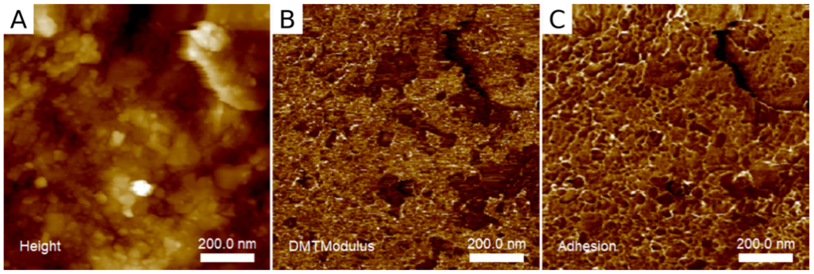
2.3. AFM Examination

A water-cooled diamond saw (314.249.524 ATZ Farmont) was used to cut out rectangular enamel slabs of approximately 5 × 5 × 1 mm3. Obtained samples were glued with highly adhesive thermoplastic material to microscope slides, and then their surfaces were prepared for examination using AFM. Preparation involved wetting of the sample surfaces with ethanol, quickly blowing off the residual alcohol and briefly drying in a stream of air followed by drying in the open air.
The atomic force microscope used in the study was Multimode 8, with a Nanoscope V controller (Bruker). The scanning probe was HQ:NSC14/ALBS (MikroMasch). The samples were imaged in the PF-QNM mode in the air; scan length was 1 µm, with the resolution 256 scan steps along each direction.
Obtained AFM images were processed in order to remove the surface tilt and to align individual image lines.
The following nanomechanical parameters were determined:
  • Sq: Surface roughness—root mean square surface roughness;
  • ISAD: Image Surface Area Difference—surface development; the ratio of the area spanned by the samples (triangulated) to the area of a flat surface with the same side length; the difference between the 3D surface area and its 2D footprint area;
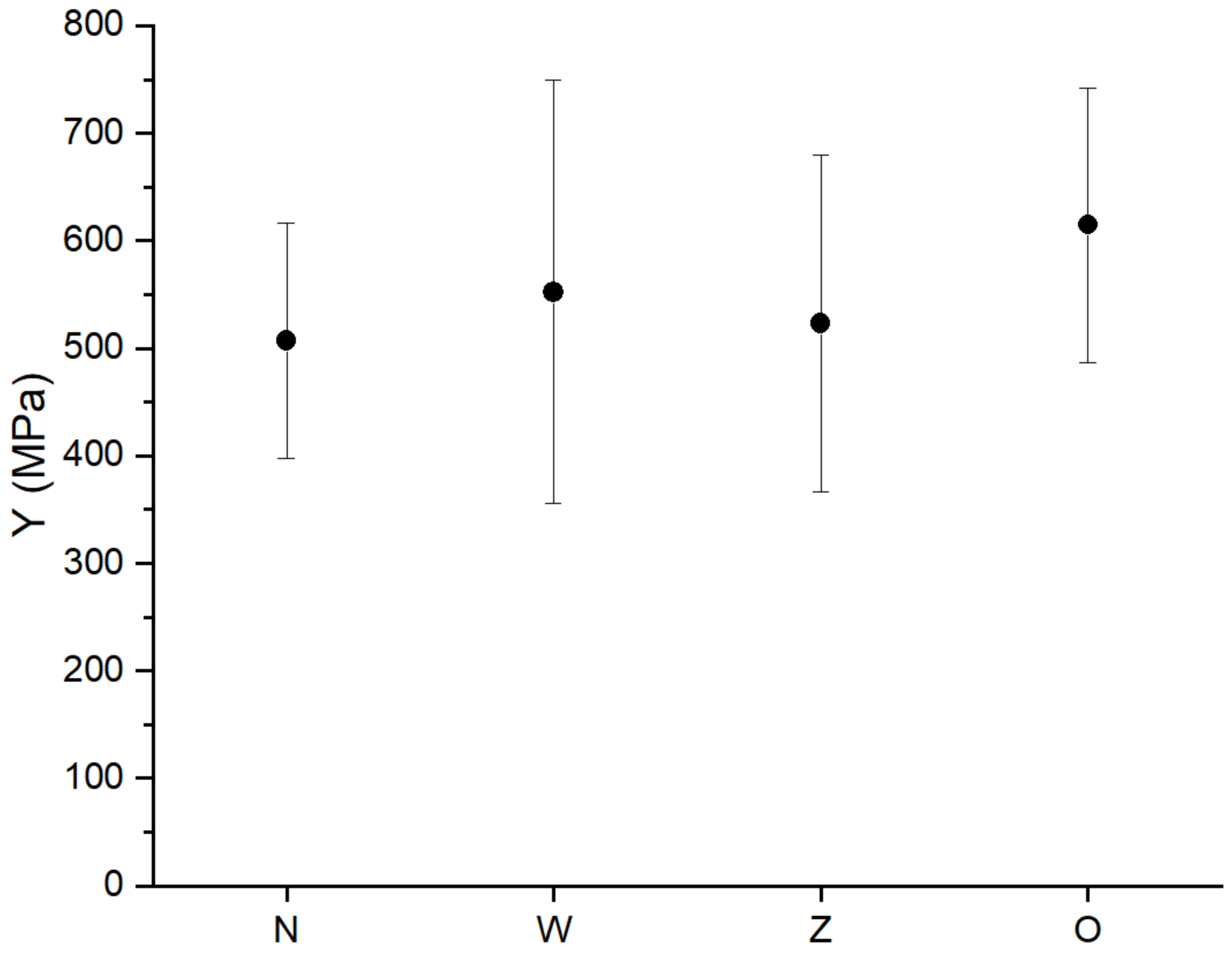
  • Ym: Mean Young’s modulus—Young’s pseudo-modulus of elasticity averaged over the image;
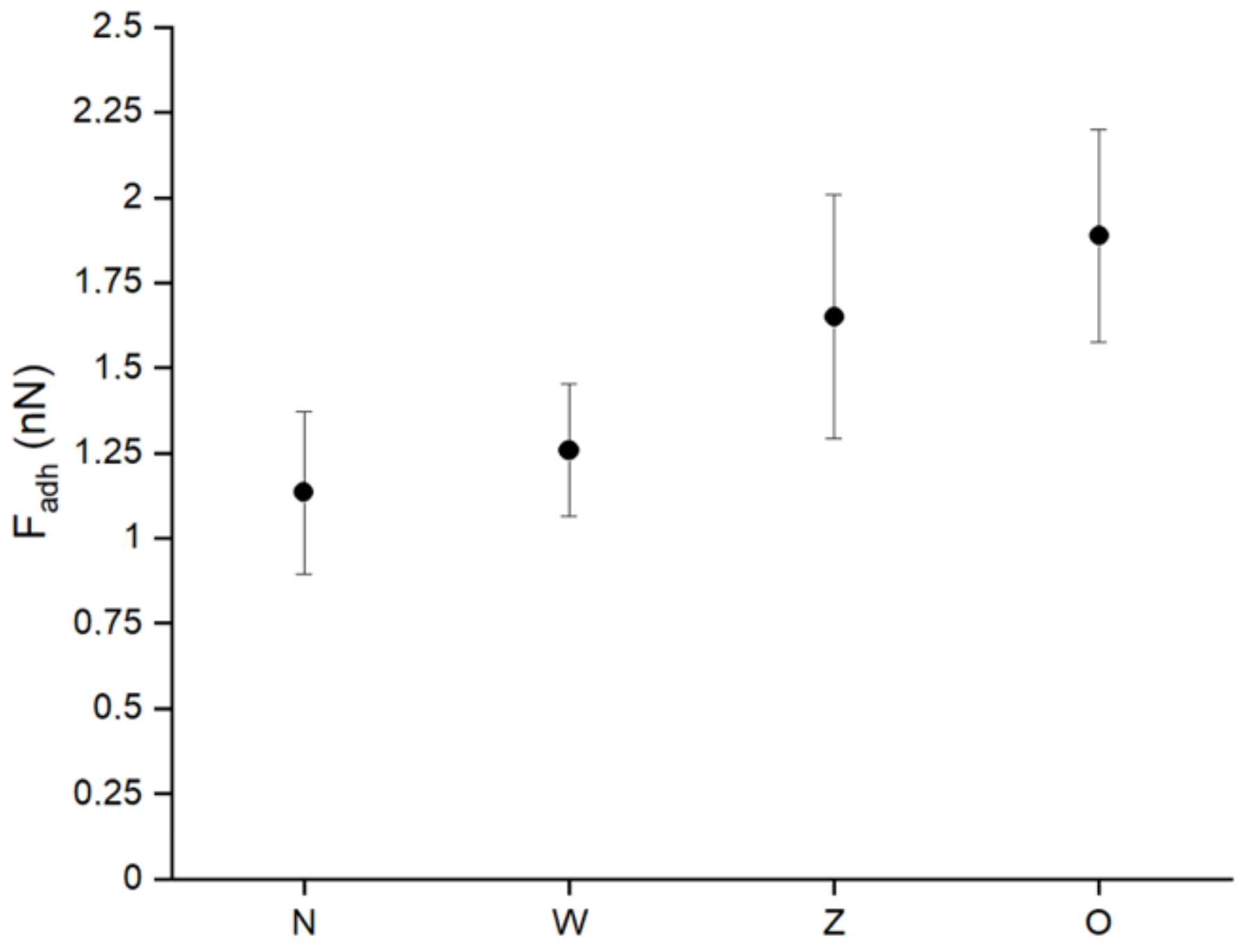
  • Fam: Mean adhesion force—the force of attraction between the scanning blade and the surface averaged over the image;
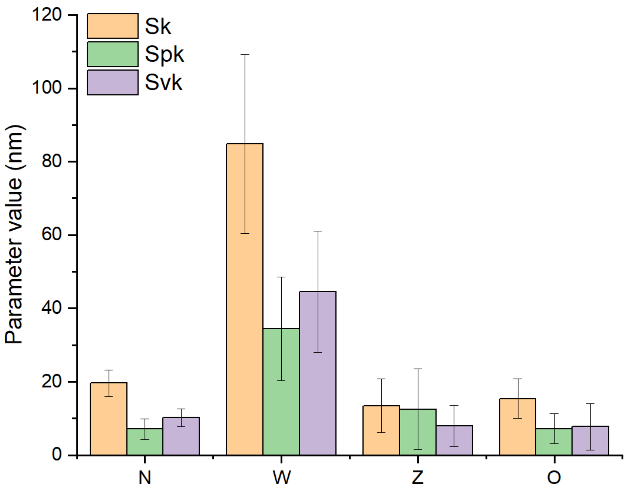
  • Sk: core—the central part of the surface responsible for load transmission;
  • Spk: peaks—top of the surface, subject to the most abrasive wear;
  • Svk: valleys—the lower part of the surface responsible for the transport of fluids.
Figure 1 shows the sample image of the AFM mapping.

2.4. Statistical Analysis

To compare the test results in the intact, cleaned, etched, and resin groups, the Kruskal-Wallis, ANOVA and median tests as well as multiple comparisons of mean ranks for all the samples were used. The level of statistical significance was p < 0.05.

3. Results

The obtained mean values of nanomechanical parameters in all groups are presented in Table 1.
(1) Sq: surface roughness
The roughness parameter analysis showed that
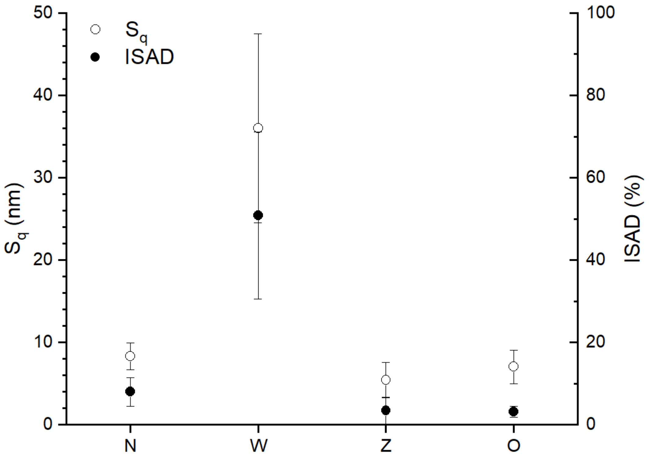
  • the highest roughness occurred after etching the surface and was on average 36 nm,
  • the resin-coated surface showed roughness of 5.4 nm and was only slightly (but statistically significantly, p < 0.05) less rough than the intact enamel, where the roughness was 8.3 nm and
  • the roughness in the group of teeth with cleaned enamel was 7.0 nm and was lower than in the intact enamel group and similar to the resin-coated enamel group. These differences were statistically significant (p < 0.05).
The results of the above analysis are presented graphically in Figure 2.
(2) Ym: mean Young’s modulus—pseudo-modulus of elasticity
Enamel etching and its cleaning of the resin are the steps of the orthodontic abutment bonding procedure, which significantly increase the elasticity coefficient.
The mean value of the pseudo-modulus of elasticity is shown in Figure 3.
The Young’s pseudo-modulus is the largest after enamel etching and has a mean value of 350–750 MPa; after enamel cleaning, it is 500–750 MPa (p < 0.05).
The pseudo-modulus in the intact enamel is on average 400–620 MPa; after using the resin, it is 350–650 MPa.
(3) Fam: mean adhesion force—the force of attraction between the scanning blade and the surface averaged over the image are presented in Figure 4.
The samples with the intact enamel show an adhesion strength of 0.9–1.3 Fadh (nN) and the samples with the etched enamel show a force of 1.0–1.5 Fadh (nN), whereas the samples from groups 3 and 4 show a greater range of adhesion: 1.25–2.0 Fadh (nN) and 1.5–2.25 Fadh (nN), respectively.
(4) Surface development
The graph in Figure 5 correlates with Figure 1, which results from the direct relationship between the surface roughness (height amplitude) and the thickness of the individual components. The strong effect of etching on exposing the porous surface structure is again confirmed.

4. Discussion

The iatrogenic effect of orthodontic treatment on tooth tissues has been studied for many years. So far, studies have been carried out with the use of a profilometer, SEM [22], OCT [1] and AFM [23].
Various aspects of the negative impact of fixing orthodontic brackets have been investigated, such as the influence of etchants on the enamel (classic etching method, self-etching system) [24], the presence and type of abutments [13], contact time with the enamel and methods of their removal. The effects of cleaning of the composite resin remnants and the adhesive from the enamel with rotating tools [25] were also analysed.
The research described in this paper focuses on the assessment of changes in the topography and nanomechanical properties of enamel during orthodontic treatment with fixed braces and aligners. Due to the use of additional attachments, the aligners also require enamel surface preparation in the same way as the attachments of fixed braces.
The first stage of enamel preparation is etching. The effect of etching on the human enamel depends on nanochemical and nanophysical interactions between acids and the enamel. These interactions may lead to the loss of hydroxyapatite crystals on the surface and the erosion process, resulting in a rough enamel surface [26]. AFM studies confirm the highest enamel roughness after etching. There are reports indicating that there is a significant difference in the acid etching patterns on different teeth, which suggests that in vitro tests should be performed on one tooth type or with the same number of different tooth types, which was done in the described experiment [27,28,29,30].
The resin-coated surface showed slightly lower roughness than the intact enamel. The roughness parameter in the group of teeth with cleaned enamel was lower than in the intact enamel group and similar to the group of surfaces covered with resin.
The results support the assumption that enamel etching removes organic matter, leaving a porous surface structure. Noteworthy is the strong surface development after etching, probably associated with removing all loosely connected elements and exposing the hard, porous structure of the sample. The applied resin refills the pores, which reduces the roughness and development ratio to previous values. An interesting result is the reduced roughness of the enamel after it has been cleaned of the orthodontic adhesive remnants using a rubber containing aluminum oxides. Many studies show an increase in surface roughness after using cemented carbide, composite or glass fibre drills [31,32,33]. This suggests that the use of rubbers with alumina, despite the extended cleaning time, is safer for the tissue surface than drills of any kind. Some studies [34] question the validity of using AFM to assess enamel roughness. Due to the fact that the pits between enamel crystals formed by acid etching are deeper than the AFM needle length, it is not possible to reach all the crystallite surfaces of the acid-etched enamel ends, and this leads to the profilometric underestimation of the roughness parameters of the acid-etched enamel. Therefore, AFM images at low magnification appear much smoother than SEM images at the appropriate magnification. However, at higher magnification, the AFM images of acid-etched enamel reflect the geometry of the probe tip. As a consequence, all roughness parameters measured with AFM on acid-etched tooth enamel surfaces are largely underestimated. However, the main advantage of AFM over other technologies is that it provides quantitative roughness data [15,35]. This method has proven effective in assessing micro-damage on hard surfaces. The advantages of AFM include the simultaneous delivery of 2D and 3D images and minimal sample preparation. Moreover, this method does not require staining or coating of the tested samples [36,37,38]. Similar tests were made according to restorative dentistry [39].
The enamel microstructure can withstand repeated mechanical stress. Although significant progress has been made in understanding the mechanical properties of enamel, there are still challenges in testing and interpreting its nanomechanical properties. For example, enamel studied in vitro is tested under dry conditions, which are significantly different from the natural environment; the measurement site differs, and the thickness and strength of the enamel vary depending on the area of the tooth it covers. The conducted research has shown that the Young’s pseudo-modulus is the highest after enamel etching and after enamel cleaning. These results differ from the studies by Ioannidis et al. [40], which showed no change in the nanomechanical properties of enamel during bonding and disassembly of orthodontic abutments. Differences in results between SEM and AFM may be due to a different test environment. AFM tests are carried out in an environment similar to the natural conditions for enamel, according to a study by G. W. Marshall Jr., which showed fracture toughness values for enamel of 0.6–0.9 MPa·m1/2 [40].
Fluctuations in the adhesion force during imaging in air are related to the geometry of the blade-surface contact and the thickness of the capillary layer. Assuming constant blade geometry, it can be assumed that the changes observed here correspond to changes in the hydrophilicity/hydrophobicity of the surface because the samples in the etched enamel group, characterized by strong surface development (and large depth of gaps that retain the capillary layer), have adhesion strength similar to that of samples with intact enamel, whose roughness is 10 times lower and thus are more hydrophobic. In turn, samples from groups 3 and 4 show greater adhesion; therefore, they are more hydrophilic. The clear upward tendency of the adhesive forces indicates the increasing force of the blade-surface interaction, which may be related to porosity. The high porosity of the untreated surface makes it difficult to form a capillary layer, unlike the smooth surface after applying the resin.

5. Conclusions

  • Etching results in removing organic substances of enamel, which increases its roughness and reduces tissue hardness.
  • The use of resin reduces roughness of the etched surface and increases hardness compared to the etched enamel.
  • The intact enamel reveals the highest hardness.
  • The enamel smoothness is greater after polishing the enamel with an alumina rubber than in the intact enamel, which suggests the legitimacy of dental polishing of healthy teeth in order to reduce the deposition of biofilms and plaque.
  • The highest adhesion in the resin-coated enamel group confirms the legitimacy of using the procedure of etching and bonding the enamel in order to increase the strength of the bond between the enamel and the composite material.

Author Contributions

Conceptualization, M.M.; methodology, M.M.; software, S.W.; validation, L.S.-S. and K.W.; formal analysis, L.S.-S. and A.D.; investigation, M.M. and S.W.; resources, K.W.; data curation, M.M., A.D. and S.K.; writing—original draft preparation, M.M.; writing—review and editing, M.M.; visualization, S.W.; supervision, S.W. and K.W.; project administration, M.M.; funding acquisition, K.W. and S.W. All authors have read and agreed to the published version of the manuscript.

Funding

This study was financially supported by Medical University of Silesia, grant no. PCN-1-013/K/0/O.

Institutional Review Board Statement

Not applicable.

Informed Consent Statement

Not applicable.

Data Availability Statement

The data are available and archived in database of Faculty of Technical Sciences, Warmia and Mazury University in Olsztyn by prof. Kulesza.

Conflicts of Interest

The authors declare no conflict of interest.

References

  1. Machoy, M.E.; Seeliger, J.; Szyszka-Sommerfeld, L.; Koprowski, R.; Gedrange, T.; Woźniak, K. Evaluation of changes in enamel thickness after orthodontic treatment depending on the force applied to remove orthodontic brackets: OCT analysis and universal testing machine. Adv. Clin. Exp. Med. 2019, 28, 807–813. [Google Scholar] [CrossRef] [PubMed]
  2. Hobson, R.S.; Mccabe, J.F. Relationships between enamel etch characteristics and resin-enamel bond strength. Br. Dent. J. 2002, 192, 463–468. [Google Scholar] [CrossRef] [PubMed]
  3. Fujita, K.; Nishiyama, N. 13C NMR analysis of the etching efficacy of acidic monomers in self-etching primers. J. Dent. 2006, 34, 123–133. [Google Scholar] [CrossRef] [PubMed]
  4. Horiuchi, S.; Kaneko, K.; Mori, H.; Kawakami, E.; Tsukahara, T.; Yamamoto, K.; Hamada, K.; Asaoka, K.; Tanaka, E. Enamel bonding of self-etching and phosphoric acid-etching orthodontic adhesives in simulated clinical conditions: Debonding force and enamel surface. Dent. Mater. J. 2009, 28, 419–425. [Google Scholar] [CrossRef] [Green Version]
  5. Scougall Vilchis, R.J.; Yamamoto, S.; Kitai, N.; Hotta, M.; Yamamoto, K. Shear bond strength of a new fluoride-releasing orthodontic adhesive. Dent. Mater. J. 2007, 26, 45–51. [Google Scholar] [CrossRef] [Green Version]
  6. Vicente, A.; Bravo, L.A.; Romero, M. Influence of a nonrinse conditioner on the bond strength of brackets bonded with a resin adhesive system. Angle Orthod. 2005, 75, 400–405. [Google Scholar]
  7. Bishara, S.E.; VonWald, L.; Laffoon, J.F.; Warren, J.J. Effect of a self-etch primer/adhesive on the shear bond strength of orthodontic brackets. Am. J. Orthod. Dentofac. Orthop. 2001, 119, 621–624. [Google Scholar] [CrossRef]
  8. Hosein, I.; Sherriff, M.; Ireland, A.J. Enamel loss during bonding, debonding, and cleanup with use of a self-etching primer. Am. J. Orthod. Dentofac. Orthop. 2004, 126, 717–724. [Google Scholar] [CrossRef]
  9. Seeliger, J.H.; Botzenhart, U.U.; Gedrange, T.; Kozak, K.; Stepien, L.; Machoy, M. Enamel shear bond strength of different primers combined with an orthodontic adhesive paste. Biomed. Tech. 2017, 62, 415–420. [Google Scholar] [CrossRef]
  10. Machoy, M.E.; Koprowski, R.; Szyszka-Sommerfeld, L.; Safranow, K.; Gedrange, T.; Woźniak, K. Optical coherence tomography as a non-invasive method of enamel thickness diagnosis after orthodontic treatment by 3 different types of brackets. Adv. Clin. Exp. Med. 2019, 28, 211–218. [Google Scholar] [CrossRef]
  11. Maas, M.C.; Dumont, E.R. Built to last: The structure, function, and evolution of primate dental enamel. Evol. Anthropol. 1999, 8, 133–152. [Google Scholar] [CrossRef]
  12. Zarrinnia, K.; Eid, N.M.; Kehoe, M.J. The effect of different debonding techniques on the enamel surface: An in vitro qualitative study. Am. J. Orthod. Dentofac. Orthop. 1995, 108, 284–293. [Google Scholar] [CrossRef]
  13. Cochrane, N.J.; Ratneser, S.; Reynolds, E.C. Effect of different orthodontic adhesive removal techniques on sound, demineralized and remineralized enamel. Aust. Dent. J. 2012, 57, 365–372. [Google Scholar] [CrossRef] [Green Version]
  14. Habelitz, S.; Marshall, S.J., Jr.; Marshall, G.W.; Balooch, M. Mechanical properties of human dental enamel on the nanometre scale. Arch. Oral Biol. 2001, 46, 173–183. [Google Scholar] [CrossRef]
  15. Loyola-Rodriguez, J.P.; Zavala-Alonso, V.; Reyes-Vela, E.; Patiño-Marin, N.; Ruiz, F.; Anusavice, K.J. Atomic force microscopy observation of the enamel roughness and depth profile after phosphoric acid etching. J. Electron Microsc. 2010, 59, 119–125. [Google Scholar] [CrossRef]
  16. Kirkham, J.; Brookes, S.J.; Zhang, J.; Wood, S.R.; Shore, R.C.; Smith, D.A.; Wallwork, M.L.; Robinson, C. Effect of experimental fluorosis on the surface topography of developing enamel crystals. Caries Res. 2001, 35, 50–56. [Google Scholar]
  17. Watari, F. In-situ etching observation of human teeth in acid agent by atomic force microscopy. J. Electron Microsc. 1999, 48, 537–544. [Google Scholar] [CrossRef]
  18. Watari, F. In situ quantitative analysis of etching process of human teeth by atomic force microscopy. J. Electron Microsc. 2005, 54, 299–308. [Google Scholar] [CrossRef]
  19. Batina, N.; Renugopalakrishnan, V.; Casillas Lavin, P.N.; Guerrero, J.C.H.; Morales, M.; Garduno-Juarez, R.; Lakka, S.L. Ultrastructure of dental enamel afflicted with hypoplasia: An atomic force miscroscopic study. Calcif. Tissue Int. 2004, 74, 294–301. [Google Scholar]
  20. Wen, H.B.; Fincham, A.G.; Morodian-Oldak, J. Progressive accretion of amelogenin molecules during nanospheres assembly revealed by atomic force microscopy. Matrix Biol. 2001, 20, 387–395. [Google Scholar] [CrossRef]
  21. Moradian-Oldak, J.; Paine, M.L.; Lei, Y.P.; Fincham, A.G.; Snead, M.L. Self-assembly properties of recombinant engineered amelogenin proteins analyzed by dynamic light scattering and atomic force microscopy. J. Struct. Biol. 2000, 131, 27–37. [Google Scholar] [CrossRef]
  22. Eliades, T.; Gioka, C.; Eliades, G.; Makou, M. Enamel surface roughness following debonding using two resin grinding methods. Eur. J. Orthod. 2004, 26, 333–338. [Google Scholar] [CrossRef] [Green Version]
  23. Mohebi, S.; Shafiee, H.A.; Ameli, N. Evaluation of enamel surface roughness after orthodontic bracket debonding with atomic force microscopy. Am. J. Orthod. Dentofac. Orthop. 2017, 151, 521–527. [Google Scholar] [CrossRef]
  24. Machoy, M.; Seeliger, J.; Koprowski, R.; Safranow, K.; Gedrange, T.; Woźniak, K. Corrigendum to “Enamel Thickness before and after Orthodontic Treatment Analysed in Optical Coherence Tomography”. BioMed Res. Int. 2020, 2020. [Google Scholar] [CrossRef]
  25. Machoy, M.; Machoy-Mokrzynska, A.; Szyszka-Sommerfeld, L.; Woźniak, K. Evaluation of the influence of the types of orthodotic materials on the enamel surface clean-up after fixed appliances removal. Probl. Nauk Stosow. 2018, 8, 177–184. [Google Scholar]
  26. Beyer, M.; Reichert, J.; Bossert, J.; Sigusch, B.W.; Watts, D.C.; Jandt, K.D. Acids with an equivalent taste lead to different erosion of human dental enamel. Dent. Mater. 2011, 27, 1017–1023. [Google Scholar] [CrossRef]
  27. Hannig, M.; Hannig, C. Nanomaterials in preventive dentistry. Nat. Nanotechnol. 2010, 5, 565–569. [Google Scholar] [CrossRef]
  28. Hobson, R.S.; Rugg-Gunn, A.J.; Booth, T.A. Acid-etch patterns on the bucal surface of human permanent teeth. Arch. Oral. Biol. 2002, 47, 407–412. [Google Scholar] [CrossRef]
  29. Wennerberg, A.; Sawase, T.; Kultje, C. The influence of Carisolv on enamel and dentine surface topography. Eur. J. Oral Sci. 1999, 107, 297–306. [Google Scholar] [CrossRef]
  30. Zanet, C.G.; Arana-Chavez, V.E.; Fava, M. Scanning electron microscopy evaluation of the effect of etching agents on human enamel surface. J. Clin. Pediatr. Dent. 2006, 30, 247–250. [Google Scholar] [CrossRef]
  31. Moura, S.K.; Pelizzaro, A.; Dal Bianco, K.; De Goes, M.F.; Loguercio, A.D.; Reis, A.; Grande, R.H. Does the acidity of self-etching primers affect bond strength and surface morphology of enamel? J. Adhes. Dent. 2006, 8, 75–83. [Google Scholar] [PubMed]
  32. Garg, R.; Dixit, P.; Khosla, T.; Gupta, P.; Kalra, H.; Kumar, P. Enamel Surface Roughness after Debonding: A Comparative Study using Three Different Burs. J. Contemp. Dent. Pract. 2018, 19, 521–526. [Google Scholar] [PubMed]
  33. Pont, H.B.; Ozcan, M.; Bagis, B.; Ren, Y. Loss of surface enamel after bracket debonding: An in-vivo and ex-vivo evaluation. Am. J. Orthod. Dentofac. Orthop. 2010, 138, 387.e1–387.e9. [Google Scholar] [CrossRef] [PubMed]
  34. Ozer, T.; Basaran, G.; Kama, J. Surface roughness of the restored enamel after orthodontic treatment. Am. J. Orthod. Dentofac. Orthop. 2010, 137, 368–374. [Google Scholar] [CrossRef]
  35. Vitkov, L.; Kastner, M.; Kienberger, F.; Hinterdorfer, P.; Schilcher, K.; Grunert, I.; Dumfahrt, H.; Krautgartner, W.D. Correlations Between AFM and SEM Imaging of Acid-Etched Tooth Enamel. Ultrastruct. Pathol. 2008, 32, 1–14. [Google Scholar] [CrossRef] [PubMed]
  36. Mattick, C.R.; Hobson, R.S. A comparative micro-topographic study of the buccal enamel of different tooth types. J. Orthod. 2000, 27, 143–149. [Google Scholar] [CrossRef] [PubMed]
  37. Kakaboura, A.; Fragouli, M.; Rahiotis, C.; Silikas, N. Evaluation of surface characteristics of dental composites using profilometry, scanning electron, atomic force microscopy and gloss-meter. J. Mater. Sci. Mater. Med. 2007, 18, 155–163. [Google Scholar] [CrossRef]
  38. De Vasconcellos, B.T.; Miranda-Junior, W.G.; Prioli, R.; Thompson, J.; Oda, M. Surface roughness in ceramics with different finishing techniques using atomic force microscope and profilometer. Oper. Dent. 2006, 31, 442–449. [Google Scholar]
  39. Serino, G.; Bignardi, C.; Boccafoschi, C.; Scotti, N.; Berutti, E.; Audenino, A.L. Collagen cross-linker effect on the mechanical properties of the radicular hybrid layer in restorative dentistry: A nanoindentation study. WIT Trans. Eng. Sci. 2019, 124, 195–203. [Google Scholar]
  40. Marshall, G.W., Jr.; Balooch, M.; Gallagher, R.R.; Gansky, S.A.; Marshall, S.J. Mechanical properties of the dentinoenamel junction: AFM studies of nanohardness, elastic modulus and fracture. J. Biomed. Mater. Res. 2001, 54, 87–95. [Google Scholar] [CrossRef]
Figure 1. Visualization of topography, Mean Young’s modulus and adhesion in atomic force microscopy.
Applsci 11 03918 g001
Figure 2. Surface roughness and development averaged over the samples (N: intact, W: etched, Z: resin, O: cleaned).
Applsci 11 03918 g002
Figure 3. Graph of the averaged Young’s pseudo-modulus of elasticity. The black dot means mean value.
Applsci 11 03918 g003
Figure 4. Graph of the averaged blade-surface adhesion force. The black dot means mean value.
Applsci 11 03918 g004
Figure 5. Graphs showing the values of functional parameters of the surface texture describing thicknesses of the surface components: core (Sk), peaks (Spk) and valleys (Svk).
Applsci 11 03918 g005
Table 1. Mean values of nanomechanical parameters in groups 1, 2, 3, 4.
SampleSq
[nm]
ISAD [%]Sk
[nm]
Spk
[nm]
Svk
[nm]
Ym
[MPa]
FAm
[nN]
18.3(1.7)8.0(3.5)19.6(3.6)7.1(2.9)10.2(2.5)510(110)1.13(0.24)
236(12)51(21)85(25)34(15)45(17)550(200)1.26(0.20)
35.4(2.2)3.4(3.4)13.5(7.4)13(11)8.0(5.6)520(160)1.65(0.36)
47.0(2.1)3.2(1.3)15.4(5.3)7.2(4.2)7.7(6.3)610(130)1.89(0.32)
Sq: surface roughness—root mean square surface roughness; ISAD: Image Surface Area Difference—surface development (the ratio of the area spanned by the samples (triangulated) to the area of a flat surface with the same side length); Ym: mean Young’s modulus—Young’s pseudo-modulus of elasticity averaged over the image; Fam: mean adhesion force—the force of attraction between the scanning blade and the surface averaged over the image; Sk: core—the central part of the surface responsible for load transmission.
Publisher’s Note: MDPI stays neutral with regard to jurisdictional claims in published maps and institutional affiliations.

