Article

Effectiveness of Shock Wave Therapy versus Intra-Articular Corticosteroid Injection in Diabetic Frozen Shoulder Patients’ Management: Randomized Controlled Trial

by
Ahmed Ebrahim Elerian
1,*,
David Rodriguez-Sanz
2,
Abdelaziz Abdelaziz Elsherif
3,
Hend Adel Dorgham
4,
Dina Mohamed Ali Al-Hamaky
5,
Mahmoud S. El Fakharany
6 and
Mahmoud Ewidea
7,*
1
Department of Basic Science for Physical Therapy, Faculty of Physical Therapy, Cairo University, Ad Doqi, Giza District, Giza Governorate 11432, Egypt
2
Faculty of Nursing, Physical Therapy and Podiatry, Universidad Complutense de Madrid, 28040 Madrid, Spain
3
Department of Physical Therapy for Neuromuscular Disorders and Its Surgery, Faculty of Physical Therapy, Cairo University, Ad Doqi, Giza District, Giza Governorate 11432, Egypt
4
Faculty of Physical Therapy, Cairo University, Ad Doqi, Giza District, Giza Governorate 11432, Egypt
5
Department of Physical Therapy for Musculoskeletal Disorders and Its Surgery, Faculty of Physical Therapy, Cairo University, Ad Doqi, Giza District, Giza Governorate 11432, Egypt
6
Department of Physical Therapy for Pediatrics, Faculty of Physical Therapy, Cairo University, Ad Doqi, Giza District, Giza Governorate 11432, Egypt
7
Department of Basic Science for Physical Therapy, Faculty of Physical Therapy, Kafrelsheikh University, Kafrelsheikh Government 33511, Egypt
*
Authors to whom correspondence should be addressed.
Appl. Sci. 2021, 11(8), 3721; https://doi.org/10.3390/app11083721
Submission received: 11 February 2021 / Revised: 16 March 2021 / Accepted: 15 April 2021 / Published: 20 April 2021
(This article belongs to the Special Issue Physical Therapy and Health)

Abstract

:

Featured Application

Adhesive Capsulitis is one of the most common, yet challenging clinical disorders presenting to the orthopedic surgeon. Shock wave therapy was demonstrated as a conservative intervention that provides good results for the treatment of frozen shoulder. Although injection of corticosteroids is an invasive procedure but associated with many risks. This study aimed to investigate the effectiveness of shock wave versus intra-articular corticosteroid injection with evaluation of their side effects.

Abstract

Frozen shoulder is a major musculoskeletal illness in diabetic patients. This study aimed to compare the effectiveness of shock wave and corticosteroid injection in the management of diabetic frozen shoulder patients. Fifty subjects with diabetic frozen shoulder were divided randomly into group A (the intra-articular corticosteroid injection group) and group B that received 12 sessions of shock wave therapy, while each patient in both groups received the traditional physiotherapy program. The level of pain and disability, the range of motion, as well as the glucose triad were evaluated before patient assignment to each group, during the study and at the end of the study. Compared to the pretreatment evaluations there were significant improvements of shoulder pain and disability and in shoulder flexion and abduction range of motion in both groups (p < 0.05). The shock wave group revealed a more significant improvement the intra-articular corticosteroid injection group, where p was 0.001 for shoulder pain and disability and shoulder flexion and abduction. Regarding the effect of both interventions on the glucose triad, there were significant improvements in glucose control with group B, where p was 0.001. Shock waves provide a more effective and safer treatment modality for diabetic frozen shoulder treatment than corticosteroid intra-articular injection.

1. Introduction

Many painful musculoskeletal conditions that interfere with regular daily activity, including trigger finger, carpal tunnel syndrome, osteoarthritis, and frozen shoulder, are associated with diabetes mellitus (DM) [1]. Adhesive capsulitis (AC) (also referred to as frozen shoulder) is characterized by severe shoulder pain [2] with advanced limitation of the joint range of motion associated with functional disability [3], that negatively affects life quality and care costs [4]. AC is caused by inflammation and results in tissue fibrosis, which might lead to joint capsule tightness [5].
Many studies had approved positive effects of oral nonsteroidal anti-inflammatory agents and oral glucocorticoids on pain management and the improvement of the range of motion in diabetic frozen shoulder patient, but without any longer effect than a few weeks [6]; hydro-dilatation accompanied with manipulation under anesthesia is also considered an effective method for resolving the condition of AC, but with the risk of anesthesia complications [6], while several studies approved many physiotherapy modalities such as ultrasound therapy, stretching exercises, manual mobilization and strengthening exercises as well-established conservative management techniques [7,8].
Multiple studies have reported the short-term benefits of corticosteroid injections, including the improvement of the range of motion (ROM) in the shoulder and pain reduction [9,10], through the reduction of the synovial inflammation by decreasing the capsular fibrosis, thereby improving joint movements and providing pain relief [11]. Corticosteroid injections are also associated with complications, such as fat atrophy, skin color changes, infected arthritis, post injection symptom flare/synovitis, allergic reaction, and steroid chondro-calcific arthropathy, as well as increasing the blood glucose level, which is one of the most dangerous side effects among diabetic patients. Therefore, it would be better to avoid steroids and search for alternative therapies [12,13].
Shock wave therapy (SWT) is a physical therapy modality that consists of very short energy waves; the waves are faster than the speed of sound and it is considered a novel treatment in a wide variety of musculoskeletal dysfunction and illness [14,15,16]. Shock wave application has been reported to result in good clinical satisfaction in patients with many conditions that include pain and disability associated with ligament or muscle injuries, joint arthritis, tendon entrapment, or inflammation [17,18,19].
Therefore, the current study’s principal aim was to investigate the effectiveness of SWT versus intra-articular corticosteroid injection when each modality was separately added to traditional physiotherapy in the management of diabetic shoulder AC patients. Additionally, evaluating both techniques’ side effects on blood glucose levels helps identify the most appropriate treatment plan for diabetic shoulder AC patients.

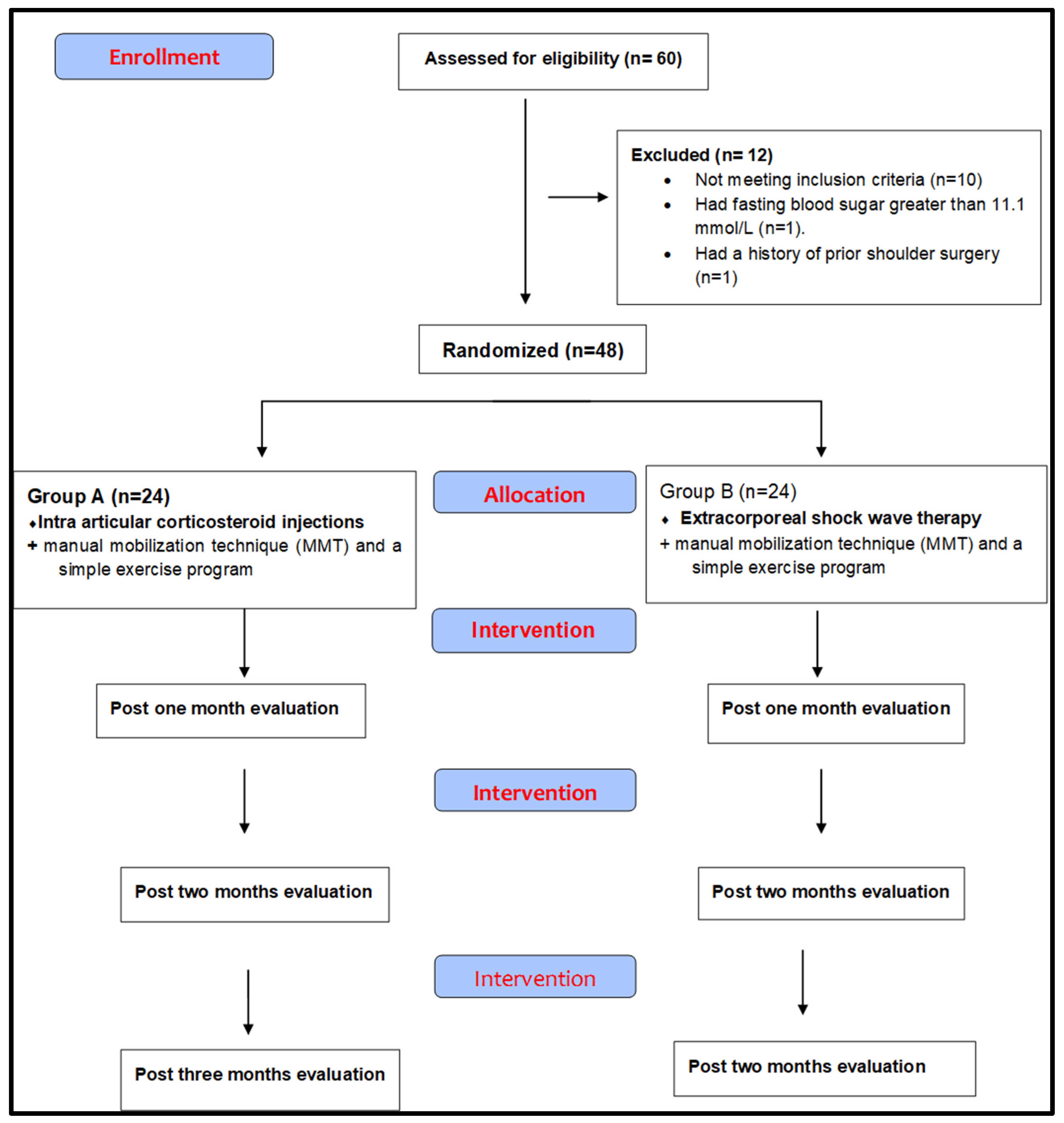
2. Materials and Methods

2.1. Design of the Study

A randomized controlled trial, in which participants were recruited from the Faculty of Physical Therapy Outpatient Clinic, in Cairo University Egypt. The SWT, traditional physiotherapy protocol, and the physical assessment of participants were conducted at the same outpatient clinic between April 2019 and February 2020. The Ethical Committee approved the study for Human Research at the faculty of Physical Therapy, Cairo University, Egypt. The study was registered at Pan African Clinical Trials Registry (PACTR) with a registration number PACTR201811597207089. All participants signed the consent form before enrolling in the study, according to the principles of the Helsinki Declaration 1975.

2.2. Participants

A specialized orthopedist referred 50 adult diabetic patients suffering from shoulder AC to our physical therapy outpatient clinic based on the diagnostic criteria for diabetic shoulder AC, described by Lebiedz and Kay 2010 and according to inclusion criteria matching [20]. The study inclusion criterion was any diabetic frozen shoulder with age range between 45 and 64 years from both sexes. The exclusion criteria were patients with rheumatologic and neurological disorders, under oral corticosteroids, intra-articular injection of hyaluronic acid agents during the 6 months before treatment, as well as any patient who had poorly controlled diabetes mellitus with fasting blood sugar greater than 11.1 mmol/L, a history of prior shoulder surgery, blood coagulation disorders, or tumors, and any pregnant or breastfeeding patients.

2.3. Sample Size and Randomization

Sample size in the present study was calculated using G*Power software (Dusseldorf, Germany) to measure a variation of 15° in shoulder range of motion between groups, measured using an electrogoniometer with an estimated standard deviation of 15°, 80% power analysis, and 5% significance level [7]. The number of patients needed in each group was calculated as 21; to account for patient attrition during the study, this number was increased to be 30 subjects. Ten subjects did not meet the inclusion criteria, while two diabetic frozen shoulder patients were excluded from the study before allocation to each group; one of them had fasting blood sugar greater than 11.1 mmol/L, and the other one had a history of prior shoulder surgery. The remaining 48 diabetic frozen shoulder patients were assigned randomly to receive intra-articular corticosteroid injection accompanied by traditional physical therapy sessions (Group A = 24) or received 12 sessions of a shock wave in addition to the traditional physiotherapy sessions (Group B = 24) as shown in Figure 1. Participants were allocated using a random numbers table generated by computer and sealed in opaque envelopes for concealed allocation. The patients and practitioners and the outcome assessors were blinded to the patient groupings.

2.4. Outcome Measurements

All outcome measures except Hemoglobin A1c (HbA1c) were evaluated for all patients four times, first before being allocated in their group, then two times during the study at 1-month intervals, and again after the end of the study (3 months). Hemoglobin A1c (HbA1c) measurement was evaluated only twice: just before allocating the study group and after 3 months.

2.4.1. The Primary Outcome Measurements

The Shoulder Pain and Disability Index (SPADI) is a valid and reliable questionnaire that measures the level of shoulder joint pain and disability [21]. This questionnaire consists of 13 items; five items addressed pain, while eight items addressed the function. All items were scored on a visual analog scale; each domain score was equally weighted. The total percentage score ranged from 0 to 100, where 0 was no pain and maximum function while 100 was maximum pain and disability [22].
A Smarttool Digital Electrogoniometer (Oklahoma City, OK, USA) was used for shoulder flexion and abduction ROM measurements [23]. For shoulder flexion ROM measurement, the digital electro goniometer was placed along the patient’s arm’s posterior aspect. In contrast, for shoulder abduction ROM measurement, the digital electrogoniometer was placed along the medial aspect of the patient’s arm [24]. All primary outcome measurements either before or during and at the end of study duration were measured by the same physical therapist who did not share in the intervention procedure.

2.4.2. Secondary Outcome Measurements

Glucose triad (fasting, postprandial glucose levels and HbA1c) was used to evaluate both treatment protocols’ side effects on blood glucose control in diabetic patients.
Blood glucose level assessment was done using a Bio-Systems Photometer (BTS 330, Spain). Blood samples were withdrawn by a laboratory technician specialist from the antecubital vein of each patient for evaluation of fasting blood glucose level (FBGL) and hemoglobin A1c (HbA1c) after fasting for 8 h, while for postprandial blood glucose level (PPBGL), 75 g of glucose mixed with water was given to each patient to drink, and 2 h later, a blood sample was withdrawn at the Cairo University hospital clinical lab [25].

2.5. Interventions

2.5.1. Traditional Physiotherapy Protocol

In both groups, patients received a traditional physiotherapy program in the manual mobilization technique (MMT) and a simple exercise program by a manual therapist with 10 years of clinical experience [26]. The MMT in the form of end-range mobilization (ERM) and the simple exercise program in the form of pendulum free range of motion exercises and isometric exercise for scapular retraction muscles for 30 min twice per week, and for 12 weeks, intensive mobilization with repetitions of 10–15 times was applied to the glenohumeral joint, with the humerus in a maximal range position in different directions [27,28]. Participants in the study were instructed to avoid any home exercise.

2.5.2. Intraarticular Corticosteroid Injection

Each patient in Group A received a mix of 2 mL of 2% lidocaine and 40 mg/mL of methylprednisolone (1 mL) shoulder intra-articular corticosteroid injection using 3-mL and 1-inch syringe, after detection and sterilization of injection area using a marker and alcohol swabs at next day after each time of physical and laboratory evaluations for all outcomes of the study. With the patient in a sitting position, the index of the free hand of a specialized orthopedist was placed on the tip of the coracoid process with the thump at the angle of the acromion and the spine of the scapula. With an imaginary line between the index finger and the thump crossing the glenoid cavity, the needle entry for injection would be near the orthopedist’s thump and just laterally to the tip of the index finger [29]. The injections were given at the beginning of the study, then after 4 and 8 weeks [30].

2.5.3. Shock Wave Therapy

A Gymna Shock master 500 Radial Shockwave Device (Gymna Uniphy-Pasweg 6a, 3740 Bilzen, Belgium) was used. All patients in Group B received one SWT session per week for 12 sessions by a physical therapist who did not share in evaluation procedures; each session involved 2000 shots on the anterior aspect of the shoulder joint crossing the midline of the joint with energy flux density ranged from low to moderate according to each patient tolerance of pain (0.06–0.14 m J/mm2) with a pressure of 4 bar and at 8 Hz frequency [31].

2.6. Data Analysis

Statistical Analysis

SPSS software for Windows, version 25.0 (Armonk, NY, USA) was used to conduct the statistical analysis. Descriptive statistics were performed at baseline, after 1 month, 2 months, and 3 months of the study. The normal distribution of data was checked using Shapiro–Wilk’s test. A mixed-design multivariate analyses of variance (MANOVA) were used to detect any differences between the mean change scores of the groups regarding SPADI scores, shoulder flexion ROM, shoulder abduction ROM, FBG level, PPBG level, and HbA1c level. Wilks’ lambda was used to detect F value. When the MANOVA revealed a significant effect (p < 0.05), a follow-up univariate mixed design ANOVAs were done with Bonferroni correction to avoid of type I error as the results of pairwise comparisons. A repeated measure ANOVAs, with a Bonferroni correction for multiple comparisons, was done to determine if there were differences in SPADI scores, shoulder flexion ROM, shoulder abduction ROM, FBG level, PPBG level, and HbA1c level in each group.

3. Results

3.1. Between-Groups Effect

After 1 month of treatment, there was no significant statistically difference between Group A and B regarding the SPADI, FBG, and PPBG (p = 0.29, 0.77, and 0.09, respectively), but flexion and abduction ROM of the shoulder was higher in group B than group A (113.8 ± 19.85 vs. 66.67 ± 15.23, p < 0.001; 67.5 ± 13.19 vs. 51.25 ± 7.41, p < 0.001, respectively) (Table 1). After 2 and 3 months of treatment, Group A and Group B showed a significant difference in SPADI score, shoulder flexion ROM, shoulder abduction ROM, FBG, and PPBG (all p-values < 0.001), as well as also for HbA1c after 3 months of treatment (<0.001). The negative mean difference scores (95% CI) on SPAD score, FBG, PPBG, and HbA1c outcome measures indicated more improvement. In comparison, positive mean difference scores (95% CI) regarding the shoulder flexion and abduction ROM outcome measures indicated more improvement, so Group B (Shock wave) had superior results to group A (intraarticular corticosteroid injection) regarding all outcome measures after 2 and 3 months of intervention and after 1 month of intervention for shoulder flexion and abduction ROM (p < 0.001) (Table 1).

3.2. Within-Group Effect

Within-group comparison for SPADI score revealed that there was a significant decrease in post-treatment measurements in both groups (p < 0.001), while for shoulder flexion and abduction ROM revealed that there was a significant increase post-treatment in both groups (p < 0.05) except flexion ROM in group A (p > 0.05) as shown in Table 2.
The results of the present study showed significant increase in FBG and PPBG at post-treatment measurements in group A (p < 0.001), with a greater increase at 3-month post-treatment measurement, with mean values of 196.9 ± 25.49, 286.7 ± 33.74 for FBG, PPBG, respectively. Additionally, as shown in Table 2, there was a significant increase in HbA1c after 3 months, with mean values of 6.53 ± 0.85 vs. 8.71 ± 1.01, (p < 0.001), at baseline and after 3 months, respectively. Regarding FBG and PPBG in Group B, the results found that the post-treatment measurements for FBG and PPBG decreased significantly only with p value p = 0.03 for FBG and p < 0.001, respectively, for PPBG as shown on Table 3, while in Group B, this study detected significant differences in HbA1c between the pretreatmentpre and post-treatment measurements (p > 0.21) as shown in Table 4.

4. Discussion

Diabetic patients can develop many musculoskeletal syndromes or symptoms; these musculoskeletal disorders have been associated with the severity and duration of diabetes mellitus. Shoulder AC is a musculoskeletal disorder associated with DM, in which the patients typically present with painful glenohumeral joint limitation and abduction and flexion ROM restriction in a capsular pattern.
The present study aimed to investigate the effectiveness of SWT versus intra-articular corticosteroid injection when each modality (separately) was added to the traditional physiotherapy in the management of diabetic shoulder AC patients. In addition to evaluating both techniques, we assess the side effects on blood glucose levels, thereby helping to describe the most effective and safest treatment plan for diabetic shoulder AC patients. The percent of female subjects in each group was 58.3% and 62.5% for Group A and B, respectively, with an age range from 48 to 59 years and a mean BMI of 37.63 kg/m2. These patient characteristics are consistent with other studies, where AC was more common in obese women in the fifth decade of age [32,33] and can affect daily living activities with a reduction in the quality of life due to different degrees of shoulder ROM limitation [3].
The present study’s primary finding is that the SPADI scores decreased significantly in post-treatment measurement compared with pretreatment measurement in both groups. This was established in many studies that approved the corticosteroid injection effect on pain management with frozen shoulder patients [30,34]. Additionally, the shock wave application resulted in pain reduction and the improvement of daily activities’ performance, as well as quality-of-life improvement on the frozen shoulder [35,36], in addition to the basic effect of traditional physiotherapy in the form of manual mobilization techniques, and a simple exercise program which was approved as an effective modality for pain-relief and the improvement of the range of motion and the function of the shoulder joint in frozen shoulder patients [27,28,29].
The present study revealed that Group B (Shock wave) had more superiority than group A (intraarticular corticosteroid injection) regarding all outcome measures after two and three months of intervention and after one month of intervention for shoulder flexion and abduction ROM. This improvement in the SWT group might be due to sound waves that could be transmitted without loss of energy and their fine and repetitive stimulations, which effectively reduced pain [37]. Shock wave stimulations could generate blood vessels around an affected region, which eventually stimulate and reactivate the tendons’ healing process and the tissues around them, thereby stabilizing the tissues [38]. Additionally, the shockwaves enhanced the secretion of substances that induce lymph-angiogenesis through the up-regulation of vascular endothelial growth factor expression and basic fibroblast growth factor [39].
In the present study, the side effects of both treatment protocols were assessed through the glucose triad (the fasting, postprandial glucose levels, and HbA1c), which seems to be the best assessment tool of glycemic control in patients with type 2 diabetes; this was supported in a recent study concluding that the measurement of HbA1c was the best method for assessing blood glucose control and for the prediction of microvascular complications [40].
The current study results not only reveal a significant increase in FBG, PPBG, and HbA1c posttreatment measurements compared with pretreatment measurements in the group (A) (with a maximum increase in the measurements which was performed at three months after the intervention), but there was also a significant decrease in the postintervention measurements of FBG, PPBG, and HbA1c compared with those pretreatment measurements in group B only. Additionally, there was no significant difference between the two groups in FBG and PPBG after one month of treatment, while there was a significant difference between the groups in FBG, PPBG and HbA1c after two and three months of treatment. The present study results revealed that the shock wave therapy had superior results to the intraarticular injection group after two and three months of intervention for all glucose triad measures.
This agreed with a previous study, which reported that diabetic patients and patients with glucose intolerance might exhibit higher blood glucose levels while taking glucocorticoids, leading to increased difficulty with glycemic control [41]. The higher blood glucose levels associated with the intra-articular corticosteroids might be due to the systemic absorption of some of the corticosteroid molecules and might cause various effects depending on the dose, the solubility of the preparation, and the number of joints injected [41]. Otherwise, ESWT was likely a safe treatment for many musculoskeletal disorders with minimal or no complications [42]. The present study results contrasted with another retrospective study with a relatively small sample size that concluded that steroid intra-articular injection led to a greater elevation in blood glucose one day after steroid injection [43].
The results of the present study were supported by a recent study that revealed that ESWT was not only a safe noninvasive modality to treat musculoskeletal disorders in diabetic patients, but also could be a treatment modality for type 2 diabetes mellitus disease itself through increasing the pancreatic islets area, c-peptide, GLP-1, and the production of insulin in the rat model of DM; ESWT also enhanced the numbers of beta cells and reduced pancreatic tissue inflammation, apoptosis, and oxidative stress as well as increasing angiogenesis, cell proliferation, and tissue repair potency [43,44].
The present study’s results were in contrast with other retrospective study with relatively small sample size that concluded that steroid intra-articular injection led to greater elevation in blood glucose one day only after steroid injection [45].
This study had limitations that should be considered. First, this study measured the intermediate effects after one, two, and three months during the treatment protocol. Further studies are now needed to determine the long-term effect of adding shock waves and intra-articular injection to the traditional therapy program. Furthermore, this study did not investigate the effect of different gender on the results of the study, so future research is recommended to examine the effect of gender on the result of the study.

5. Conclusions

ESWT could be a safe, noninvasive treatment for diabetic frozen shoulder patients. The SWT group showed better improvement of shoulder pain and disability level, and in shoulder range of motion than intraarticular articular corticosteroid injection group after three months of intervention, and this improvement was enhanced with more glycemic control in diabetic AC patients.

Author Contributions

Conceptualization, A.E.E. and M.E.; Data curation, A.A.E., H.A.D. and M.S.E.F.; Formal analysis, A.A.E.; Investigation, A.E.E., M.E., and D.M.A.A.-H.; Methodology, A.A.E. and M.E.; Project administration, M.S.E.F. and H.A.D.; Software, D.R.-S., and D.M.A.A.-H.; Supervision, D.R.-S.; Validation, D.R.-S., H.A.D. and A.A.E.; Writing—original draft, M.E. and M.S.E.F.; Writing—review & editing, A.E.E., and D.R.-S. All authors have read and agreed to the published version of the manuscript.

Funding

This research received no external funding.

Institutional Review Board Statement

Ethical Committee Approval: The study was approved by the Ethical Committee for Human Research at faculty of Physical therapy, Cairo University, Egypt.

Informed Consent Statement

Informed consent was obtained from all subjects involved in the study.

Data Availability Statement

Not applicable.

Acknowledgments

The authors appreciate the effort of Hany Hamed “Associate Professor at Orthopedic Surgery Department, Faculty of Medicine, Kafr-elshiek University, Egypt” for his effort in all intraarticular corticosteroid injection procedures, also the authors thank all patients who participated in this study.

Conflicts of Interest

The authors declare no conflict of interest.

Clinical Trial Registration

The study was registered at Pan African Clinical Trials Registry (PACTR) with a registration number PACTR201811597207089.

References

  1. Pai, L.-W.; Li, S.-F.; Chen, L.-L.; Liu, H.-L.; Hung, C.-T.; Chung, Y.-C. Musculoskeletal pain in people with and without type 2 diabetes in Taiwan: A population-based, retrospective cohort study. BMC Musculoskelet. Disord. 2015, 16, 364. [Google Scholar] [CrossRef] [PubMed] [Green Version]
  2. Larkin, M.E.; Barnie, A.; Braffett, B.H. Diabetes Control and Complications Trial/ Epidemiology of Diabetes Interventions and Complications Research Group. Musculoskeletal complications in type 1 diabetes. Diabetes Care 2014, 37, 1863–1869. [Google Scholar] [CrossRef] [PubMed] [Green Version]
  3. Zreik, N.H.; Malik, R.; Charalambous, C. Adhesive capsulitis of the shoulder and diabetes: A meta-analysis of prevalence. Muscle Ligaments Tendons J. 2019, 6, 26. [Google Scholar] [CrossRef]
  4. Laslett, L.L.; Burnet, S.P.; Jones, J.; Redmond, C.L.; McNeil, J.D. Musculoskeletal morbidity: The growing burden of shoulder pain and disability and poor quality of life in diabetic outpatients. Clin. Exp. Rheumatol. 2007, 25, 422–429. [Google Scholar] [PubMed]
  5. Le, H.V.; Lee, S.J.; Nazarian, A.; Rodriguez, E.K. Adhesive capsulitis of the shoulder: Review of pathophysiology and current clinical treatments. Shoulder Elb. 2017, 9, 75–84. [Google Scholar] [CrossRef] [PubMed] [Green Version]
  6. Jainandn, K.; Sharma, K. The effectiveness of physiotherapeutic interventions in treatment of frozen shoulder/adhesive cap-sulitis: A systematic review. J. Back Musculoskelet. Rehabil. 2014, 27, 247–273. [Google Scholar]
  7. Elhafez, H.M.; Elhafez, S.M. Axillary Ultrasound and Laser Combined With Postisometric Facilitation in Treatment of Shoulder Adhesive Capsulitis: A Randomized Clinical Trial. J Manip. Physiol Ther. 2016, 39, 330–338. [Google Scholar] [CrossRef] [PubMed]
  8. Kumar, N.; Baliwan, V.; Sen, S.; Badoni, N.; Khatri, N. Prevalence and management of the diabetic frozen shoulder. Int. J. Adv. Res. 2020, 8, 944–954. [Google Scholar] [CrossRef]
  9. Lorbach, O.; Anagnostakos, K.; Scherf, C.; Seil, R.; Kohn, D.; Pape, D. Nonoperative management of adhesive capsulitis of the shoulder: Oral cortisone application versus intra-articular cortisone injections. J. Shoulder Elb. Surg. 2010, 19, 172–179. [Google Scholar] [CrossRef]
  10. Sun, Y.; Chen, J.; Li, H. Steroid injection and nonsteroidal antiinflammatory agents for shoulder pain: Systematic review and meta-analysis of randomized controlled trials. Medicine 2015, 94, e2216. [Google Scholar] [CrossRef] [PubMed]
  11. Jacobs, L.G.; Smith, M.G.; Khan, S.A.; Smith, K.; Joshi, M. Manipulation or intra-articular steroids in the management of adhesive capsulitis of the shoulder? A prospective randomized trial. J. Shoulder Elb. Surg. 2009, 18, 348–353. [Google Scholar] [CrossRef] [PubMed]
  12. Wang, C.T.; Lin, J.; Chang, C.J.; Lin, Y.T.; Hou, S.M. Therapeutic effects of hyaluronic acid on osteoarthritis of the knee: A me-ta-analysis of randomized controlled trials. J. Bone Jt. Surg. Am. 2004, 86, 538–545. [Google Scholar] [CrossRef] [PubMed] [Green Version]
  13. Hannafinja, J.A.; Chiaiata, T.A. Adhesive capsulitis. A treatment approach. Clin. Orthop. Relat. Res. 2000, 372, 95–109. [Google Scholar] [CrossRef]
  14. Grecco, M.V.; Brech, G.C.; Greve, J.M.D. One-year treatment follow-up of plantar fasciitis: Radial shockwaves vs. conventional physiotherapy. Clinics 2013, 68, 1089–1095. [Google Scholar] [CrossRef]
  15. Elerian, A.E.; Ewidea, T.M.; Nour, A. Effect of shock wave therapy versus corticosteroid injection in management of knee os-teoarthritis. Int. J. Physiother. 2016, 68, 246–251. [Google Scholar]
  16. Moya, D.; Ramón, S.; Schaden, W.; Wang, C.-J.; Guiloff, L.; Cheng, J.-H. The Role of Extracorporeal Shockwave Treatment in Musculoskeletal Disorders. J. Bone Jt. Surg. 2018, 100, 251–263. [Google Scholar] [CrossRef] [PubMed] [Green Version]
  17. Saggini, R.; Di Stefano, A.; Saggini, A.; Bellomo, R.G. Clinical Application of Shock Wave Therapy in Muscu-Loskeletal Disorders: Part I. J. Biol. Regul. Homeost. Agents 2015, 29, 533–545. [Google Scholar]
  18. Elerian, A.E. Effect of shockwaves versus traditional physiotherapy in treating de quervain tenosynovitis. Int. J. Recent Sci. Res. 2016, 7, 9902–9906. [Google Scholar]
  19. Aydın, C.G.; Örsçelik, A.; Gök, M.C.; Akman, Y.E. The Efficacy of Extracorporeal Shock Wave Therapy for Chronic Coccydynia. Med. Princ. Pract. 2020, 29, 444–450. [Google Scholar] [CrossRef]
  20. Lebiedz, O.; Kay, J. Rheumatic manifestations of diabetes mellitus. Rheum. Dis. Clin. 2010, 36, 681–699. [Google Scholar] [CrossRef] [PubMed]
  21. Macdermid, J.C.; Solomon, P.; Prkachin, K. The Shoulder Painand Disability Index demonstrates factor, construct and longitu-dinal validity. BMC Musculoskelet Disord. 2006, 7, 12. [Google Scholar] [CrossRef] [PubMed] [Green Version]
  22. Roy, J.-S.; MacDermid, J.C.; Woodhouse, L.J. Measuring shoulder function: A systematic review of four questionnaires. Arthritis Rheum. 2009, 61, 623–632. [Google Scholar] [CrossRef]
  23. Mullaney, M.; McHugh., M.P.; Johnson, C.P.; Tyler, T.F. Reliability of shoulder range of motion comparing a goniometer to a digital level. Physiother Theory Pract. 2010, 26, 327–333. [Google Scholar] [CrossRef]
  24. Gutiérrez, E.; Héctor, A.; Felipe, P.; Zavala, G.; Gana, H.; Cavero-Redondo, I.; Álvarez-Bueno, C. Effectiveness of supervised early exercise program in patients with arthroscopic rotator cuff repair. Medicine 2020, 99, e18846. [Google Scholar] [CrossRef]
  25. Kashid, M.; Rai, S.K.; Chakrabarty, B.; Upreti, V.; Shaki, O. Is it necessary to screen patient with adhesive capsulitis of shoulder for diabetes mellitus? J. Fam. Med. Prim. Care 2019, 8, 2927–2932. [Google Scholar] [CrossRef] [PubMed]
  26. Nakandala, P.; Nanayakkara, I.; Wadugodapitiya, S.; Gawarammana, I. Physiotherapy Interventions for Adhesive Capsulitis of Shoulder: A Systematic Review. Int. J. Physiother. Res. 2015, 3, 1318–1325. [Google Scholar] [CrossRef]
  27. Vermeulen, H.M.; Obermann, W.R.; Burger, B.J. End-range mobilization techniques in adhesive capsulitis of the shoulder joint: A multiple-subject case report. Phys. Ther. 2000, 80, 1204–1213. [Google Scholar] [CrossRef] [PubMed] [Green Version]
  28. Yang, J.-L.; Chang, C.-W.; Chen, S.-Y.; Wang, S.-F.; Lin, J.-J. Mobilization Techniques in Subjects With Frozen Shoulder Syndrome: Randomized Multiple-Treatment Trial. Phys. Ther. 2007, 87, 1307–1315. [Google Scholar] [CrossRef] [PubMed] [Green Version]
  29. Çeliker, R.; Arslan, S. Comparison of the efficacy of local corticosteroid injection and physical therapy for the treatment of adhesive capsulitis. Rheumatol. Int. 2001, 21, 20–23. [Google Scholar] [CrossRef]
  30. Cho, C.-H.; Bae, K.-C.; Kim, D.-H. Treatment Strategy for Frozen Shoulder. Clin. Orthop. Surg. 2019, 11, 249–257. [Google Scholar] [CrossRef] [PubMed]
  31. Santoboni, F.; Balducci, S.; D’Errico, V.; Haxhi, J.; Vetrano, M.; Piccinini, G.; Ferretti, A.; Pugliese, G.; Vulpiani, M.C. Extracorporeal Shockwave Therapy Improves Functional Outcomes of Adhesive Capsulitis of the Shoulder in Patients with Diabetes. Diabetes. Care Feb. 2017, 40, e12–e13. [Google Scholar] [CrossRef] [PubMed] [Green Version]
  32. Dias, R.; Cutts, S.; Massoud, S. Clinical Review: Frozen Shoulder. Br. Med. J. 2005, 331, 1453–1456. [Google Scholar] [CrossRef] [PubMed]
  33. Rizk, T.E.; Pinals, R.S. Frozen shoulder. Semin. Arthritis Rheum. 1982, 11, 440–452. [Google Scholar] [CrossRef]
  34. Koh, K.H. Corticosteroid injection for adhesive capsulitis in primary care: A systematic review of randomised clinical trials. Singap. Med. J. 2016, 57, 646–657. [Google Scholar] [CrossRef] [PubMed] [Green Version]
  35. Vahdatpour, B.; Taheri, P.; Zade, A.Z.; Moradian, S. Efficacy of Extracorporeal Shockwave Therapy in Frozen Shoulder. Int. J. Prev. Med. 2014, 5, 875–881. [Google Scholar] [PubMed]
  36. Lee, S.; Lee, S.; Jeong, M.; Oh, H.; Lee, K. The effects of extracorporeal shock wave therapy on pain and range of motion in patients with adhesive capsulitis. J. Phys. Ther. Sci. 2017, 29, 1907–1909. [Google Scholar] [CrossRef] [PubMed] [Green Version]
  37. Loew, M.; Daecke, W.; Kusnierczak, D.; Rahmanzadeh, M.; Ewerbeck, V. Shock-wave therapy is effective for chronic calcifying tendinitis of the shoulder. J. Bone Jt. Surgery. Br. Vol. 1999, 81, 863–867. [Google Scholar] [CrossRef]
  38. Hammer, D.S.; Rupp, S.; Ensslin, S.; Kohn, D.; Seil, R. Extracorporal shock wave therapy in patients with tennis elbow and painful heel. Arch. Orthop. Trauma Surg. 2000, 120, 304–307. [Google Scholar] [CrossRef] [PubMed]
  39. Serizawa, F.; Ito, K.; Matsubara, M.; Sato, A.; Shimokawa, H.; Satomi, S. Extracorporeal Shock Wave Therapy Induces Therapeutic Lymphangiogenesis in a Rat Model of Secondary Lymphoedema. Eur. J. Vasc. Endovasc. Surg. 2011, 42, 254–260. [Google Scholar] [CrossRef] [Green Version]
  40. Mark, E. Current methods of assessing blood glucose control in diabetes. Br. J. Diabetes 2016, 16 (Suppl. 1), S7–S9. [Google Scholar]
  41. Choudhry, M.N.; Malik, R.A.; Charalambos, P. Blood glucose levels following intra-articular steroid injections in patients with diabetes: A systematic review. JBJS Rev. 2016, 4, 3. [Google Scholar] [CrossRef]
  42. Li, H.; Xiong, Y.; Zhou, W.; Liu, Y.; Liu, J.; Xue, H.; Hu, L.; Panayi, A.C.; Mi, B.; Liu, G. Shock-wave therapy improved outcome with plantar fasciitis: A meta-analysis of randomized controlled trials. Arch. Orthop. Trauma Surg. 2019, 139, 1763–1770. [Google Scholar] [CrossRef] [PubMed]
  43. Hsiao, C.-C.; Lin, C.-C.; Hou, Y.-S.; Ko, J.-Y.; Wang, C.-J. Low-Energy Extracorporeal Shock Wave Ameliorates Streptozotocin Induced Diabetes and Promotes Pancreatic Beta Cells Regeneration in a Rat Model. Int. J. Mol. Sci. 2019, 20, 4934. [Google Scholar] [CrossRef] [PubMed] [Green Version]
  44. Hsiao, C.-C.; Huang, W.-H.; Cheng, K.-H.; Lee, C.-T. Low-Energy Extracorporeal Shock Wave Therapy Ameliorates Kidney Function in Diabetic Nephropathy. Oxid. Med. Cell. Longev. 2019, 2019, 1–12. [Google Scholar] [CrossRef] [PubMed]
  45. Shin, W.-Y.; An, M.-J.; Im, N.-G.; Oh, K.-R.; Choe, Y.; Yoon, S.-R.; Ryu, S.-R. Changes in Blood Glucose Level After Steroid Injection for Musculoskeletal Pain in Patients with Diabetes. Ann. Rehabil. Med. 2020, 44, 117–124. [Google Scholar] [CrossRef] [PubMed]
Figure 1. Flow chart of the study.
Applsci 11 03721 g001
Table 1. Primary and Secondary Outcomes Data for 1-, 2-, and 3-months Follow-Up in Both Groups of the Study *.
Outcome VariablesGroup A (n = 24)Group B (n = 24)MD(95%CI)p-Value **
SPADI Scorepost 1 month7.33 ± 1.037.04 ± 0.860.29 (−0.26, 0.84)0.29
post 2 months6.23 ± 1.134.23 ± 1.22.0 (1.32, 2.67)<0.0001
post 3 months4.77 ± 1.341.98 ± 0.742.79 (2.16, 3.42)<0.0001
Shoulder Flexion ROM (deg.)Post 1 month66.67 ± 15.23113.8 ± 19.85−47.1 (−57.4, −36.8)<0.0001
Post 2 months74.38 ± 18.32136.3 ± 18.31−61.9 (−72.5, −51.2)<0.0001
Post 3 months91.67 ± 21.2151.7 ± 8.93−60.0 (−69.5, −50.5)<0.0001
Shoulder Abduction ROM (deg.)Post 1 month51.25 ± 7.4167.5 ± 13.19−16.3 (−22.5, −10.0)<0.0001
Post 2 months56.67 ± 6.8682.5 ± 15.46−25.8 (−32.8, −18.9)<0.0001
Post 3 months62.21 ± 12.47101.5 ± 15.78−63.3 (−44.5, −28.0)<0.0001
FBG (mg/dL)Post 1 month148.1 ± 22.93146.5 ± 14.561.67 (−9.49, 12.83)0.77
Post 2 months181.3 ± 23.6142.5 ± 12.9438.75 (27.69, 49.81)<0.0001
Post 3 months196.9 ± 25.49136.7 ± 28.9260.21 (44.37, 76.05)<0.0001
PPBG (mg/dL)Post 1 month201.7 ± 23.48213.8 ± 24.99−12.1 (−26.2, 2.01)0.09
Post 2 months252.5 ± 19.39209.8 ± 22.4842.71 (30.51, 54.91)<0.0001
Post 3 months286.7 ± 33.74195 ± 8.20.7890.83 (74.55, 107.1)<0.0001
HbA1C (mg/dL)Post 3 months8.71 ± 1.016.91 ± 0.661.8 (1.3, 2.29)<0.0001
SPADI, Shoulder pain and disability index; ROM, Range of motion; FBG, Fasting blood glucose, PPBG, Post prandial blood glucose; HbAIc, Hemoglobin A1c; MD, mean difference; CI, confidence interval. * Data are mean± SD, p-Value < 0.05 indicate statistical significance. ** p-Value adjusted for multiple comparison: Bonferroni.
Table 2. Basal Characteristics of The Subjects of the study *.
CharacteristicsGroup A (n = 24)Group B (n = 24)
Age(years)51.33 ± 4.0152.13 ± 3.06
BMI (Kg/m2)37.65 ± 0.8537.7 ± 1.07
SPADI Score8.92 ± 0.768.92 ± 0.78
Shoulder Flexion ROM (deg.)62.29 ± 14.8264.38 ± 13.28
Shoulder ABDUCTION ROM (deg.)45.00 ± 8.4748.13 ± 13.97
FBG (mg/dL)149.13 ± 23.93151 ± 19.07
PPBG mg/dL203.76 ± 24.5228.54 ± 36.04
HbA1c %6.53 ± 0.857.30 ± 0.83
BMI, Body mass index; SPADI, Shoulder pain and disability index; ROM, Range of motion; FBG, Fasting blood glucose, PPBG, Postprandial blood glucose; HbAIc, Hemoglobin A1c.* Data are mean± SD.
Table 3. Pairwise Comparisons for Group A Pretreament, after 1 Month, 2 Months and 3 Months *.
Baseline vs. 1 MonthBaseline vs. 2 MonthsBaseline vs. 3 Months
OutcomesMD (95% CI)p-ValueMD (95% CI)p-ValueMD (95% CI)p-Value
SPADI Score1.63 (1.13, 2.12)0.00012.73 (1.99, 3.46)0.00014.19 (3.47, 4.91)0.0001
Flexion ROM (deg.)28.96 (−36.9, 94.81)0.9921.25 (−45.0, −87.47)0.993.96 (−61.8, 69.69)0.99
Abduction ROM (deg.)−6.25 (−11.0, −1.46)0.005−11.67 (−19.0, −4.29)0.0004−20.21 (−29.1, −11.3)0.0001
FBG (mg/dL)0.01 (−3.69,3.69)0.99−33.13 (−41.3, −25.0)0.0001−48.75 (−62.2, −35.3)0.0001
PPBG mg/dL0.01 (−7.25,7.25)0.99−50.83 (−64.3, −37.4)0.0001−85.0 (−103.3, −66.7)0.0001
HbA1c%----−2.18 (−2.67, −1.68)0.0001
SPADI, Shoulder pain and Disability index; ROM, Range of Motion; FBG, Fasting blood glucose, PPBG, Post prandial Blood Glucose; HbAIc, Hemoglobin A1c; MD, Mean difference; CI, confidence interval. * Data are mean ± SD, p-Values < 0.05 indicate statistical significance. ** p-Value adjusted for multiple comparison: Bonferroni/
Table 4. Pairwise Comparisons for Group B Pretreatment, after 1 Month, 2 Months and 3 Months *.
Baseline vs. 1 MonthBaseline vs. 2 MonthsBaseline vs. 3 Months
OutcomesMD (95% CI)p-ValueMD (95% CI)p-ValueMD (95% CI *)p-Value **
SPADI Score1.88 (1.38, 2.37)0.00014.69 (3.95, 5.42)0.00016.94 (6.22, 7.66)0.0001
Flexion ROM (deg.)0.69−60.88 (−127.0, 5.35)0.09−76.29 (−142.0, −10.6)0.01
Abduction ROM (deg.)−19.38 (−24.2, −14.6)0.0001−34.38 (−41.8, −27.0)0.0001−53.33 (−62.2, −44.5)0.0001
FBG (mg/dL)4.79 (1.11,8.48)0.0058.75 (0.58, 16.92)0.0314.58 (1.11, 28.06)0.03
PPBG mg/dL14.79 (7.55,22.04)0.000118.75 (5.32, 32.18)0.00232.71 (14.37, 51.05)0.0001
HbA1c %----0.39 (−0.11, 0.89)0.21
SPADI, Shoulder pain and disability index; ROM, Range of motion; FBG, Fasting blood glucose, PPBG, Postprandial blood glucose; HbAIc, Hemoglobin A1c; MD, Mean difference; CI, confidence interval.* Data are mean ± SD, p-Values < 0.05 indicate statistical significance. ** p-Value adjusted for multiple comparison: Bonferroni.
Publisher’s Note: MDPI stays neutral with regard to jurisdictional claims in published maps and institutional affiliations.

