Article

Spondias mombin Seed Oil Compounds Identification by Raman Spectroscopy and NMR

by
Perla Yolanda López-Camacho
1,
Juan Carlos Martínez-Espinosa
2,*,
Gustavo Basurto-Islas
3,
Andrea Torres-Zarraga
2,
José Martín Márquez-Villa
4,
Mariana Macías-Alonso
2 and
Joaquin G. Marrero
2
1
Departamento de Ciencias Naturales, Universidad Autónoma Metropolitana, Av. Vasco de Quiroga No. 4871, Mexico City 05370, Mexico
2
Instituto Politécnico Nacional-UPIIG, Av. Mineral de Valenciana No. 200, Fraccionamiento Industrial Puerto Interior, Guanajuato 36275, Mexico
3
Division of Science and Engineering of University of Guanajuato, Loma del Bosque No. 103, Fracc. Lomas del Campestre, Guanajuato 37150, Mexico
4
Research and Assistance Center for Technology and Design of the State of Jalisco, A.C. Av. Normalistas 800, Guadalajara 44270, Mexico
*
Author to whom correspondence should be addressed.
Appl. Sci. 2021, 11(6), 2886; https://doi.org/10.3390/app11062886
Submission received: 3 August 2020 / Revised: 31 August 2020 / Accepted: 3 September 2020 / Published: 23 March 2021
(This article belongs to the Special Issue Biological and Medical Applications of Vibrational Spectroscopy)

Abstract

:
Spondias mombin L. has been used in traditional medicine to treat some cases such as infections and inflammations. Some researchers have reported that its biological components, such as carotenoids, carotenes, and phenols, have been characterized primarily by HPLC analysis. Here, we report on the characterization of Spondias mombin L. seed oil by Raman spectroscopy, and the profile identification of fatty acids by 1H-NMR and 13C-NMR spectroscopy. The oil was extracted from different weight volumes of seeds using organic solvent, and each batch was characterized. The analysis of the fatty acid profile by NMR indicated that the seed oil is highly unsaturated (monounsaturated: 29.4% and polyunsaturated: 43.5%). Molecular Raman vibrations at 1006, 1158 and 1523 cm−1 showed the presence of carotenoids, which in turn performed an antioxidant activity. This was demonstrated by a 2,2′-Azino-bis (3-ethylbenzothiazoline-6-sulfonic acid) diammonium salt (ABTS) method. The cell viability in colon cancer cells was promoted in the presence of the oil. The compounds identified in this study from seed oil could be an interesting proposal for food or pharmaceutical applications.

1. Introduction

Vegetable oils constitute a major source of nutritional lipids, and supply a great amount of fats and oils consumed worldwide. Several oils have shown to be relevant for medical treatments due to their composition, containing monounsaturated fatty acids and antioxidants which can be used to prevent cardiovascular diseases, osteoporosis and cancer [1,2].
Spondias mombin L. is a tropical fruit that has been widely used for its medicinal and nutritional values. The pulp and leaves of Spondias mombin L. are important in food and biomedical industries, and their use in traditional medicine has been reported for the treatment of stomach infection and inflammation [3,4]. In addition, other studies have revealed antibacterial activity against various strains such as Bacillus cereus, Streptococcus pyogenes and Mycobacterium fortuitum. Likewise, it has demonstrated sedative, antipsychotic, diuretic, gastroprotective and antioxidant activity in mammals [4,5,6]. The essential oils composition of Spondias mombin L. shows more than 54 compounds [7]. Particularly, the content of the seed oil is remarkable, as it is present in 31.5% w/w yield [8].
Some molecules contained in oils, such as triacylglycerol (TAG) and unsaturated fatty acids, indicate the quality of the sample and its potential for pharmaceutical applications. Several methods have been developed for the determination of TAG profiles in oils. Here, we use the proton and carbon nuclear magnetic resonance technique (1H-NMR and 13C-NMR, respectively) as an analytical method to determine the structural analysis and quantification of fatty acids and their derivatives. The analysis focuses on characterizing several specific signals such as those of the olefinic, allylic and bis-allylic, methylene and the terminal methyl group protons [9,10].
Raman spectroscopy (RS) is an analytical method that provides many advantages compared to traditional analysis methods, as it is precise and easy to use while not requiring sample preprocessing. It has been used in chemical, medical, biological, environmental and food applications, sometimes in combination with chemometric methods [11,12,13,14,15]. This method has been used to provide an accurate identification of compounds present in oils and is therefore useful to identify purity or possible adulterations [16,17,18,19,20]. RS has been used for the identification of carotenoid compounds of different vegetable oils such as goji berries, buriti and passionflower, as well as the petals of some flowers [21,22,23]. Furthermore, in samples of fruit and vegetable juices such as tomatoes, carrots, oranges, spinach and black grapes, they have been analyzed to identify main carotenoids such as lycopene, lutein, β-carotene, α-carotene, β-cryptoxanthin and zeaxanthin [24].
This work is intended to contribute to the development of feasible and accurate methods to characterize natural product composition, and relate them with biological activities and biocompatibility. We performed RS and NMR (1H- and 13C-) as noninvasive and nondestructive analytical techniques to identify molecule profiles through the characterization of molecular vibrations and resonant frequencies associated with the carotenoids and fatty acid profile of Spondias mombin L. seed oil.

2. Materials and Methods

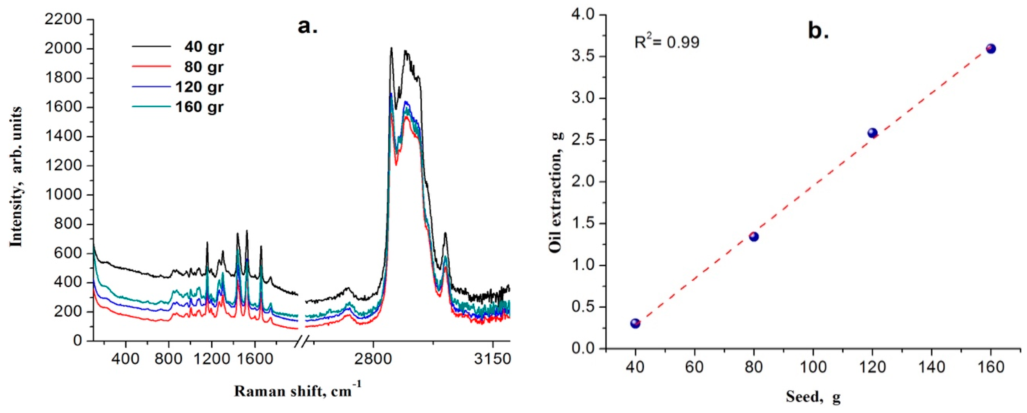
2.1. Seed collection and Oil Extraction

Spondias mombin L. fruits were collected in Sierra de Otontepec, located in the northern region of Veracruz State, Mexico. The seeds were separated from the fruit and oven-dried at 65 °C for 72 h. Different amounts of seeds (40, 80, 120 and 160 g) were submitted to hexane Soxhlet extraction and the yield was calculated. Each extract was evaporated under reduced pressure in a rotary evaporator (Büchi Rotavapor RII) followed by oven evaporation (Universal oven, Memmert UN30) at 60 °C to extract the solvent excess.

2.2. Acquisition of the Raman Spectral Block

The spectral recording was carried out using an integrated RS system (DXR Thermo Scientific) The excitation source was a 780 nm fiber laser operating at 20 mW average optical power. For each batch, 10 spectra were recorded in a range from 100 to 3000 cm−1 with 15 s of exposure time (focusing on different points of the sample through a 20× resolution objective). A total of 120 spectra were recorded and averaged for each batch for their comparative analysis regarding molecular vibrations.

2.3. NMR Experiments

1H-NMR and 13C-NMR spectra were obtained from a Varian 500 MHz instrument. Experiments using samples (approx. 50 mg) in CDCl3 with TMS as internal standard were carried out. Chemical shifts were recorded in ppm, using the solvent proton signal as standard. Data were analyzed by using the equipment software.
1H-NMR (500 MHz, CDCl3): δ 0.87–0.92 (m, terminal CH3), 1.25–1.41 (m, CH2), 1.59–1.66 (m, CH2–CH2–COO), 2.00–2.09 (m, CH2–CH = CH), 2.31–2.37(t, J = 6.0 Hz, CH2–COO), 2.77–2.80 (t, J = 3 Hz, HC = HC–CH2-CH = CH), 4.14–4.19 (dd, J1 = 9 Hz, J2 = 6 Hz, glyceryl CH2O), 4.29–4.33 (dd, J1 = 9 Hz, J2 = 3 Hz, glyceryl CH2O), 5.25–5.29 (m, glyceryl CHO), 5.30–5.41 (olefinic H).
13CNMR (150 MHz, CDCl3): δ 14.02 (α-CH3), 22.54 (β-CH3), 24.82 (C-3), 25.62 (C-11, diallylic), 27.16 (overlapping signals: C8-11 (oleyl), C8-14 (linoleyl)), 29.07-29.68 (CH2n), 31.51 (C-16, linoleyl), 34.01 (α- C-2), 34.17 (β-C-2), 62.09 (glyceryl CH2O), 68.91 (glyceryl CHO), 127.88 (C-12, linoleyl), 128.06 (C-13, linoleyl), 128.06 (C-13), 129.68 (C-9, oleyl), 129.95 (C-9, linoleyl, 129.98 (α-C-10, oleyl), 129.99 (β-C-10, oleyl), 130.20 (C-10, linoleyl), 172.79 (α-C-1), 173.25 (β-C-1), 173.21 (β-C-1).
To determine the relative percentage of saturated and unsaturated fatty acids in the oil using 1H-NMR, we followed the methodology described by Thoss et al. [25], who established the following equations using the integrals of different proton environments: polyunsaturated fatty acids: PUFA = F/E; monounsaturated fatty acids: MUFA = [C/2E]-PUFA; saturated fatty acids: SFA = 1-[D/2E], where D: α-allylic proton environment at δ 2.05 ppm; E: integrations of the acyl group at δ 2.34 ppm; F: protons attached to the bis-allylic carbon at δ 2.79 ppm.

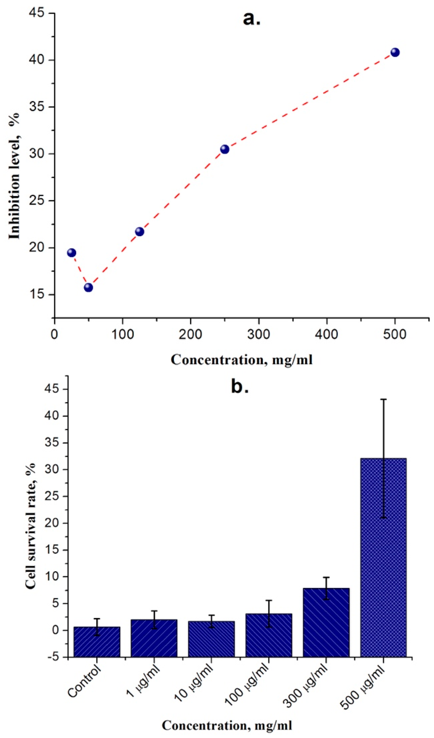
2.4. ABTS Radical Scavenging Activity Assay

The radical cation scavenging capacity of Spondias mombin L. seed oil was examined against 2,2’-azino-bis (3-ethylbenzothiazoline-6-sulphonic acid) radical cation (ABTS•+). A stock solution was prepared by reacting 7 mM ABTS (Sigma-Aldrich, Saint Louis, MO, USA) solution with 2.5 mM potassium persulfate as a final concentration, followed by 16 h incubation at room temperature in the dark. The ABTS• + stock solution was diluted with ethanol to an absorbance of 1.0 at 734 nm. Then, antioxidant activity was evaluated by incubating 150 µL of ethanol-ABTS• + with 50 µL of oil solution in ethanol at different concentrations (25–500 µg mL−1). After 30 min, the percentage inhibition of ABTS• + at 734 nm was calculated with the equation % Inhibition = [(AC—AO)/AC] × 100, where AC is the absorbance of the control (ABTS• + solution), and AO is the absorbance of the oil. Trolox and catechol were used as positive controls.

2.5. Sample Preparation and Cell Culture

A stock solution (100 mg mL−1) of Spondias mombin L. seed oil solubilized with ethyl alcohol (1:4) was prepared. Prior to the cell experiments, these samples were diluted to final concentrations of 1, 10, 50, 100, and 500 μg/mL using culture medium. HTC 116 cells (obtained from American Type Culture Collection, ATCC) were grown in a 100 mm cell culture dish at 37 °C with 5% CO2 in DMEM medium, supplemented with 10% fetal bovine serum. The cells were placed on a 96-well cell culture plate for cell proliferation assay. The oil was free of any solvent except for the alcohol used for the final dilution, in a concentration range from 0.00075% to 0.375%. These concentrations did not interfere with the assay. Standard protocols contain ≈2% alcohol concentration as previously reported [26,27]. Effects on cell viability by ethyl alcohol have been reported at a concentration of 5% and above [28].

2.6. Cell Proliferation Assay

The analysis of cell proliferation in cultured cells was performed by a water-soluble tetrazolium salt (WST) assay, following the manufacturer’s instructions (Abcam). Briefly, 5 × 10⁴/well cells were cultured in a 96-well microtiter plate in a final volume of 100 μL per well 24 h prior the experiment. Cells were incubated with oil sample solutions for 24 h at the five different concentrations described previously. WST-1 reagent was added to each well and incubated in standard culture conditions for 1 h. Absorbance of samples was measured using a microtiter plate reader at 420–480 nm. Nontreated cells were the reference of 100% viability; each treatment was compared to this control.

3. Results and Discussion

Spondias mombin L. has a variety of applications; indeed, previous reports demonstrated that the leaf and stem bark extracts have been used in traditional medicine [3,4]. In this study, we described the molecular vibrations and functional groups contained in the oil extracted from the seed of Spondias mombin L. identified by Raman spectroscopy, 1H-NMR and 13C-NMR.
Figure 1a shows the Raman spectrum for Spondias mombin L. oil seed obtained from different weights (40, 80, 120 and 160 g of seeds, processed separately). Figure 1b indicates a linear correlation between mass and yield. The corresponding yield is 2.58%.
Different amounts of extracted oil exhibit slight changes in peak intensities in the Raman spectrum; however, no significant changes are shown in the Raman frequencies’ positions. Spondias mombin L. oil measurements were carried out, which allowed a qualitative analysis through the identification of the characteristic Raman frequencies associated with carotenoids [29] (Figure 2).
Indeed, no literature on the characterization of Spondias mombin L. seed oil using the RS technique was found. The characteristic frequencies identified in the Raman spectra of Spondias mombin L. oil are shown in Table 1. The frequencies are related to previous reports from other vegetable oils [30,31,32,33,34].
Raman spectra recorded from Spondias mombin L. vegetable oil samples showed signals associated with the vibrational modes C-C (ν), C-H (δ), C = O (ν), C = C (ν), as well as bands assigned to functional groups such as CH2 and CH3 [32,35]. Registered Raman spectra showed very strong characteristic bands in the range of 2850 and 2900 cm−1, which are associated with lipids, lignins and the functional groups C-CH3, CH2 and OH [34]. Raman frequency region between 1006 and 1300 cm−1 showed characteristic shifts corresponding to alkyl aryl ether; in particular, at the frequency of 1006 cm−1, it showed a stretching vibration corresponding to a C-O group. On the other hand, these shifts can also be attributed to vibrations of groups CH3, HC-CH3, aromatic rings and υ C-C aliphatic chains [35]. In addition, the level of unsaturation of the fatty acids can be determined by the vibrational band at 1300 cm−1 by the group =CH- [30]. The relatively weak band at 1080 cm−1 is characteristic of vegetable oils, attributed to the presence of the terpene 1,8-cineole, aromatic rings or υ C-C aliphatic chains [35,36]. Bands at 1006 (bending), 1158 (stretching) and 1523 cm−1 (stretching) correspond to CH3, C-C and C=C, respectively, and can also be attributed to carotenoids [34,37]. CH3 and CH2 are associated with the vibrational band at 1440 cm−1 and are related to modes of inflection between both groups and phenolic compounds. The shift at 1657 cm−1 was associated with the stretching of the functional group C = C assigned to monocyclic monoterpenes and showed relative intensity in relation to the level of unsaturation of fatty acids. The band located at 1746 cm−1 was assigned to the stretching of group C = O in triglycerides structures and the presence of polyesters, pectins and lignins [30,38]. The vibrational frequency located at 3010 cm−1 was associated with unsaturated fatty acids and stretching groups CH = CH, C-H aromatic, =CH2 and =C-H [39]. In each batch of fruit seed oil, the frequencies associated with carotenoids were observed (Lutein, α-carotene and β-carotene) at 1006, 1158 and 1523 cm−1 [30,40,41].
NMR is a powerful and widely analytical method used in structure elucidation protocols. NMR spectroscopy constitutes an accurate, sensitive method to supplement chemical information for structural identification. Here, we obtained the 500 MHz 1H-NMR spectra of the crude oil, showing a characteristic signal pattern found in other seed oils [42] (Table 2).
The olefinic protons for all unsaturated fatty acids appeared at δ 5.30–5.41 ppm. In the low-field region of the spectra, a multiplet at δ 5.25–5.29 ppm was assigned to the proton attached to C-2 of the glycerol. The chemical shifts and multiplicities of coupling constants for the signals at δ 4.14–4.19 ppm (J1 = 9 Hz, J2 = 6 Hz, dd) and δ 4.29–4.33 ppm (J1 = 9 Hz, J2 = 3 Hz, dd) are characteristic for H-1 and H-3 protons of glycerol system. Bis-allylic and allylic proton signals appeared as a triplet at δ 2.77–2.80 ppm (J = 3 Hz) and a complex multiplet at δ 2.00–2.09 ppm. A triplet at δ 2.31–2.37 ppm (J = 6.0 Hz) was designated to methylene H-2 protons adjacent to the acyl group, a multiplet at δ 1.59–1.66 ppm was assigned to H-3, while the other methylene protons appeared between δ 1.25 and 1.41 ppm. The terminal methyl protons appeared at δ 0.87–0.92 ppm. The downfield-shifted proton signal at δ 6.25 indicated the presence of linoleic acid [43]. The relative % of SFA, MUFA and PUFA present in the sample was calculated following the method described by Thoss et al. (Figure 3) [25].
The chemical shift differences between some fatty acids depend on substitution on glycerol backbone. The 13C NMR spectra (Table 3) showed signals at δ 173.25 and 173.21 ppm assigned to carbonyl carbons attached to 1,3-glycerol positions, and a signal at δ 172.79 ppm corresponding to a fatty acid esterified at position two of glycerol, while no signal was observed for free fatty acids (δ 177.0–182.0 ppm). The C-1 and C-3 terminal carbons for the triglyceride moiety were found at the same chemical shift, 62.09 ppm, whereas signal for C-2 was present at 68.91 ppm. The olefinic carbons appeared in the range from δ127.88 to 130.20 ppm. For most components, such as oleic acid, α and β carbon signals could be distinguished, such as C-10 at δα = 129.98 and δβ = 129.99 ppm. As expected, most of the signals were observed at the low field region (δ 14.02–34.17 ppm), corresponding to methyl, methylene and allylic carbons. All of the above NMR signals were in agreement with the structure of a triacylglycerol bearing unsaturated fatty acid chains.
It has been demonstrated that antioxidant compounds are present in natural products such as vegetables, fruits, and oil seeds [44]. Several investigations have determined the antioxidant activity of carotenoids [45,46], and it has been possible to state the carotenoid content as responsible for the antioxidant activity. On the other hand, it has been shown that there is a high carotenoid content in plants from the Anacardiaceae family [46,47], such as Mexican plum (Spondias purpurea), ambarella (Spondias dulcis) and yellow mombin (Spondias mombin L.). Chemical composition in these plants varies according to the season of the year, where it is harvested, state of maturation, etc. [48].
Some authors have shown antioxidant capacity for Spondias mombin L. pulp under certain conditions [49,50], and in this study we demonstrated a higher efficiency. Spondias mombin L. seed oil had concentration-dependent antioxidant activity as shown in Figure 4a, with a maximum ABTS• + radical inhibition of 45%.
Our data showed a free radical scavenging effect in Spondias mombin L. oil, suggesting a protective activity against alterations in oxidant/antioxidant imbalance, promoting an increase of cell viability in the presence of higher oil concentration. Previous reports support this finding; pomegranate seed oil demonstrated an antioxidant effect on a diabetes model in kidney, heart and mitochondria from rats and H9c2 cell line [51]. The antioxidant capacity has been highly correlated with cell viability. Several studies demonstrated that carotenoids promote a protective effect resulting in cell viability against different insults such as UV light [52,53]. Other reports indicated that safflower seed oil increased significantly the viability and proliferation of embryonic neural stem cells [54]. Antioxidant compounds promote cell viability by several pathways. Some studies involved antioxidants in the maintenance of the genomic integrity (DNA) during proliferation stages [55]. Other reports have demonstrated that antioxidants prevent cells from oxidant-mediated cell death, through reducing membrane lipid peroxidation [56] and oxidative damage to cellular organelles [57].
The content of compounds in crude extracts depends on the extraction solvent used. In this case, we used hexane to extract mainly nonpolar molecules, including carotenoids and fatty acids, as it was demonstrated by the applied spectroscopy technics. In general, these compounds constitute only a few necessary sources of nutrients for cell proliferation, as cells mainly need carbohydrates. Indeed, different research groups report antiproliferative and apoptotic effects for oils extracted from plants evaluated on different cell lines [58,59,60]. As a result, the proposed molecular mechanism associated with increased cell viability has a cytoprotective effect. Moreover, it has been demonstrated by Giusti et al. that molecules in natural product oils increase cell viability revealed by proteomic analyses [61].
Previous reports have demonstrated the Spondias mombin L. cytotoxic effect. Indeed, the antimicrobial activity has been reported from herbalists through ethnobotanical surveys [62]; a study demonstrated antibacterial capacity from leaf and bark extracts, comparable to antibiotics. Supporting this evidence, another study of aqueous and ethanolic leaf extracts revealed vibriocidal and antimicrobial activities [63]. An in vitro study on rat red blood cells indicated an increase in hemolysis in the presence of the aforementioned extracts [64]. In this study, the seed oil of Spondias mombin L. was tested for cell viability, where, surprisingly, the treated cells incubated with oil showed more viability than the control cells without treatment. Likewise, Figure 4b shows an increment in cell viability according to oil concentration.

4. Conclusions

In the present work, we demonstrated the use of Raman and 1H- and 13C-NMR spectroscopy as noninvasive and nondestructive analytical techniques, which allowed for the characterization of the oil extracted from the seed of Spondias mombin L. Through the use of these two techniques, the characteristic vibrational frequencies of the Spondias mombin L. seed oil were identified, as well as its fatty acid profile. Raman vibratory frequencies at 1006, 1158 and 1523 cm−1 were identified and assigned to carotenoid compounds with antioxidant activity. On the other hand, NMR experiments proved to be a valuable tool to obtain the fatty acid profile of crude oil from the seed. Selected peak integrals were used to calibrate the composition of MUFA, PUFA and SFA. Applying this approach to a Spondias mombin L. seed oil sample, we obtained values for MUFA and PUFA contents of 29.4 and 43.5%, which indicated that the oil is highly unsaturated (>70%). The oil showed high biocompatibility and promoted cell viability in the HTC 116 cell line.
Due to its properties, the seed oil of this tropical fruit could have a potential application in cosmetic and pharmaceutical products as an antioxidant agent. Finally, the combination of Raman spectroscopy and the NMR technique are very powerful and useful tools for the characterization of organic compounds, particularly in natural vegetable oils.

Author Contributions

Antioxidant evaluation of compounds, P.Y.L.-C. and A.T.-Z.; cytotoxic evaluation, G.B.-I.; NMR analysis, M.M.-A. and J.G.M.; writing—review and editing, J.M.M.-V.; Raman spectroscopy analysis and project administration, J.C.M.-E. Authorship is limited to those who have contributed substantially to the work reported. All authors have read and agreed to the published version of the manuscript.

Funding

This work was funded by the Research and Post-Graduate Secretary of the National Polytechnic Institute (IPN) for financial support through the SIP-2019044 and SIP-20200637 project, Institutional Research Announcement 2018 from University of Guanajuato, and projects CONACyT CB-2012/182003 and A1-S-29906.

Acknowledgments

We thank the company Nabicron & DM (Don Mon) for the use of laboratory equipment for the oil extraction and logistics in the transport of Spondias mombin L. seed.

Conflicts of Interest

The authors declare no conflict of interest.

References

  1. Ramirez Niño, M.Á.; Jiménez Forero, J.A.; Bernal Salazar, J.P.; Osorio Dueñas, M.D. Characterization of oil extracted from the kernel of the fruit of cumare’s palm (Astrocaryum chambira Barret). Rev. Fac. Nac. Agron. 2018, 71, 8415–8422. [Google Scholar] [CrossRef] [Green Version]
  2. Saraiva, S.A.; Cabral, E.C.; Eberlin, M.N.; Catharino, R.R. Amazonian Vegetable Oils and Fats: Fast Typification and Quality Control via Triacylglycerol (TAG) Profiles from Dry Matrix-Assisted Laser Desorption/lonization Time-Of-Flight (MALDI-TOF) Mass Spectrometry Fingerprinting. J. Agric. Food Chem. 2009, 57, 4030–4034. [Google Scholar] [CrossRef] [PubMed]
  3. Ayoka, A.O.; Akomolafe, R.O.; Akinsomisoye, O.S.; Ukponmwan, O.E. Medicinal and Economic Value of Spondias mombin. Afr. J. Biomed. Res. 2008, 11, 129–136. [Google Scholar] [CrossRef]
  4. Ayoka, A.O.; Akomolafe, R.O.; Iwalewa, E.O.; Ukponmwan, O.E. Studies on the Anxiolytic Effect of Spondias mombin L. (Anacardiaceae) Extracts. Afr. J. Tradic. Complement. Altern. Med. 2005, 2, 153–155. [Google Scholar] [CrossRef] [Green Version]
  5. Osuntokun, O.T.; To, I.; Cristina, G.M. Antibacterial and Antifungal Efficacy of Partially Partitioned Fractions of Spondias mombin (Linn) Extracts (Root, Leaf and Stem Bark) Against Clinical and Environmental Isolates. Med. Chem. 2018, 8, 10–17. [Google Scholar] [CrossRef]
  6. Senes-Lopes, T.F.; López, J.A.; do Amaral, V.S.; Brandão-Neto, J.; de Rezende, A.A.; da Luz, J.R.D.; Almeida, M.d.G. Genotoxicity of Turnera subulata and Spondias mombin × Spondias tuberosa Extracts From Brazilian Caatinga Biome. J. Med. Food 2018, 21, 1–8. [Google Scholar] [CrossRef]
  7. Olufunke, M.D.; Kasali, A.A.; Olusegun, E. Constituents of the Spondias mombin Linn and the Comparison between its Fruit and Leaf essential oils. J. Essent Oil-Bear. Plants 2003, 6, 148–152. [Google Scholar] [CrossRef]
  8. Eromosele, C.O.; Paschal, N.H. Characterization and Viscosity Parameters of Seed Oils From Wild Plants. Bioresour. Technol. 2003, 86, 203–205. [Google Scholar] [CrossRef]
  9. Knothe, G.; Kenar, J.A. Determination of the fatty acid profile by 1H-NMR spectroscopy. Eur. J. Lipid Sci. Technol. 2004, 106, 88–96. [Google Scholar] [CrossRef]
  10. Di Pietro, M.E.; Mannu, A.; Mele, A. NMR Determination of Free Fatty Acids in Vegetable Oils. Processes 2020, 8, 410. [Google Scholar] [CrossRef] [Green Version]
  11. Huck, C.W.; Bec, K.B.; Grabska, J. The use of vibrational spectroscopy in medicinal plant analysis: Current and future directions. Planta Med. 2019, 85, 1408–1409. [Google Scholar] [CrossRef]
  12. Krafft, C.; Popp, J. Micro-Raman spectroscopy in medicine. Phys. Sci. Rev. 2019, 4, 1–15. [Google Scholar] [CrossRef]
  13. De Silva, I.W.; Kretsch, A.R.; Lewis, H.M.; Bailey, M.; Verbeck, G.F. True one cell chemical analysis: A review. Analyst 2019, 144, 4733–4749. [Google Scholar] [CrossRef] [PubMed]
  14. Nims, C.; Cron, B.; Wetherington, M.; Macalady, J.; Cosmidis, J. Low frequency Raman spectroscopy for micron-scale and in vivo characterization of elemental sulfur in microbial samples. Sci. Rep. 2019, 9, 1–12. [Google Scholar] [CrossRef] [PubMed] [Green Version]
  15. He, H.; Sun, D.W.; Pu, H.; Chen, L.; Lin, L. Applications of Raman spectroscopic techniques for quality and safety evaluation of milk: A review of recent developments. Crit. Rev. Food Sci. Nutr. 2019, 59, 770–793. [Google Scholar] [CrossRef]
  16. Hu, R.; He, T.; Zhang, Z.; Yang, Y.; Liu, M. Safety analysis of edible oil products via Raman spectroscopy. Talanta 2019, 191, 324–332. [Google Scholar] [CrossRef]
  17. Meenu, M.; Cai, Q.; Xu, B. A critical review on analytical techniques to detect adulteration of extra virgin olive oil. Trends Food Sci. Technol. 2019, 91, 391–408. [Google Scholar] [CrossRef]
  18. Berghian-Grosan, C.; Magdas, D.A. Raman spectroscopy and Machine-Learning for edible oils evaluation. Talanta 2020, 121176. [Google Scholar] [CrossRef]
  19. Kwofie, F.; Lavine, B.K.; Ottaway, J.; Booksh, K. Incorporating brand variability into classification of edible oils by Raman spectroscopy. J. Chemom. 2020, 34, e3173. [Google Scholar] [CrossRef]
  20. Saraiva, A.G.Q.; Saraiva, G.D.; Albuquerque, R.L.; Nogueira, C.E.S.; Teixeira, A.M.R.; Lima, L.B.; Cruz, B.G.; de Sousa, F.F. Chemical analysis and vibrational spectroscopy study of essential oils from Lippia sidoides and of its major constituent. Vib. Spectrosc. 2020, 103111. [Google Scholar] [CrossRef]
  21. Pedro, A.C.; Bach, F.; Stafussa, A.P.; Menezes, L.R.A.; Barison, A.; Maciel, G.M.; Haminiuk, C.W.I. 1H NMR and Raman spectroscopy of oils and extracts obtained from organic and conventional goji berries: Yield, fatty acids, carotenoids and biological activities. Int. J. Food Sci. Technol. 2019, 54, 282–290. [Google Scholar] [CrossRef] [Green Version]
  22. Ferreira, B.S.; de Almeida, C.G.; Le Hyaric, M.; de Oliveira, V.E.; Edwards, H.G.; de Oliveira, L.F.C. Raman spectroscopic investigation of carotenoids in oils from Amazonian products. Spectrosc. Lett. 2013, 46, 122–127. [Google Scholar] [CrossRef]
  23. Gamsjaeger, S.; Baranska, M.; Schulz, H.; Heiselmayer, P.; Musso, M. Discrimination of carotenoid and flavonoid content in petals of pansy cultivars (Viola × wittrockiana) by FT-Raman spectroscopy. J. Raman Spectrosc. 2011, 42, 1240–1247. [Google Scholar] [CrossRef]
  24. Bhosale, P.; Ermakov, I.V.; Ermakova, M.R.; Gellermann, W.; Bernstein, P.S. Resonance Raman quantification of nutritionally important carotenoids in fruits, vegetables, and their juices in comparison to high-pressure liquid chromatography analysis. J. Agric. Food Chem. 2004, 52, 3281–3285. [Google Scholar] [CrossRef]
  25. Thoss, V.; Myrphy, P.J.; Marriott, R.; Wilson, T. Triacylglycerol composition of British bluebell (Hyacinthoides non-scripta) seed oil. RSC Adv. 2012, 12, 5314–5322. [Google Scholar] [CrossRef]
  26. Döll-Boscardin, P.M.; Sartoratto, A.; Sales Maia, B.H.L.D.N.; Padilha de Paula, J.; Nakashima, T.; Farago, P.V.; Kanunfre, C.C. In vitro cytotoxic potential of essential oils of Eucalyptus benthamii and its related terpenes on tumor cell lines. Evid.-Based Complement. Altern. Med. 2012, 2012, 342652. [Google Scholar] [CrossRef] [Green Version]
  27. Virador, V.M.; Kobayashi, N.; Matsunaga, J.; Hearing, V.J. A standardized protocol for assessing regulators of pigmentation. Anal. Biochem. 1999, 270, 207–219. [Google Scholar] [CrossRef]
  28. Forman, S.; Káˇs, J.; Fini, F.; Steinberg, M.; Ruml, T. The effect of different solvents on the ATP/ADP content and growth properties of HeLa cells. J. Biochem. Mol. Toxicol. 1999, 13, 11–15. [Google Scholar] [CrossRef]
  29. de Oliveira, V.E.; Castro, H.V.; Edwards, H.G.M.; de Oliveira, L.F.C. Carotenes and carotenoids in natural biological samples: A Raman spectroscopic analysis. J. Raman Spectrosc. 2010, 41, 642–650. [Google Scholar] [CrossRef]
  30. Seidler-Lozykowska, K.; Baranska, M.; Baranski, R.; Krol, D. Raman Analysis of Caraway (Carum carvi L.) Single Fruits. Evaluation of Essential Oil Content and Its Composition. J. Agric. Food Chem. 2010, 58, 5271–5275. [Google Scholar] [CrossRef]
  31. Baranska, M.; Schütze, W.; Schulz, H. Determination of Lycopene and β-Carotene Content in Tomato Fruits and Related Products: Comparison of FT-Raman, ATR-IR, and NIR Spectroscopy. Anal. Chem. 2006, 78, 8456–8461. [Google Scholar] [CrossRef] [PubMed]
  32. Jentzsch, P.; Ramos, L.; Ciobotă, V. Handheld Raman Spectroscopy for the Distinction of Essential Oils Used in the Cosmetics Industry. Cosmetics 2015, 2, 162–176. [Google Scholar] [CrossRef] [Green Version]
  33. Nogales-Bueno, J.; Baca-Bocanegra, B.; Rooney, A.; Hernández-Hierro, J.M.; Byrne, H.J.; Heredia, F.J. Study of phenolic extractability in grape seeds by means of ATR-FTIR and Raman spectroscopy. Food Chem. 2017, 232, 602–609. [Google Scholar] [CrossRef]
  34. El-Abassy, R.M.; Donfack, P.; Materny, A. Visible Raman spectroscopy for the discrimination of olive oils from different vegetable oils and the detection of adulteration. J. Raman Spectrosc. 2009, 40, 1284–1289. [Google Scholar] [CrossRef]
  35. Baranska, M.; Schulz, H.; Krüger, H.; Quilitzsch, R. Chemotaxonomy of Aromatic Plants of the Genus Origanum via Vibrational Spectroscopy. Anal. Bioanal. Chem. 2005, 381, 1241–1247. [Google Scholar] [CrossRef] [PubMed]
  36. Smith, E.; Dent, G. Modern Raman Spectroscopy: A Practical Approach; John Wiley and Sons, Ltd.: Chichester, UK, 2005. [Google Scholar] [CrossRef] [Green Version]
  37. Baeten, V.; Dardenne, P.; Aparicio, R. Interpretation of Fourier transform Raman Spectra of the Unsaponifiable Matter in a Selection of Edible Oils. J. Agric. Food Chem. 2001, 49, 5098–5107. [Google Scholar] [CrossRef] [PubMed]
  38. Lin-Vien, D.; Colthup, N.B.; Fateley, W.G.; Grasselli, J.G. The Handbook of Infrared and Raman Characteristic Frequencies of Organic Molecules, 1st ed.; Academic Press: San Diego, CA, USA, 1991. [Google Scholar]
  39. Martini, W.S.; Porto, B.L.S.; De Oliveira, M.A.L.; Sant’Ana, A.C. Comparative Study of the Lipid Profiles of Oils from Kernels of Peanut, Babassu, Coconut, Castor and Grape by GC-FID and Raman Spectroscopy. J. Braz. Chem. Soc. 2018, 29, 390–397. [Google Scholar] [CrossRef]
  40. Afseth, N.K.; Wold, J.P.; Segtnan, V.H. The Potential of Raman Spectroscopy for Characterization of the Fatty Acid Unsaturation of Salmon. Anal. Chim. Acta 2006, 572, 85–92. [Google Scholar] [CrossRef]
  41. Tiburski, J.H.; Rosenthal, A.; Deliza, R.; de Oliveira Godoy, R.L.; Pacheco, S. Nutritional Properties of Yellow Mombin (Spondias mombin L.) pulp. Food Res. Int. 2011, 44, 2326–2331. [Google Scholar] [CrossRef] [Green Version]
  42. Vlahov, G. Application of NMR to the Study of Olive Oils. Prog. Nucl. Magn. Reson Spectrosc. 1999, 35, 341–357. [Google Scholar] [CrossRef]
  43. Scano, P.; Anedda, R.; Melis, M.P.; Dessi, M.A.; Lai, A.; Roggio, T. 1H- and 13C-NMR Characterization of the Molecular Components of the Lipid Fraction of Pecorino Sardo Cheese. J. Am. Oil Chem. Soc. 2011, 88, 1305–1316. [Google Scholar] [CrossRef]
  44. Matthäus, B. Antioxidant Activity of Extracts Obtained from Residues of Different Oilseeds. J. Agric. Food Chem. 2002, 50, 3444–3452. [Google Scholar] [CrossRef] [PubMed]
  45. Stahl, W.; Sies, H. Antioxidant Activity of Carotenoids. Mol. Asp. Med. 2003, 24, 345–351. [Google Scholar] [CrossRef]
  46. You, J.S.; Jeon, S.; Byun, Y.J.; Koo, S.; Choi, S.S. Enhanced Biological Activity of Carotenoids Stabilized by Phenyl Groups. Food Chem. 2015, 177, 339–345. [Google Scholar] [CrossRef]
  47. Giuffrida, D.; Menchaca, D.; Dugo, P.; Donato, P.; Cacciola, F.; Murillo, E. Study of the Carotenoid Composition in Membrillo, Guanabana Toreta, Jobo and Mamey Fruits. Fruits 2015, 70, 163–172. [Google Scholar] [CrossRef] [Green Version]
  48. Perry, N.B.; Anderson, R.E.; Brennan, N.J.; Douglas, M.H.; Heaney, A.J.; McGimpsey, J.A.; Smallfield, B.M. Essential Oils from Dalmatian Sage (Salvia officinalis L.): Variations Among Individuals, Plant Parts, Seasons, and Sites. J. Agric. Food Chem. 1999, 47, 2048–2054. [Google Scholar] [CrossRef]
  49. Maria do Socorro, M.R.; Alves, R.E.; de Brito, E.S.; Pérez-Jiménez, J.; Saura-Calixto, F.; Mancini-Filho, J. Bioactive Compounds and Antioxidant Capacities of 18 non-traditional Tropical Fruits from Brazil. Food Chem. 2010, 121, 996–1002. [Google Scholar] [CrossRef] [Green Version]
  50. Vasco, C.; Ruales, J.; Kamal-Eldin, A. Total Phenolic Compounds and Antioxidant Capacities of Major Fruits from Ecuador. Food Chem. 2008, 111, 816–823. [Google Scholar] [CrossRef]
  51. Mollazadeh, H.; Boroushaki, M.T.; Soukhtanloo, M.; Afshari, A.R.; Vahedi, M.M. Effects of Pomegranate Seed Oil on Oxidant/Antioxidant Balance in Heart and Kidney Homogenates and Mitochondria of Diabetic Rats and High Glucose-Treated H9c2 Cell Line. Avicenna J. Phytomed. 2017, 7, 317–333. [Google Scholar]
  52. Petruk, G.; Del Giudice, R.; Rigano, M.M.; Monti, D.M. Antioxidants from Plants Protect against Skin Photoaging. Oxid. Med. Cell. Longev. 2018, 2018, 1454936. [Google Scholar] [CrossRef] [Green Version]
  53. Fernández-García, E.; Carvajal-Lérida, I.; Pérez-Gálvez, A. Carotenoids Exclusively Synthesized in Red Pepper (capsanthin and capsorubin) Protect Human Dermal Fibroblasts against UVB Induced DNA Damage. Photochem. Photobiol. Sci. 2016, 15, 1204–1211. [Google Scholar] [CrossRef] [PubMed] [Green Version]
  54. Ghareghani, M.; Zibara, K.; Azari, H.; Hejr, H.; Sadri, F.; Jannesar, R.; Ghanbari, A. Safflower Seed Oil, Containing Oleic Acid and Palmitic Acid, Enhances the Stemness of Cultured Embryonic Neural Stem Cells Through Notch1 and Induces Neuronal Differentiation. Front. Neurosci. 2017, 11, 1–10. [Google Scholar] [CrossRef] [PubMed]
  55. Pucci, B.; Kasten, M.; Giordano, A. Cell cycle and apoptosis. Neoplasia 2000, 2, 291–299. [Google Scholar] [CrossRef] [PubMed] [Green Version]
  56. Ali, S.S.; Ahsan, H.; Zia, M.K.; Siddiqui, T.; Khan, F.H. Understanding oxidants and antioxidants: Classical team with new players. J. Food Biochem. 2020, 44, e13145. [Google Scholar] [CrossRef] [PubMed]
  57. Ruhee, R.T.; Ma, S.; Suzuki, K. Protective effects of sulforaphane on exercise-induced organ damage via inducing antioxidant defense responses. Antioxidants 2020, 9, 136. [Google Scholar] [CrossRef] [Green Version]
  58. Jayathilake, A.G.; Senior, P.V.; Su, X.Q. Krill oil extract suppresses cell growth and induces apoptosis of human colorectal cancer cells. BMC Complement. Altern. Med. 2016, 16, 328. [Google Scholar] [CrossRef] [Green Version]
  59. Jayathilake, A.G.; Kadife, E.; Luwor, R.B.; Nurgali, K.; Su, X.Q. Krill oil extract suppresses the proliferation of colorectal cancer cells through activation of caspase 3/9. Nutr. Metab. 2019, 16, 1–15. [Google Scholar] [CrossRef]
  60. De Stefanis, D.; Scimè, S.; Accomazzo, S.; Catti, A.; Occhipinti, A.; Bertea, C.M.; Costelli, P. Anti-proliferative effects of an extra-virgin olive oil extract enriched in ligstroside aglycone and oleocanthal on human liver cancer cell lines. Cancers 2019, 11, 1640. [Google Scholar] [CrossRef] [Green Version]
  61. Giusti, L.; Angeloni, C.; Barbalace, M.C.; Lacerenza, S.; Ciregia, F.; Ronci, M.; Urbani, A.; Manera, C.; Digiacomo, M.; Macchia, M.; et al. A Proteomic Approach to Uncover Neuroprotective Mechanisms of Oleocanthal against Oxidative Stress. Int. J. Mol. Sci. 2018, 19, 2329. [Google Scholar] [CrossRef] [Green Version]
  62. Abo, K.A.; Ogunleye, V.O.; Ashidi, J.S. Antimicrobial Potential of Spondias mombin, Croton zambesicus and Zygotritonia crocea. Phytother. Res. 1999, 13, 494–497. [Google Scholar] [CrossRef]
  63. Bolatito, O.; Olugbenga, O.; Mobolaji, A.; Adeniran, S. Phytochemical and antimicrobial screening of Spondias mombin, Senna occidentalis and Musa sapientum against Vibrio cholerae O1. Int. J. Curr. Microbiol. Appl. Sci. 2014, 3, 948–961. [Google Scholar]
  64. Moussa, G.; Alassane, T.; Yaoemile, K.; Jean, O.N. Toxicity Assessment of an Aqueous Extract of the Stem Bark of Spondias mombin (Anacardiaceae) in wistar albino rats. Int. J. Curr. Microbiol. Appl. Sci. 2018, 7, 3625–3635. [Google Scholar] [CrossRef]
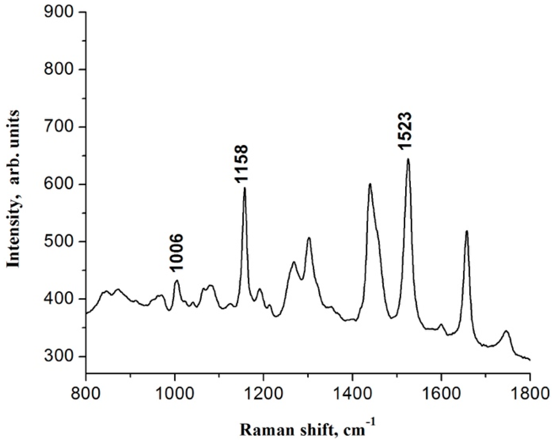
Figure 1. Raw Raman spectra recorded. (a) Average Raman spectra recorded for each batch of oil extracted with different amount of seed. (b) The weight ratio between the processed seed versus extracted oil shows a linear tendency.
Applsci 11 02886 g001
Figure 2. Region of the Raman spectrum of an oil sample from Sondias mombin L. where Raman frequencies are located at 1006, 1158 and 1523 cm−1, which are associated with carotenoids compounds.
Applsci 11 02886 g002
Figure 3. Relative % of saturated (SFA), monounsaturated (MUFA) and polyunsaturated fatty acids (PUFA) as determined using 1H-NMR.
Applsci 11 02886 g003
Figure 4. Antioxidation and cell viability tests. (a) Spondias mombin L. seed oil shows concentration-dependent antioxidant activity. It was measured by 2,2′-Azino-bis (3-ethylbenzothiazoline-6-sulfonic acid) diammonium salt (ABTS) antioxidant activity assay incubated for 30 min. (b) Spondias mombin L. seed oil increases cell viability. HTC 116 cells were cultured and incubated with different concentrations of vegetable oil. After 24 h, a water-soluble tetrazolium salt (WST) assay showed the increase of cell viability according to oil concentration. Nontreated cells were used as control referring 100% viability.
Applsci 11 02886 g004
Table 1. Assignment of Raman frequencies that identify the vegetable oil of Spondias mombin L. Abbreviations: vs, very strong; m, medium; w, weak, mw, medium weak; sh, shoulder; ν, stretching; δ, bending.
Raman Frequency (cm−1)Functional GroupVibrational Mode
871 sh, w-(CH2)n-C-C, ν
968 sh, wTrans RHC = CHRC = C, δ
1006 wHC-CH3CH3, δ
1080 sh, w-(CH2)n-C-C, ν
1158 m-(CH2)n-C-C, ν
1267 mwcis RHC = CHR=C-H, δ
1300 mw-CH2C-H, δ
1440 m-CH2C-H, δ
1523 mRHC = CHRC = C, ν
1657 mcis RHC = CHRC = C, ν
1746 wRC = OORC = O, ν
2850 vs-CH2C-H, ν
2900 sh, vs-CH3C-H, ν
3010 mcis RHC = CHR=C-H, ν
Table 2. Chemical shifts and assignments of the characteristic resonances in the 1H-NMR spectrum of crude seed oil.
Chemical Shift (ppm)ProtonFunctional Group
0.87–0.92–CH3Terminal methyl protons of saturated and unsaturated chains
1.25–1.41CH2Protons of methylene envelop
1.59–1.66CH2–CH2–COOH-3 protons of acyl moieties in triacylglycerols
2.00–2.09CH2–CH = CHAllylic methylenes
2.31–2.37CH2–COOH-2 protons of acyl moieties in triacylglycerols
2.77–2.80C = C–CH2-C = CProtons attached to bis allylic carbon
4.14–4.19
4.29–4.33
CH2OH-1 and H-3 protons of glycerol
5.25–5.29CHO (β)H-2 of the glycerol backbone
5.30–5.41CH = CHOlefinic protons of unsaturated Fatty acids
Table 3. Chemical shifts and assignments of the characteristic signals in the carbon nuclear magnetic response (13C-NMR) spectrum of crude seed oil.
Chemical Shift (ppm)CarbonAssignment
14.02α-CH3All acyl chains
22.54β-CH3All acyl chains
24.82C-3All acyl chains
25.62C-11Diallylic
27.16C8-11 (oleyl), C8-14 (linoleyl)Allylic
29.07–29.68CH2nAll acyl chains
31.51C-16Linoleyl
34.01α-C-2All acyl chains
34.17β-C-2All acyl chains
62.09α-CH2OGlycerol (triacylglycerols)
68.91β-CHOGlycerol (triacylglycerols)
127.88C-12Linoleyl
128.06C-13Linoleyl
129.68C-9Oleyl
129.95C-9Linoleyl
129.98α-C-10Oleyl
129.99β-C-10Oleyl
130.20C-10Linoleyl
172.79α-C-1 GlycerolTriacylglycerols
2xC, oleoyl and linoleoyl
173.25 and 173.21β-C-1 GlycerolTriacylglycerols

