Article

In Vitro Cytotoxic Evaluation and Apoptotic Effects of Datura innoxia Grown in Saudi Arabia and Phytochemical Analysis

1
Biology Department, College of Science, Imam Mohammad ibn Saud Islamic University (IMSIU), Riyadh 11623, Saudi Arabia
2
Medicinal Aromatic, and Poisonous Plants Research Centre, College of Pharmacy, King Saud University, Riyadh 11451, Saudi Arabia
3
Department of Pharmacognosy, College of Pharmacy, King Saud University, Riyadh 11451, Saudi Arabia
4
Basic Science Department, College of Medicine, Princess Nourah bint Abdulrahman University, Riyadh 11671, Saudi Arabia
5
Department of Zoology, College of Science, King Saud University, Riyadh 11451, Saudi Arabia
6
Department of Biological Sciences, College of Science, Taibah University, Madina 41321, Saudi Arabia
*
Authors to whom correspondence should be addressed.
Appl. Sci. 2021, 11(6), 2864; https://doi.org/10.3390/app11062864
Submission received: 1 February 2021 / Revised: 9 March 2021 / Accepted: 10 March 2021 / Published: 23 March 2021
(This article belongs to the Special Issue Application of Plant Natural Compounds)

Abstract

:
Datura innoxia is an important species of Solanaceae family with several purposes in folk medicine. This study intends to explore the cytotoxic effect of D. innoxia on various cancer cell proliferation. D. innoxia ethanolic extract’s effect on the progression of the cell cycle and the induction of apoptosis were investigated by flow cytometry. Further, real-time PCR was employed to confirm apoptosis initiation. In addition, active phytochemicals of D. innoxia was identified by gas chromatography–mass spectroscopy (GC-MS). The cell viability study revealed that the ethanolic extract of D. innoxia demonstrated potent cytotoxicity, with an IC50 value of 10 μg/mL against LoVo colon cancer cells. Cell cycle staining with propidium iodide revealed that D. innoxia treatment leads to cell accumulation in the sub-G1 phase. Using the Annexin V-FITC/PI assay, the ethanolic extract was found to cause a dose-dependent increase in early and late apoptosis when compared to control cells. Apoptosis as the mode of cell death was also confirmed by the increased expression of p53, bax and caspase-8, -9, and -3 along with downregulation of Bcl-2. GC-MS analysis displayed that 3,5-Dihydroxybenzoic acid (16.53%), heneicosyl formate (14.14%), 2,3-dimethyl-3-pentanol (12.89%), 2-hydroxy-4-methyl pentanoic acid (5.19%) were the main phytoconstituents. These findings conclude that D. innoxia causes cell death through apoptosis, suggesting more attention should be paid to further exploration of the active components from D. innoxia responsible for the observed activities.

1. Introduction

Cancer is still a major health issue, and the incidence of cancer related deaths is increasing around the globe [1]. The female breast, lung and colorectal cancers types are among the most commonly diagnosed cancers globally and continue to be the leading cause of cancer related death worldwide [2]. Although mainly unsuccessful and leading to many deaths due to their side effects, the use of conventional treatments to treat cancer is still the most common option of treatment. However, the development of new drugs from natural sources with fewer side effects is becoming promising field in cancer research [3]. Plants are still a promising reservoir for a novel chemical compound in the cancer research area [4]. Many well-known anticancer drugs, such as paclitaxel and camptothecins, are isolated from plant-derived products [5]. With a different biogeographic region, Saudi Arabia provides a remarkably rich source for medicinal plants that have anticancer properties [6,7].
The genus Datura belongs to Solanaceae family and comprises about 20 species that grow worldwide [8]. These plants have been used in folk medicine to treat different diseases, including asthma, gastric pain, and indigestion. Other reported medicinal uses of the plant are anti-inflammatory properties, stimulation of the nervous system, and the treatment of dental and skin infections [9]. Various phytoconstituents have been reported in this genus including alkaloids, atropine and scopolamine [10], carotenes and coumarins [11] and saponins [12].
Datura innoxia belongs to the Solanaceae family and it is used in folk medicine to treat several ailments, including skin eruptions, colds, and nervous disorders [13]. Additionally, several medicinal purposes for D. innoxia have been reported, such as antispasmodic, pacifying, pain relief, and treating respiratory ailments [14]. The other phytochemicals documented in the plant like phenols, flavonoids, saponins, tannins, glycosides, and resins have analgesic anti-inflammatory, antibacterial as well as antipyretic activities [15,16,17].
The cytotoxicity of D. innoxia against different cancer cells has been reported [18,19]. However, exploring the mode of cell death induced by D. innoxia extract still needs more investigation. To our knowledge, there are no reports regarding the activity of D. innoxia related to cell cycle arrest and apoptosis induction in LoVo colon cancer cells. Additionally, this study is the initial report documented the phytoconstituents present in D. innoxia grown in Saudi Arabia.

2. Materials and Methods

2.1. Plant Collection and Authentication

The fresh aerial parts of D. innoxia were collected from Al-Madinah Al-Munawara, Saudi Arabia, in March 2019. The plant material was authenticated by Professor Sami Zalat, Biology Department, College of Science, Taibah University, Saudi Arabia. The plant material was cleaned with water and then rinsed with distilled water. Thereafter, it was dried under shade at room temperature for 10 days. Three hundred grams of powdered plant material were extracted by submerging the plant powder with ethanol and deionized water (70/30 v/v) for 48 h using a Soxhlet apparatus. Next, the mixture was centrifuged at 2500× g, and the collected supernatant was concentrated under reduced pressure in a rotary evaporator. The crude extract was kept at −20 °C for future use.

2.2. Cell Viability (MTT Assay)

Different human cancer cell lines of various origin was maintained in appropriate medium DMEM (Gibco, USA). The medium was complemented with 10% fetal bovine serum (FBS) and 1% penicillin/streptomycin (Gibco, USA). Cells were maintained in an incubator containing humidified atmosphere of 95% air and 5% CO2 at 37 °C. MTT reduction assay was carried out to assess the antiproliferation activity as previously described [20]. Briefly, A549 (lung), LoVo (colon), MCF-7 and MDA-MB-231 (breast) cancer cells were plated in 24-multiwell culture plates at 5 × 10⁴ cells per ml and allowed to adhere for 24 h. Cells were incubated with extract at various concentrations (100, 50, 25 and 12.5 μg/mL) as well as DMSO as a vehicle control (0.1 v/v final concentration). At the end of incubation (48 h), 100 μL of MTT (Sigma Aldrich, St. Louis, MO, USA) was added to each well. After 4 h, the medium was discarded, and the formazan crystals were dissolved with isopropanol. The absorbance was measured at 570 nm with BioTek plate reader (USA). Extract concentration that decreases the number of viable cells into half (IC50) was computed using the program OriginPro 8.5 Software.

2.3. Cell Cycle Analysis

Cell cycle analysis was conducted by flow cytometry using propidium iodide (PI, BioLegend, San Diego, CA, USA) staining. It was done by plating the LoVo cells in 6-multiwell culture plates at 5 × 10⁴ cells per ml for 24 h. After treatment with D. innoxia ethanolic extract (5 and 10 μg/mL) as well as a vehicle for 48 h, both floating and attached cells were collected, and washed with cold PBS. The cells were then permeabilized and fixed in ice cold, 70% (v/v) ethanol for 1 h at 4 °C. Finally, cells were incubated with staining solution (50 μg/mL PI and 20 μg/mL RNase A in PBS) at 37 °C for 15 min. Cell cycle phase analysis by flow cytometry was done using a Flow Cytometer (Beckman Coulter Inc. Brea, CA, USA)

2.4. FITC Annexin V/PI Apoptosis Detection

LoVo cells were seeded at 5 × 10⁴ cells/mL in 6 well culture plates, using 2 mL per well, and treated with 5 and 10 μg/mL of ethanolic D. innoxia extract and vehicle control. After 48 h of treatment and incubation, the LoVo cells were washed with ice-cold PBS, then the cells were stained following the Annexin V-FITC/PI Kit protocol (BioLegend, CA, USA). In brief, after centrifugation, the supernatant was discarded and cell pellets were resuspended in ice-cold 1X binding buffer (100 µL). Annexin V-FITC (5 μL) and PI (5 μL) were added to each tube. Tubes were then mixed and incubated for 15 min in the dark. Samples were read on a Beckman Coulter FACScan flow cytometer (Cytomics FC 500; Beckman Coulter, Brea, CA, USA).

2.5. Gene Quantification by qRT-PCR

TRIzol reagent (Invitrogen, Waltham, MA, USA) was used for the total RNA extraction [21] of untreated cells and treated cells, with D. innoxia ethanolic extract at 5 and 10 µg/mL and vehicle for 48 h. Total extracted RNA (1 μg) was reverse transcribed using Promega cDNA Synthesis kit (Promega, CA, USA). Real time PCR master mix for apoptosis related genes was prepared according to the previous protocol [22]. The amounts of the mRNA transcripts were measured using the Applied Biosystems 7500 Fast real time PCR detection system (Applied Biosystems, Foster City, CA, USA). The thermo cycling conditions were established as 5 min at 95 °C, followed by 36 cycles of 15 s at 95 °C, 30 s at 60 °C and 30 s at 72 °C. Each reaction was conducted in triplicate and the 2−ΔΔCt method was applied to quantify the mean difference between control and treated samples and GAPDH was used as an internal control. The designed forward (F) and reverse (R) primers sequences are listed in (Table 1).

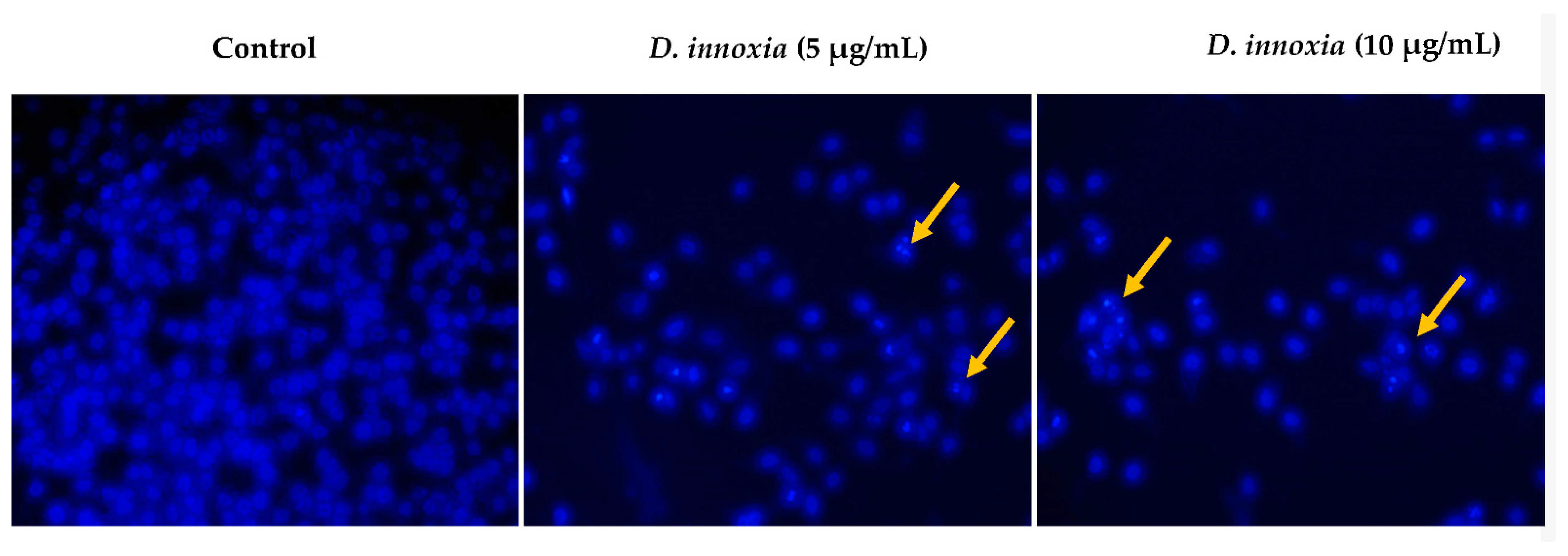
2.6. Hoechst 33258 Staining

LoVo cells were grown on 12-well plate for 24 h, and then treated with vehicle as control or with D. innoxia ethanolic extract at (5 and 10 μg/mL). After rinsing with PBS, cells were fixed in 4% paraformaldehyde followed by permeabilization in cold methanol. Cells were then washed with PBS, and then incubated with Hoechst 33258 (Sigma, St. Louis, MO, USA) which was prepared in PBS (0.5 μg/mL) and added to LoVo cells for 15 min at the dark. Finally, cells were examined under Zeiss fluorescence microscope a (Zeiss, Wetzlar, Germany).

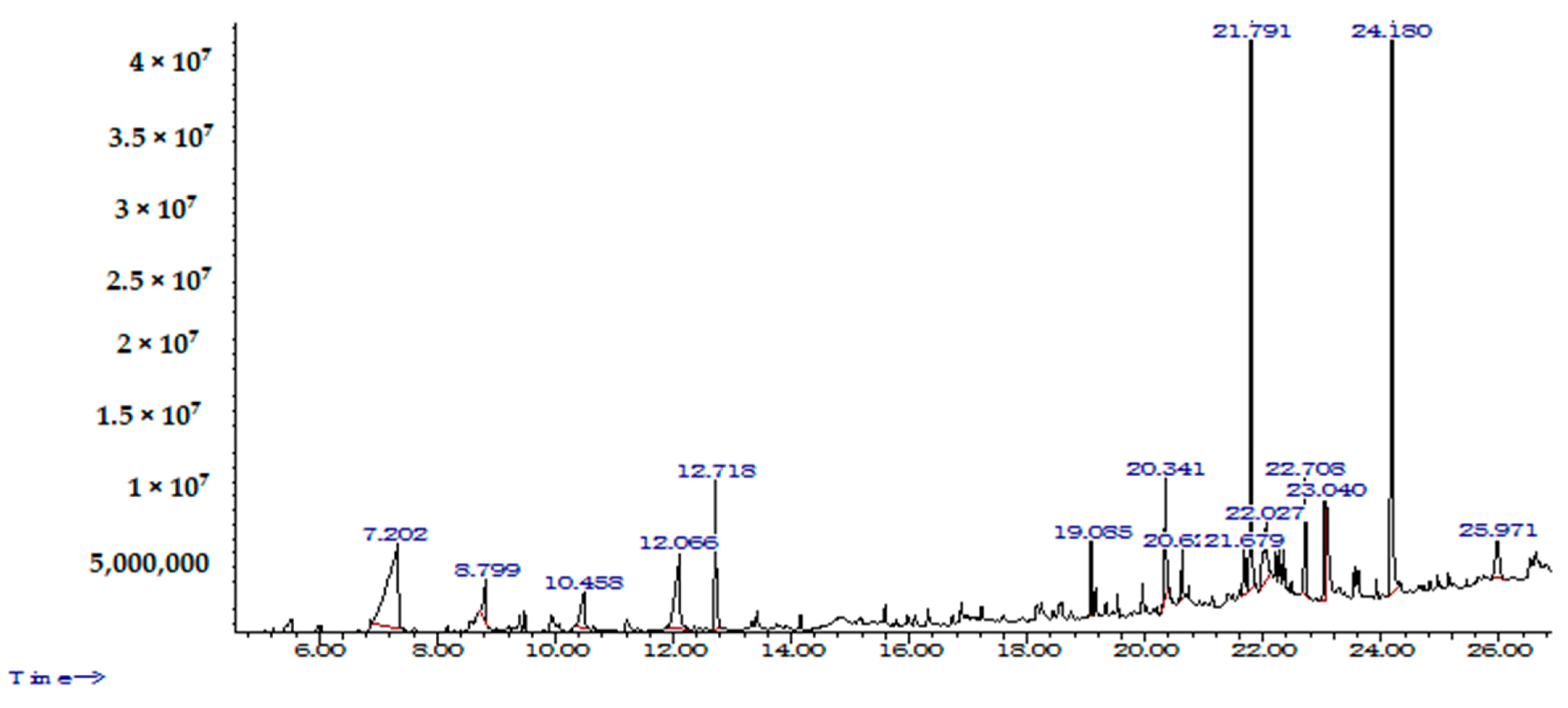
2.7. GC-MS Analysis of D. innoxia Constituents

GC-MS analysis of the D. innoxia ethanolic extract was conducted using a Perkin Elmer Clarus 600 GC-MS (PerkinElmer, Waltham, MA, USA). Two microliters of D. innoxia extract were placed into the Elite-5MS column (30 m, 0.25 µm thickness, 0.25 µm internal diameter) and components separation was conducted using helium gas as a carrier at a flow of 1 mL/min. The oven temperature was programmed as described previously [20]. The Wiley GC-MS [23] and Adams [24] mass spectral libraries were used to compare comparable mass spectra found for D. innoxia constituents.

2.8. Statistical Analysis

All treatments were conducted in triplicates and the standard deviation (SD) of the mean of the three experiments was computed. The Student’s two-tailed t-test was performed for each assay to determine significance at p < 0.05.

3. Results

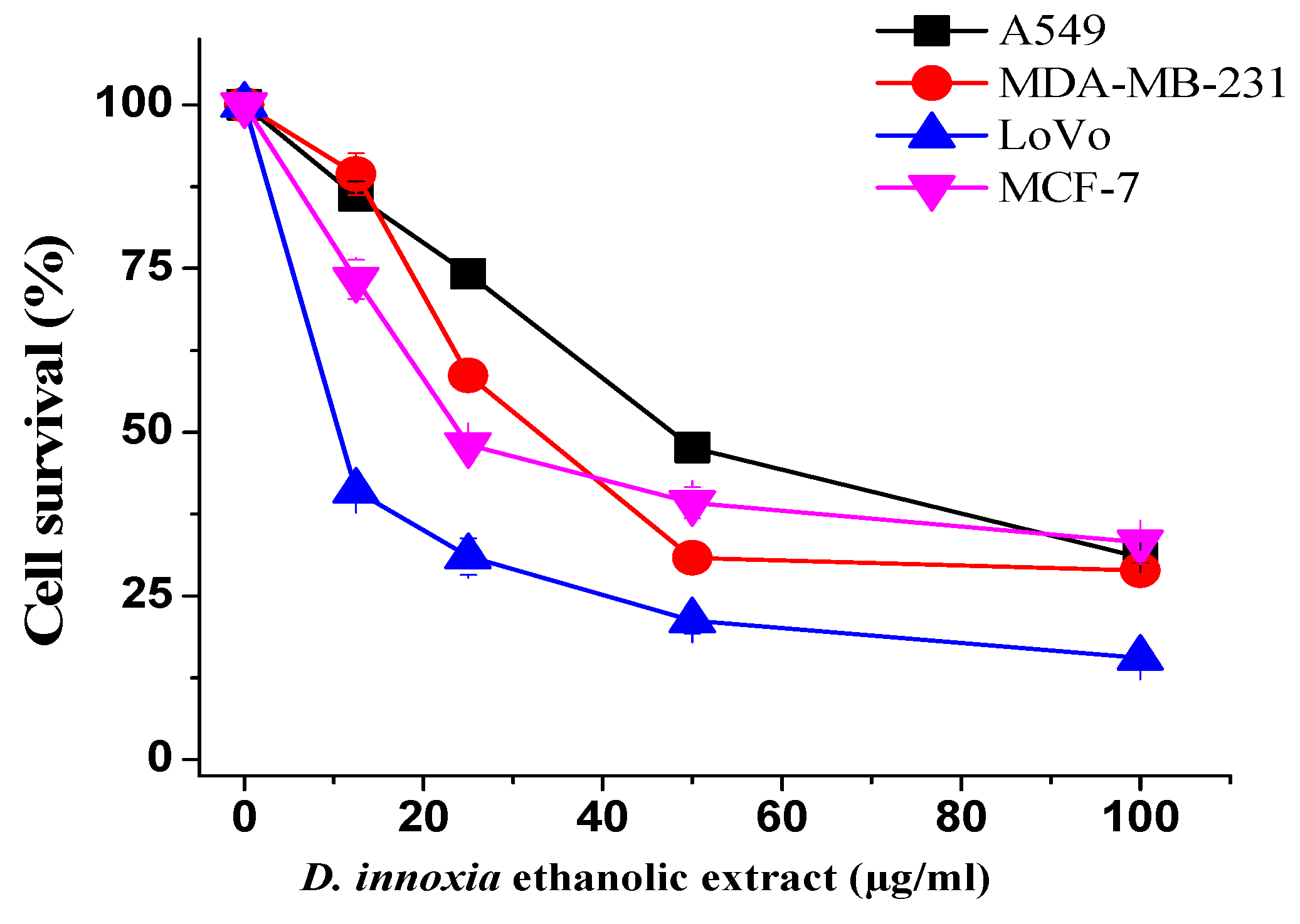
3.1. Ethanol Extract of D. innoxia Inhibit Cells Proliferation

A549, LoVo, MCF-7 and MDA-MB-231 cancer cells were treated with different D. innoxia ethanol extract concentrations to determine its cytotoxic abilities, and thereafter half maximal inhibitory concentration (IC50) values were computed for each cell line. After 48 h of treatment, values were obtained using a dose–response inhibition curve (Table 2). Figure 1 is a dose–response curve for the four cancer cell lines treated with ethanolic extract. The ethanol extract of D. innoxia was cytotoxic against all tested cell lines and the LoVo colon cancer cells was the most sensitive cells in terms of IC50 values (IC50 = 10 μg/mL), therefore, it was selected to conduct the remaining assays.

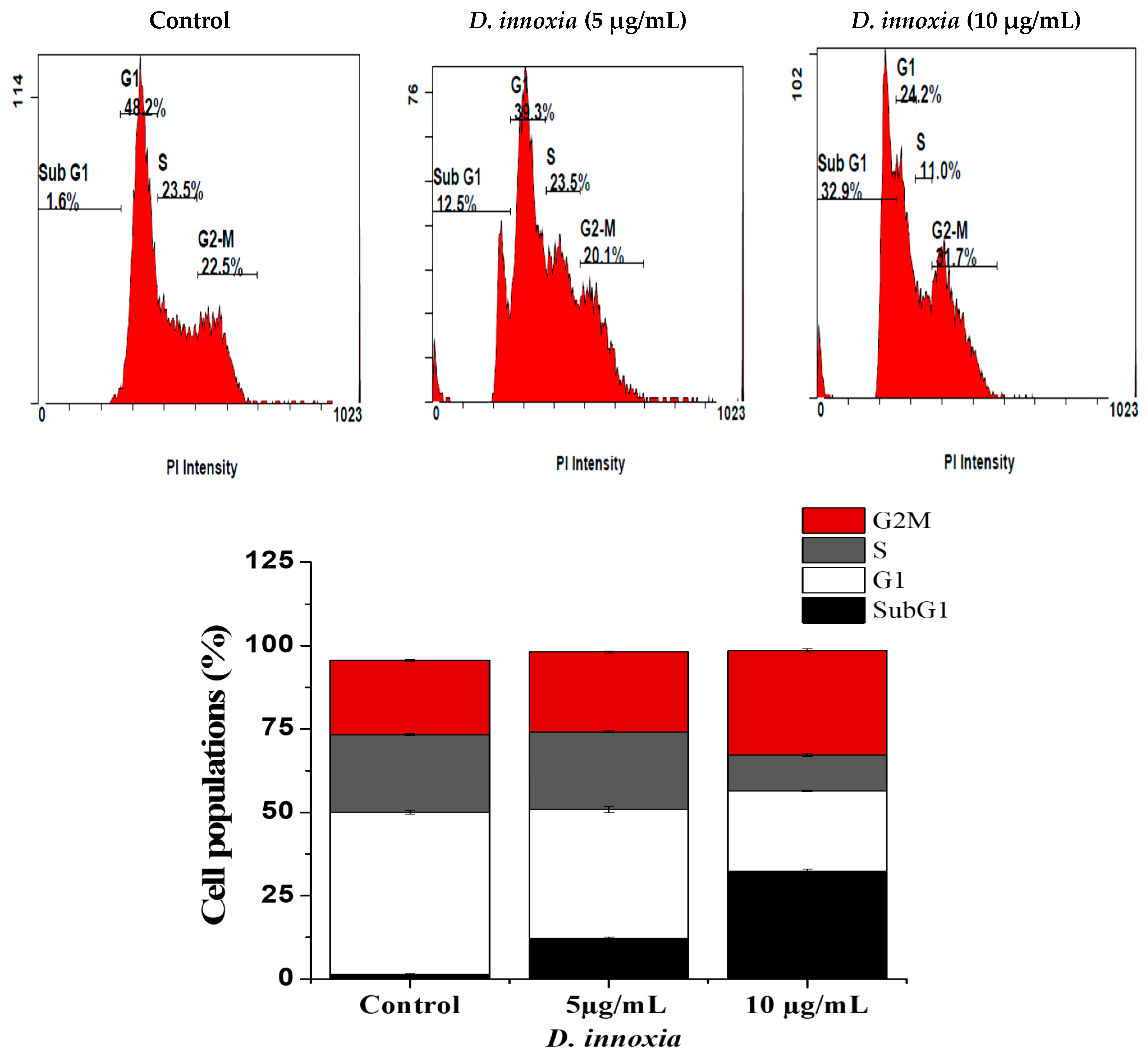
3.2. D. innoxia Causes a Sub-G1 Cells Accumulation

Effect of D. innoxia ethanolic extract on cell cycle of LoVo cells was analyzed using propidium iodide via flow cytometry. After treatment with 5 and 10 µg/mL for 48 h, the sub-G1 phase cells were accumulated to 12.5 ± 0.5% and 32.9 ± 1, respectively, (Figure 2), while in the control group it was only 1.6 ± 0.1%. We also observed that the G2/M phase was affected at 10 µg/mL, indicating a cell cycle block or delay at this stage.

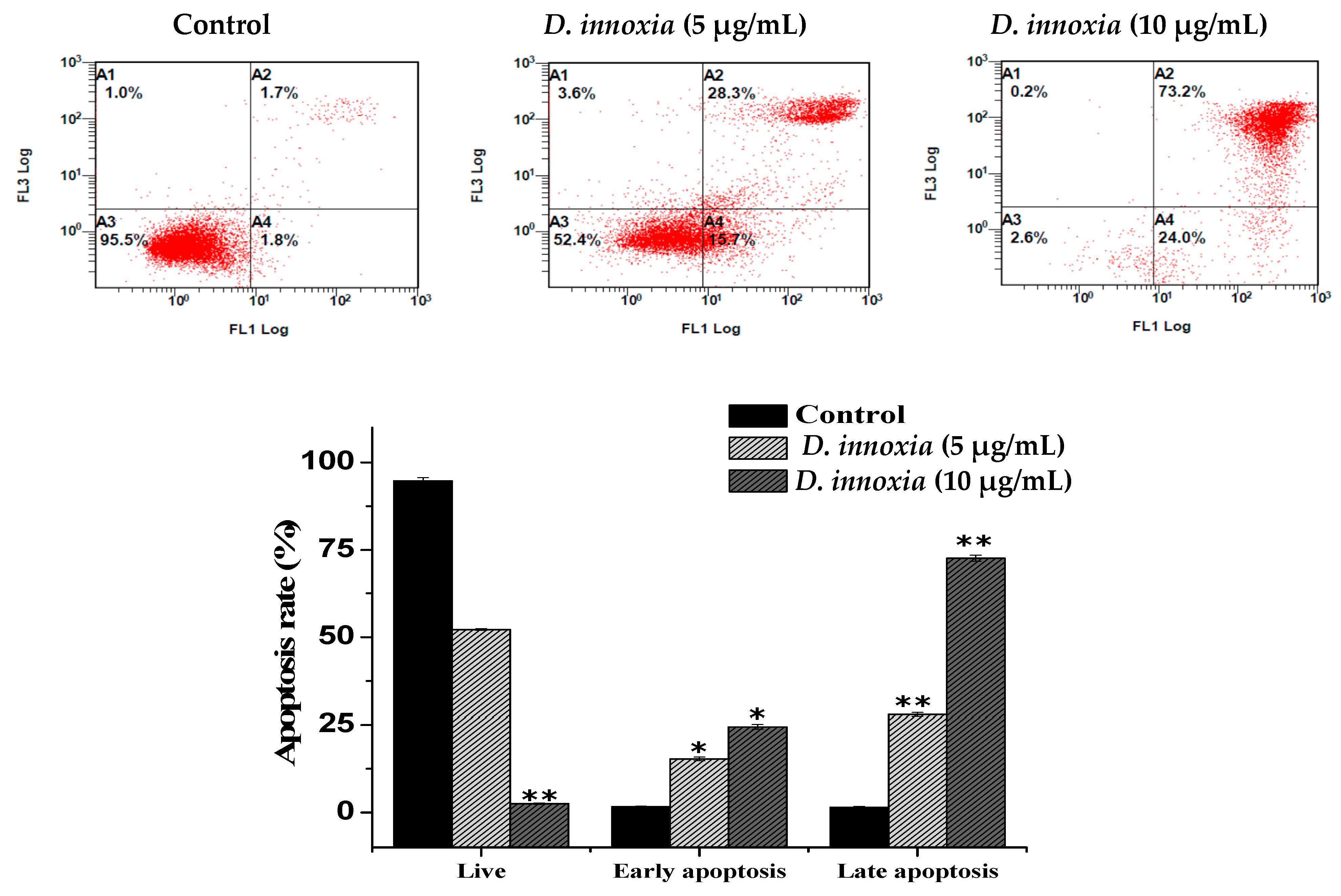
3.3. Apoptotic Cell Death Quantification of D. innoxia Treated Cells

Staining with Annexin V-FITC and propidium iodide was also used to confirm apoptosis induction on LoVo cells by ethanolic D. innoxia extract. The results obtained from this experiment are depicted in (Figure 3). A significant increase from 1.7 ± 0.14% to 15.35 ± 0.5% and 27.9 ± 0.5% (p < 0.05) of early and late apoptotic cells respectively was recorded after treatment with 5 µg/mL. In the same manner, an increase to 24.5 ± 0.7% and 72.6 ± 0.84% (p < 0.01) was recorded for both stages after treatment with 10 µg/mL (Figure 3).

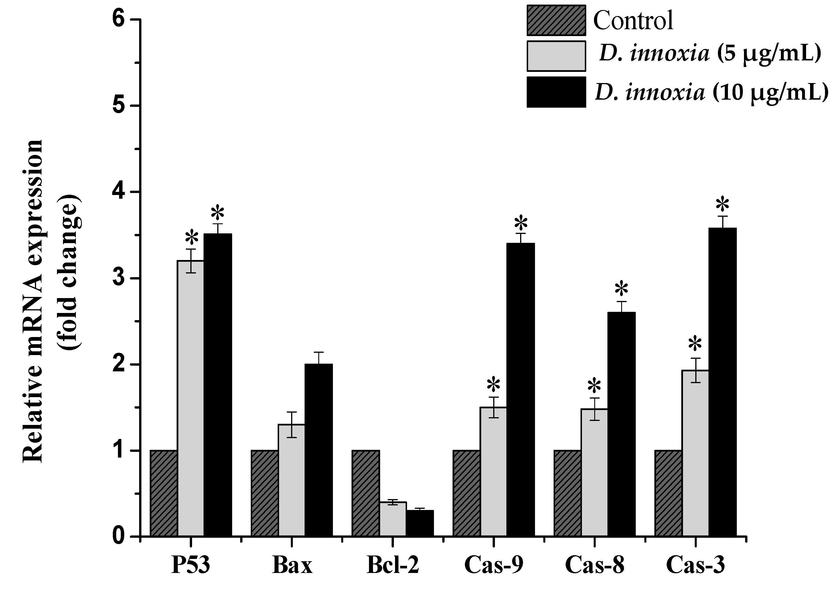
3.4. The Quantitative Real-Time PCR Analysis of Apoptosis-Related Genes

Our results indicate that the D. innoxia treated cells showed a dose-dependent increase in the levels of apoptosis related genes. The results showed that the p53 and caspase-3, -9, and -8 showed higher upregulation and were significantly upregulated (p < 0.5). However, no significant expression was reported for Bax gene in cells treated with D. innoxia ethanolic extract. Furthermore, the expression of antiapoptotic Bcl-2 gene was inhibited after treatment in dose-dependent manner (Figure 4).

3.5. D. innoxia Causes a DNA Fragmentation

To explore the morphological alterations caused by D. innoxia ethanolic extract treatment, LoVo cells were monitored using Hoechst 33258 staining. As compared with the control cells, the features of apoptotic cell death, including nuclear fragmentation in the D. innoxia treated cells, were noticed (Figure 5). These results elucidated that the inhibition of D. innoxia ethanolic extract on LoVo cells growth was linked with its induction of apoptosis.

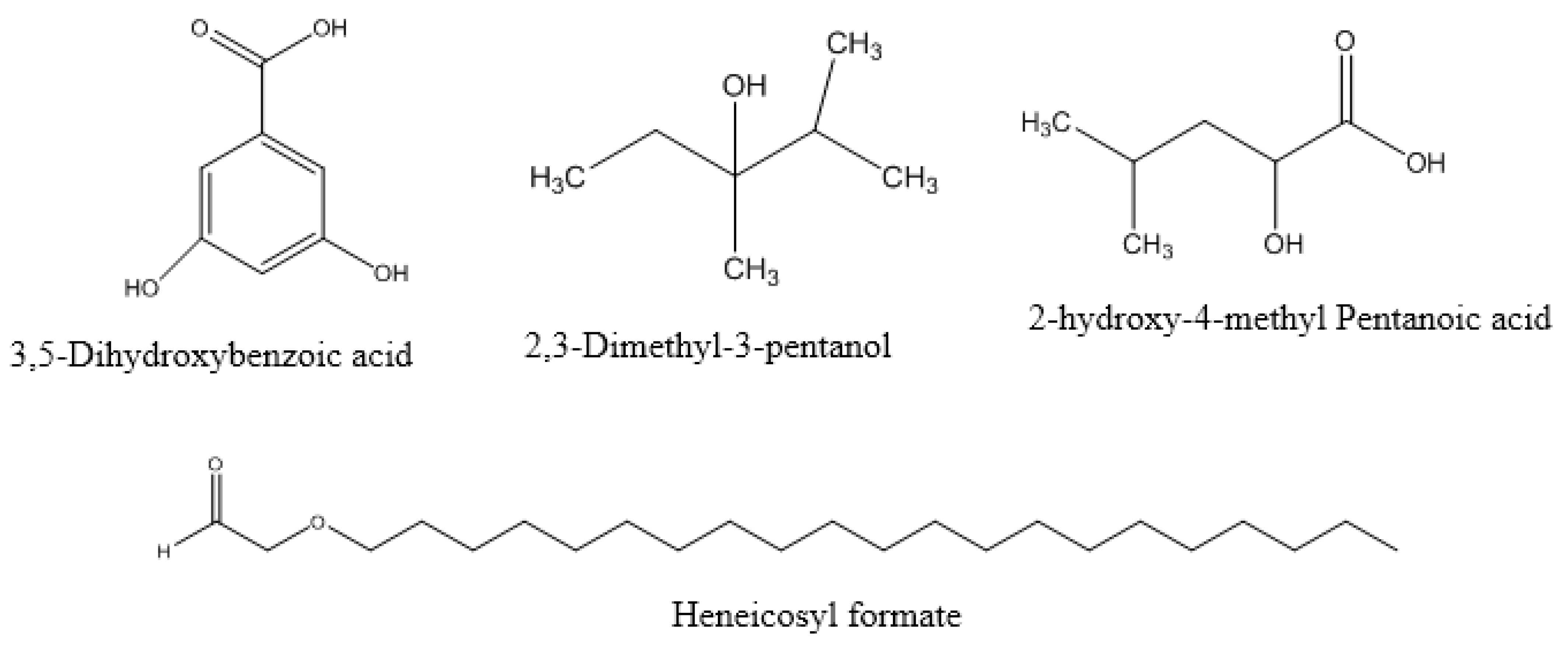
3.6. Identification of D. innoxia Components by GC-MS

The analysis of the D. innoxia by GC-MS identified 50 compounds from the ethanolic extract. The retention time and the percentage amounts of the compositions are displayed in (Table 3). The identified compounds are represented in the order of their elution on the HP Innowax column (Figure 6). 3,5-dihydroxybenzoic acid (16.53%), Heneicosyl formate (14.14%) and 2,3-dimethyl-3-pentanol (12.89%) and 2-hydroxy-4-methyl pentanoic acid (5.19%) were the primary constituents (Figure 7). The remaining compounds are present in small proportions (Table 3).

4. Discussion

Datura innoxia is a valuable medicinal plant rich in different phytochemicals with a broad medicinal property [25]. In this study, D. innoxia was extracted by hydroalcoholic solvent in order to obtain the maximum amount and diversity of biologically active phytochemicals. The results of the present study presented that the D. innoxia ethanolic extract exerted a cytotoxic effect on various cancer cells, with high potent activity against LoVo human colon cancer cells. We have also demonstrated that D. innoxia ethanolic extract induces a cell cycle arrest at the sub-G1 phase and more precisely an apoptotic cell death. This finding is supported by several pieces of evidence including Annexin V-FITC/PI labeling as well as the activation of the apoptosis signaling molecules. This finding is in line with a prior study that reported a strong antiproliferative effect of D. innoxia leaf methanolic extracts against colon (HCT-15) and liver HepG-2 cancer cells [26]. A potent cytotoxic effect against the THP-1 (human leukemia) cell line with a close IC50 value (4.52 μg/mL) was also documented for the D. innoxia grown in Pakistan [27]. In the same manner, the cytotoxic effect against the MCF-7 cell line was also reported [18] for D. innoxia grown in Iran. This variation in IC50 values may result from different D. innoxia geographical sources, growing conditions and various cell types used [28]. National Cancer Institute (NCI, USA) criteria considered a plant crude extract with IC50 value less than 20 μg/mL a suitable extract for in vitro cytotoxic activity [29]. Based on this, D. innoxia displayed potent cytotoxic activity against LoVo with IC50 10 μg/mL; therefore, LoVo cells were selected to assert the inhibition mechanisms exerted by this extract.
The cell cycle distribution of LoVo cells treated with D. innoxia was analyzed to explore the growth inhibition mechanism. It was observed that cells treatments with D. innoxia accumulated at the sub-G1 cells phase, and this result proposed that LoVo cells undergo apoptosis since the accumulation of the cells in this stage is considered as a biomarker for DNA damage as well as an apoptotic cell death marker [30]. Confirmation of apoptosis induction was then verified through Annexin V-FITC/PI apoptosis detection assay, which is extensively used to discriminate living cells from both early and late apoptosis [31]. It was noted that D. innoxia resulted in a strong shift to early and late apoptotic cell populations and no necrotic cells were detected, which emphasizes that D. innoxia extract exerted apoptosis cell death mode. The indications of the dual staining of Annexin-V/PI and the accumulation of cells in the sub-G1 phase, as well as nuclear derangement which was observed with Hochest staining after treatment with D. innoxia ethanolic extract, clearly indicates that the cells are going in apoptosis. In fact, the induction of the apoptotic signaling pathways to trigger cancer cell death is the main mechanism for most anticancer drugs [32].
Apoptosis is a highly gene regulated process and various gene families, such as the P53 and the Bcl-2 family, are implicated in apoptotic cell death [33]. To support the flow cytometry results, the expression of the genes involved in the apoptosis pathway was analyzed using the qRT-PCR technique. Our results clearly proved that the extract treatments led to a decrease in the antiapoptotic Bcl-2 gene and an increase in the proapoptotic Bax gene, as well as activating caspase genes in the treated groups compared to the control, suggesting that D. innoxia acts through a caspase-dependent apoptosis pathway. A similar event was observed in a recent study which reported that D. innoxia extract induced apoptosis through increasing the expression of P53, BAX/BCL2 ratio, caspase 9, 3, 6, 7 on human chronic myeloid leukemia cells (K562 cells) [34]. Altogether this study and our study support the apoptosis-inducing effect of D. innoxia against different cancer cells.
Many species of the Solanaceae family are rich in calystegin, which has an inhibitory property against cancer [35]. The Lectin was also isolated from D. innoxia seeds and it has also shown antiproliferative activity against human cancer cell lines [36]. GC/MS is also a useful and reliable method for the rapid identification of complex plant extracts [37]. The 3,5-dihydroxybenzoic acid, which was detected as a major constituent of D. innoxia in this study, has shown an inhibitory effect against colorectal cancer cells’ growth [38], suggesting a possible role for this compound in the observed activity against LoVo colon cells.
The plant extracts’ medicinal effects could be primarily attributed to their secondary products, which act synergistically rather than as a single compound [39]. As aforementioned, D. innoxia contains a combination of several compounds that have anticancer effects, so it can be concluded that the anticancer effects observed in the extract are associated with the presence of these compounds.

5. Conclusions

The D. innoxia ethanolic extract exerted antiproliferation capacity against various cancer cells. In particular, we have illustrated that D. innoxia ethanolic extract affects the growth of the LoVo colon cancer cell line successfully by inducing cell death. Our data have shown the potential role of D. innoxia in activating apoptosis via increasing the SubG1 phase as well as causing dose-dependent increases in early and late apoptosis cells population. Besides, apoptosis initiation induced by D. innoxia was confirmed by the upregulation of apoptosis gene markers in LoVo cells. This study provides preliminary data that proposes D. innoxia as a valuable source of potentially new natural anticolon cancer compound(s) that act by triggering apoptotic cell death. Further research is required to find effective compounds as well as the cellular and molecular mechanisms involved.

Author Contributions

Conceptualization, M.A.-Z. and A.S.A.; methodology, F.A.N., M.A.-Z., I.O.B. and H.M.D.; validation, A.S.A. and F.A.N.; investigation, writing—original draft preparation; M.A.-Z., F.A.N.; writing—review and editing, M.A.W.C. and A.A.A.; project administration, S.A. and A.D.; funding acquisition A.B. and A.A.A. All authors have read and agreed to the published version of the manuscript.

Funding

This work was funded by the Deanship of Scientific Research at Princess Nourah bint Abdulrahman University, through the Research Groups Program Grant no. (RGP-1442-0033).

Institutional Review Board Statement

Not applicable.

Informed Consent Statement

Not applicable.

Data Availability Statement

The raw data used and/or analyzed during the current study will be available from the corresponding author on reasonable request.

Acknowledgments

This work was funded by the Deanship of Scientific Research at Princess Nourah bint Abdulrahman University, through the Research Groups Program Grant no. (RGP-1442-0033).

Conflicts of Interest

The authors declare that they have no competing interests.

References

  1. Bray, F.; Ferlay, J.; Soerjomataram, I.; Siegel, R.L.; Torre, L.A.; Jemal, A. Global cancer statistics 2018: GLOBOCAN estimates of incidence and mortality worldwide for 36 cancers in 185 countries. CA Cancer J. Clin. 2018, 68, 394–424. [Google Scholar] [CrossRef] [PubMed] [Green Version]
  2. Sung, H.; Ferlay, J.; Siegel, R.L.; Laversanne, M.; Soerjomataram, I.; Jemal, A.; Bray, F. Global cancer statistics 2020: GLOBOCAN estimates of incidence and mortality worldwide for 36 cancers in 185 countries. CA Cancer J. Clin. 2021. [Google Scholar] [CrossRef]
  3. Zhang, Q.-Y.; Wang, F.-X.; Jia, K.-K.; Kong, L.-D. Natural Product Interventions for Chemotherapy and Radiotherapy-Induced Side Effects. Front. Pharm. 2018, 9, 1253. [Google Scholar] [CrossRef] [Green Version]
  4. Iqbal, J.; Abbasi, B.A.; Mahmood, T.; Kanwal, S.; Ali, B.; Shah, S.A.; Khalil, A.T. Plant-derived anticancer agents: A green anticancer approach. Asian Pac. J. Trop. Biomed. 2017, 7, 1129–1150. [Google Scholar] [CrossRef]
  5. Desai, A.G.; Qazi, G.N.; Ganju, R.K.; El-Tamer, M.; Singh, J.; Saxena, A.K.; Bedi, Y.S.; Taneja, S.C.; Bhat, H.K. Medicinal plants and cancer chemoprevention. Curr. Drug Metab. 2008, 9, 581–591. [Google Scholar] [CrossRef] [Green Version]
  6. Al-Eisawi, D.M.; Al-Ruzayza, S. The flora of holy Mecca district, Saudi Arabia. Int. J. Biodivers. Conserv. 2015, 7, 173–189. [Google Scholar]
  7. Al-Zahrani, A.A. Saudi anti-human cancer plants database (SACPD): A collection of plants with anti-human cancer activities. Oncol. Rev. 2018, 12, 349. [Google Scholar] [CrossRef] [Green Version]
  8. Evans, W.; Ghani, A.; Woolley, V.A. Distribution of littorine and other alkaloids in the roots of Datura species. Phytochemistry 1972, 11, 2527–2529. [Google Scholar] [CrossRef]
  9. Pandey, M.; Saraswati, S.; Agrawal, S. Antiproliferative effects of Datura innoxia extract in cervical HeLa cell line. J. Pharm. Res. 2011, 4, 1124–1126. [Google Scholar]
  10. Giral, F.; Hidalgo, C. Presence of alkaloids in Mexican plants. Int. J. Crude Drug Res. 1983, 21, 1–13. [Google Scholar] [CrossRef]
  11. El-Tawil, B. Chemical constituents of indigenous plants used in native medicine of Saudi Arabia. II. Arab Gulf J. Sci. Res. 1983. [Google Scholar]
  12. Aynehchi, Y.; Salehi Sormaghi, M.; Amin, G.; Khoshkhow, M.; Shabani, A. Survey of Iranian plants for saponins, alkaloids, flavonoids and tannins. III. Int. J. Crude Drug Res. 1985, 23, 33–41. [Google Scholar] [CrossRef]
  13. Shama, A.I.; Abd-Kreem, Y.; Fadowa, A.; Samar, R.; Sabahelkhier, M. In vitro antibacterial and antifungal activity and Datura innoxia extracts. Int. J. Environ. 2014, 3, 173–185. [Google Scholar] [CrossRef] [Green Version]
  14. Gajendran, B.; Durai, P.; Varier, K.M.; Liu, W.; Li, Y.; Rajendran, S.; Nagarathnam, R.; Chinnasamy, A. Green synthesis of silver nanoparticle from Datura inoxia flower extract and its cytotoxic activity. BioNanoScience 2019, 9, 564–572. [Google Scholar] [CrossRef]
  15. Cook, N.C.; Samman, S. Flavonoids—chemistry, metabolism, cardioprotective effects, and dietary sources. J. Nutr. Biochem. 1996, 7, 66–76. [Google Scholar] [CrossRef]
  16. Manach, C.; Williamson, G.; Morand, C.; Scalbert, A.; Rémésy, C. Bioavailability and bioefficacy of polyphenols in humans. I. Review of 97 bioavailability studies. Am. J. Clin. Nutr. 2005, 81, 230S–242S. [Google Scholar] [CrossRef] [PubMed] [Green Version]
  17. Hostettmann, K.; Marston, A. Saponins; Cambridge University Press: Cambridge, UK, 2005. [Google Scholar]
  18. Cheshomi, H.; Aldaghi, L.S.; Rezaei Seresht, H. Cytotoxicity of the methanol extract of datura innoxia petals on MCF-7 and HEK-293 cell lines. J. Biomed. 2016, 1, e6623. [Google Scholar] [CrossRef] [Green Version]
  19. Chamani, E.; Ebrahimi, R.; Khorsandi, K.; Meshkini, A.; Zarban, A.; Sharifzadeh, G. In vitro cytotoxicity of polyphenols from Datura innoxia aqueous leaf-extract on human leukemia K562 cells: DNA and nuclear proteins as targets. Drug Chem. Toxicol. 2020, 43, 138–148. [Google Scholar] [CrossRef]
  20. Nasr, F.A.; Noman, O.M.; Alqahtani, A.S.; Qamar, W.; Ahamad, S.R.; Al-Mishari, A.A.; Alyhya, N.; Farooq, M. Phytochemical constituents and anticancer activities of Tarchonanthus camphoratus essential oils grown in Saudi Arabia. Saudi Pharm. J. 2020, 28, 1474–1480. [Google Scholar] [CrossRef] [PubMed]
  21. Rio, D.C.; Ares, M., Jr.; Hannon, G.J.; Nilsen, T.W. Purification of RNA using TRIzol (TRI reagent). Cold Spring Harb. Protoc. 2010, 2010. [Google Scholar] [CrossRef]
  22. Abutaha, N.; Nasr, F.A.; Al-zharani, M.; Alqahtani, A.S.; Noman, O.M.; Mubarak, M.; Abdelhabib, S.; Wadaan, M.A. Effects of hexane root extract of ferula hermonis boiss. On human breast and colon cancer cells: An in vitro and in vivo study. Biomed. Res. Int. 2019, 2019, 3079895. [Google Scholar] [CrossRef] [PubMed] [Green Version]
  23. McLafferty, F.W.; Stauffer, D.B. The Wiley/NBS Registry of Mass Spectral Data; Wiley: New York, NY, USA, 1989; Volume 1. [Google Scholar]
  24. Adams, R.P. Identification of Essential oil Components by Gas Chromatography/Mass Spectrometry; Allured Publishing Corporation: Carol Stream, IL, USA, 2007; Volume 456. [Google Scholar]
  25. Jaafar, F.R.; Ajeena, S.J.; Mehdy, S.S. Anti-inflammatory impacts and analgesiac activity of aqueous extract Datura innoxia leaves against induced pain and inflammation in mice. J. Entomol. Zool. Stud. 2018, 6, 1894–1899. [Google Scholar]
  26. Arulvasu, C.; Babu, G.; Manikandan, R.; Srinivasan, P.; Sellamuthu, S.; Prabhu, D.; Dinesh, D. Anti-cancer effect of Datura innoxia P. Mill. Leaf extract in vitro through induction of apoptosis in human Colon Adenocarcinoma and larynx cancer cell lines. J. Pharm. Res. 2010, 3, 1485–1488. [Google Scholar]
  27. Fatima, H.; Khan, K.; Zia, M.; Ur-Rehman, T.; Mirza, B.; Haq, I.-u. Extraction optimization of medicinally important metabolites from Datura innoxia Mill.: An in vitro biological and phytochemical investigation. BMC Complement. Altern. Med. 2015, 15, 376. [Google Scholar] [CrossRef] [Green Version]
  28. Uğur, D.; Güneş, H.; Güneş, F.; Mammadov, R. Cytotoxic Activities of Certain Medicinal Plants on Different Cancer Cell Lines. Turk. J. Pharm. Sci. 2017, 14, 222–230. [Google Scholar] [CrossRef]
  29. Boik, J. Natural Compounds in Cancer Therapy; Oregon Medical Press: Princeton, MN, USA, 2001. [Google Scholar]
  30. Riccardi, C.; Nicoletti, I. Analysis of apoptosis by propidium iodide staining and flow cytometry. Nat. Protoc. 2006, 1, 1458–1461. [Google Scholar] [CrossRef] [PubMed]
  31. Demchenko, A.P. Beyond annexin V: Fluorescence response of cellular membranes to apoptosis. Cytotechnology 2013, 65, 157–172. [Google Scholar] [CrossRef] [PubMed] [Green Version]
  32. Pistritto, G.; Trisciuoglio, D.; Ceci, C.; Garufi, A.; D’Orazi, G. Apoptosis as anticancer mechanism: Function and dysfunction of its modulators and targeted therapeutic strategies. Aging 2016, 8, 603–619. [Google Scholar] [CrossRef] [PubMed] [Green Version]
  33. Kiraz, Y.; Adan, A.; Kartal Yandim, M.; Baran, Y. Major apoptotic mechanisms and genes involved in apoptosis. Tumour Biol. 2016, 37, 8471–8486. [Google Scholar] [CrossRef] [Green Version]
  34. Chamani, E.; Rezaei, Z.; Dastjerdi, K.; Javanshir, S.; Khorsandi, K.; Mohammadi, G.A. Evaluation of some genes and proteins involved in apoptosis on human chronic myeloid leukemia cells (K562 cells) by datura innoxia leaves aqueous extract. J. Biomol. Struct. Dyn. 2020, 38, 4838–4849. [Google Scholar] [CrossRef] [PubMed]
  35. Diker, D.; Markovitz, D.; Rothman, M.; Sendovski, U. Coma as a presenting sign of Datura stramonium seed tea poisoning. Eur. J. Intern. Med. 2007, 18, 336–338. [Google Scholar] [CrossRef] [PubMed]
  36. Singh, R.; Nawale, L.; Sarkar, D.; Suresh, C. Two chitotriose-specific lectins show anti-angiogenesis, induces caspase-9-mediated apoptosis and early arrest of pancreatic tumor cell cycle. PLoS ONE 2016, 11, e0146110. [Google Scholar] [CrossRef] [PubMed]
  37. Iordache, A.; Culea, M.; Gherman, C.; Cozar, O. Characterization of some plant extracts by GC–MS. Nucl. Instrum. Methods Phys. Res. Sect. B Beam Interact. Mater. At. 2009, 267, 338–342. [Google Scholar] [CrossRef]
  38. Sankaranarayanan, R.; Valiveti, C.K.; Dachineni, R.; Kumar, D.R.; Lick, T.; Bhat, G.J. Aspirin metabolites 2,3-DHBA and 2,5-DHBA inhibit cancer cell growth: Implications in colorectal cancer prevention. Mol. Med. Rep. 2020, 21, 20–34. [Google Scholar] [CrossRef] [Green Version]
  39. Jigna, P.; Rathish, N.; Sumitra, C. Preliminary screening of some folklore medicinal plants from western India for potential antimicrobial activity. Indian J. Pharmacol. 2005, 37, 408–409. [Google Scholar]
Figure 1. Cytotoxic effect of D. innoxia ethanolic extract on various cancer cell lines after 48 h of exposure. Cells were treated as described in the Methods section and cell viability was determined using the MTT assay. Data are expressed as the mean + standard deviation of three experiments.
Applsci 11 02864 g001
Figure 2. Histograms representing DNA contents analysis after 48 h of LoVo cells treatment. LoVo cells were treated with 5 and 10 μg/mL D. innoxia and vehicle control. Cell cycle analysis was recorded on a Beckman Coulter Cytomics FC500 after PI staining.
Applsci 11 02864 g002
Figure 3. Dot plots of Annexin V-FITC stained LoVo cells after 48 h exposure to 5 and 10 μg/mL of D. innoxia. (A3: Annexin V-negative; PI-negative), early apoptotic cells (A4: Annexin V-positive; PI-negative), late apoptotic cells (A2: Annexin V-positive; PI-positive) and necrotic cells (A1: Annexin V-negative; PI-positive). A minimum of 20,000 events were read (n = 3). (One representative for 3 individual experiments, each performed in triplicate). Data are presented as means ± SD and the difference was statistically significant at (* p < 0.05, ** p < 0.01).
Applsci 11 02864 g003
Figure 4. Gene expression levels in LoVo cells, treated with 5 and 10 μg/mL of D. innoxia for 48 h. Apoptosis related gene expression levels of p53, bax, bcl-2 and caspases 9, 8, 3 were determined by quantitative real-time PCR. GAPDH was used as an internal control. The significant differences from control are indicated by * p < 0.05.
Applsci 11 02864 g004
Figure 5. Efficacy of D. innoxia in inducing apoptosis in LoVo cells. The cells were stained with Hoechst dye for nuclear morphology after treatments with D. innoxia ethanolic extract and fluorescent images were obtained. Characteristics of apoptosis, such as nuclear derangement, are observed in LoVo treated cells (arrows) compared to control cells.
Applsci 11 02864 g005
Figure 6. GC-MS chromatogram of D. innoxia ethanolic extract.
Applsci 11 02864 g006
Figure 7. Major constituent in D. innoxia.
Applsci 11 02864 g007
Table 1. Primer sequences for quantitative real-time polymerase chain reaction (qRT-PCR).
Gene NameForward Primer SequencesReverse Primer Sequences
1P535′-TGGCTCTGACTGTACCACCATCC-3′5′-CAGCTCTCGGAACATCTCGAAGC-3′
2Bax5′-GGA TGC GTC CAC CAA GAA G-3′5′-CCT CTG CAG CTC CAT GTT AC-3′
3Bcl-25′-GTG GAT GAC TGA GTA CCT GAA C-3′5′-GCC AGG AGA ATT CAA ACA GAG G-3′
4Caspase 95′-CAG GCC CCA TAT GAT CGA GG-3′5′-TCG ACA ACT TTG CTG CTT GC-3′
5Caspase 85′-CTG GTC TGA AGG CTG GTT GT-3′5′-CAG GCT CAG GAA CTT GAG GG-3′
6Caspase 35′-CTG GTT TTC GGT GGG TGT G-3′5′-ACG GCA GGC CTG AAT AAT GAA
7GAPDH5′-GGT ATC GTG GAA GGA CTC ATG AC-3′5′-ATG CCA GTG AGC TTC CCG TTC AGC
Table 2. Cytotoxic activity of D. innoxia ethanolic extract against different cancer cells.
SampleCell Lines and IC50 (µg/mL)
A549MDA-MB-231LoVoMCF-7
D. innoxia47.76 ± 1.7832.61 ± 1.2910.1 ± 0.5523.97 ± 0.66
Doxorubicin1.2 ± 0.061.3 ± 0.081.1 ± 0.81.5 ± 0. 6
Table 3. The identified compounds from D. innoxia ethanolic extract by GC-MS.
Compound NameChemical FormulaMolecular Weight (g/mol)RT (min)Area%
Bicyclo [3.2.0]heptane C7H1296.175.410.26
1-Propene-1-thiolC3H6S74.155.5250.73
3-Methyl-isoxazol-5(4H)-oneC4H5NO299.096.0270.32
(Z)-3-Methyl-2-hexeneC7H1498.196.670.31
Alpha.Aminooxy-propionic acidC3H7NO3105.096.8610.35
2,3-Dimethyl-3-pentanolC7H16O116.27.33812.89
3,4,6-Tri-O-methyl-d-glucoseC9H18O6222.248.6930.51
3,3-Dimethyl-2-pentanolC7H16O116.28.8142.09
2,6-diethyl-BenzenamineC10H15N149.239.3930.32
3-(1-Cyclopentenyl) furanC9H10O134.179.4750.44
N,N-dimethyl-Propanamide C5H11NO101.159.9331.03
4-methyl-1,3-Dioxane C5H10O2102.1310.4932.2
3-methyl-Benzoyl chlorideC8H7ClO154.5911.2120.65
2-hydroxy-4-methyl Pentanoic acid C6H12O3132.1612.1095.19
PhenoxyacetamideC8H9NO2151.1612.7143.65
1-HeptynylbenzeneC13H16172.2713.420.5
Palmitoleic acidC16H30O2254.4115.9710.3
3-(2-furanyl)-2-PropenalC7H6O2122.1216.0980.36
MegastigmatrienoneC13H18O190.2816.3210.46
2-nitrophenyl azideC6H4N4O2164.1216.8810.5
Spiro [2.3] hexan-4-one, 5,5-diethylC10H16O152.2317.2240.33
3-Nonyn-1-olC9H16O140.2217.60.29
Methyl 3-methoxy-4-nitrobenzoateC9H9NO5211.1718.1660.61
CyclopropaneoctanalC11H20O168.2818.2420.6
(6R)-6alpha-[(Z)-1,3-Butadienyl]-1,4-cycloheptadieneC11H14146.2318.5410.39
2,3,3,4,5-pentaethyl 1,2,5-OxadiborolaneC12H26O2O207.9619.0821.26
CyclotridecaneC13H26182.3519.1580.6
DolichodialC10H14O2166.2119.5270.44
p-Menth-8(10)-en-9-olC10H18O154.2519.960.61
1,4-IcosanediolC20H42O2314.520.6280.95
13-Tetradece-11-yn-1-olC14H24O208.3420.7360.26
9,17-OctadecadienalC18H32O264.421.1370.29
1,19-EicosadieneC20H38278.521.6781.35
Heneicosyl formateC22H44O2340.621.79214.14
8-Tetradecen-1-olC14H28O212.3722.0534.21
3-MethoxybenzylamineC8H11NO137.1822.2820.7
Linoleic acidC18H32O2280.422.3520.86
Bicyclo [6.1.0] non-1-eneC9H14122.2122.4730.33
4-IsopropenylcyclohexanoneC9H14O138.2122.7082.89
Cyclohexane, 1,1′-methylenebisC13H24180.3323.0452.55
Z, Z-3,13-Octadecadien-1-ol acetateC20H36O2308.523.5480.6
9-Methyl-Z, Z-10,12-hexadecadien-1-ol acetate C19H34O2294.523.6180.54
2,10-dimethyl-9-UndecenalC13H24O196.3323.9170.35
3,5-Dihydroxybenzoic acidC7H6O4154.1224.18416.53
7-PentadecyneC15H28208.3824.9540.25
1,3,12-NonadecatrieneC19H34262.525.1390.49
1,5,9-CyclododecatrieneC12H18162.2726.5320.79
MonoelaidinC21H40O4356.527.411.23
2-methyl-5-(1-methyl ethenyl)-CyclohexanolC10H18O154.2527.761.21
Z-(13,14-Epoxy) tetradec-11-en-1-ol acetateC16H28O3268.3927.8681.24
Publisher’s Note: MDPI stays neutral with regard to jurisdictional claims in published maps and institutional affiliations.

