
Effect of Non-Thermal Atmospheric Pressure Plasma on Differentiation Potential of Human Deciduous Dental Pulp Fibroblast-like Cells

Abstract
:1. Introduction
2. Materials and Methods
2.1. Cell Culture
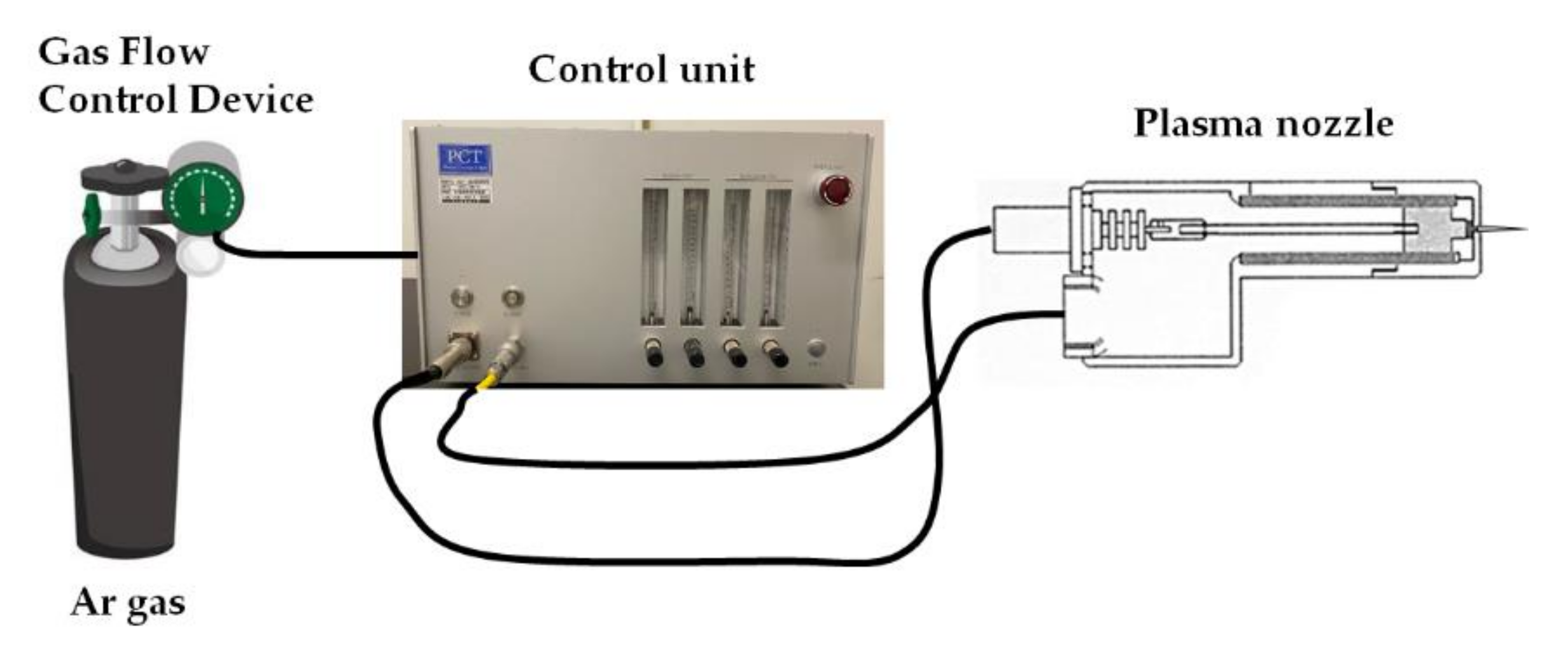
2.2. NTAPP Stimulation Device
2.3. Cell Proliferation Assay
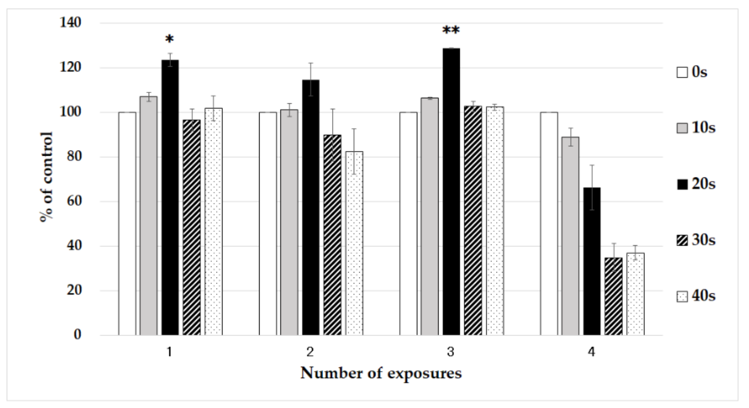
2.3.1. Investigation of NTAPP Irradiation Conditions
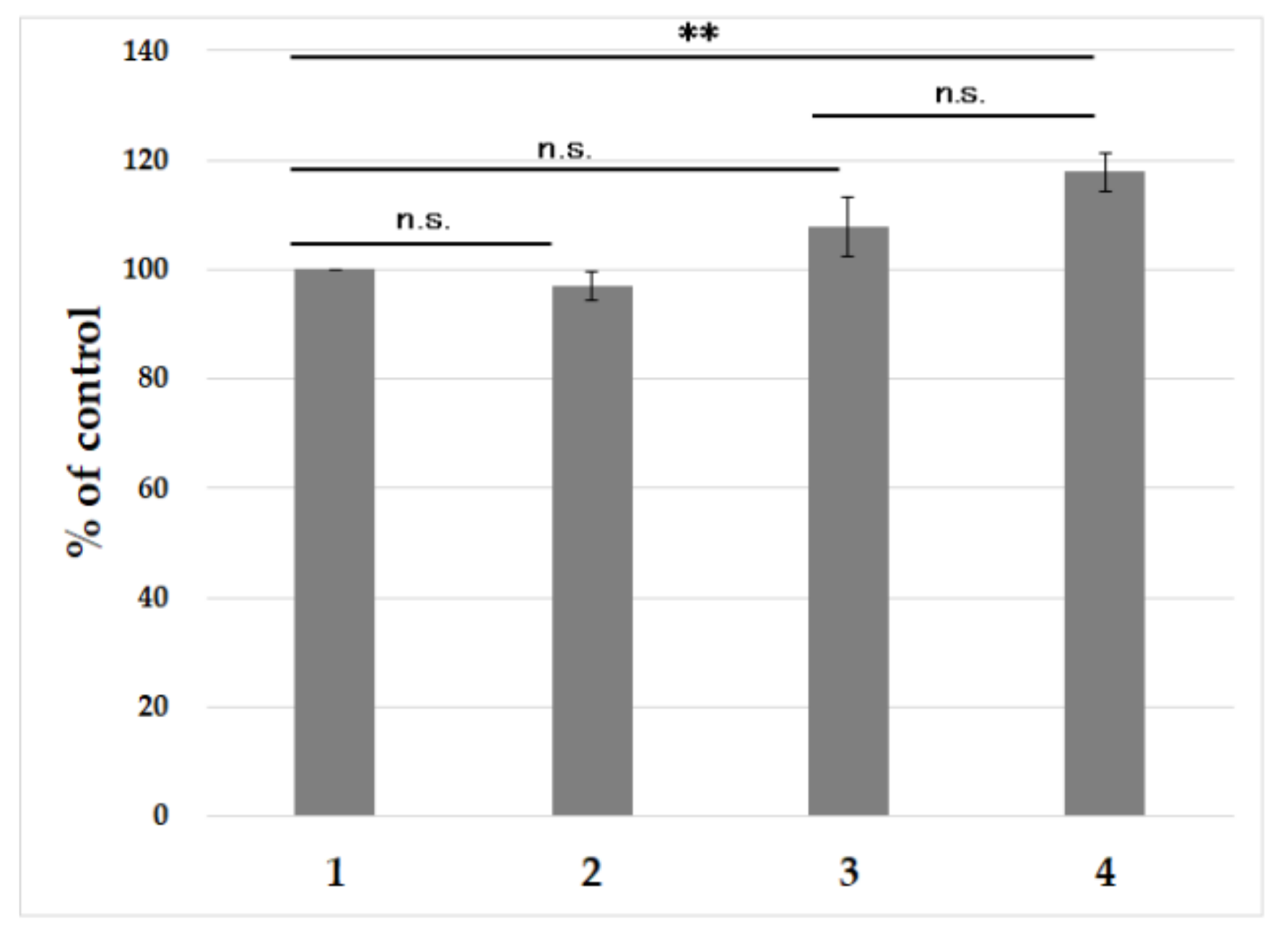
2.3.2. Effects of NTAPP on Culture Medium
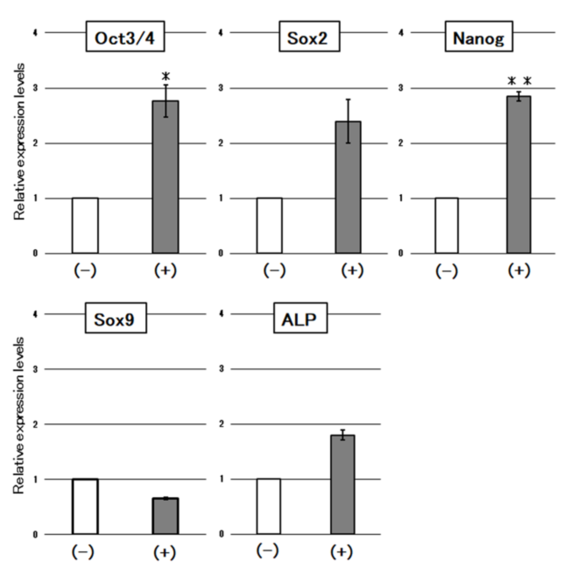
2.4. Reverse-Transcription Polymerase Chain Reaction (RT-PCR)
2.5. Flow Cytometry
2.6. Statistical Analysis
3. Results
3.1. Cell Proliferation Assay
3.1.1. Investigation of NTAPP Irradiation Conditions
3.1.2. Effects on the Culture Medium
3.2. RT-PCR
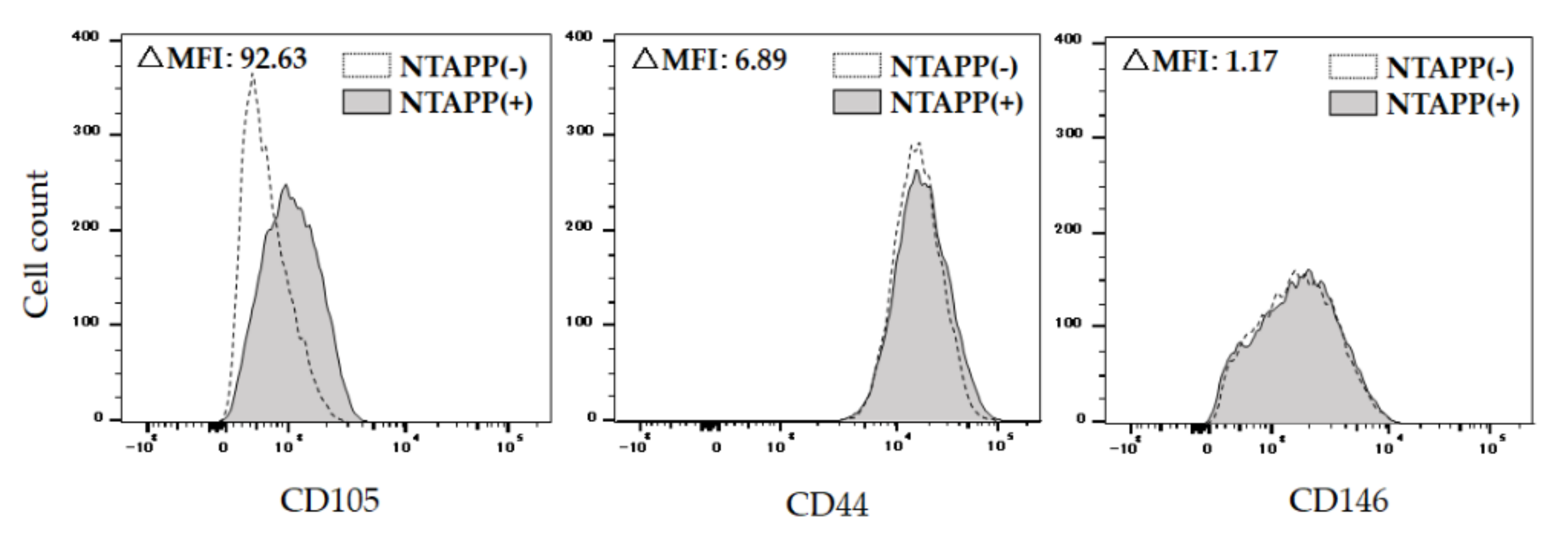
3.3. Flow Cytometry
4. Discussion
5. Conclusions
Author Contributions
Funding
Institutional Review Board Statement
Informed Consent Statement
Data Availability Statement
Acknowledgments
Conflicts of Interest
References
- Prockop, D.J. Marrow stromal cells as stem cells for nonhematopoietic tissues. Science 1997, 276, 71–74. [Google Scholar] [CrossRef] [PubMed] [Green Version]
- Yamashita, K.; Dennis, J.E.; Lennon, D.P.; Morimoto, H.; Kitamura, S.; Caplan, A.I. Dental pulp cells with multi-potential for differentiation to odontoblast and chondroblast. J. Hard Tissue Biol. 2003, 12, 49–55. [Google Scholar] [CrossRef]
- Baghaban, E.M.; Vahabi, S.; Shariati, M.; Nazarian, H. In vitro growth and characterization of stem cells from human dental pulp of deciduous versus permanent teeth. J. Dent. Res. 2010, 7, 185–195. [Google Scholar]
- Gronthos, S.; Mankani, M.; Brahim, J.; Robey, P.G.; Shi, S. Postnatal human dental pulp stem cells (DPSCs) in vitro and in vivo. Proc. Natl. Acad. Sci. USA 2000, 97, 13625–13630. [Google Scholar] [CrossRef] [Green Version]
- Cordeiro, M.M.; Dong, Z.; Kaneko, T.; Zhang, Z.; Miyazawa, M.; Shi, S.; Smith, A.J.; Nör, J.E. Dental pulp tissue engineering with stem cells from exfoliated deciduous teeth. J. Endod. 2008, 34, 962–969. [Google Scholar] [CrossRef] [PubMed]
- Huang, G.T.; Yamaza, T.; Shea, L.D.; Djouad, F.; Kuhn, N.Z.; Tuan, R.S.; Shi, S. Stem/progenitor cell–mediated de novo regeneration of dental pulp with newly deposited continuous layer of dentin in an in vivo model. Tissue Eng. Part A 2010, 16, 605–615. [Google Scholar] [CrossRef] [Green Version]
- d’Aquino, R.; Graziano, A.; Sampaolesi, M.; Laino, G.; Pirozzi, G.; De Rosa, A.; Papaccio, G. Human postnatal dental pulp cells co-differentiate into osteoblasts and endotheliocytes: A pivotal synergy leading to adult bone tissue formation. Cell Death Dis. 2007, 14, 1162–1171. [Google Scholar] [CrossRef] [PubMed] [Green Version]
- Miura, M.; Gronthos, S.; Zhao, M.; Lu, B.; Fisher, L.W.; Robey, P.G.; Shi, S. SHED: Stem cells from human exfoliated deciduous teeth. Proc. Natl. Acad. Sci. USA 2003, 100, 5807–5812. [Google Scholar] [CrossRef] [PubMed] [Green Version]
- Nakamura, S.; Yamada, Y.; Katagiri, W.; Sugito, T.; Ito, K.; Ueda, M. Stem cell proliferation pathways comparison between human exfoliated deciduous teeth and dental pulp stem cells by gene expression profile from promising dental pulp. J. Endod. 2009, 35, 1536–1542. [Google Scholar] [CrossRef]
- Honda, M.J.; Nakashima, F.; Satomura, K.; Shinohara, Y.; Tsuchiya, S.; Watanabe, N.; Ueda, M. Side population cells expressing ABCG2 in human adult dental pulp tissue. Int. Endod. J. 2007, 40, 949–958. [Google Scholar] [CrossRef] [PubMed]
- Kenmotsu, M.; Matsuzaka, K.; Kokubu, E.; Azuma, T.; Inoue, T. Analysis of side population cells derived from dental pulp tissue. Int. Endod. J. 2010, 43, 1132–1142. [Google Scholar] [CrossRef] [PubMed] [Green Version]
- Kawai, S.; Harada, K.; Nagata, S.; Ohura, K.; Arita, K. Effect of 6-bromoindirubin-3′-oxime on human deciduous tooth dental pulp cells. Jpn. Pharmacol. Ther. 2012, 31, 87–95. [Google Scholar]
- Shi, X.M.; Xu, G.M.; Zhang, G.J.; Liu, J.R.; Wu, Y.M.; Gao, L.G.; Yang, Y.; Chang, Z.S.; Yao, C.W. Low-temperature plasma promotes fibroblast proliferation in wound healing by ROS-activated NF-κB signaling pathway. Curr. Med. Sci. 2018, 38, 107–114. [Google Scholar] [CrossRef] [PubMed]
- Chang, J.W.; Kang, S.U.; Shin, Y.S.; Kim, K.I.; Seo, S.J.; Yang, S.S.; Lee, J.S.; Moon, E.; Baek, S.J.; Lee, K.; et al. Non-thermal atmospheric pressure plasma induces apoptosis in oral cavity squamous cell carcinoma: Involvement of DNA-damage-triggering sub-G (1) arrest via the ATM/p53 pathway. Arch. Biochem. Biophys. 2014, 545, 133–140. [Google Scholar] [CrossRef] [PubMed]
- Park, J.; Lee, H.; Lee, H.J.; Kim, G.C.; Kim, D.Y.; Han, S.; Song, K. Non-thermal atmospheric pressure plasma efficiently promotes the proliferation of adipose tissue-derived stem cells by activating NO-response pathways. Sci. Rep. 2016, 6, 39298. [Google Scholar] [CrossRef] [PubMed] [Green Version]
- Kajiyama, H.; Utsumi, F.; Nakamura, K.; Tanaka, H.; Toyokuni, S.; Hori, M.; Kikkawa, F. Future perspective of strategic non-thermal plasma therapy for cancer treatment. J. Clin. Biochem. Nutr. 2017, 60, 33–38. [Google Scholar] [CrossRef] [PubMed] [Green Version]
- Ikawa, S.; Kitano, K.; Hamaguchi, S. Effects of pH on bacterial inactivation in aqueous solutions due to low-temperature atmospheric pressure plasma application. Plasma Process. Polym. 2010, 7, 33–42. [Google Scholar] [CrossRef]
- Hoffmann, C.; Berganza, C.; Zhang, J. Cold atmospheric plasma: Methods of production and application in dentistry and oncology. Med. Gas Res. 2013, 3, 21. [Google Scholar] [CrossRef] [Green Version]
- Park, J.; Lee, H.; Lee, H.J.; Kim, G.C.; Kim, S.S.; Han, S.; Song, K. Non-thermal atmospheric pressure plasma is an excellent tool to activate proliferation in various mesoderm-derived human adult stem cells. Free Radic. Biol. Med. 2019, 134, 374–384. [Google Scholar] [CrossRef]
- Hamaguchi, S. Interaction of plasmas with biological objectsin plasma medicine. Plasma Fusion Res. 2011, 87, 696–703. [Google Scholar]
- Tasaki, T.; Ohshima, T.; Usui, E.; Ikawa, S.; Kitano, K.; Maeda, N.; Momoi, Y. Plasma-treated water eliminates Streptococcus mutans in infected dentin model. Dent. Mater. J. 2017, 36, 422–428. [Google Scholar] [CrossRef] [PubMed] [Green Version]
- Yanagida, T.; Yamamoto, S.; Akisaka, T.; Sawada, T. Tooth Development Tissue Lesion, 1st ed.; Ishiyaku Publishers, Inc.: Tokyo, Japan, 1995; p. 92. [Google Scholar]
- Shi, G.; Jin, Y. Role of Oct4 in maintaining and regaining stem cell pluripotency. Stem Cell Res. Ther. 2010, 1, 39. [Google Scholar] [CrossRef] [Green Version]
- Tsai, C.C.; Hung, S.C. Functional roles of pluripotency transcription factors in mesenchymal stem cells. Cell Cycle 2012, 11, 3711–3712. [Google Scholar] [CrossRef] [PubMed] [Green Version]
- Han, S.M.; Han, S.H.; Coh, Y.R.; Jang, G.; Ra, J.C.; Kang, S.K.; Lee, H.W.; Youn, H.Y. Enhanced proliferation and differentiation of Oct4-And Sox2-overexpressing human adipose tissue mesenchymal stem cells. Exp. Mol. Med. 2014, 46, e101. [Google Scholar] [CrossRef] [PubMed]
- Sun, Z.; Han, Q.; Zhu, Y.S.; Li, Z.Y.; Chen, B.; Liao, L.M.; Bian, C.J.; Li, J.; Shao, C.S.; Zhao, R.C. NANOG has a role in mesenchymal stem cells’ immunomodulatory effect. Stem Cells Dev. 2011, 20, 1521–1528. [Google Scholar] [CrossRef] [PubMed]
- Fujita, N.; Takayasu, M.; Daito, M. In vitro chondrogenic differentiation potential of dental pulp stem cells. Pediatric Dent. J. 2008, 46, 548–554. [Google Scholar]
- Mortada, I.; Mortada, R. Dental pulp stem cells and osteogenesis: An update. Cytotechnology 2018, 70, 1479–1486. [Google Scholar] [CrossRef]
- Sato, R.; Namura, Y.; Tanabe, N.; Sakai, M.; Utsu, A.; Tomita, K.; Suzuki, N.; Motoyoshi, M. Atmospheric pressure plasma treatment with nitrogen induces osteoblast differentiation and reduces iNOS and COX-2 expressions. J. Hard Tissue Biol. 2021, 30, 131–136. [Google Scholar] [CrossRef]
- Makoto, H. Biological effect of calcium ion released from mineral trioxide aggregate. J. Jpn. Endod. Assoc. 2019, 40, 1–6. [Google Scholar] [CrossRef]
- Sato, M.; Ishida, C.; Iwasa, S.; Takei, H.; Honda, M.; Tetsuo Shirakawa, T. Characterization of cultured human mesenchymal stem cells from deciduous and permanent teeth. Pediatr. Dent. J. Jpn. 2014, 52, 417–424. [Google Scholar]
- Beeravolu, N.; Khan, I.; McKee, C.; Dinda, S.; Thibodeau, B.; Wilson, G.; Perez-Cruet, M.; Bahado-Singh, R.; Chaudhry, G.R. Isolation and comparative analysis of potential stem/progenitor cells from different regions of human umbilical cord. Stem Cell Res. 2016, 16, 696–711. [Google Scholar] [CrossRef] [Green Version]
- Beeravolu, N.; McKee, C.; Alamri, A.; Mikhael, S.; Brown, C.; Perez-Cruet, M.; Chaudhry, G.R. Isolation and characterization of mesenchymal stromal cells from human umbilical cord and fetal placenta. J. Vis. Exp. 2017, 122, e55224. [Google Scholar] [CrossRef] [PubMed] [Green Version]
- Takamatsu, T.; Hirai, H.; Sasaki, R.; Miyahara, H.; Okino, A. Surface hydrophilization of polyimide films using atmospheric damage-free multigas plasma jet source. IEEE Trans. Plasma Sci. 2013, 41, 119–125. [Google Scholar] [CrossRef]
- Takamatsu, T.; Uehara, K.; Sasaki, Y.; Miyahara, H.; Matsumura, Y.; Iwasawa, A.; Ito, N.; Azuma, T.; Kohno, M.; Okino, A. Investigation of reactive species using various gas plasmas. RSC Adv. 2014, 4, 39901–39905. [Google Scholar] [CrossRef] [Green Version]
- Nomura, Y.; Takamatsu, T.; Kawano, H.; Miyahara, H.; Okino, A.; Yoshida, M.; Azuma, T. Investigation of blood coagulation effect of nonthermal multigas plasma jet in vitro and in vivo. J. Surg. Res. 2017, 219, 302–309. [Google Scholar] [CrossRef] [PubMed]


| Group | Cell | Medium |
|---|---|---|
| 1 | NTAPP(-) | NTAPP(-) |
| 2 | NTAPP(-) | NTAPP(+) |
| 3 | NTAPP(+) | NTAPP(-) |
| 4 | NTAPP(+) | NTAPP(+) |
Publisher’s Note: MDPI stays neutral with regard to jurisdictional claims in published maps and institutional affiliations. |
© 2021 by the authors. Licensee MDPI, Basel, Switzerland. This article is an open access article distributed under the terms and conditions of the Creative Commons Attribution (CC BY) license (https://creativecommons.org/licenses/by/4.0/).
Share and Cite
Okuno, M.; Aoki, S.; Kawai, S.; Imataki, R.; Abe, Y.; Harada, K.; Arita, K. Effect of Non-Thermal Atmospheric Pressure Plasma on Differentiation Potential of Human Deciduous Dental Pulp Fibroblast-like Cells. Appl. Sci. 2021, 11, 10119. https://doi.org/10.3390/app112110119
Okuno M, Aoki S, Kawai S, Imataki R, Abe Y, Harada K, Arita K. Effect of Non-Thermal Atmospheric Pressure Plasma on Differentiation Potential of Human Deciduous Dental Pulp Fibroblast-like Cells. Applied Sciences. 2021; 11(21):10119. https://doi.org/10.3390/app112110119
Chicago/Turabian StyleOkuno, Masae, Sho Aoki, Saki Kawai, Rie Imataki, Yoko Abe, Kyoko Harada, and Kenji Arita. 2021. "Effect of Non-Thermal Atmospheric Pressure Plasma on Differentiation Potential of Human Deciduous Dental Pulp Fibroblast-like Cells" Applied Sciences 11, no. 21: 10119. https://doi.org/10.3390/app112110119