Brief Report

The Presence of Marine Filamentous Fungi on a Copper-Based Antifouling Paint

by
Sergey Dobretsov
1,2,*,
Hanaa Al-Shibli
3,
Sajeewa S. N. Maharachchikumbura
4 and
Abdullah M. Al-Sadi
3
1
Department of Marine Science and Fisheries, College of Agricultural and Marine Sciences, Sultan Qaboos University, P.O. Box-34, Al-Khod 123, Oman
2
Centre of Excellence in Marine Biotechnology, Sultan Qaboos University, P.O. Box-50, Al-Khod 123, Oman
3
Department of Crop Sciences, College of Agricultural and Marine Sciences, Sultan Qaboos University, P.O. Box-34, Al-Khod 123, Oman
4
School of Life Science and Technology, University of Electronic Science and Technology of China, Chengdu 611731, China
*
Author to whom correspondence should be addressed.
Appl. Sci. 2021, 11(18), 8277; https://doi.org/10.3390/app11188277
Submission received: 24 June 2021 / Revised: 19 August 2021 / Accepted: 20 August 2021 / Published: 7 September 2021
(This article belongs to the Special Issue Discovery and Research on Aquatic Microorganisms)

Abstract

:
Marine biofouling is undesirable growth on submerged substances, which causes a major problem for maritime industries. Antifouling paints containing toxic compounds such as copper are used to prevent marine biofouling. However, bacteria and diatoms are usually found in biofilms developed on such paints. In this study, plastic panels painted with a copper-based self-polishing antifouling paint were exposed to biofouling for 6 months in the Marina Bandar Rowdha, Sea of Oman. Clean panels were used as a control substratum. Marine filamentous fungi from protected and unprotected substrate were isolated on a potato dextrose agar. Pure isolates were identified using sequences of the ITS region of rDNA. Six fungal isolates (Alternaria sp., Aspergillus niger, A. terreus, A. tubingensis, Cladosporium halotolerans, and C. omanense) were obtained from the antifouling paint. Four isolates (Aspergillus pseudodeflectus, C. omanense, and Parengyodontium album) were isolated from clean panels and nylon ropes. This is the first evidence of the presence of marine fungi on antifouling paints. In comparison with isolates from the unprotected substrate, fungi from the antifouling paint were highly resistant to copper, which suggests that filamentous fungi can grow on marine antifouling paints.

Graphical Abstract

1. Introduction

Marine biofouling is defined as the “undesirable accumulation and growth of organisms on submerged surfaces” [1]. Usually, biofouling organisms are divided by their size onto microfouling and macrofouling. Microfouling is composed of microscopic (<0.5 mm) organisms, mainly bacteria and diatoms [2,3]. Macrofouling, on the other hand, is composed of macroscopic organisms (>0.5 mm) visible by the naked eye, such as barnacles, mussels, bryozoans, macroalgae and others [4,5,6]. Microfouling has a significant impact on the recruitment of spores and larvae of algae and invertebrates (reviewed by [4,7]).
Marine biofouling causes significant problems for maritime industries [8,9]. It can increase the fuel consumption of ships, clog membranes and pipes, increase corrosion, decrease buoyancy, and destroy nets and cages [10,11]. Countries worldwide spend more than USD 7 billion per year in order to protect from biofouling and deal with is consequences [9].
In order to prevent submerged structures like boats and ships from biofouling companies are using antifouling coatings [9,12]. These antifouling coatings usually contain biocides that kill biofouling organisms. Currently, the most effective biocide is copper or cuprous oxide [12].
While antifouling paints are supposed to prevent biofouling, most of them have biofilms on their surfaces [3,13,14]. There is limited information about the biofilm composition of antifouling paints (see [15,16]). It has been shown that biofilms on antifouling paints consist of diverse species of bacteria and diatoms [17,18,19]. Up to now, filamentous fungi on antifouling paints have not been observed. Previous studies showed that biocides of antifouling paints and environmental conditions shaped the structure of the microbial communities [20,21].
Marine fungi are an important component of the marine environment [22,23]. While marine filamentous fungi are not very well studied, they are widely distributed and associated with sediments, sand grains, seaweeds, submerged wood, sea animals and plants [24]. Chytidiomycota and Ascomycota are fungal divisions dominated in samples from six European near-shore sites [22]. Forty-six fungal isolates belonging to the genera Cladosporium, Paraphaeosphaeria, Trichoderma, Alternaria, Phoma, and Arthrinium were isolated from marine biofilms developed on different submerged substrata [25]. Chytidiomycetes fungi dominated in marine biofilms developed on glass and plastic substrates [26]. To our knowledge, marine fungi have never been recorded on antifouling paints, especially copper-based ones. However, it was observed using metabarcoding that most of the fungal species in marine periphyton biofilms were not affected by 10 μM of copper [27]. Some species of marine filamentous fungi are highly resistant to high concentrations of copper. For example, Penicillium chrysogenum was able to tolerate concentrations of 500 mg L−1 of copper [28].
The main aim of this study was to identify the fungal isolates from biofilms developed on the surface of a cooper-based antifouling paint and demonstrate their copper resistance.

2. Materials and Methods

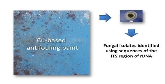
2.1. Antifouling Paint and Other Substrata

A commercial copper self-polishing paint Interspeed® BRA640 (International Paint, Gateshead, UK) was used in this study. The antifouling paint contains about 25–50% of cuprous oxide by weight [29]. An average release rate of copper from the paint was 3.8 μg cm−2 day−1 [30]. The paint was manually applied (thickness 125 μm) onto plastic fiberglass panels (15 cm × 28 cm) cleaned with ethanol (96%, Sigma, Ronkonkoma, NY, USA) in the laboratory. Fiberglass was obtained from a local Omani manufacturer (Al Kaboura, Muscat, Oman). This material was selected because it is used to make boats and it has a high biofouling potential. All coated panels were air-dried for several days at ambient temperature prior to deployment. All substrates (panels and ropes) were cleaned with 96% ethanol before the experiment to eliminate bacteria and fungi. No fungi were found on these substrates prior to the experiment.

2.2. Experiment and Testing Site

Three panels covered with the antifouling paint were exposed vertically to biofouling at the depth of 1 m for 6 months in Marina Bandar Rowdha (23,035′07″ N 58,036′48″ E), Muscat, Oman. As a control, uncoated fiberglass panels were used. Panels were fixed at the desired depth using a nylon rope (RopeNet, Taishan, China) attached to a pontoon. At the end of the rope, a weight was attached to keep the panels in a vertical position.
Marina Bandar Rowdha is a semi-enclosed bay for private recreational boats and yachts. It has a relatively high hydrocarbon and heavy metal pollution with one of the highest concentrations of TBT in Oman’s waters [31]. This marina was selected due to its (very high) biofouling rates and a history of biofouling and antifouling investigations [21,32]. The experiment in the marina was conducted in 2018 between the months of February and September. During the study the seawater temperature varied from 24 to 30 °C, pH was about 8.2 and the salinity varied from 37 to 38 ppt. During the experiment, the seawater turbidity was 2–3 NTU (Nephelometric Turbidity Units).

2.3. Isolation of Fungi

Biofouled panels (painted and not) and ropes holding panels were collected from the marina in September 2018. At the marina, the ropes and the panels were individually packed into sterile bags and brought on ice to the laboratory. In the laboratory, the panels and ropes were washed several times with sterile distilled water (SDI). Using sterile scissors, the ropes were cut into 1.0 cm pieces. Surfaces of the panels and the ropes were disinfected using 1% sodium hypochlorite solution to eliminate bacteria (NaClO, Zhengzhou Sino Chemical Ltd., Beijing, China). Then, the panels and the ropes were washed three times with SDI. After that, the biofilms were removed from the panels using sterile cotton swabs. Finally, one piece from each rope or an individual swab was placed into a Petri dish containing a 2.5% potato dextrose agar (PDA, Merck, Kenilworth, NJ, USA) prepared using filtered (0.45 μm cellulose nitrate filter, Sartorius, Germany) and autoclaved seawater from the marina. As a control, PDA Petri dishes containing autoclaved seawater were used. Visible growth of fungi was checked after incubation at 25 °C for up to three weeks. Each individual fungal colony was transferred into a new fresh PDA plate. Pure fungal colonies were stored on PDA slants with 10% glycerol for further genetic identification (see below).

2.4. Identification of Fungi

Before the identification, the isolate was grown on PDA. The identification of filamentous fungal isolates was done based on sequences of the internal transcribed spacer region (ITS) of the ribosomal DNA [33]. Firstly, 80 g of fungal mycelia were harvested and freeze-dried. Then, its DNA was further extracted [34]. Secondly, the ITS rDNA region was amplified using the primer pairs of ITS4 (TCCTCCGCTTATTGATATGC) and ITS5 (GGAAGTAAAAGT CGTAACAAGG). The PCR program followed the conditions of [35]. MACROGEN, Korea sequenced the PCR products. In order to obtain the ITS rRNA sequence, two complementary sequences for each fungal isolate were aligned using MEGA v.6 [36]. Fungal isolates were identified based on a comparison of the ITS rRNA sequences against the National Center for Biotechnology Information (NCBI) database. Sequences of fungal isolates were deposited in the NCBI GenBank database with accession numbers MN947598–MN947607. For phylogenetic trees, maximum likelihood analysis with 1000 bootstrap replicates based on ITS sequence data was done using RaxmlGUI v. 1.3 [37]. The final phylogenetic tree was selected by comparing the likelihood scores using the GTR+GAMMA substitution model.

2.5. Copper Resistance of Fungal Isolates

In order to prove that fungal isolates are able to grow on antifouling paints, we tested their sensitivity to copper by an agar diffusion technique. Because copper oxide is not soluble in water, copper sulfate (CuSO4, Sigma Aldrich, Ronkonkoma, NY, USA) was used. Firstly, different concentrations (500–0.01 g L−1) of copper sulfate in autoclaved seawater were prepared. Secondly, fungal isolates were grown onto 2.5% potato dextrose agar (PDA, Merck, Kenilworth, NJ, USA) for 3 days. PDA was made using autoclaved seawater from the marina. A four mm disc of each fungal isolate was cut and individually placed onto the PDA Petri dish. Thirdly, 10 μL of copper sulfate solution was added to a sterile paper disk (diameter 6 mm). As a control, disks with 10 μL of seawater were used. The disks were air-dried at room temperature and placed in the middle of a PDA Petri dish two cm away from the isolate. The dishes were incubated at 25 °C for 5 days. The experiment was made in triplicate. The presence or absence of an inhibition zone was detected. Finally, the minimal inhibitory concentration of copper (II) sulfate (μg cm−2) for each fungal isolate was calculated.

3. Results and Discussion

3.1. Biofouling on Different Substrata

Biofouling on the antifouling paint was minimal and only biofilms were observed. In opposite, the ropes and unprotected panels were completely covered with macrofouling organisms, dominated by Tunicata and Bryozoa. This supports our previous data about performance of different antifouling paints in Oman waters [21,32]. The 1-year field experiment showed that copper-based antifouling paints have only diatom and bacterial biofouling [21]. A previous experiment with unprotected fiberglass and acrylic panels demonstrated dominance of Bryozoa, barnacles and sponges [38]. The absence of sponges and barnacles in the current study could be due to differences in the substratum chemistry (nylon versus acrylic) and shape (flat plates versus cylindrical ropes).

3.2. Species of Fungi Isolated from Different Substrata

In total, six fungal isolates were obtained from the antifouling paint and four were isolated from unprotected substrata (Table 1). Based on the phylogenetic analysis, the majority of isolates belonged to Aspergillus and Cladosporium genera (Supplement Figures S1–S4). The genera Aspergillus and Cladosporium are commonly found in the marine environment [39,40,41]. Additionally, Aspergillus and Cladosporium are associated with marine sponges [42,43]. There is limited information about marine derived filamentous fungi in Oman, but we have been able to isolate Aspergillus terreus from mangrove areas [44]. Cladosporium omanense found in this study (Table 1) was previously isolated from living leaves of Zygophyllum coccineum in Oman [45]. The presence of C. omanense on all investigated substrates could be due to several reasons. It could suggest that this species is very common in Omani waters and can colonize protected and unprotected substrata. Alternatively, it could be due to contamination of our culture by spores of this fungus. This is highly unlikely, as there were no fungi recovered from the control plates with autoclaved seawater.
Filamentous fungi belonging to the genera Parengyodontium were isolated from biofouled ropes only (Table 1). Parengyodontium album is an environmental saprobic mold and an opportunistic pathogen [46]. This species has been observed on buildings composed of limestone and plaster [47]. Additionally, P. album was found in sediments of polar-boreal White Sea [48]. The presence of this fungus in Oman waters suggests that this species can be found in tropical waters as well.
The genera Aspergillus, Cladosporium and Alternaria were found on the copper-based antifouling paint (Table 1). Moreover, A. tubingensis, A. terreus, A. niger and C. halotolerans were found only on the antifouling paint. Alternaria isolates were obtained exclusively from the paint. Previously, the fungi Alternaria were isolated from soft corals [49], sponges [50] and algae [51]. While 18S RNA of fungi belonging to the class Agaricomycetes was detected on an antifouling paint using Illumina amplicon sequencing [52], fungal isolates were obtained from antifouling paints for the first time in this study. Previously, only bacteria and diatoms were detected in biofilms on antifouling paints [3,21].

3.3. Copper Resistance of Fungal Isolates

In order to prove that fungal isolates are able to grow on antifouling paints, their sensitivity to different copper concentrations is tested in laboratory experiments (Table 2). Due to low solubility of CuO, CuSO4 was used in this experiment. Previous studies suggest that CuSO4 is more toxic compare to CuO [53]. Thus, the isolates are more resistant to CuO than is reported in Table 2. Generally, isolates from antifouling paint can tolerate higher concentrations of copper. Five out of six isolates from the antifouling paint can tolerate an average daily release rate of copper 3.8 μg cm−2 day−1 [30] from the tested paint (Table 2). The highest copper resistance was observed for Aspergillus terreus. This fungus can tolerate 2% of CuCl2 in a polyvinyl chloride coating in a laboratory experiment [54] and can be used to remove heavy metals from water [54]. In opposite, fungal isolates from unprotected substrata had low tolerance to copper (Table 2). This suggests that isolates from the copper-based antifouling paint are adapted to high copper concentrations and could grow and play an important role in biofilms.

3.4. Importance of This Study

Our finding has very important implications for antifouling industries. Firstly, it demonstrates that some fungal species can live on antifouling paints and tolerate relatively high copper concentrations. Compared to the isolates from unprotected substrata (ropes and panels), fungi from the antifouling paint were highly resistant to copper. Previous studies suggested copper resistance of some fungal species that bind copper to cell walls [55,56]. Additionally, fungi can produce copper-binding proteins and chelating compounds in response to elevated concentrations of copper [55]. Secondly, the role of fungal species on antifouling paints requires further investigations. It is possible to propose that filamentous fungi can degrade organic matrix of the paint, composed of vinyl or acrylic resin or silicone polymers, which in turn can affect release of the biocide and the life span of the antifouling paint. It has been shown that filamentous fungi can deteriorate synthetic paints [57,58]. Additionally, filamentous fungi can degrade biocides of antifouling paints, such as Irgarol 1051 [59] and TBT [60]. In opposite, some marine fungal species produce antimicrobial and antifouling compounds (see review [61]). Presence of these strains on paints could be beneficial and enhance their antifouling properties. Finally, more research is needed to understand if marine fungi can be found on other antifouling paints exposed to biofouling in different seas and investigate possible mechanisms whereby fungi transform these paints. Additionally, it is important to investigate the role of marine fungi on antifouling paints and possible mechanisms of their resistance.

Supplementary Materials

The following are available online at https://www.mdpi.com/article/10.3390/app11188277/s1, Figure S1: Phylogram generated from maximum likelihood analysis based on ITS sequence data of analyzed Alternaria species. Isolates derived from this study are in red. The tree is rooted to A. alternantherae (CBS124392), Figure S2: Phylogram generated from maximum likelihood analysis based on ITS sequence data of analyzed Aspergillus species. Isolates derived from this study are in red. The tree is rooted to Penicillium herquei (CBS 336.48), Figure S3: Phylogram generated from maximum likelihood analysis based on ITS sequence data of analyzed Cladosporium species. Isolates derived from this study are in red, Figure S4: The tree is rooted to Cercospora beticola (CBS 116456), Phylogram generated from maximum likelihood analysis based on ITS sequence data of analyzed Parengyodontium species. Isolates derived from this study are in red. The tree is rooted to Purpureocillium lilacinum (CBS 284.36).

Author Contributions

Conceptualization, S.D. and A.M.A.-S.; methodology, S.D. and A.M.A.-S.; formal analysis, H.A.-S., S.D. and S.S.N.M.; investigation, H.A.-S. and S.S.N.M.; writing—original draft preparation, S.D. and A.M.A.-S.; writing—review and editing, S.D., A.M.A.-S., H.A.-S. and S.S.N.M.; supervision, S.D. and A.M.A.-S.; project administration, S.D. and A.M.A.-S.; funding acquisition, S.D. and A.M.A.-S. All authors have read and agreed to the published version of the manuscript.

Funding

This research was funded by the TRC grant RC/AGR/FISH/16/01, Omantel grant EG/SQU-OT/20/01, SQU internal grant IG/AGR/FISH/18/01 and the grant EG/AGR/CROP/16/01.

Institutional Review Board Statement

Not applicable.

Informed Consent Statement

Not applicable.

Data Availability Statement

Sequences of fungal isolates can be found in the NCBI GenBank database with accession numbers MN947598–MN947607.

Acknowledgments

S.D. acknowledged financial support by the TRC grant RC/AGR/FISH/16/01 and SQU internal grant IG/AGR/FISH/18/01. A.M.A. research was supported by the grant EG/AGR/CROP/16/01 and RC/AGR/FISH/16/01.

Conflicts of Interest

The authors declare no conflict of interest.

References

  1. Wahl, M. Marine epibiosis. I. Fouling and antifouling: Some basic aspects. Mar. Ecol. Prog. Ser. 1989, 58, 175–189. [Google Scholar] [CrossRef] [Green Version]
  2. Dobretsov, S.; Dahms, H.-U.; Qian, P.-Y. Inhibition of biofouling by marine microorganisms and their metabolites. Biofouling 2006, 22, 43–54. [Google Scholar] [CrossRef] [PubMed]
  3. Salta, M.; Wharton, J.A.; Blache, Y.; Stokes, K.R.; Briand, J.-F. Marine biofilms on artificial surfaces: Structure and dynamics. Environ. Microbiol. 2013, 15, 2879–2893. [Google Scholar] [CrossRef] [PubMed]
  4. Hadfield, M.G. Biofilms and marine invertebrate larvae: What bacteria produce that larvae use to choose settlement sites. Annu. Rev. Mar. Sci. 2011, 3, 453–470. [Google Scholar] [CrossRef]
  5. Venkatesan, R.; Murthy, P.S. Macrofouling control in power plants. In Marine and Industrial Biofouling; Flemming, H.C., Murthy, P.S., Venkatesan, R., Cooksey, K., Eds.; Springer Series on Biofilms; Springer: Berlin/Heidelberg, Germany, 2009; Volume 4, pp. 1–27. [Google Scholar]
  6. Wieczorek, S.K.; Todd, C.D. Inhibition and facilitation of settlement of epifaunal marine invertebrate larvae by microbial biofilm cues. Biofouling 1998, 12, 81–118. [Google Scholar] [CrossRef]
  7. Dobretsov, S.; Rittschof, D. Love at first taste: Induction of larval settlement by marine microbes. Int. J. Mol. Sci. 2020, 21, 731. [Google Scholar] [CrossRef] [Green Version]
  8. Schultz, M.P.; Bendick, J.A.; Holm, E.R.; Hertel, W.M. Economic impact of biofouling on a naval surface ship. Biofouling 2011, 27, 87–98. [Google Scholar] [CrossRef]
  9. Yebra, D.M.; Kiil, S.; Dam-Johansen, K. Antifouling technology—Past, present and future steps towards efficient and environmentally friendly antifouling coatings. Prog. Org. Coat. 2004, 50, 75–104. [Google Scholar] [CrossRef]
  10. Bott, T.R. Biofouling control in cooling water. Int. J. Chem. Eng. 2009, 2009. [Google Scholar] [CrossRef] [Green Version]
  11. Jones, G. The battle against marine biofouling: A historical review. In Advances in Marine Antifouling Coatings and Technologies; Hellio, C., Yebra, D., Eds.; Woodhead publishing series in metals and surface engineering; CRC Press: Boca Ration, FL, USA, 2009; pp. 19–45. [Google Scholar]
  12. Maréchal, J.-P.; Hellio, C. Challenges for the development of new non-toxic antifouling solutions. Int. J. Mol. Sci. 2009, 10, 4623–4637. [Google Scholar] [CrossRef] [Green Version]
  13. Dempsey, M.J. Colonisation of antifouling paints by marine bacteria. Bot. Mar. 2009, 24, 185–192. [Google Scholar] [CrossRef]
  14. Thomas, T.E.; Robinson, M.G. The role of bacteria in the metal tolerance of the fouling diatom Amphora coffeaeformis Ag. J. Exp. Mar. Biol. Ecol. 1987, 107, 291–297. [Google Scholar] [CrossRef]
  15. Yang, J.-L.; Li, Y.-F.; Liang, X.; Guo, X.-P.; Ding, D.W.; Zhang, D.; Zhou, S.; Bao, W.-Y.; Bellou, N.; Dobretsov, S. Silver nanoparticles impact biofilm communities and mussel settlement. Sci. Rep. 2016, 6, 37406. [Google Scholar] [CrossRef] [Green Version]
  16. Yang, J.-L.; Li, Y.-F.; Guo, X.-P.; Liang, X.; Xu, Y.-F.; Ding, D.-W.; Bao, W.-Y.; Dobretsov, S. The effect of carbon nanotubes and titanium dioxide incorporated in PDMS on biofilm community composition and subsequent mussel plantigrade settlement. Biofouling 2016, 32, 763–777. [Google Scholar] [CrossRef]
  17. Chen, C.-L.; Maki, J.S.; Rittschof, D.; Teo, S.L.-M. Early marine bacterial biofilm on a copper-based antifouling paint. Int. Biodeterior. Biodegrad. 2013, 83, 71–76. [Google Scholar] [CrossRef]
  18. Pelletier, É.; Bonnet, C.; Lemarchand, K. Biofouling growth in cold estuarine waters and evaluation of some chitosan and copper anti-fouling paints. Int. J. Mol. Sci. 2009, 10, 3209–3223. [Google Scholar] [CrossRef] [Green Version]
  19. Winfield, M.O.; Downer, A.; Longyer, J.; Dale, M.; Barker, G.L.A. Comparative study of biofilm formation on biocidal antifouling and fouling-release coatings using next-generation DNA sequencing. Biofouling 2018, 34, 464–477. [Google Scholar] [CrossRef]
  20. Briand, J.-F.; Djeridi, I.; Jamet, D.; Cope, S.; Bressy, C.; Molmeret, M.; Le Berre, B.; Rimet, F.; Bouchez, A.; Blache, Y. Pioneer marine biofilms on artificial surfaces including antifouling coatings immersed in two contrasting French Mediterranean coast sites. Biofouling 2012, 28, 453–463. [Google Scholar] [CrossRef] [PubMed]
  21. Muthukrishnan, T.; Abed, R.M.M.; Dobretsov, S.; Kidd, B.; Finnie, A.A. Long-term microfouling on commercial biocidal fouling control coatings. Biofouling 2014, 30, 1155–1164. [Google Scholar] [CrossRef]
  22. Richards, T.A.; Leonard, G.; Mahé, F.; del Campo, J.; Romac, S.; Jones, M.D.M.; Maguire, F.; Dunthorn, M.; De Vargas, C.; Massana, R.; et al. Molecular diversity and distribution of marine fungi across 130 European environmental samples. Proc. R. Soc. B Biol. Sci. 2015, 282, 20152243. [Google Scholar] [CrossRef] [PubMed]
  23. Tisthammer, K.H.; Cobian, G.M.; Amend, A.S. Global biogeography of marine fungi is shaped by the environment. Fung. Ecol. 2016, 19, 39–46. [Google Scholar] [CrossRef]
  24. Overy, P.D.; Rämä, T.; Oosterhuis, R.; Walker, K.A.; Pang, K.-L. The neglected marine fungi, sensu stricto, and their isolation for natural products’ discovery. Mar. Drug. 2019, 17, 42. [Google Scholar] [CrossRef] [PubMed] [Green Version]
  25. Miao, L.; Qian, P.-Y. Antagonistic antimicrobial activity of marine fungi and bacteria isolated from marine biofilm and seawaters of Hong Kong. Aquat. Microb. Ecol. 2005, 38, 231–238. [Google Scholar] [CrossRef]
  26. Kirstein, I.V.; Wichels, A.; Krohne, G.; Gerdts, G. Mature biofilm communities on synthetic polymers in seawater-specific or general? Mar. Environ. Res. 2018, 142, 147–154. [Google Scholar] [CrossRef] [PubMed]
  27. Corcoll, N.; Yang, J.; Backhaus, T.; Zhang, X.; Eriksson, K.M. Copper affects composition and functioning of microbial communities in marine biofilms at environmentally relevant concentrations. Front. Microb. 2019, 9, 3248. [Google Scholar] [CrossRef]
  28. Lotlikar, N.; Damare, S.; Meena, R.M.; Jayachandran, S. Variable protein expression in marine-derived filamentous fungus Penicillium chrysogenum in response to varying copper concentrations and salinity. Metallomics 2020, 12, 1083–1093. [Google Scholar] [CrossRef] [PubMed]
  29. Interspeed BRA640 Red Safety Sheet. Available online: http://datasheets1.international-coatings.com/msds/BRA640_USA_eng_B4.pdf (accessed on 22 July 2020).
  30. Valkirs, A.O.; Seligman, P.F.; Haslbeck, E.; Caso, J.S. Measurement of copper release rates from antifouling paint under laboratory and in situ conditions: Implications for loading estimation to marine water bodies. Mar. Poll. Bull. 2003, 46, 763–779. [Google Scholar] [CrossRef]
  31. Jupp, B.P.; Fowler, S.W.; Dobretsov, S.; van der Wiele, H.; Al-Ghafri, A. Assessment of heavy metal and petroleum hydrocarbon contamination in the Sultanate of Oman with emphasis on harbours, marinas, terminals and ports. Mar. Pollut. Bull. 2017, 121, 260–273. [Google Scholar] [CrossRef]
  32. Muthukrishnan, T.; Dobretsov, S.; De Stefano, M.; Abed, R.M.M.; Kidd, B.; Finnie, A.A. Diatom communities on commercial biocidal fouling control coatings after one year of immersion in the marine environment. Mar. Environ. Res. 2017, 129, 102–112. [Google Scholar] [CrossRef]
  33. Al-Sadi, A.M.; Al-Mazroui, S.S.; Phillips, A.J.L. Evaluation of culture-based techniques and 454 pyrosequencing for the analysis of fungal diversity in potting media and organic fertilizers. J. Appl. Microb. 2015, 119, 500–509. [Google Scholar] [CrossRef]
  34. Al-Sadi, A.M.; Al-Ghaithi, A.G.; Al-Balushi, Z.M.; Al-Jabri, A.H. Analysis of diversity in Pythium aphanidermatum populations from a single greenhouse reveals phenotypic and genotypic changes over 2006 to 2011. Plant Dis. 2012, 96, 852–858. [Google Scholar] [CrossRef] [Green Version]
  35. White, T.J.; Bruns, T.D.; Lee, S.; Taylor, J. Amplification and direct sequencing of fungal ribosomal RNA genes for phylogenetics. In PCR Protocols: A Guide to Methods and Applications; Innis, M.A., Gelfand, D.H., Sninsky, J.J., White, T.J., Eds.; Academic Press: New York, NY, USA, 1990; pp. 315–322. [Google Scholar]
  36. Tamura, K.; Stecher, G.; Peterson, D.; Filipski, A.; Kumar, S. MEGA6: Molecular evolutionary genetics analysis version 6.0. Mol. Biol. Evol. 2013, 30, 2725–2729. [Google Scholar] [CrossRef] [Green Version]
  37. Silvestro, D.; Michalak, I. RaxmlGUI: A graphical front-end for RAxML. Org. Divers. Evol. 2012, 12, 335–337. [Google Scholar] [CrossRef]
  38. Dobretsov, S. Biofouling on artificial substrata in Muscat waters. J. Agric. Mar. Sci. JAMS 2015, 20, 24–29. [Google Scholar] [CrossRef] [Green Version]
  39. Chung, D.; Kim, H.; Choi, H.S. Fungi in salterns. J. Microbiol. Seoul Korea 2019, 57, 717–724. [Google Scholar] [CrossRef]
  40. Corinaldesi, C.; Barone, G.; Marcellini, F.; Dell’Anno, A.; Danovaro, R. Marine microbial-derived molecules and their potential use in cosmeceutical and cosmetic products. Mar. Drugs 2017, 15, 118. [Google Scholar] [CrossRef]
  41. Lee, Y.M.; Kim, M.J.; Li, H.; Zhang, P.; Bao, B.; Lee, K.J.; Jung, J.H. Marine-derived Aspergillus species as a source of bioactive secondary metabolites. Mar. Biotechnol. 2013, 15, 499–519. [Google Scholar] [CrossRef]
  42. Paulino, G.V.B.; Félix, C.R.; Landell, M.F. Diversity of filamentous fungi associated with coral and sponges in coastal reefs of northeast Brazil. J. Basic Microbiol. 2019, 60, 103–111. [Google Scholar] [CrossRef]
  43. Wang, G.; Li, Q.; Zhu, P. Phylogenetic diversity of culturable fungi associated with the Hawaiian sponges Suberites zeteki and Gelliodes fibrosa. Antonie Van Leeuwenhoek 2008, 93, 163–174. [Google Scholar] [CrossRef] [PubMed]
  44. Al-Shibli, H.; Dobretsov, S.; Al-Nabhani, A.; Maharachchikumbura, S.S.N.; Rethinasamy, V.; Al-Sadi, A.M. Aspergillus terreus obtained from mangrove exhibits antagonistic activities against Pythium aphanidermatum-induced damping-off of cucumber. PeerJ 2019, 7, e7884. [Google Scholar] [CrossRef] [Green Version]
  45. Halo, B.A.; Maharachchikumbura, S.S.N.; Al-Yahyai, R.A.; Al-Sadi, A.M. Cladosporium omanense, a new endophytic species from Zygophyllum coccineum in Oman. Phytotaxa 2019, 388, 145–154. [Google Scholar] [CrossRef]
  46. Tsang, C.-C.; Chan, J.F.W.; Pong, W.-M.; Chen, J.H.K.; Ngan, A.H.Y.; Cheung, M.; Lai, C.K.C.; Tsang, D.N.C.; Lau, S.K.P.; Woo, P.C.Y. Cutaneous hyalohyphomycosis due to Parengyodontium album gen. et comb. nov. Med. Mycol. 2016, 54, 699–713. [Google Scholar] [CrossRef] [Green Version]
  47. Ponizovskaya, V.B.; Rebrikova, N.L.; Kachalkin, A.V.; Antropova, A.B.; Bilanenko, E.N.; Mokeeva, V.L. Micromycetes as colonizers of mineral building materials in historic monuments and museums. Fungal Biol. 2019, 123, 290–306. [Google Scholar] [CrossRef]
  48. Khusnullina, A.I.; Bilanenko, E.N.; Kurakov, A.V. Microscopic fungi of White Sea sediments. Contemp. Probl. Ecol. 2018, 11, 503–513. [Google Scholar] [CrossRef]
  49. Zhao, D.-L.; Cao, F.; Wang, C.-Y.; Yang, L.-J.; Shi, T.; Wang, K.-L.; Shao, C.-L.; Wang, C.-Y. Alternatone A, an unusual perylenequinone-related compound from a soft-coral-derived strain of the fungus Alternaria alternata. J. Nat. Prod. 2019, 82, 3201–3204. [Google Scholar] [CrossRef]
  50. Chen, Y.; Chen, R.; Xu, J.; Tian, Y.; Xu, J.; Liu, Y. Two new altenusin/thiazole hybrids and a new benzothiazole derivative from the marine sponge-derived fungus Alternaria sp. SCSIOS02F49. Molecules 2018, 23, 2844. [Google Scholar] [CrossRef] [Green Version]
  51. Shi, Z.-Z.; Fang, S.-T.; Miao, F.-P.; Ji, N.-Y. Two new tricycloalternarene esters from an alga-epiphytic isolate of Alternaria alternata. Nat. Prod. Res. 2018, 32, 2523–2528. [Google Scholar] [CrossRef]
  52. Dobretsov, S.; Abed, R.M.M.; Muthukrishnan, T.; Sathe, P.; Al-Naamani, L.; Queste, B.Q.; Piontkovski, S. Living on the edge: Biofilms developing in oscillating environmental conditions. Biofouling 2018, 34, 1064–1077. [Google Scholar] [CrossRef]
  53. Rotini, A.; Tornambè, A.; Cossi, R.; Iamunno, F.; Benvenuto, G.; Berducci, M.T.; Maggi, C.; Thaller, M.C.; Cicero, A.M.; Manfra, L.; et al. Salinity-based toxicity of CuO nanoparticles, CuO-bulk and Cu ion to Vibrio anguillarum. Front. Microbiol. 2017, 8, 2076. [Google Scholar] [CrossRef]
  54. Nasrallah, D.A.; Morsi, M.A.; El-Sayed, F.; Metwally, R.A. Structural, optical and electrical properties of copper chloride filled polyvinyl chloride/polystyrene blend and its antifungal properties against Aspergillus avenaceus and Aspergillus terreus. Compos. Commun. 2020, 22. [Google Scholar] [CrossRef]
  55. Dias, M.A.; Lacerda, I.C.A.; Pimentel, P.F.; De Castro, H.F.; Rosa, C.A. Removal of heavy metals by an Aspergillus terreus strain immobilized in a polyurethane matrix. Lett. Appl. Microb. 2002, 34, 46–50. [Google Scholar] [CrossRef]
  56. Cervantes, C.; Gutierrez-Corona, F. Copper resistance mechanisms in bacteria and fungi. FEMS Microbiol. Rev. 1994, 14, 121–137. [Google Scholar] [CrossRef]
  57. Cappitelli, F.; Vicini, S.; Piaggio, P.; Abbruscato, P.; Princi, E.; Casadevall, A.; Nosanchuk, J.D.; Zanardini, E. Investigation of fungal deterioration of synthetic paint binders using vibrational spectroscopic techniques. Macromol. Biosci. 2005, 5, 49–57. [Google Scholar] [CrossRef]
  58. Saad, D.S.; Kinsey, G.C.; Kim, S.; Gaylarde, C.C. Extraction of genomic DNA from filamentous fungi in biofilms on water-based paint coatings; 12th International Biodeterioration and Biodegradation Symposium (Biosorption and Bioremediation III). Int. Biodeterior. Biodegrad. 2004, 54, 99–103. [Google Scholar] [CrossRef]
  59. Ogawa, N.; Okamura, H.; Hirai, H.; Nishida, T. Degradation of the antifouling compound Irgarol 1051 by manganese peroxidase from the white rot fungus Phanerochaete chrysosporium. Chemosphere 2004, 55, 487–491. [Google Scholar] [CrossRef] [Green Version]
  60. Bernat, P.; Długoński, J. Acceleration of tributyltin chloride (TBT) degradation in liquid cultures of the filamentous fungus Cunninghamella elegans. Chemosphere 2006, 62, 3–8. [Google Scholar] [CrossRef]
  61. Liu, L.L.; Wu, C.-H.; Qian, P.Y. Marine natural products as antifouling molecules—A mini-review (2014–2020). Biofouling 2020, 36, 1210–1226. [Google Scholar]
Table 1. The list of fungal isolates from panels painted with the antifouling paint and not painted (control) panels and ropes.
SpeciesSubstrateNoGenBank Accession NumberPicture
Aspergillus tubingensisAntifouling paintH1MN947598 Applsci 11 08277 i001
Aspergillus terreusAntifouling paintH2MN947599 Applsci 11 08277 i002
Alternaria sp.Antifouling paintH3MN947600 Applsci 11 08277 i003
Aspergillus nigerAntifouling paintH4MN947601 Applsci 11 08277 i004
Cladosporium halotoleransAntifouling paintH6MN947602 Applsci 11 08277 i005
Cladosporium omanenseAntifouling paintH7MN947603 Applsci 11 08277 i006
Aspergillus pseudodeflectusNot painted panel (control)H90MN947605 Applsci 11 08277 i007
Cladosporium omanenseNot painted panel (control)H89MN947604 Applsci 11 08277 i008
Cladosporium omanenseRopesH91MN947606 Applsci 11 08277 i009
Parengyodontium albumRopesH92MN947607 Applsci 11 08277 i010
Table 2. The minimal inhibitory concentration of copper (II) sulfate (μg cm−2) for fungal isolates from panels painted with the antifouling paint, not painted (control), and ropes. Highlighted values exceed an average release rate of copper from the paint (3.8 μg cm−2 day−1 [30]).
SpeciesSubstrateMinimal Inhibitory Concentration
Aspergillus tubingensisAntifouling paint4.3
Aspergillus terreusAntifouling paint5.2
Alternaria sp.Antifouling paint3.9
Aspergillus nigerAntifouling paint4.3
Cladosporium halotoleransAntifouling paint3.9
Cladosporium omanenseAntifouling paint1.3
Aspergillus pseudodeflectusControl0.17
Cladosporium omanenseControl0.87
Cladosporium omanenseRopes1.3
Parengyodontium albumRopes1.3
Publisher’s Note: MDPI stays neutral with regard to jurisdictional claims in published maps and institutional affiliations.

